User login
Sulforaphane for autism? Maybe
LOS ANGELES –
Sulforaphane is a compound in cruciferous vegetables, especially 3-day-old broccoli sprouts. It’s sold widely online and in stores, often as broccoli sprout extract, for anticancer and other effects.
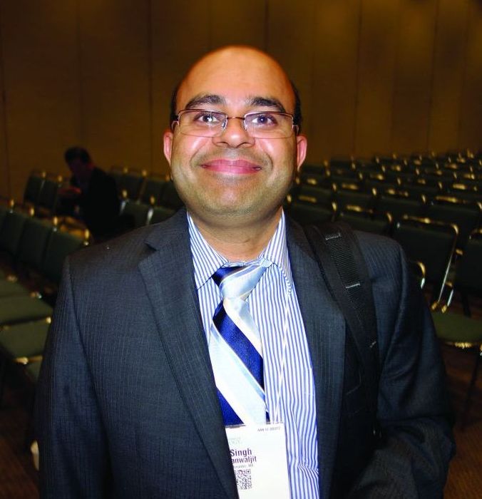
The idea of using it for autism came about after investigators noticed that, in the lab, it induced some of the cellular changes associated with fever, including upregulation of heat shock proteins, according to Kanwaljit Singh, MD, a pediatrics instructor at the University of Massachusetts, Worcester.
Fever has been reported to improve autism symptoms. So, several years ago, “we decided to do a pilot study with sulforaphane” to see if it had a similar effect, Dr. Singh said at the annual meeting of the American Academy of Neurology.
At 18 weeks, 29 young autistic men randomized to the supplement outperformed 15 randomized to placebo on the Aberrant Behavior Checklist and other measures. It was the first study of sulforaphane for autism, and it got a good deal of press attention when it was published in 2014; Dr. Singh was the lead author (Proc Natl Acad Sci U S A. 2014 Oct 28;111[43]:15550-5).
“Because we had a very good signal in our pilot study, we decided to do a slightly larger, slightly more complex clinical trial, which is ongoing right now,” he said. The results aren’t due until the second half of 2018, but he gave an interim report at the meeting.
There are 50 children with autism in the new study, aged 3-12 years. Half are randomized to sulforaphane, half to placebo, for the first 15 weeks, then all are switched to open-label sulforaphane for 15 weeks more, followed by a 6-week washout period.
The randomized portion is still blinded. But so far, 31% have responded positively at 15 weeks, meaning a much or very much improved score in at least two domains on the Ohio Autism Clinical Global Impressions Scale; domains cover social interaction, violent behavior, communication, and other areas.
Among the patients who have completed the study, the response rate at week 30 almost doubled, to 56%. “We don’t know which patients were on sulforaphane and which were on placebo” in the randomized phase, Dr. Singh said. “But we think because the response doubled” when the second half of the children were switched to sulforaphane, “there should probably not be a very large placebo effect here.”
Meanwhile, after the washout period, “some patients still do well, but many more [go] back to baseline,” added Dr. Singh, the senior investigator in the new trial.
The most common side effects are insomnia (28%), vomiting (19%), flatulence (17%), diarrhea (15%), and constipation (13%). A few patients have dropped out because of insomnia and diarrhea; more have dropped out because they simply didn’t want to take the pills – 125 mg of broccoli seed powder three to eight times a day, depending on weight.
Other groups are looking into sulforaphane, too – not just for autism, but also for schizophrenia, prostate cancer, and other indications.
The U.S. Department of Defense is funding the research. The investigators said they have no relevant disclosures.
SOURCE: Zimmerman A et al. Neurology. 2018 Apr 22; 90(15 Suppl.):N1.002.
LOS ANGELES –
Sulforaphane is a compound in cruciferous vegetables, especially 3-day-old broccoli sprouts. It’s sold widely online and in stores, often as broccoli sprout extract, for anticancer and other effects.
The idea of using it for autism came about after investigators noticed that, in the lab, it induced some of the cellular changes associated with fever, including upregulation of heat shock proteins, according to Kanwaljit Singh, MD, a pediatrics instructor at the University of Massachusetts, Worcester.
Fever has been reported to improve autism symptoms. So, several years ago, “we decided to do a pilot study with sulforaphane” to see if it had a similar effect, Dr. Singh said at the annual meeting of the American Academy of Neurology.
At 18 weeks, 29 young autistic men randomized to the supplement outperformed 15 randomized to placebo on the Aberrant Behavior Checklist and other measures. It was the first study of sulforaphane for autism, and it got a good deal of press attention when it was published in 2014; Dr. Singh was the lead author (Proc Natl Acad Sci U S A. 2014 Oct 28;111[43]:15550-5).
“Because we had a very good signal in our pilot study, we decided to do a slightly larger, slightly more complex clinical trial, which is ongoing right now,” he said. The results aren’t due until the second half of 2018, but he gave an interim report at the meeting.
There are 50 children with autism in the new study, aged 3-12 years. Half are randomized to sulforaphane, half to placebo, for the first 15 weeks, then all are switched to open-label sulforaphane for 15 weeks more, followed by a 6-week washout period.
The randomized portion is still blinded. But so far, 31% have responded positively at 15 weeks, meaning a much or very much improved score in at least two domains on the Ohio Autism Clinical Global Impressions Scale; domains cover social interaction, violent behavior, communication, and other areas.
Among the patients who have completed the study, the response rate at week 30 almost doubled, to 56%. “We don’t know which patients were on sulforaphane and which were on placebo” in the randomized phase, Dr. Singh said. “But we think because the response doubled” when the second half of the children were switched to sulforaphane, “there should probably not be a very large placebo effect here.”
Meanwhile, after the washout period, “some patients still do well, but many more [go] back to baseline,” added Dr. Singh, the senior investigator in the new trial.
The most common side effects are insomnia (28%), vomiting (19%), flatulence (17%), diarrhea (15%), and constipation (13%). A few patients have dropped out because of insomnia and diarrhea; more have dropped out because they simply didn’t want to take the pills – 125 mg of broccoli seed powder three to eight times a day, depending on weight.
Other groups are looking into sulforaphane, too – not just for autism, but also for schizophrenia, prostate cancer, and other indications.
The U.S. Department of Defense is funding the research. The investigators said they have no relevant disclosures.
SOURCE: Zimmerman A et al. Neurology. 2018 Apr 22; 90(15 Suppl.):N1.002.
REPORTING FROM AAN 2018
Key clinical point: The dietary supplement is showing benefit in an ongoing trial, but there are a lot of pills.
Major finding: The response rate at 30 weeks was 56%.
Study details: A randomized trial involving 50 children with autism.
Disclosures: The U.S. Department of Defense is funding the research. The investigators said they have no relevant disclosures.
Source: Zimmerman A et al. Neurology. 2018 Apr;90(15 Suppl.):N1.002.
Adjuvant trastuzumab for breast cancer: 6 months may suffice
Shortening the duration of adjuvant trastuzumab (Herceptin) therapy for early-stage HER2+ breast cancer from the current standard of 12 months to 6 months yields similar efficacy but halves the incidence of cardiac toxicity, the PERSEPHONE trial found.
“In 2005, trastuzumab was licensed with a standard of three weekly injections for 12 months, and this was the duration used empirically in pivotal registration studies,” lead study author Helena Earl, MD, professor of clinical cancer medicine at the University of Cambridge, England, said in a press briefing leading up to the annual meeting of the American Society of Clinical Oncology.
However, cardiac toxicity has been particularly problematic with this regimen. Furthermore, the Fin-HER trial, while small, suggested that only 9 weeks of adjuvant trastuzumab was possibly as efficacious (N Engl J Med. 2006 Feb;354[8]:809-20).
Dr. Earl and her coinvestigators enrolled in their phase 3 noninferiority, randomized, controlled trial 4,089 women with early-stage HER2+ breast cancer, randomizing them to either 6 months or 12 months of trastuzumab, mapped onto standard U.K. real-world practice.
The main findings showed that the 4-year rate of disease-free survival, the trial’s primary endpoint, was nearly 90% in both groups, with the absolute difference of just 0.4% falling well within the predefined 3% margin for noninferiority.
Moreover, the rate of stopping trastuzumab because of cardiotoxicity was half as high with the shorter-duration therapy; patients in that arm had more rapid recovery of cardiac function, too.
“The PERSEPHONE trial’s first results demonstrate that 6 months of adjuvant trastuzumab is noninferior to 12 months; 6 months, compared with 12 months, of treatment reduces cardiac and other toxicities and costs both to patients and health care systems,” Dr. Earl summarized. “We are confident [these results] will mark the first steps towards reduction of treatment duration for many women with HER2+ breast cancer.”
The investigators are still analyzing quality of life, patient-reported outcomes, and health economic data, she said. In addition, they are performing translational studies to look for biomarkers that may identify subgroups who fare better with one or the other duration of trastuzumab.
Will the standard change?
At present, the PERSEPHONE findings are not sufficient to change the existing standard of care of 12 months of adjuvant trastuzumab, according to Dr. Earl. “We need to be very careful and cautious about coming out at this point and saying, ‘Yes, 6 months is enough,’ ” she maintained. “At the moment, I do think we need to wait for longer follow-up and we need to take a real close look at the data. Changing from an established treatment that works is always going to be a very complex and very challenging thing to do.”
“Personally, I find the results quite compelling, and I think that it is likely that they will signal a shift even in the U.S. oncology community toward shorter duration of Herceptin adjuvant therapy,” commented Richard L. Schilsky, MD, FACP, FASCO, chief medical officer of ASCO and press briefing moderator. However, “we don’t have data yet on overall survival. Survival in this study is still relatively short for a breast cancer population, although patients with HER2+ disease oftentimes have a somewhat more aggressive course,” he noted. In addition, the ongoing translational studies will be critical to any decisions about changing the standard of care because some subgroups of patients will probably not fare as well with the shorter-duration therapy.
U.S. payers are unlikely to start covering only 6 months of trastuzumab unless the drug’s label is changed based on new data or clinical practice guidelines begin to endorse that shorter duration, according to Dr. Schilsky. “Until one of those things occurs, there is not really a rationale for a payer to mandate that a physician undertake a course of treatment that they may not think is in the patient’s best interest,” he elaborated.
As roughly 12% to 15% of women with early breast cancer have HER2+ disease, the PERSEPHONE findings could have considerable implications for treatment costs, noted Bruce E. Johnson, MD, FASCO, president of ASCO.
However, longer follow-up will be needed before any change to the standard is made, he agreed. “One has to be circumspect about how long you wait and how much more data one has before making a definitive statement. With 8% deaths and 12% recurrences, it may be a bit early to make a definite change in practice.”
While important, the more favorable safety of the 6-month therapy is not sufficient, according to Dr. Johnson, who is also a professor of medicine at the Dana-Farber Cancer Institute and a leader of the Dana-Farber/Harvard Cancer Center Lung Cancer Program, both in Boston. “In my opinion, the efficacy drives most of the therapeutic decision making. We are encouraged by this, and 5-year follow-up is certainly a reasonable initial step. But to be sure of the efficacy, you probably need a bit more time and a few more events,” he explained.
Study details
Patients enrolled in PERSEPHONE had stage Ia to IIIa breast cancer. They were randomized evenly to either 6 months (9 cycles) or 12 months (18 cycles) of adjuvant trastuzumab, given with or after completion of chemotherapy.
Main results showed that the 4-year rate of disease-free survival was 89.8% with 12 months of trastuzumab and 89.4% with 6 months of trastuzumab (hazard ratio, 1.07; P for noninferiority = .01), Dr. Earl reported.
Cardiotoxicity data for the trial population, previously reported (Br J Cancer. 2016 Dec 6;115[12]:1462-70), showed that the rate of stopping trastuzumab because of this adverse effect was 8% with the standard-duration therapy and 4% with the shorter-duration therapy (P less than .0001). Patients saw recovery of cardiac function after stopping the drug (P less than .0001), with more rapid recovery in the shorter-duration group (P = .02).
The group given 6 months of trastuzumab also had lower rates of grade 3 or 4 cough, fatigue, pain, chills, and palpitations, problems which patients reported were having an impact on their lives, Dr. Earl noted. “Perhaps more importantly, patients given 6 months of treatment will be able to return more quickly to their normal lives once their treatment is completed.”
Dr. Earl disclosed that she has a consulting or advisory role with Celgene, Pfizer, Roche, and AstraZeneca; receives travel, accommodations, or expenses, and receives honoraria from Pfizer, Daiichi Sankyo, Amgen, and AstraZeneca; and receives research funding from Roche and Sanofi Pasteur. The study was funded by the National Institute for Health Research in the United Kingdom.
SOURCE: Earl H et al. ASCO 2018, Abstract 506.
REPORTING FROM ASCO 2018
Key clinical point:
Major finding: The 4-year rate of disease-free survival was 89.8% with 12 months of trastuzumab and 89.4% with 6 months of trastuzumab (P for noninferiority = .01)
Study details: Phase 3 noninferiority, randomized, controlled trial among 4,089 women with early HER2+ breast cancer (PERSEPHONE trial).
Disclosures: Dr. Earl disclosed that she has a consulting or advisory role with Celgene, Pfizer, Roche, and AstraZeneca; receives travel, accommodations, or expenses, and honoraria from Pfizer, Daiichi Sankyo, Amgen, and AstraZeneca; and receives research funding from Roche and Sanofi Pasteur. The study was funded by the National Institute for Health Research in the United Kingdom.
Source: Earl H et al. ASCO 2018, Abstract 506.
Specialty practices hire more physician assistants and nurse practitioners
based on data from a review of approximately 90% of physician practices in the United States.
The employment of advanced practice clinicians in primary care continues to grow, but their presence in specialty practices has not been well studied, wrote Grant R. Martsolf, PhD, of the University of Pittsburgh, and his colleagues. In a study published in JAMA Internal Medicine, the researchers used the proprietary SK&A data set to examine employment in specialty practices between 2008 and 2016.
Overall, 28% of all specialty practices employed advanced practice clinicians in 2016 – a 22% increase from 2008. Nearly half of multispecialty practices (49%) employed advanced practice clinicians, as did at least 25% of dermatology, cardiology, obstetrics-gynecology, orthopedic surgery, and gastroenterology practices.
Plastic surgery and ophthalmology practices were the least likely to employ advanced practice clinicians. Surgical practices were more likely to employ physician assistants than nurse practitioners, but the other specialty practices were more likely to employ NPs than PAs.
The growth in employment of advanced practice clinicians may be driven by factors such as economics and the expanding roles for advanced practice clinicians in specialty practice, the researchers said.
The findings were limited by the inclusion of outpatient providers only, and by the lack of information about the exact duties of advanced practice clinicians in each practice, the researchers noted. However, the results suggest that advanced practice clinicians will become even more prevalent in specialty care, and “future research will need to understand their contributions to access, quality, and value,” they wrote.
The researchers had no financial conflicts to disclose.
SOURCE: Martsolf GR et al. JAMA Intern Med. 2018 Apr 30. doi: 10.1001/jamainternmed.2018.1515 .
FROM JAMA INTERNAL MEDICINE
Breath test may detect esophagogastric cancer
A breath test that analyzes volatile organic compounds to detect esophagogastric cancer showed similar diagnostic accuracy to an existing test for endoscopy referral based on clinical parameters, based on a study in 335 patients – 163 with esophagogastric cancer and 172 controls.
The resulting test, which examined the concentrations of volatile organic compounds including butyric acid, hexanoic acid, butanal, and decanal, had a sensitivity of 80%, a specificity of 81%, and an area under the curve of 0.85. However, all of the patients had T3-stage esophagogastric cancer, so there is no indication about whether the breath test would be effective at picking up earlier T1-stage cancers, the authors wrote in the study, published online May 17 in JAMA Oncology.
By comparison, the clinical parameters test based on the NICE guidelines for endoscopy referral has a sensitivity of 59%, a specificity of 81% and an area under the curve of 0.72. These guidelines use age thresholds and symptom criteria such as dyspepsia, but the authors commented that there still remains a huge degree of variability in referral patterns for endoscopy.
“The breath test for esophagogastric cancer aims to provide clinicians with an objective assessment of need for endoscopic referral,” wrote Sheraz R. Markar, PhD, of the department of surgery & cancer at Imperial College London and his coauthors.
The authors said the diagnostic accuracy of the breath test also compared favorably with other cancer diagnostic technologies such as the fecal occult blood test – for which the sensitivity ranges from 30% to 70% – and the Cytosponge test for Barrett esophagus, which has a sensitivity of 73%.
Because all five volatile organic compounds showed an association with esophagogastric cancer, the authors suggested that there could be the possibility of calculating a more stratified risk of cancer for individual patients.
The study found no significant differences in the concentration of the five volatile organic compounds used in the test between patients with esophageal or those with gastric cancer.
The authors noted that with fecal occult blood testing and the Cytosponge test, multiple episodes of testing were known to increase the sensitivity, so this could be another area for future research in breath testing.
The breath test was seen as something that could be used in primary care to identify patients with nonspecific symptoms who should be referred for endoscopy.
“This view has been endorsed by our recent finding that the diagnostic model for OGC [oesophagogastric cancer] is different from that for colorectal cancer, providing the concept for a single breath test for multiple gastrointestinal cancers,” the authors wrote.
“If a clinician is presented with a patient with gastrointestinal symptoms that do not prompt referral based on NICE [National Institute for Health and Care Excellence] criteria, he/she would not need to watch and wait to see if symptoms worsen but could offer the exhaled breath test immediately.”
This approach could help avoid unnecessary endoscopies, which are expensive and have a low diagnostic yield. The breath test could also be administered by a nurse.
One author declared support from the National Institute of Health Research, and the study was supported by the National Institute for Health Research, the Rosetrees Trust and Stoneygate Trust. No conflicts of interest were declared.
SOURCE: Markar SR et al. JAMA Oncol. 2018 May 17. doi: 10.1001/jamaoncol.2018.0991.
FROM JAMA ONCOLOGY
Key clinical point: A breath test could help detect esophagogastric cancer.
Major finding:
Study details: Study in 163 with esophagogastric cancer and 172 controls.
Disclosures: One author declared support from the National Institute of Health Research, and the study was supported by the National Institute for Health Research, the Rosetrees Trust and Stoneygate Trust. No conflicts of interest were declared.
Source: Markar SR et al. JAMA Oncol. 2018 May 17. doi:10.1001/jamaoncol.2018.0991.
New regimens for youth with T-cell malignancies yield best outcomes yet
A set of novel chemotherapy regimens yield excellent outcomes—the best yet—among pediatric and young adult patients with T-cell malignancies, finds a phase 3 randomized controlled trial conducted by the Children’s Oncology Group (ALL0434).
“Despite very intense and complex chemotherapy, 20% of children and adolescents enrolled in Children’s Oncology Group T-cell leukemia trials between 2000 and 2005 did not survive. New drugs were needed to improve survival rates for T-cell malignancies,” lead study author Kimberly P. Dunsmore, MD, a professor at Virginia Tech, Roanoke, said in a press briefing leading up to the annual meeting of the American Society of Clinical Oncology.
The ALL0434 trial tested the addition of methotrexate (Trexall) and/or nelarabine (Arranon), a T cell–specific drug known to be efficacious in relapsed disease, to standard chemotherapy, with tailoring of the regimen to recurrence risk. Analyses were based on 1,545 patients with T-cell acute lymphoblastic leukemia (T-ALL) or T-cell lymphoblastic lymphoma (T-LL).
Results for all patients with T-ALL showed that, with addition of either or both drugs, more than 90% were alive at 4 years and more than 80% were leukemia free. Adding nelarabine to standard chemotherapy improved disease-free survival among the subset having intermediate- or high-risk disease, and the best outcomes were seen with addition of both nelarabine and an escalating dose of methotrexate.
Although patients with T-LL did not see benefit from addition of nelarabine, they still had an 85% rate of overall survival at 4 years.
“ALL0434 is the largest trial for children and young adults with T-cell malignancy ever conducted. It has the best-ever survival data,” Dr. Dunsmore commented.
“Our next steps will be to examine what implications and benefits may accrue when using nelarabine in protocols without cranial irradiation. This is to decrease long-term neurologic side effects, and we think it may be possible since nelarabine also reduces CNS relapses,” she said.
“This trial highlights how effective our pediatric and young adult oncologists are at accruing: This is a rare disease, and they were able to put more than 1,500 patients on trial with this rare disease over the course of time,” commented ASCO President Bruce E. Johnson, MD, FASCO.
The new combination regimens are noteworthy in that they improved survival by an absolute 10% without minimal increase in toxicity, he maintained.
“This is part of the paradigm where nelarabine had been approved [by the FDA] for relapsed or recurrent disease, and in this particular setting, it has been moved upfront, closer to the initial treatment, improving the outcomes for those patients,” elaborated Dr. Johnson, who is also a professor of medicine at the Dana-Farber Cancer Institute and a leader of the Dana-Farber/Harvard Cancer Center Lung Cancer Program, both in Boston.
Study details
The ALL0434 trial enrolled patients aged 1-30 years with newly diagnosed T-ALL or T-LL. After induction chemotherapy, all patients received standard chemotherapy, the Children’s Oncology Group augmented Berlin-Frankfurt-Munster regimen (N Engl J Med. 1998;338:1663-71), and depending on recurrence risk, cranial irradiation.
In addition to that regimen, they were randomly assigned to four arms, according to methotrexate dosing (high dose with leucovorin rescue in the inpatient setting vs. escalating dose in the outpatient setting) and nelarabine therapy (receipt vs. nonreceipt).
Among patients with T-ALL, those with low-risk disease were ineligible for nelarabine and did not receive cranial irradiation, whereas those with intermediate- and high-risk disease were randomized to all four arms, Dr. Dunsmore explained. In addition, those who did not achieve remission on induction chemotherapy were nonrandomly assigned to the high-dose methotrexate plus nelarabine arm.
Patients with T-LL were ineligible for high-dose methotrexate and were randomized to escalating-dose methotrexate with or without nelarabine.
Among all patients with T-ALL, the 4-year rate of overall survival was 90.2%, and the 4-year rate of disease-free survival was 84.1%, Dr. Dunsmore reported.
Disease-free survival was better with escalating-dose methotrexate than with high-dose methotrexate (89.8% vs. 78%).
Addition of nelarabine for patients with T-ALL having intermediate- or high-risk disease improved disease-free survival, from 83% without the drug to 88% with the drug (P = .0450), and reduced the rate of CNS relapse. Disease-free survival was highest, at 91%, among those who received both escalating-dose methotrexate and nelarabine.
Among the patients who did not achieve remission from induction chemotherapy, the 4-year rate of overall survival was 54%. “This is important because it’s more than double the past survival rates,” Dr. Dunsmore noted.
Patients with T-LL fared similarly well whether they received nelarabine or not; fully 85% overall were still alive at 4 years.
In terms of adverse effects of nelarabine therapy, the rate of peripheral neuropathy (motor or sensory), one of the more problematic adverse effects of the drug, was 8% in the trial population overall and did not exceed 9% in any treatment arm, she reported.
Dr. Dunsmore disclosed that an immediate family member is an employee of and has a leadership role with TypeZero Technologies; that she receives travel, accommodations, and/or expenses from Novo Nordisk; and that an immediate family member receives travel, accommodations, and/or expenses from Tandem Diabetes Care. The study received funding from the Cancer Therapy Evaluation Program within the National Cancer Institute/National Institutes of Health and received support from the St. Baldrick’s Foundation.
SOURCE: Dunsmore KP et al. ASCO 2018, Abstract 10500.
REPORTING FROM ASCO 2018
Key clinical point:
Major finding: Patients with T-ALL had a 4-year rate of overall survival of 90.2% and disease-free survival of 84.1%; patients with T-LL had a 4-year rate of overall survival of 85%.
Study details: Phase 3 randomized controlled trial among 1,545 youth with T-ALL or T-LL testing various regimens of methotrexate and/or nelarabine with standard chemotherapy (ALL0434).
Disclosures: Dr. Dunsmore disclosed that an immediate family member is an employee of and has a leadership role with TypeZero; that she receives travel, accommodations, and/or expenses from Novo Nordisk; and that an immediate family member receives travel, accommodations, and/or expenses from Tandem Diabetes Care. The study received funding from the Cancer Therapy Evaluation Program within the National Cancer Institute/National Institutes of Health and received support from the St. Baldrick’s Foundation.
Source: Dunsmore KP et al. ASCO 2018, Abstract 10500.
VIDEO: Skin exam crucial in rheumatic diseases, expert says
SANDESTIN, FLA. – Even when you know a patient’s serology and hear their symptoms and think you have a bead on their rheumatic disease, you might not. It’s vital to check the skin in patients with rheumatic disease to be sure the right disease is being treated and that they don’t actually have a more severe condition that might progress suddenly if left unchecked, said Alisa Femia, MD, assistant professor of dermatology at the annual Congress of Clinical Rheumatology.
In a session filled with pearls for rheumatologists on what to look for on their patients’ skin to help guide diagnosis and treatment, she told the story of a woman whom a rheumatologist colleague had correctly diagnosed with dermatomyositis. She was started on prednisone and mycophenolate mofetil, but her skin disease did not clear.
After examining her skin, Dr. Femia became immediately concerned.
“Despite prednisone, despite mycophenolate, here not only does she have Gottron’s papules, but she has erosions within her Gottron’s papules,” Dr. Femia said. The woman also had erosions within papules on her palms.
These were telltale signs of MDA5-associated dermatomyositis, which studies have found to be linked with interstitial lung disease (J Am Acad Dermatol. 2011 Jul;65[1]:25-34). Under her care, these patients ideally undergo lung monitoring every 3 months, Dr. Femia said.
“That is a form of dermatomyositis that you cannot miss,” she said.
The effects of discoid lupus are another reason to take special care in skin examination. Once the disease, which involves a scaling of the skin, is obvious, there can be permanent aesthetic effects that could have been avoided with earlier detection and treatment, Dr. Femia said.
Clinicians should also be on the lookout for volume loss, or contour change, in discoid lupus patients, because that’s a sign of lupus panniculitis, which involves deeper lesions mainly to fatty areas such as the cheeks or thighs. The disease can progress fast, with sudden, massive loss of body volume, so therapy should be escalated quickly, she said.
“We want to treat these patients aggressively in order to avoid this.”
SOURCE: Femia A. CCR 2018.
EXPERT ANALYSIS AT CCR 18
VIDEO: Researchers seek end to early corticosteroid use in AAV
SANDESTIN, FLA. – Clinicians have long wanted to avoid using corticosteroids in the treatment of ANCA-associated vasculitis (AAV). They’re drawing closer to getting their wish, said Christian Pagnoux, MD, of the department of internal medicine at Mount Sinai Hospital in Toronto.
The drugs have been a cornerstone in the treatments of these diseases – including granulomatosis with polyangiitis (GPA) and microscopic polyangiitis (MPA) – for decades, but they come at the price of osteoporosis, cardiovascular comorbidities, diabetes, increased infection risk, and other problems.
The video associated with this article is no longer available on this site. Please view all of our videos on the MDedge YouTube channel
The emergence of newer therapies such as rituximab and complement C5a-blocker avacopan could mean less of a reliance on corticosteroids, Dr. Pagnoux said. The ongoing ADVOCATE trial is assessing the efficacy of avacopan with rituximab or cyclophosphamide, with or without a tapered dose of prednisone for the first 21 weeks.
“Whether we can use a lighter, briefer, shorter corticosteroid regimen for induction is really a burning question,” Dr. Pagnoux said. Avacopan “may totally replace corticosteroids in the very near future,” he said.
Another trial taking an intense look at winnowing corticosteroids from GPA and MPA treatment is the eagerly awaited PEXIVAS trial, an international effort of 700 patients that is the largest ever in AAV, Dr. Pagnoux said.
The primary endpoint in the trial is assessing plasma exchange versus no plasma exchange, but the use of corticosteroids is being assessed as well.
“The PEXIVAS [trial] may give you some additional information,” Dr. Pagnoux said. “Patients were not only randomized to receive plasma exchange or no plasma exchange, but they were also randomized to receive the standard regimen of corticosteroids with a slow taper ... or a much faster regimen with a much faster tapering of the corticosteroids.” The fast taper involves a steep drop every week, so that, after just 1 month, doses have fallen from 60 mg to 10 mg.
Dr. Pagnoux said he can imagine the day when corticosteroids can be completely eliminated from induction treatment for GPA and MPA. But he added there are studies looking at the efficacy and safety of the drugs in maintenance treatment even once they’re eliminated from induction, but at far lower doses.
“The good news is that it would only be 5 mg per day, for example.”
SOURCE: Pagnoux C. CCR 2018.
EXPERT ANALYSIS AT CCR 18
Hematocrit improvement with SGLT2 inhibitor: Not just a diuretic effect?
BOSTON – The SGLT2 inhibitor dapagliflozin may increase red blood cell production by suppressing plasma levels of hepcidin, a proinflammatory inhibitor of iron transport, according to results of a randomized study.
This reduction in hepcidin provides a new mechanistic explanation for the improvement in hematocrit seen with SGLT2 inhibitor treatment and suggests a role for use of these drugs beyond their current indications, according to researcher Husam A. Ghanim, PhD, of the State University of New York at Buffalo.

To see whether there were other mechanisms involved beyond hemoconcentration caused by diuretic effects of the drugs, Dr. Ghanim and his colleagues investigated the possibility that dapagliflozin might suppress concentrations of hepcidin concentrations, thereby increasing erythropoiesis.
Their study included 22 patients with type 2 diabetes and normal renal function randomized to dapagliflozin 10 mg daily or placebo for 12 weeks.
They found that the plasma concentration of hepcidin fell significantly over that time period, from 265 to 215 ng/mL in dapagliflozin-treated patients. They also saw significant decreases in hemoglobin A1c, hemoglobin concentration, and hematocrit, as well as an increase in transferrin, the major transporter of iron in the circulation, over 12 weeks.
No such significant changes in those measures were seen in the placebo group, Dr. Ghanim said.
There was a modest but nonsignificant increase in erythropoietin concentrations in the dapagliflozin-treated group, according to the researcher.
Circulating ferritin also fell by about 40% over the course of the study. “Circulating ferritin doesn’t have a clear indication or implication on iron transport,” Dr. Ghanim said. “However, it gets secreted from macrophages and from the liver, and it gets used as a marker for inflammation, and it’s also used as a marker of liver function. So a reduction in ferritin levels may have some clinical implication to what’s going on in the liver.”
On the basis of these findings, it appears that SGLT2 inhibition might increase hematocrit via anti-inflammatory effects and increased erythropoiesis, Dr. Ghanim said.
The increase in oxygenated blood available to tissues might contribute to the beneficial effects of SGLT2 inhibitors on cardiovascular disease, he added.
Also, it’s possible that SGLT2 inhibitors could have a “major impact” on the liver since hepcidin and ferritin are secreted mainly by the liver: “This could also lead us to think that it is possible that we could use SGLT2 inhibitors in conditions of liver inflammation like nonalcoholic steatohepatitis and fatty liver disease,” Dr. Ghanim said in his presentation. “These are future ideas we could explore, based on our data.”
Dr. Ghanim had no disclosures to report.
SOURCE: Ghanim HA et al. AACE 2018, Abstract 228.
BOSTON – The SGLT2 inhibitor dapagliflozin may increase red blood cell production by suppressing plasma levels of hepcidin, a proinflammatory inhibitor of iron transport, according to results of a randomized study.
This reduction in hepcidin provides a new mechanistic explanation for the improvement in hematocrit seen with SGLT2 inhibitor treatment and suggests a role for use of these drugs beyond their current indications, according to researcher Husam A. Ghanim, PhD, of the State University of New York at Buffalo.
To see whether there were other mechanisms involved beyond hemoconcentration caused by diuretic effects of the drugs, Dr. Ghanim and his colleagues investigated the possibility that dapagliflozin might suppress concentrations of hepcidin concentrations, thereby increasing erythropoiesis.
Their study included 22 patients with type 2 diabetes and normal renal function randomized to dapagliflozin 10 mg daily or placebo for 12 weeks.
They found that the plasma concentration of hepcidin fell significantly over that time period, from 265 to 215 ng/mL in dapagliflozin-treated patients. They also saw significant decreases in hemoglobin A1c, hemoglobin concentration, and hematocrit, as well as an increase in transferrin, the major transporter of iron in the circulation, over 12 weeks.
No such significant changes in those measures were seen in the placebo group, Dr. Ghanim said.
There was a modest but nonsignificant increase in erythropoietin concentrations in the dapagliflozin-treated group, according to the researcher.
Circulating ferritin also fell by about 40% over the course of the study. “Circulating ferritin doesn’t have a clear indication or implication on iron transport,” Dr. Ghanim said. “However, it gets secreted from macrophages and from the liver, and it gets used as a marker for inflammation, and it’s also used as a marker of liver function. So a reduction in ferritin levels may have some clinical implication to what’s going on in the liver.”
On the basis of these findings, it appears that SGLT2 inhibition might increase hematocrit via anti-inflammatory effects and increased erythropoiesis, Dr. Ghanim said.
The increase in oxygenated blood available to tissues might contribute to the beneficial effects of SGLT2 inhibitors on cardiovascular disease, he added.
Also, it’s possible that SGLT2 inhibitors could have a “major impact” on the liver since hepcidin and ferritin are secreted mainly by the liver: “This could also lead us to think that it is possible that we could use SGLT2 inhibitors in conditions of liver inflammation like nonalcoholic steatohepatitis and fatty liver disease,” Dr. Ghanim said in his presentation. “These are future ideas we could explore, based on our data.”
Dr. Ghanim had no disclosures to report.
SOURCE: Ghanim HA et al. AACE 2018, Abstract 228.
REPORTING FROM AACE 2018
Key clinical point: The SGLT2 inhibitor dapagliflozin suppressed hepcidin, a proinflammatory inhibitor of iron transport.
Major finding: Hepcidin plasma concentration fell from 265 to 215 ng/mL (P < 0.05) in dapagliflozin-treated patients.
Study details: A study of 22 patients with type 2 diabetes randomized to either dapagliflozin 10 mg daily or placebo for 12 weeks.
Disclosures: Dr. Ghanim had no disclosures related to the presentation.
Source: Ghanim HA et al. AACE 2018, Abstract 228.
Cannabidiol gel for osteoarthritis knee pain gives lukewarm results
LIVERPOOL, ENGLAND – There was no significant reduction in pain from knee osteoarthritis (OA) with the use of investigational cannabidiol (CBD) gel ZYN002 in a phase 2a trial presented at the World Congress on Osteoarthritis.
The mean reductions in baseline knee pain scores from study entry to a 12-week assessment were –2.4 for placebo and –2.6 (P = .5) and –2.8 (P = .25), respectively, for a 250-mg and a 500-mg formulation of the gel.
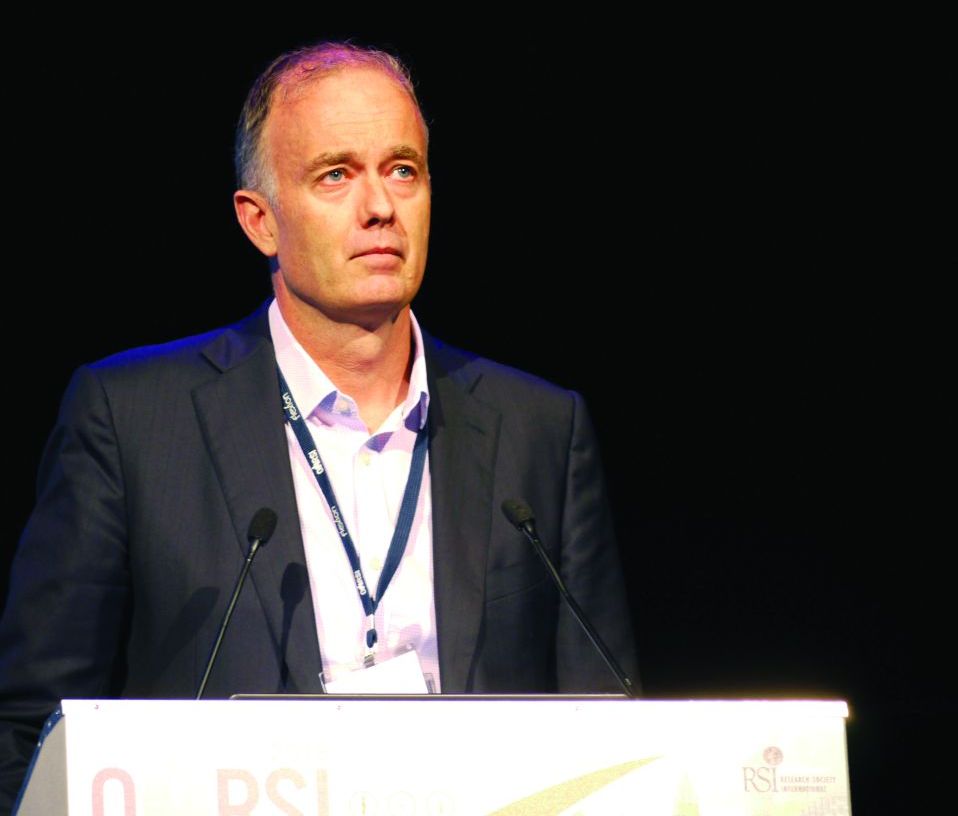
While there was a trend for benefit, it was “neither statistically or clinically significant,” reported David Hunter, MBBS, PhD.
However, he observed that a significantly (P = .016) greater number of patients who received the 250-mg dose (52.7%) were “composite responders,” compared with patients who received placebo (34.1%). A composite response was defined as at least a 30% reduction in pain, and a 20% decrease in WOMAC physical function subscale score at the last observation.
Although the percentage of composite responders was also higher than placebo with the 500-mg dose, the difference wasn’t significant (45.1% vs. 34.1%; P = .0169).
Post-hoc analyses also suggested that perhaps some patients may benefit more than others, reported Dr. Hunter, professor of medicine at the University of Sydney and the Royal North Shore Hospital, Sydney.
For example, patients with baseline pain scores or 7 or more had greater mean reduction in pain at 12 weeks with both doses of the gel combined than placebo at week 4 (–2.2 vs. –1.6; P = .029), although the difference was not significant at week 8 (–3.0 vs. –2.2; P = .05) or 12 (–3.3 vs. –2.5; P = .086).
Women also exhibited a greater placebo response than did men, and “patients with less variability in baseline pain scores may have had greater separation between placebo and the treatment,” Dr. Hunter said. Indeed, 50%-52% of patients with less than 33% variation in baseline scores had a composite response to the gel, versus 27% for the placebo arm.
Evidence from preclinical models suggest that cannabinoids have antinociceptive and antihyperalgesic effects, Dr. Hunter explained at the congress, sponsored by the Osteoarthritis Research Society International. CBD has also been shown to have broad anti-inflammatory effects, and it may even promote osteoclast cell function and decrease bone resorption.
ZYN002 is a synthetic CBD formulated for transdermal delivery using a patented method to enhance its permeation through the skin. According to the manufacturer, Zynerba, it was developed for neuropsychiatric disorders, including fragile X syndrome, adult refractory epilepsy, and developmental and epileptic encephalopathies.
The primary aim of the phase 2 trial reported by Dr. Hunter was to assess ZYN002’s efficacy in managing osteoarthritis knee pain. Secondary objectives were to assess the gel’s safety and tolerability.
The STOP 1 (Synthetic Transdermal Cannabidiol for the Treatment of Knee Pain Due to Osteoarthritis) trial was a double-blind, placebo-controlled trial. For inclusion in the study, patients had to be between age 40 and 75 years and have had knee pain for at least 12 months because of primary OA, based on clinical and x-ray data as per American College of Rheumatology criteria. Anyone with a history of fibromyalgia or epilepsy was excluded.
A total of 320 patients with painful knee OA, with a mean age of 62 years, were randomized and underwent a 1-week washout period in which all their analgesic medications being used for osteoarthritis knee pain, except acetaminophen, were stopped. That was followed by a 7- to 10-day period when baseline daily worst pain levels were captured using a 0-10 numeric rating scale. Patients then underwent 12 weeks of treatment with either a high (500 mg) or a low (250 mg) dose of the gel, or placebo, given in twice-daily doses.
Just over a third (34%) of patients in the placebo arm discontinued the study, compared with 22% and 24% of those in the high- and low-dose gel arms. The main reason for discontinuation was withdrawn consent because of lack of efficacy in the placebo arm, with 8%, 8%, and 4% of patients, respectively, discontinuing because of adverse effects.
“Treatment-emergent adverse effects were roughly equally distributed across the three groups,” Dr. Hunter reported. The adverse events of more interest, he noted, were application site dryness, reaction, or pain. There was “a slight predisposition” to each of these in the 250-mg gel arm (5%, 3%, and 3% of patients affected) versus the 500-mg gel (3%, 0%, and 0%) and placebo (1%, 1%, 0%) arms.
SOURCE: Hunter D et al. Osteoarthritis Cartilage 2018:26(1):S26. Abstract 30.
LIVERPOOL, ENGLAND – There was no significant reduction in pain from knee osteoarthritis (OA) with the use of investigational cannabidiol (CBD) gel ZYN002 in a phase 2a trial presented at the World Congress on Osteoarthritis.
The mean reductions in baseline knee pain scores from study entry to a 12-week assessment were –2.4 for placebo and –2.6 (P = .5) and –2.8 (P = .25), respectively, for a 250-mg and a 500-mg formulation of the gel.
While there was a trend for benefit, it was “neither statistically or clinically significant,” reported David Hunter, MBBS, PhD.
However, he observed that a significantly (P = .016) greater number of patients who received the 250-mg dose (52.7%) were “composite responders,” compared with patients who received placebo (34.1%). A composite response was defined as at least a 30% reduction in pain, and a 20% decrease in WOMAC physical function subscale score at the last observation.
Although the percentage of composite responders was also higher than placebo with the 500-mg dose, the difference wasn’t significant (45.1% vs. 34.1%; P = .0169).
Post-hoc analyses also suggested that perhaps some patients may benefit more than others, reported Dr. Hunter, professor of medicine at the University of Sydney and the Royal North Shore Hospital, Sydney.
For example, patients with baseline pain scores or 7 or more had greater mean reduction in pain at 12 weeks with both doses of the gel combined than placebo at week 4 (–2.2 vs. –1.6; P = .029), although the difference was not significant at week 8 (–3.0 vs. –2.2; P = .05) or 12 (–3.3 vs. –2.5; P = .086).
Women also exhibited a greater placebo response than did men, and “patients with less variability in baseline pain scores may have had greater separation between placebo and the treatment,” Dr. Hunter said. Indeed, 50%-52% of patients with less than 33% variation in baseline scores had a composite response to the gel, versus 27% for the placebo arm.
Evidence from preclinical models suggest that cannabinoids have antinociceptive and antihyperalgesic effects, Dr. Hunter explained at the congress, sponsored by the Osteoarthritis Research Society International. CBD has also been shown to have broad anti-inflammatory effects, and it may even promote osteoclast cell function and decrease bone resorption.
ZYN002 is a synthetic CBD formulated for transdermal delivery using a patented method to enhance its permeation through the skin. According to the manufacturer, Zynerba, it was developed for neuropsychiatric disorders, including fragile X syndrome, adult refractory epilepsy, and developmental and epileptic encephalopathies.
The primary aim of the phase 2 trial reported by Dr. Hunter was to assess ZYN002’s efficacy in managing osteoarthritis knee pain. Secondary objectives were to assess the gel’s safety and tolerability.
The STOP 1 (Synthetic Transdermal Cannabidiol for the Treatment of Knee Pain Due to Osteoarthritis) trial was a double-blind, placebo-controlled trial. For inclusion in the study, patients had to be between age 40 and 75 years and have had knee pain for at least 12 months because of primary OA, based on clinical and x-ray data as per American College of Rheumatology criteria. Anyone with a history of fibromyalgia or epilepsy was excluded.
A total of 320 patients with painful knee OA, with a mean age of 62 years, were randomized and underwent a 1-week washout period in which all their analgesic medications being used for osteoarthritis knee pain, except acetaminophen, were stopped. That was followed by a 7- to 10-day period when baseline daily worst pain levels were captured using a 0-10 numeric rating scale. Patients then underwent 12 weeks of treatment with either a high (500 mg) or a low (250 mg) dose of the gel, or placebo, given in twice-daily doses.
Just over a third (34%) of patients in the placebo arm discontinued the study, compared with 22% and 24% of those in the high- and low-dose gel arms. The main reason for discontinuation was withdrawn consent because of lack of efficacy in the placebo arm, with 8%, 8%, and 4% of patients, respectively, discontinuing because of adverse effects.
“Treatment-emergent adverse effects were roughly equally distributed across the three groups,” Dr. Hunter reported. The adverse events of more interest, he noted, were application site dryness, reaction, or pain. There was “a slight predisposition” to each of these in the 250-mg gel arm (5%, 3%, and 3% of patients affected) versus the 500-mg gel (3%, 0%, and 0%) and placebo (1%, 1%, 0%) arms.
SOURCE: Hunter D et al. Osteoarthritis Cartilage 2018:26(1):S26. Abstract 30.
REPORTING FROM OARSI 2018
Key clinical point:
Major finding: Mean knee pain scores at 12 weeks fell by –2.4 for placebo, and –2.6 (P = .5) and –2.8 (P = .25) for a 250-mg and a 500-mg formulation of the gel.
Study details: A 12-week, randomized, double-blind, placebo-controlled, phase 2, multidose study involving 320 patients with osteoarthritis knee pain for at least 12 months.
Disclosures: Dr. Hunter has consulted for Flexion, Merck Serono, TissueGene, and Zynerba, and has received royalties from DJO for a patellofemoral brace.
Source: Hunter D et al. Osteoarthritis Cartilage 2018:26(1):S26. Abstract 30.
VIDEO: Real-world findings on hybrid closed-loop insulin system
BOSTON – Real-world experience with the Medtronic MiniMed 670G, a hybrid closed-loop insulin delivery system, showed the device was associated with improved average glucose readings and more time in euglycemia in 26 patients with type 1 diabetes.
The findings go beyond the safety data from the clinical trial of the MiniMed 670G system, Kathryn Weaver, MD, of the University of Washington, Seattle, and her colleagues reported in a poster presented at the annual meeting of the American Association of Clinical Endocrinologists.

The clinical trial included a 2-week run-in period during which the system was used in manual mode before it was switched to automated mode. Mean sensor glucose readings for participants went from 150.2 mg/dL during run-in to 150.8 mg/dL at the end of 3 months, which was not a statistically significant difference (JAMA. 2016;316[13]:1407-8).
In the real-world study, average sensor glucose readings dropped from a mean 169.46 mg/dL at baseline to 157.08 mg/dL at the end of the 3-month study period (P = .05). Also, the time spent with blood glucose levels greater than 180 mg/dL fell from 26.5% to 20% (P = .007), while the amount of time with glucose readings between 70 and 180 mg/dL increased from 61.7% to 71.1% (P = .02). Periods of hypoglycemia and severe hypoglycemia were already low at baseline and did not change, Dr. Weaver said.
“It is important to note that the initial pivotal trial was a study designed to evaluate safety not a study designed to evaluate effectiveness. And the [trial] group did demonstrate safety; they had a very significant reduction in the amount of hypoglycemia” with the pump, said Dr. Weaver. “We did not show a significant reduction in hypoglycemia in our [real-world] group, likely because we had a very low rate of hypoglycemia going into the study.”
Two of the study coauthors are employees of Medtronic, which manufactures the MiniMed 670G insulin pump/continuous glucose monitor. Medtronic did not provide funding support for the study or provide the closed-loop systems, and Dr. Weaver reported that she had no relevant financial disclosures.
SOURCE: Weaver K et al. AACE 2018, Abstract 210.
BOSTON – Real-world experience with the Medtronic MiniMed 670G, a hybrid closed-loop insulin delivery system, showed the device was associated with improved average glucose readings and more time in euglycemia in 26 patients with type 1 diabetes.
The findings go beyond the safety data from the clinical trial of the MiniMed 670G system, Kathryn Weaver, MD, of the University of Washington, Seattle, and her colleagues reported in a poster presented at the annual meeting of the American Association of Clinical Endocrinologists.
The clinical trial included a 2-week run-in period during which the system was used in manual mode before it was switched to automated mode. Mean sensor glucose readings for participants went from 150.2 mg/dL during run-in to 150.8 mg/dL at the end of 3 months, which was not a statistically significant difference (JAMA. 2016;316[13]:1407-8).
In the real-world study, average sensor glucose readings dropped from a mean 169.46 mg/dL at baseline to 157.08 mg/dL at the end of the 3-month study period (P = .05). Also, the time spent with blood glucose levels greater than 180 mg/dL fell from 26.5% to 20% (P = .007), while the amount of time with glucose readings between 70 and 180 mg/dL increased from 61.7% to 71.1% (P = .02). Periods of hypoglycemia and severe hypoglycemia were already low at baseline and did not change, Dr. Weaver said.
“It is important to note that the initial pivotal trial was a study designed to evaluate safety not a study designed to evaluate effectiveness. And the [trial] group did demonstrate safety; they had a very significant reduction in the amount of hypoglycemia” with the pump, said Dr. Weaver. “We did not show a significant reduction in hypoglycemia in our [real-world] group, likely because we had a very low rate of hypoglycemia going into the study.”
Two of the study coauthors are employees of Medtronic, which manufactures the MiniMed 670G insulin pump/continuous glucose monitor. Medtronic did not provide funding support for the study or provide the closed-loop systems, and Dr. Weaver reported that she had no relevant financial disclosures.
SOURCE: Weaver K et al. AACE 2018, Abstract 210.
REPORTING FROM AACE 2018