User login
Complete MUS mesh removal not linked to incontinence
SAN FRANCISCO – Stress urinary incontinence (SUI) following removal of a mid-urethral sling (MUS) mesh is not necessarily associated with increased risk of postsurgical urinary incontinence, according to a retrospective study at a high-volume, tertiary medical center.
Follow-up procedures occurred more often in women with preoperative urodynamic SUI and less often in women who were stress continent.

Among women who were stress continent, obesity and postmenopausal status were linked to postsurgical SUI. There was no association between postsurgical SUI and the extent of mesh excision or prior revisions.
The study grew out of observations that SUI occurred less often than expected.
“There’s an increasing recognition of complications related to synthetic MUS,” said Janine Oliver, MD, who presented the study at the annual meeting of the American Urological Association. “As mesh removal procedures were being performed, we assumed that the majority of patients, if not all, would be incontinent afterward, since we were removing the sling that was put in to fix stress incontinence in most cases.”
In a patient who would benefit from a complete mesh removal, “the fear that it may lead to a higher risk of urinary incontinence is not a good justification to not do it,” Dr. Oliver said. She did note, however, that the procedures were done by specialists, so findings may not be applicable to general practitioners.
The study was performed while Dr. Oliver was a fellow at the University of California, Los Angeles. She is now with the division of urology at the University of Colorado, Anschutz, in Aurora.
Dr. Oliver and her colleagues analyzed data from 233 patients who underwent MUS excision at UCLA for MUS-related complications during 2013-2015, and who had at least 3 months of follow-up. The average patient age was 55.4 years; an average of 5.4 years passed between the placement and excision of MUS. The mean body mass index was 28.9, and mean follow-up was 23.5 months.
A total of 84% of patients underwent a total excision; 45% of MUS were retropubic, 35% were transobturator, 10% were single incision, and 10% were multiple incision.
Nearly half (49%) of patients required a second procedure for SUI, such as bulking agent injection, bladder neck suspension, or repeat sling procedure.
In the entire cohort, multivariate analyses found significant associations between heightened risk of postoperative SUI and increasing time to MUS excision (odds ratio, 1.16; 95% confidence interval, 1.03-1.30), total MUS excision (OR, 4.14; 95% CI, 1.38-12.37), and preoperative urodynamic SUI (OR, 4.66; 95% CI, 2.13-10.19).
Of 51 patients who had preoperative urodynamic SUI, 39 (76%) ultimately underwent another surgery. Although increased time to MUS excision and total mesh removal were associated with urinary incontinence in this group in univariate analyses, they were no longer significant following a multivariate analysis.
Of 140 patients with a negative preoperative urodynamic testing for SUI, 59 (42%) went on to have another SUI procedure. After multivariate analysis, the only risk factors for urinary incontinence were obesity (OR, 4.74; 95% CI, 1.73-13.02) and postmenopausal status (OR, 3.78; 95% CI, 1.16-12.33).
“I think there’s a lot of fear, even among urologists and specialists who see these problems, that complete mesh removal is associated with a higher risk of complications and a higher risk of incontinence,” said Dr. Oliver. “These data would suggest that, in certain subgroups, that’s not true. The risks factors that we identified in a multivariate analysis were being obese and being postmenopausal, but not complete mesh removal.”
The study received no external funding. Dr. Oliver reported having no financial conflicts of interest.
SOURCE: Oliver J et al. AUA Annual Meeting. Abstract PD05-10.
SAN FRANCISCO – Stress urinary incontinence (SUI) following removal of a mid-urethral sling (MUS) mesh is not necessarily associated with increased risk of postsurgical urinary incontinence, according to a retrospective study at a high-volume, tertiary medical center.
Follow-up procedures occurred more often in women with preoperative urodynamic SUI and less often in women who were stress continent.
Among women who were stress continent, obesity and postmenopausal status were linked to postsurgical SUI. There was no association between postsurgical SUI and the extent of mesh excision or prior revisions.
The study grew out of observations that SUI occurred less often than expected.
“There’s an increasing recognition of complications related to synthetic MUS,” said Janine Oliver, MD, who presented the study at the annual meeting of the American Urological Association. “As mesh removal procedures were being performed, we assumed that the majority of patients, if not all, would be incontinent afterward, since we were removing the sling that was put in to fix stress incontinence in most cases.”
In a patient who would benefit from a complete mesh removal, “the fear that it may lead to a higher risk of urinary incontinence is not a good justification to not do it,” Dr. Oliver said. She did note, however, that the procedures were done by specialists, so findings may not be applicable to general practitioners.
The study was performed while Dr. Oliver was a fellow at the University of California, Los Angeles. She is now with the division of urology at the University of Colorado, Anschutz, in Aurora.
Dr. Oliver and her colleagues analyzed data from 233 patients who underwent MUS excision at UCLA for MUS-related complications during 2013-2015, and who had at least 3 months of follow-up. The average patient age was 55.4 years; an average of 5.4 years passed between the placement and excision of MUS. The mean body mass index was 28.9, and mean follow-up was 23.5 months.
A total of 84% of patients underwent a total excision; 45% of MUS were retropubic, 35% were transobturator, 10% were single incision, and 10% were multiple incision.
Nearly half (49%) of patients required a second procedure for SUI, such as bulking agent injection, bladder neck suspension, or repeat sling procedure.
In the entire cohort, multivariate analyses found significant associations between heightened risk of postoperative SUI and increasing time to MUS excision (odds ratio, 1.16; 95% confidence interval, 1.03-1.30), total MUS excision (OR, 4.14; 95% CI, 1.38-12.37), and preoperative urodynamic SUI (OR, 4.66; 95% CI, 2.13-10.19).
Of 51 patients who had preoperative urodynamic SUI, 39 (76%) ultimately underwent another surgery. Although increased time to MUS excision and total mesh removal were associated with urinary incontinence in this group in univariate analyses, they were no longer significant following a multivariate analysis.
Of 140 patients with a negative preoperative urodynamic testing for SUI, 59 (42%) went on to have another SUI procedure. After multivariate analysis, the only risk factors for urinary incontinence were obesity (OR, 4.74; 95% CI, 1.73-13.02) and postmenopausal status (OR, 3.78; 95% CI, 1.16-12.33).
“I think there’s a lot of fear, even among urologists and specialists who see these problems, that complete mesh removal is associated with a higher risk of complications and a higher risk of incontinence,” said Dr. Oliver. “These data would suggest that, in certain subgroups, that’s not true. The risks factors that we identified in a multivariate analysis were being obese and being postmenopausal, but not complete mesh removal.”
The study received no external funding. Dr. Oliver reported having no financial conflicts of interest.
SOURCE: Oliver J et al. AUA Annual Meeting. Abstract PD05-10.
REPORTING FROM THE AUA ANNUAL MEETING
Key clinical point: There was no association between postsurgical incontinence risk and complete mesh removal.
Major finding: In patients without presurgical urodynamic incontinence, only obesity (OR, 4.74) and postmenopausal status (OR, 3.78) were linked to incontinence risk.
Study details: A retrospective analysis of 233 patients.
Disclosures: The study received no external funding. Dr. Oliver reported having no financial conflicts of interest.
Source: Oliver J et al. AUA Annual Meeting. Abstract PD05-10.
VIDEO: Calming microglia might control fibromyalgia
SANDESTIN, FLA. – Activated microglia may be a root cause of fibromyalgia, and bringing them back to a resting state an effective path to symptom relief.
Jarred Younger, PhD, is particularly interested in dextronaltrexone, the right-handed isomer of the drug commonly employed in addiction medicine, for calming microglia in fibromyalgia.
The video associated with this article is no longer available on this site. Please view all of our videos on the MDedge YouTube channel
Unlike the commercially available levo-naltrexone, which binds at both the mu-opioid receptor and Toll-like receptor 4 (TLR4), dextronaltrexone blocks only TLR4. Blocking this receptor interferes with the cells’ ability to recruit peripheral immune cells, which may enter the brain, release cytokines, and induce a proinflammatory environment. By targeting only TLR4 and sparing opioid receptors, , Dr. Younger said in a video interview at the annual Congress of Clinical Rheumatology.
He already has investigated low-dose levo-naltrexone in a small positive crossover trial in 31 fibromyalgia patients. While taking the drug, patients reported significantly less pain and improved mood.
Dr. Younger also recently published a study suggesting that low-dose naltrexone actively improves peripheral proinflammatory cytokine levels.
The placebo-controlled crossover trial enrolled eight women with moderately severe fibromyalgia who took 4.5 mg naltrexone daily for 8 weeks. Compared with baseline, they had significantly reduced plasma levels of a variety of interleukin (IL) subtypes. Also reduced were interferon-alpha, transforming growth factor-alpha and -beta, TNF-alpha, and granulocyte-colony stimulating factor. Patients experienced a mean 15% reduction in fibromyalgia pain and an 18% reduction in overall symptoms.
But proving the drug’s method of action continues to be a challenge, he admitted. It’s not easy to observe microglial trafficking and cellular response to immune signaling in the brain.
Dr. Younger is preparing to launch an innovative PET study that should prove whether activated microglia are recruiting peripheral leukocytes into the brains of fibromyalgia patients. He intends to isolate T and B cells from blood, tag them with a PET radioligand, and reinject them into the subject.
“Since those cells are tagged, a few days later, we can scan the person and see if those cells made it into the brain,” Dr. Younger explained. “If we find T cells and B cells in the brain, that’s clear evidence that the peripheral immune system is attacking and infiltrating the brain, which would be very good in telling us what’s going on in fibromyalgia.”
Low-dose naltrexone is not approved for treating fibromyalgia, he noted. However, during the discussion period after Dr. Younger’s presentation, a number of physicians said they have been using the drug in fibromyalgia patients; some said it has been useful for patients with multiple sclerosis, as well.
Dr. Younger had no relevant financial disclosures.
REPORTING FROM CCR 2018
VIDEO: Second wave of psoriatic arthritis therapies
SANDESTIN, FLA. – An array of potential new options for psoriatic arthritis offers new targeted options and poses challenges for how to use the drugs, Arthur Kavanaugh, MD, professor of medicine at the University of California, San Diego, said in a video interview at the annual Congress of Clinical Rheumatology.
“We’re seeing a second wave – a second wave driven by the additional ways that we have to target aspects of the immune system relevant to psoriatic arthritis,” he said.
First used to treat rheumatoid arthritis, monoclonal antibodies to interleukin targets, including IL12 and IL23 (ustekinumab) and IL17 (secukinumab and ixekizumab), have become established psoriatic arthritis therapies. Additionally, the Janus kinase (JAK) inhibitor tofacitinib has become an option.
Other options in the pipeline include the JAK inhibitor baricitinib; the anti-IL23 monoclonal antibodies guselkumab, risankizumab, and tildrakizumab; and even more anti-IL17 therapies, including brodalumab and bimekizumab .
“Now we have the synergy of having novel therapeutic approaches to maybe address some of the different domains of disease,” he said. Despite efforts to develop better biomarkers, it’s hard to predict how an individual patient will respond to a specific therapy. The longer the menu of therapeutic options, the better it is for patients.
As methotrexate remains a go-to treatment for many patients, new data from the SEAM trial assessing etanercept and methotrexate will address the question of whether the conventional drug and tumor necrosis factor inhibitors create therapeutic synergy in patients with psoriatic arthritis.
Dr. Kavanaugh discussed the implications of the trial’s findings, which are expected to go public this summer.
REPORTING FROM CCR
VIDEO: Diabetes patients achieve lipid goals on alirocumab
BOSTON – The PCSK9 inhibitor alirocumab was superior to ezetimibe in meeting multiple lipid goals In patients with type 2 diabetes, according to results from a pooled analysis of randomized clinical trials.
“Alirocumab is an efficient therapy to get patients at target, which is our clinical daily business and the reason to treat patients,” said investigator Dirk Müller-Wieland, MD, an internist at University Hospital Aachen, Germany.
Dr. Müller-Wieland and his colleagues conducted a pooled analysis of 407 individuals with type 2 diabetes enrolled in one of three randomized trials who had hypercholesterolemia despite background lipid-lowering treatments. They found a total of 241 patients with diabetes who had received alirocumab in the trials, and 166 who had received ezetimibe.
With alirocumab on top of statins, 75.0% of patients met a combined LDL cholesterol, non–HDL cholesterol, and apolipoprotein B threshold after 24 weeks of treatment, compared with 56.7% of patients receiving ezetimibe along with their statins, a significant difference, it was reported at the annual meeting of the American Association of Clinical Endocrinologists.
The proportion of patients achieving LDL levels of less than 70 or 100 mg/dL (depending on cardiovascular risk) was significantly larger in the alirocumab group than in the ezetimibe group, at 80.8% versus 64.3%, Dr. Müller-Wieland reported.
In patients with extreme cardiovascular risk, the proportion of patients achieving LDL levels of less than 55 mg/dL was 66.0% in the alirocumab group, compared with 36.6% in the ezetimibe group, suggesting the PCSK9 inhibitor was “much more efficient than ezetimibe” in reaching that goal, Dr. Müller-Wieland said in a video interview.
For patients in the extreme cardiovascular risk category, as defined in recent guidelines, the AACE recommends a new LDL treatment goal of less than 55 mg/dL, Dr. Müller-Wieland noted.
Significant differences in favor of alirocumab were also reported for the proportion of patients achieving non-HDL and ApoB goals, the report showed.
Adverse events related to treatment occurred in a similar proportion of patients in the alirocumab and ezetimibe groups, according to the investigators.
SOURCE: Müller-Wieland D et al. AACE 2018. Abstract #402.
REPORTING FROM AACE 2018
Key clinical point:
Major finding: 75.0% of alirocumab-treated individuals met a combined LDL-C, non–HDL-C, and ApoB threshold, compared with 56.7% of ezetimibe-treated individuals (P = .0003).
Study details: A pooled analysis of 407 individuals with type 2 diabetes enrolled in one of three randomized trials of alirocumab. Of them, 241 had received alirocumab, and 166 received ezetimibe.
Disclosures: Dr. Müller-Wieland reported speakers bureau and consultant/advisory board fees from Amgen, Astrazeneca, Boehringer Ingelheim, Merck Sharp & Dohme, Novartis, Novo Nordisk, and Sanofi.
Source: Müller-Wieland D et al. AACE 2018. Abstract #402.
Three days of beta-lactam beat clinically stable CAP
MADRID – Three days of beta-lactam therapy was just as effective as 8 days for clinically stable patients presenting with community-acquired pneumonia.
In a randomized, placebo-controlled trial, 15-day cure rates were 69.9% in patients who took 3 days of antibiotics and 61.2% in those who took 8 days – a nonsignificant difference, Aurélien Dinh, MD, said at the European Society of Clinical Microbiology and Infectious Diseases annual congress.
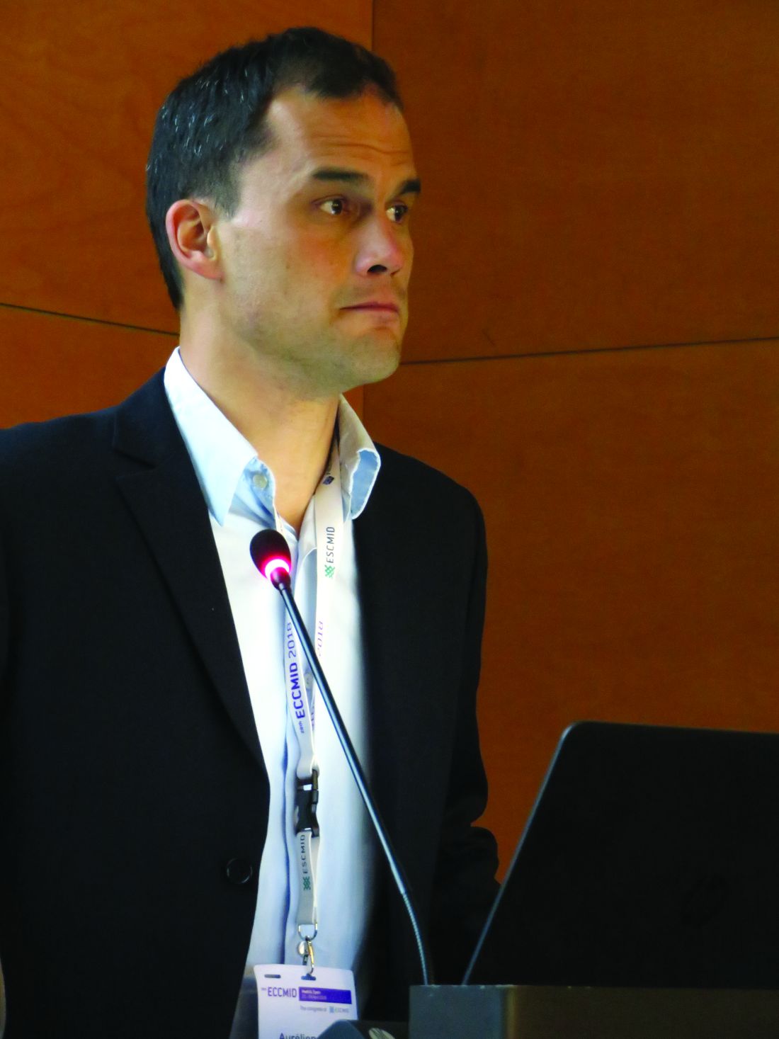
“Reducing treatment time now appears to be manageable and effective in a number of infectious diseases,” Dr. Dinh explained. “Although there are some limits, surely, this change in practice might lead to reduced rates of multidrug-resistant bacteria, fewer adverse events, and surely lower costs.”
The French PTC Trial (Short Duration Treatment of Non-Severe Community-Acquired Pneumonia) randomized 310 patients (mean age, 73.5 years) to either short- or long-course treatment with a beta-lactam antibiotic. Patients were eligible for the study if they were admitted to the hospital for community-acquired pneumonia based on respiratory signs, fever of 38° C or higher, and evidence of new infiltrate on chest radiograph.
All patients were treated with 3 days of amoxicillin/clavulanic acid (Augmentin) or third-generation cephalosporin. Those who had responded clinically by day 3 entered the 5-day randomization period, receiving placebo or 5 more days of active therapy with the same agent.
Clinical requirements for randomization included being afebrile with stable heart and respiratory rate, a systolic blood pressure of at least 90 mm Hg, and oxygen saturation of at least 90%.
The primary endpoint was clinical cure at day 15: no fever, absence of or improvement in respiratory symptoms (dyspnea, cough, purulent sputum, and cackles), and no need for additional antibiotic treatment for any indication.
Secondary endpoints were cure at day 30, 30-day mortality, adverse events, length of stay, return to usual activities by day 30, and quality of life at day 30.
Many of the generally elderly patient cohort had comorbid illnesses, including diabetes (about 20%), chronic obstructive pulmonary disease (about 35%), and coronary insufficiency (about 14%). About 20% were active smokers. Less than 10% had gotten a pneumococcal vaccine in the past 5 years.
At admission, more than half of patients were dyspneic, 80% had cough, and 39% had purulent sputum. The median PSI/PORT Score was 82.
After 3 days of treatment, clinical cure was not significantly different between the 3- and 8-day groups, either in the intent-to-treat analysis (69.9% vs. 61.2%) or in the per-protocol analysis (75.7% vs. 68.7%).
Because the trial had closed days before the ECCMID meeting, only the primary endpoints were available for discussion, Dr. Dinh said. Investigators are analyzing the secondary endpoint data, which he said would be published at a later date.
Despite the positive results, Dr. Dinh cautioned against using the study as justification for a one-size-fits-all treatment for community-acquired pneumonia.
“Although I think we demonstrated that 3 days of treatment with beta-lactam is not inferior to 8 days, this cannot be imposed without regard to individual patient status,” he cautioned. Such a treatment paradigm would not be advisable for patients with moderately severe pneumonia, who were excluded from the study, or those with compromised immune systems.
Nor does Dr. Dinh expect wholesale clinical embracing of the encouraging results, which bolster the ever-accumulating data in favor of shorter courses of antibiotics for some infectious diseases.
“I think there is a chance that clinicians who normally treat for 9 or 10 days may now feel comfortable reducing to 7,” he said with a chuckle.
The French Ministry of Health sponsored the study. Dr. Dinh had no competing financial interests.
SOURCE: Dinh et al. ECCMID 2018, Oral Abstract O1126.
MADRID – Three days of beta-lactam therapy was just as effective as 8 days for clinically stable patients presenting with community-acquired pneumonia.
In a randomized, placebo-controlled trial, 15-day cure rates were 69.9% in patients who took 3 days of antibiotics and 61.2% in those who took 8 days – a nonsignificant difference, Aurélien Dinh, MD, said at the European Society of Clinical Microbiology and Infectious Diseases annual congress.
“Reducing treatment time now appears to be manageable and effective in a number of infectious diseases,” Dr. Dinh explained. “Although there are some limits, surely, this change in practice might lead to reduced rates of multidrug-resistant bacteria, fewer adverse events, and surely lower costs.”
The French PTC Trial (Short Duration Treatment of Non-Severe Community-Acquired Pneumonia) randomized 310 patients (mean age, 73.5 years) to either short- or long-course treatment with a beta-lactam antibiotic. Patients were eligible for the study if they were admitted to the hospital for community-acquired pneumonia based on respiratory signs, fever of 38° C or higher, and evidence of new infiltrate on chest radiograph.
All patients were treated with 3 days of amoxicillin/clavulanic acid (Augmentin) or third-generation cephalosporin. Those who had responded clinically by day 3 entered the 5-day randomization period, receiving placebo or 5 more days of active therapy with the same agent.
Clinical requirements for randomization included being afebrile with stable heart and respiratory rate, a systolic blood pressure of at least 90 mm Hg, and oxygen saturation of at least 90%.
The primary endpoint was clinical cure at day 15: no fever, absence of or improvement in respiratory symptoms (dyspnea, cough, purulent sputum, and cackles), and no need for additional antibiotic treatment for any indication.
Secondary endpoints were cure at day 30, 30-day mortality, adverse events, length of stay, return to usual activities by day 30, and quality of life at day 30.
Many of the generally elderly patient cohort had comorbid illnesses, including diabetes (about 20%), chronic obstructive pulmonary disease (about 35%), and coronary insufficiency (about 14%). About 20% were active smokers. Less than 10% had gotten a pneumococcal vaccine in the past 5 years.
At admission, more than half of patients were dyspneic, 80% had cough, and 39% had purulent sputum. The median PSI/PORT Score was 82.
After 3 days of treatment, clinical cure was not significantly different between the 3- and 8-day groups, either in the intent-to-treat analysis (69.9% vs. 61.2%) or in the per-protocol analysis (75.7% vs. 68.7%).
Because the trial had closed days before the ECCMID meeting, only the primary endpoints were available for discussion, Dr. Dinh said. Investigators are analyzing the secondary endpoint data, which he said would be published at a later date.
Despite the positive results, Dr. Dinh cautioned against using the study as justification for a one-size-fits-all treatment for community-acquired pneumonia.
“Although I think we demonstrated that 3 days of treatment with beta-lactam is not inferior to 8 days, this cannot be imposed without regard to individual patient status,” he cautioned. Such a treatment paradigm would not be advisable for patients with moderately severe pneumonia, who were excluded from the study, or those with compromised immune systems.
Nor does Dr. Dinh expect wholesale clinical embracing of the encouraging results, which bolster the ever-accumulating data in favor of shorter courses of antibiotics for some infectious diseases.
“I think there is a chance that clinicians who normally treat for 9 or 10 days may now feel comfortable reducing to 7,” he said with a chuckle.
The French Ministry of Health sponsored the study. Dr. Dinh had no competing financial interests.
SOURCE: Dinh et al. ECCMID 2018, Oral Abstract O1126.
REPORTING FROM ECCMID 2018
Key clinical point: Three days of beta-lactam treatment were as effective as 8 days in curing clinically stable patients with community-acquired pneumonia.
Major finding: Cure rates at 15 days were 69.9% in the 3-day group, compared with 61.2% in the 8-day group, a nonsignificant difference.
Study details: The placebo-controlled study randomized 310 patients to treatment.
Disclosures: The French Ministry of Health sponsored the trial. Dr. Dinh had no financial disclosures.
Source: Dinh et al. ECCMID 2018, oral abstract O1126.
VIDEO: Pills alone not the answer for pain management
SANDESTIN, FLA. – More than ever, clinicians need to rely on a multimodal approach to pain management, Katherine Galluzzi, DO, said at the annual Congress of Clinical Rheumatology.
In the era of opioid addiction – in which she said physicians have sometimes been unfairly vilified – pharmaceutical options are limited not only by the threat of abuse but also by governmental regulation, explained Dr. Galluzzi, chair of geriatrics at the Philadelphia College of Osteopathic Medicine.
The underpinning of pain management in the future will need to be cognitive-behavioral therapy, such as changing behavior and meditation; physical approaches, such as exercise and acupuncture; and interventional treatments, such as nerve blocks and trigger-point injections. Pharmacotherapy can’t do it all, nor should it, she said.
“This is what we have, this is what we need to do,” Dr. Galluzzi said. “This impacts the quality of life, and patients need to begin providing self-care. It’s not going to come in the form of a pill. It has to be a commitment between the patient and the physician.”
The Centers for Medicare & Medicaid Services are proposing a new limit on opioid prescriptions for Medicare recipients – a maximum of 90 morphine mg equivalents per day for no more than 7 days. That will affect older people, who are most likely to be in need of pain management, she said. Those on hospice care and experiencing certain cancer pain will be exempt, she noted in an interview.
Concerns about addiction to drugs such as gabapentin and benzodiazepines might make these therapies less of an option in coming years, Dr. Galluzzi added.
Risk evaluation and mitigation strategy training is an important tool for helping physicians weigh the benefits and the risks of opioid prescriptions. Dr. Galluzzi particularly suggests enrolling in a 3-4 hour, in-person program, saying that it’s well worth the time.
“If you haven’t done a risk assessment and mitigation strategies course and you’re an opioid prescriber,” she said, “I highly recommend that you do that.”
EXPERT ANALYSIS FROM CCR 18
VIDEO: Characteristic flora define intestinal microbiome in scleroderma
SANDESTIN, FLA. – Scleroderma patients appear to have a characteristic microbiome composition, which is consistent in samples taken around the world.
These patients showed decreased populations of beneficial commensal flora and increased populations of proinflammatory species, Elizabeth Volkmann, MD, said at the annual Congress of Clinical Rheumatology.
Furthermore, specific species seem to correlate with specific gastrointestinal symptoms, said Dr. Volkmann of the University of California, Los Angeles. “Features also unexpectedly overlap with the consortium typical for Crohn’s disease, a disease with both inflammatory and fibrosing phenotype,” she said.
The video associated with this article is no longer available on this site. Please view all of our videos on the MDedge YouTube channel
Her recent exploration of this topic included 17 patients with scleroderma and GI symptoms and 17 matched healthy controls (BMJ Open Gastro. 2017;3:e000134). Everyone underwent a bowel prep and colonoscopy, during which cecum and sigmoid mucosal lavage samples were obtained. Those samples underwent RNA sequencing.
In addition to quantifying the species present, Dr. Volkmann sought to associate populations with symptoms. The primary assessment tool was the GIT 2.0, which measures distention/bloating; diarrhea; fecal soilage; constipation; emotional well-being; and social functioning.
Similar to the findings in inflammatory disease states, scleroderma patients had decreased levels of commensal Clostridia, a class of Firmicutes that is established in early infancy and very important in the maintenance of gut homeostasis. They also showed a decreased proportion of Faecalibacterium, a genus with anti-inflammatory activity; this finding has been observed in patients with Crohn’s disease.
Patients also showed relative increases in pathobionts. These are potentially pathological organisms that, under normal circumstances, live symbiotically. Janet Chow, PhD, who coined the term in a 2011 paper, said these species are typically proinflammatory (Curr Opin Immunol. 2011 Aug; 23[4]:473-80).
“Organisms proposed as pathobionts are associated with chronic inflammatory conditions – unlike opportunistic pathogens, which often cause acute infections and are typically acquired from the environment or other parts of the body. In addition, pathobionts are innocuous to the host under normal conditions,” wrote Dr. Chow of the California Institute of Technology, Pasadena.
In Dr. Volkmann’s study, Bifidobacterium and Lactobacillus, which are usually reduced in proinflammatory disorders, were relatively abundant in patients, compared with controls.
She noted specific associations with both symptoms. Parabacteroides and Enterobacteriaceae were associated with increased constipation. Prevotella was associated with increased diarrhea and increased distention/bloating.
Her results are consistent with a Swedish study (Arthritis Res Ther. 2016 Nov 1;18[1]:278) and three Italian studies conducted in Rome, Milan, and Piacenza.
“It’s fascinating that we seem to be identifying a consistent microbiome profile for scleroderma patients,” Dr. Volkmann said.
Dr. Volkmann had no relevant financial disclosures.
REPORTING FROM CCR 18
Malignant pleural mesothelioma guidelines often are ignored
SAN DIEGO – National guidelines for the treatment of malignant pleural mesothelioma often are not followed, a new study showed, with fewer than one-third of patients receiving cancer-directed surgery.
Another 32% received no treatment, although that didn’t seem to have an impact on median months of survival.
Still, “there can be a wide variation in median survival time, depending on clinical factors and tumor characteristics,” said study coauthor Harmik Soukiasian, MD, of Cedars-Sinai Medical Center, Los Angeles. “Given the variation in prognosis, it is quite astonishing that over 30% of MPM patients are not receiving any form of treatment. As clinicians armed with these data, we need to investigate why that is.”
Dr. Soukiasian presented the study findings at the annual meeting of the American Association for Thoracic Surgery.
MPM, a rare cancer, is mainly linked to asbestos exposure. “MPM is almost always a fatal disease, and the prognosis can only be modestly influenced by oncological treatments,” according to the authors of guidelines released in 2013. “The diagnostic process can be complex, with highly specialized advice frequently required to arrive at a definite diagnosis. Treatment varies from therapeutic nihilism to radical combined-modality treatment approaches” (J Thorac Dis. 2013 Dec;5[6]:E254-E307).
Surgical resection is a controversial treatment for MPM, Dr. Soukiasian said. It is “based on the principle of macroscopic resection of solid tumor with adjuvant therapy to treat micrometastatic disease,” he explained. “Cancer-directed surgery for MPM is usually reserved for localized epithelial type histology and is associated with a 5-year survival rate of 15%.”
For the new study, the investigators tracked 3,834 patients in the National Cancer Database (2004-2014) diagnosed with MPM clinical stages I-III. Most had epithelioid MPM (69%), with sarcomatoid (17%) and mixed subtype (15%) making up the rest. They examined whether patient treatment complied with the National Comprehensive Cancer Network (NCCN) guidelines, which recommend surgery in resectable epithelioid MPM.
“Our study revealed significant lack of compliance with NCCN guidelines, as well as many disparities in the management of MPM,” Dr. Soukiasian said. “For the overall cohort, 32.3% of patients did not receive any treatment, 18.1% had surgery plus chemotherapy, 38.6% chemotherapy alone, and only 7% received trimodality therapy. In patients with epithelial histology, surgery was significantly underutilized, with only 30% of patients receiving cancer-directed surgery.”
In addition, he said, “our study reveals several disparities that affect compliance with NCCN guidelines. Treatment disparities were observed in women, octogenarians, the uninsured, the Medicaid-insured, and in patients with comorbidities. Guideline adherence was significantly increased in academic and high-volume hospitals with an associated increase in survival.”
But the study also found that median survival estimates were similar regardless of treatment: 10 months for no treatment, 15 months for chemotherapy only, 17 months for surgery only, and 22 months for surgery plus chemotherapy.
During the AATS presentation, an audience member asked about how performance status – a measure of a person’s ability to perform everyday activities – affects the eligibility for surgery.
“It’s quite common for low performance status to exclude someone from surgery,” the audience member said. “Some of these patients are very sick.”
Dr. Soukiasian acknowledged that performance status was not included in the data. The study was focused on the gap between guidelines and real-world practice, and generated questions of why and about the potential opportunity for improved treatment of these patients.
How do patient choices, cost, and quality of life factor in? “These are very important questions and concerns,” Dr. Soukiasian said. “Although our research does not provide data or conclusions on quality of life or cost, these topics will be important to address in follow-up studies to elucidate possible barriers in the treatment of MPM and the initiation of future educational opportunities for our patients.”
No disclosures and no study funding were reported.
SOURCE: Espinoza-Mercado F et al. General Thoracic Surgery Simultaneous Scientific Session. Abstract 18.
REPORTING FROM THE AATS ANNUAL MEETING
Key clinical point:
Major finding: Guidelines recommend surgery in epithelioid MPM, but only 30% of patients received it.
Study details: Analysis of 3,834 patients diagnosed with MPM clinical stages I-III during 2004-2014.
Disclosures: No disclosures and no study funding were reported.
Source: Espinoza-Mercado F et al. General Thoracic Surgery Simultaneous Scientific Session. Abstract 18.
Novel x-ray score distinguishes psoriatic arthritis from osteoarthritis of the hand
LIVERPOOL, ENGLAND – A novel radiologic scoring system differentiated psoriatic arthritis (PsA) from nodal osteoarthritis (OA) of the hand in a pilot study.
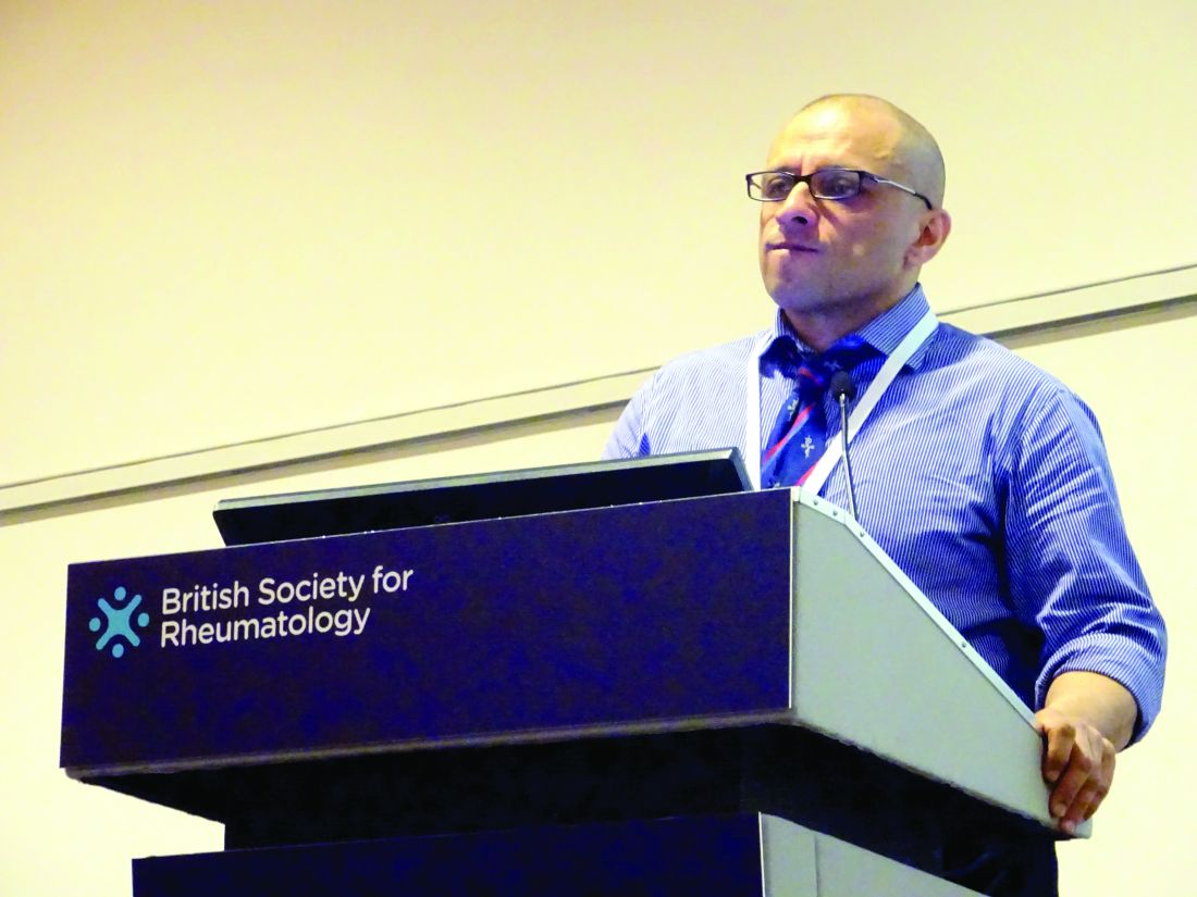
“It’s a dilemma that’s faced, perhaps every couple of weeks, by most [rheumatologists]: Is it osteoarthritis or is it early psoriatic arthritis?” said Sardar Bahadur, MD, at the British Society for Rheumatology annual conference.
Both conditions are seen in daily practice, although the prevalence of hand OA is less frequent than knee OA. Approximately one in five of all adults in the United Kingdom have OA and 1%-2% have psoriasis. Of these, the prevalence of hand OA is about 11% and 0.1%-0.3% have psoriatic arthritis.
Being able to differentiate between the two conditions has important consequences for treatment, Dr. Bahadur said.
“Getting the diagnosis wrong could have major implications,” he said. “If you miss psoriatic arthritis, then potentially you are going to find irreversible joint damage causing pain and disability, and the opposite is also true, with misdiagnosis of osteoarthritis, with overuse of immunosuppression and all the cost implications as well as medicolegal consequences.”
Dr. Bahadur of the department of rehabilitation medicine and rheumatology at the Defence Medical Rehabilitation Centre Headley Court, in Epsom, England, added: “So early diagnosis is very important, it means early treatment, it means better care, potentially preventing serious and irreversible damage.”
Together with researchers at Guy’s and St Thomas’ NHS Trust, London, Dr. Bahadur hypothesized that changes in hand x-rays were distinct and could be reliably used to differentiate between the two conditions. They developed a scoring system for hand radiographs that looked at the differences in the interphalangeal joints, soft tissue, and bone features of patients with known OA or PsA.
Dr. Bahadur noted that the aim was to focus on plain film radiographs of the hands because these were inexpensive, universally accessible, did not rely on radiologists’ interpretation, and changes in the hands were known to occur in both OA and PsA.
A total of 99 sets of hand x-rays taken between 2008 and 2016 from 50 patients with OA and 49 patients with PsA were obtained. These were anonymized and then analyzed by a musculoskeletal radiologist using the scoring system the team had developed. The radiologist was unaware of the patients’ clinical status. The results were then compared to the clinical diagnosis.
The novel method of scoring each x-ray was then taught to two rheumatology and one radiology trainee during a 1-hour training session and were then asked to score the same radiographs.
Dr. Bahadur reported that the radiologist reported normal hand radiographs in five patients and, of the remaining 94 sets of left- and right-hand radiographs, the scoring system correctly allocated 100% of images to either PsA, OA, or rheumatoid arthritis (RA).
Of note was that the radiologist correctly identified two patients with nodal hand OA who later developed PsA several years later, and one patient with RA who was initially thought to have PsA.
“The system could be successfully used by nonradiologists,” Dr. Bahadur proposed. There was good agreement between the scoring system results and the clinical diagnosis then used by the trainees, with 88% and 67% of the radiographs correctly matched to the clinical diagnosis by the rheumatology trainees, and 70% for the radiology trainee.
Dr. Bahadur noted that the features that were consistently identified as being different between hand OA and PsA patients were soft tissue changes, such as dactylitis, as well as erosions, new bone formation, and other features such as subchondral surface changes and cysts.
The results of this single-center study show that the novel radiologic scoring system of hand radiographs was effective at differentiating patients with PsA from nodal OA.
“The ambition is to make this usable by nonradiologists,” Dr. Bahadur said. A multicenter trial would be the next step to look at the use of the scoring system.
Dr. Bahadur had no conflicts of interest to disclose.
SOURCE: Bahadur S et al. Rheumatology. 2018;57[Suppl. 3]:key075.184
LIVERPOOL, ENGLAND – A novel radiologic scoring system differentiated psoriatic arthritis (PsA) from nodal osteoarthritis (OA) of the hand in a pilot study.
“It’s a dilemma that’s faced, perhaps every couple of weeks, by most [rheumatologists]: Is it osteoarthritis or is it early psoriatic arthritis?” said Sardar Bahadur, MD, at the British Society for Rheumatology annual conference.
Both conditions are seen in daily practice, although the prevalence of hand OA is less frequent than knee OA. Approximately one in five of all adults in the United Kingdom have OA and 1%-2% have psoriasis. Of these, the prevalence of hand OA is about 11% and 0.1%-0.3% have psoriatic arthritis.
Being able to differentiate between the two conditions has important consequences for treatment, Dr. Bahadur said.
“Getting the diagnosis wrong could have major implications,” he said. “If you miss psoriatic arthritis, then potentially you are going to find irreversible joint damage causing pain and disability, and the opposite is also true, with misdiagnosis of osteoarthritis, with overuse of immunosuppression and all the cost implications as well as medicolegal consequences.”
Dr. Bahadur of the department of rehabilitation medicine and rheumatology at the Defence Medical Rehabilitation Centre Headley Court, in Epsom, England, added: “So early diagnosis is very important, it means early treatment, it means better care, potentially preventing serious and irreversible damage.”
Together with researchers at Guy’s and St Thomas’ NHS Trust, London, Dr. Bahadur hypothesized that changes in hand x-rays were distinct and could be reliably used to differentiate between the two conditions. They developed a scoring system for hand radiographs that looked at the differences in the interphalangeal joints, soft tissue, and bone features of patients with known OA or PsA.
Dr. Bahadur noted that the aim was to focus on plain film radiographs of the hands because these were inexpensive, universally accessible, did not rely on radiologists’ interpretation, and changes in the hands were known to occur in both OA and PsA.
A total of 99 sets of hand x-rays taken between 2008 and 2016 from 50 patients with OA and 49 patients with PsA were obtained. These were anonymized and then analyzed by a musculoskeletal radiologist using the scoring system the team had developed. The radiologist was unaware of the patients’ clinical status. The results were then compared to the clinical diagnosis.
The novel method of scoring each x-ray was then taught to two rheumatology and one radiology trainee during a 1-hour training session and were then asked to score the same radiographs.
Dr. Bahadur reported that the radiologist reported normal hand radiographs in five patients and, of the remaining 94 sets of left- and right-hand radiographs, the scoring system correctly allocated 100% of images to either PsA, OA, or rheumatoid arthritis (RA).
Of note was that the radiologist correctly identified two patients with nodal hand OA who later developed PsA several years later, and one patient with RA who was initially thought to have PsA.
“The system could be successfully used by nonradiologists,” Dr. Bahadur proposed. There was good agreement between the scoring system results and the clinical diagnosis then used by the trainees, with 88% and 67% of the radiographs correctly matched to the clinical diagnosis by the rheumatology trainees, and 70% for the radiology trainee.
Dr. Bahadur noted that the features that were consistently identified as being different between hand OA and PsA patients were soft tissue changes, such as dactylitis, as well as erosions, new bone formation, and other features such as subchondral surface changes and cysts.
The results of this single-center study show that the novel radiologic scoring system of hand radiographs was effective at differentiating patients with PsA from nodal OA.
“The ambition is to make this usable by nonradiologists,” Dr. Bahadur said. A multicenter trial would be the next step to look at the use of the scoring system.
Dr. Bahadur had no conflicts of interest to disclose.
SOURCE: Bahadur S et al. Rheumatology. 2018;57[Suppl. 3]:key075.184
REPORTING FROM RHEUMATOLOGY 2018
Key clinical point:
Major finding: Using the scoring system, 100% of images were correctly allocated to PsA, OA, or RA.
Study details: Single center pilot study assessing 99 x-rays of both hands taken between 2008 and 2016 of patients with OA (n = 50) or PsA (n = 49).
Disclosures: Dr. Bahadur had no conflicts of interest to disclose.
Source: Bahadur S et al., Rheumatology. 2018;57[Suppl. 3]:key075.184
Patidegib, the first topical hedgehog inhibitor, scores in Gorlin syndrome
CHICAGO – in a phase 2 study, and did so without causing the problematic adverse events that prompt many Gorlin patients to discontinue systemic hedgehog inhibitor drugs, Ervin Epstein Jr., MD, said at the annual meeting of the American College of Mohs Surgery.
He characterized patidegib, a small-molecule cyclopamine derivative, as “a Goldilocks drug, a topical hedgehog inhibitor with percutaneous absorption that’s just right: sufficient to have anti-hedgehog and anti–basal cell carcinoma efficacy, but not enough to cause systemic exposure or systemic adverse events.”

A complete response – tumor clinical disappearance – occurred in 25% of the BCCs in the two active treatment arms. In contrast, none of the BCCs in the control group cleared. Patients on patidegib developed one or more new surgically eligible BCCs after study week 2 at a rate of 0.4 tumors per patient, markedly less than the rate of 1.4 tumors per patient in controls.
“We think that prevention is really the place to go with this drug,” said Dr. Epstein, cofounder and chief medical officer at PellePharm, the company based in Menlo Park, Calif., that is developing patidegib.
Indeed, he envisions patidegib gel as lifetime therapy for Gorlin patients.
Tumor shrinkage was significantly greater with 2% patidegib than with the 4% concentration. But so was treatment adherence: Patients in the 2% patidegib arm missed on average just 2 days of therapy over the course of 6 months, while those in the 4% arm missed 50 days. Dr. Epstein attributed this discrepancy to a freak of randomization that can occur in such a small study: 5 of 6 patients in the 2% patidegib group were women, while most in the 4% arm were men. And the men were far less adherent to treatment, possibly because men are less accustomed to applying a product on their face daily, he noted.
In any case, it’s the 2% gel formulation that is moving on to a phase 3, double-blind, randomized trial. The 150-patient U.S. and European study, scheduled to start this summer, will have as its primary endpoint the rate of new BCCs over the course of 1 year. The Food and Drug Administration has granted topical patidegib Breakthrough Drug and Orphan Drug status.
Dr. Epstein said that, in the phase 2 study, BCC shrinkage occurred only in patients whose hedgehog pathway activity decreased after 6 weeks of topical therapy as evidenced by a reduction in the GLI1 mRNA biomarker. Of note, circulating blood levels of patidegib in study participants were more than 500-fold lower than when the drug is given orally. And of greatest importance, rates of the hallmark side effects of oral hedgehog inhibitor therapy that cause so many patients to discontinue therapy – muscle cramps, taste loss, and hair loss – were no different in patients on patidegib gel than in those on placebo.
“In the randomized trial of vismodegib [Erivedge], half of patients stopped taking it within 1 year despite good results. And indeed, when they stopped taking the drug, the basal cell carcinomas returned,” the dermatologist noted.
Dr. Epstein is an employee of PellePharm, which is developing patidegib. Dermatologists with patients with Gorlin syndrome who are interested in participating in the phase 3 trial can contact him at [email protected].
CHICAGO – in a phase 2 study, and did so without causing the problematic adverse events that prompt many Gorlin patients to discontinue systemic hedgehog inhibitor drugs, Ervin Epstein Jr., MD, said at the annual meeting of the American College of Mohs Surgery.
He characterized patidegib, a small-molecule cyclopamine derivative, as “a Goldilocks drug, a topical hedgehog inhibitor with percutaneous absorption that’s just right: sufficient to have anti-hedgehog and anti–basal cell carcinoma efficacy, but not enough to cause systemic exposure or systemic adverse events.”
A complete response – tumor clinical disappearance – occurred in 25% of the BCCs in the two active treatment arms. In contrast, none of the BCCs in the control group cleared. Patients on patidegib developed one or more new surgically eligible BCCs after study week 2 at a rate of 0.4 tumors per patient, markedly less than the rate of 1.4 tumors per patient in controls.
“We think that prevention is really the place to go with this drug,” said Dr. Epstein, cofounder and chief medical officer at PellePharm, the company based in Menlo Park, Calif., that is developing patidegib.
Indeed, he envisions patidegib gel as lifetime therapy for Gorlin patients.
Tumor shrinkage was significantly greater with 2% patidegib than with the 4% concentration. But so was treatment adherence: Patients in the 2% patidegib arm missed on average just 2 days of therapy over the course of 6 months, while those in the 4% arm missed 50 days. Dr. Epstein attributed this discrepancy to a freak of randomization that can occur in such a small study: 5 of 6 patients in the 2% patidegib group were women, while most in the 4% arm were men. And the men were far less adherent to treatment, possibly because men are less accustomed to applying a product on their face daily, he noted.
In any case, it’s the 2% gel formulation that is moving on to a phase 3, double-blind, randomized trial. The 150-patient U.S. and European study, scheduled to start this summer, will have as its primary endpoint the rate of new BCCs over the course of 1 year. The Food and Drug Administration has granted topical patidegib Breakthrough Drug and Orphan Drug status.
Dr. Epstein said that, in the phase 2 study, BCC shrinkage occurred only in patients whose hedgehog pathway activity decreased after 6 weeks of topical therapy as evidenced by a reduction in the GLI1 mRNA biomarker. Of note, circulating blood levels of patidegib in study participants were more than 500-fold lower than when the drug is given orally. And of greatest importance, rates of the hallmark side effects of oral hedgehog inhibitor therapy that cause so many patients to discontinue therapy – muscle cramps, taste loss, and hair loss – were no different in patients on patidegib gel than in those on placebo.
“In the randomized trial of vismodegib [Erivedge], half of patients stopped taking it within 1 year despite good results. And indeed, when they stopped taking the drug, the basal cell carcinomas returned,” the dermatologist noted.
Dr. Epstein is an employee of PellePharm, which is developing patidegib. Dermatologists with patients with Gorlin syndrome who are interested in participating in the phase 3 trial can contact him at [email protected].
REPORTING FROM THE ACMS ANNUAL MEETING