User login
Former Smokers Motivate Quitters
In 2012, the CDC launched the “Tips from Former Smokers” campaign. It was memorable and emotionally forceful—one woman who had oral and throat cancer delivered her ad through an artificial voicebox—but did it have an impact on actual quitting rates?
No study had been done to assess the campaign’s combined, multiyear impact until CDC researchers looked at sustained (6 month) cigarette abstinence during the first 4 years of the campaign (2012-2015).
They found that the Tips campaign led to about 9.15 million total quit attempts. Based on an assumed 5.7% abstinence rate for people attempting to quit, this amounts to approximately 522,000 sustained quits.
The researchers say their findings indicate that the comprehensive approach combining evidence-based messages with the promotion of cessation resources was highly successful. Their finding of more than half-million sustained quits underscores the critical role of national tobacco education campaigns as a “counterpoint” to the substantial pro-tobacco advertising and promotion.
Pros and Cons of Telemental Health Care
When telemental health care (TMH) works, it works well, the research agrees. For rural patients who often do not have easy access to health care TMH can be a lifesaver. The VA uses TMH to deliver care to veterans in rural VA medical centers, community-based outpatient clinics, and residential areas. However, TMH is still relatively new in many rural communities, say researchers from University of Mississippi in Oxford and Augusta University in Georgia, and few studies have examined the delivery tool from an administrative standpoint. The literature suggests TMH will save money—the exploratory study, however, suggests otherwise.
The researchers interviewed 6 providers selected from the 15 community mental health (MH) centers (CMHCs) in rural Mississippi as well as an independent MH counselor who develops policy for the Mississippi Counselors Association. They asked respondents about the feasibility of TMH in the Mississippi Delta; in particular, the benefits, the costs, and the role of the state in facilitating the service. The researchers also collected data from a grant-funded pilot project conducted in the Mississippi Delta region by the Delta Health Alliance, a nonprofit organization in partnership with the University of Mississippi Medical Center, which ran from 2008 to 2011. Telepsychiatry sessions are not currently being used in the region, but before the project ended, it was responsible for > 1,000 videoconferencing clinical sessions.
The initial counseling sessions were “awkward” for some patients, the interviewees said, and some clients felt the consultation was “less personal.” Getting used to the technology may take some time. Once clients acclimated the feedback was positive.
The health care providers were concerned by not being able to observe in-person nonverbal clues, such as poor hygiene, that would normally help them evaluate the client’s health. The nurse at the CMHC helped fill a gap, the researchers say, created by technology.
The researchers determined that the benefit side was weighty: For instance, patients had better access to well-trained MH professionals and to the state hospital, and family could visit inpatients via videoconferencing. Staff had better access to professional development and training.
However, cost issues were a definite concern. The project would not have been feasible without grant funding, the researchers say. Medicaid reimburses for TMH services but not for technology setup costs and maintenance. Moreover, the interviews with administrators, the researchers say, indicated that TMH did not save the organization money. Costs for the equipment, installation, rent, and other supplies were prohibitive.
Although start-up costs are high, overall systematic costs go down, with savings on travel-related costs, including fewer missed appointments. Broadband technology, videoconferencing software, webcams, and education all take money. “If policymakers are serious” about TMH, the researchers conclude, they should allocate appropriate funding and resources.
Source:
Holland J, Hatcher W, Meares WL. J Health Hum Serv Adm. 2018;41(1):52-86.
Drug approved for radical cure of P vivax malaria
The US Food and Drug Administration (FDA) has approved tafenoquine (Krintafel) for the radical cure of Plasmodium vivax malaria.
Tafenoquine is a single-dose medicine that is now approved to prevent relapse of P vivax malaria in patients age 16 and older who are receiving appropriate antimalarial therapy for acute P vivax infection.
Tafenoquine is the first new treatment approved for P vivax malaria in more than 60 years.
Tafenoquine is an 8-aminoquinoline derivative with activity against all stages of the P vivax lifecycle, including hypnozoites. The product was first synthesized by scientists at the Walter Reed Army Institute of Research in 1978.
GSK began developing tafenoquine as a potential medicine for malaria more than 20 years ago. In 2008, GSK entered into a collaboration with Medicines for Malaria Venture to develop tafenoquine as an anti-relapse medicine for patients infected with P vivax.
The primary evidence for the clinical efficacy and safety of the 300 mg, single dose of tafenoquine was provided by a pair of phase 3 studies—DETECTIVE (NCT01376167, TAF112582) and GATHER (NCT02216123, TAF116564).
Results from these studies were presented at the 6th International Conference on Plasmodium vivax Research (ICPVR) in 2017 (abstract 63245 and abstract 63246).
DETECTIVE trial
In this double-blind, double-dummy study, researchers evaluated the efficacy, safety, and tolerability of tafenoquine. The trial included 522 patients with P vivax malaria who were randomized to receive one of the following:
- A single dose (1 day) of tafenoquine (300 mg)
- A 14-day course of primaquine (15 mg)
- Placebo.
All patients also received a 3-day course of chloroquine to treat the acute blood stage of the infection.
A significantly greater proportion of patients remained relapse-free over the 6-month follow-up period if they were treated with tafenoquine rather than placebo—60% and 26%, respectively—with an odds ratio for risk of relapse of 0.24 (P<0.001).
Likewise, a significantly greater proportion of patients were relapse-free when treated with primaquine rather than placebo—64% and 26%, respectively—with an odds ratio of 0.20 (P<0.001).
The frequency of adverse events (AEs) was 63% for the tafenoquine group, 59% for the primaquine group, and 65% for the placebo group. The frequency of serious AEs was 8%, 3%, and 5%, respectively.
GATHER trial
This study enrolled 251 patients, ages 16 and older, with microscopy-confirmed parasitemia.
Researchers compared how a single dose of tafenoquine (300 mg) and a 14-day course of primaquine (15 mg) affected hemoglobin levels in these patients. All patients also received a standard 3-day course of chloroquine.
The incidence of decline in hemoglobin (the primary endpoint) was similar between the 2 treatment groups—2.4% in the tafenoquine arm and 1.2% in the primaquine arm. The difference in proportion was 1.23% (95% CI, -4.16%, 4.98%).
None of the patients in this study required a blood transfusion.
The frequency of AEs was 72% for the tafenoquine group and 75% for the primaquine group. The frequency of serious AEs was 4% and 1%, respectively.
Team finds potential therapeutic targets for T-ALL

Researchers have found the NOTCH1 pathway “hijacks” heat shock transcription factor 1 (HSF1) signaling in T-cell acute lymphoblastic leukemia (T-ALL).
Therefore, blocking one or more genes in the HSF1 pathway could represent a new approach to treating T-ALL.
An experimental drug, PU-H71, is already in development against one of these targets, heat shock protein 90 (HSP90).
The researchers found that PU-H71 was active against T-ALL in vitro and in vivo.
The team recounted these findings in Nature Medicine.
“Our study shows how the NOTCH1 pathway hijacks the heat shock transcription factor 1 pathway to promote tumor growth,” said study author Iannis Aifantis, PhD, of NYU School of Medicine in New York, New York.
“The cancer cells are sending into overdrive a system that helps healthy cells respond to stress.”
Dr Aifantis and his colleagues found that HSF1 is involved in the pathogenesis of T-ALL. When they knocked down HSF1 in T-ALL cell lines, the researchers observed an increase in apoptosis, defective proteostasis, and a decrease in the growth of leukemic cells.
Similarly, HSF1 was deemed necessary for disease progression in mouse models of T-ALL. When the researchers deleted HSF1, mice experienced “striking” reductions in leukemic burden and “dramatic” improvements in survival. However, HSF1 deletion did not affect normal hematopoiesis.
Dr Aifantis and his colleagues also showed that NOTCH1 regulates the epichaperome in T-ALL. The team said previous studies have shown that, in the presence of oncogenic stress, heat shock proteins participate in a network nucleated by HSP90 and HSP70 chaperones—the epichaperome.
The researchers found that an intact epichaperome was critical for T-ALL by showing that pharmacologic inhibition of HSP90 and HSP70 significantly hindered the growth of human T-ALL in vitro. The team also found the HSP90 inhibitor PU-H71 reduced leukemic burden and extended survival in a NOTCH1-inducible T-ALL mouse model.
Then, the researchers found NOTCH1 levels could predict response to HSP90 inhibition in vitro. T-ALL patient samples expressing high levels of nuclear NOTCH1 and high levels of epichaperome were significantly more sensitive to treatment with PU-H71.
PU-H71 is already in early clinical trials of patients with breast cancer. If further testing proves successful, PU-H71 could be quickly adapted for trials in T-ALL patients, according to Dr Aifantis.
In the meantime, he and his colleagues plan to evaluate the effects of another 8 proteins produced by genes active in the HSF1 pathway to see if any show promising anticancer activity in T-ALL.
GPS receives fast track designation for MM
The US Food and Drug Administration (FDA) has granted fast track designation to the cancer vaccine galinpepimut-S (GPS) for the treatment of multiple myeloma (MM).
GPS consists of 4 modified peptide chains that induce an innate immune response (CD4+/CD8+ T cells) against the Wilms’ tumor 1 (WT1) antigen.
GPS is administered in combination with an adjuvant and an immune modulator to improve the immune response to the target.
GPS has been tested in a phase 2 trial of patients with MM. Results from this trial were presented at the 44th Annual Meeting of the EBMT in March.
Phase 2 trial
The study enrolled 19 MM patients. Fifteen of them had high-risk cytogenetics at diagnosis, and 18 were at least minimal residual disease-positive after autologous stem cell transplant (ASCT).
Patients began receiving GPS within 22 days of ASCT. Initially, they received 6 doses (administered subcutaneously with the oil emulsifier montanide) every 2 weeks. Injection sites were pre-stimulated with granulocyte-macrophage colony-stimulating factor (70 μg) on days -2 (± 1 day) and 0 of each GPS vaccination.
The patients underwent assessment 2 to 4 weeks after the 6th GPS dose. Then, they received 6 additional monthly doses of GPS in conjunction with lenalidomide maintenance (10 mg daily), starting on day 100 post-ASCT. Patients were assessed 2 to 4 weeks after the 12th GPS dose.
The researchers found that GPS stimulated time-dependent CD4+ or CD8+ T-cell immune responses specific for all 4 WT1 peptides within GPS, 2 of which are heteroclitic.
Immune responses were confirmed in up to 91% of patients, with multivalent immune responses in up to 64% of patients. Three-quarters of patients had multifunctional cross-epitope T-cell reactivity to antigenic epitopes against which the hosts were not specifically immunized, in a pattern akin to epitope spreading.
In patients who received all 12 doses of GPS (n=12), there was a “strong” association between clinical benefit—defined as complete response (CR) or very good partial response (VGPR)—and frequency of CD4/CD8 immune responses.
Of those patients who had achieved CR/VGPR upon completion of GPS treatment, 100% (n=11) had CD4 immune responses, and 81.8% (n=9) had CD8 immune responses.
The median progression-free survival was 23.6 months, and the median overall survival was not reached. At 18 months, the rate of progression-free survival was 62%, and the rate of overall survival was 88%.
About fast track designation
The FDA’s fast track development program is designed to expedite clinical development and submission of applications for products with the potential to treat serious or life-threatening conditions and address unmet medical needs.
Fast track designation facilitates frequent interactions with the FDA review team, including meetings to discuss the product’s development plan and written communications about issues such as trial design and use of biomarkers.
Products that receive fast track designation may be eligible for accelerated approval and priority review if relevant criteria are met. Such products may also be eligible for rolling review, which allows a developer to submit individual sections of a product’s application for review as they are ready, rather than waiting until all sections are complete.
Pregnancy and years of reproductive capability linked to dementia risk
CHICAGO – More pregnancies and a longer span of reproductive years appear to confer some level of protection against dementia, suggesting that lifetime estrogen exposure may be an important modulator of long-term cognitive health.
The study, presented at the Alzheimer’s Association International Conference, is the first to examine this question in a large cohort, comprising more than 14,500 women with up to 50 years of follow-up data. It is one of the first explorations in a renewed interest in the link between hormones and cognition, said Suzanne Craft, PhD, who moderated a press briefing on the topic.
“WHI [Women’s Heath Initiative] had a completely chilling effect on research into estrogen and cognition completely,” Dr. Craft of Wake Forest University, Winston-Salem, N.C., said in an interview. “Many well-regarded researchers simply couldn’t get funding and had to close their programs and move on to other areas. But now I think the pendulum is slowly moving back. It’s clear that something is going on, that there is a link. I’m glad we’re starting to explore this again.”
The investigation of women’s total reproductive experience affects dementia risk found several intriguing associations, said Paola Gilsanz, ScD, of Kaiser Permanente, Oakland, Calif. Earlier age of menarche, later menopause, and more completed pregnancies all independently reduced the risk of dementia in women.
Her study comprised 14,595 women, all members of the Kaiser Permanente health care database. All of them had completed a comprehensive health checkup sometime between 1964 and 1973, when they were 40-55 years old. They reported their number of miscarriages, number of children, ages at first and last menstrual period, and the total number of years in their reproductive period. Most of the group (68%) was white, but 16% were black, 6% Asian, and 5% Hispanic. Dr. Gilsanz looked at rates of dementia during 1996-2017, when the women were 62-86 years old.
A multivariate regression model controlled for age, race, education, midlife health issues (hypertension, smoking, and body mass index), hysterectomy, and late-life health issues (stroke, heart failure, and diabetes).
Half of the cohort (50%) had at least three children, and 75% had experienced at least one miscarriage. The average age at menarche was 13 years, and the average age at last natural menstrual period was 47 years. This equated to an average reproductive period of 34 years.
At the end of follow-up, 36% of the cohort had developed some type of dementia.
Women with at least three children were 12% less likely to develop dementia, compared with those with one child (hazard ratio, 0.88). The association remained significant even after researchers controlled for age, race, educational level, and hysterectomy.
Miscarriages also influenced the risk of dementia. Those who didn’t report one were 20% less likely to develop dementia than were those who had experienced at least one (HR, 0.81). The benefit of no miscarriage was even greater among women with at least three children, conferring a 28% reduced risk (HR, 0.72).
A shortened reproductive period increased the risk of dementia. Those who experienced menarche at 16 years or older had a 31% increased risk of dementia (HR, 1.31), and those who experienced their last period at age 45 or younger faced a 28% greater risk (HR, 1.28). Each additional year of reproductive capability was associated with a 2% decreased risk (HR, 0.98).
Women with a reproductive period of 21-30 years were 33% more likely to develop dementia than were those with a longer period (HR, 1.33)
“Reproductive events that signal different exposures to estrogen, like pregnancy and reproductive period may play a role in modulating dementia risk,” Dr. Gilsanz said. “Women who are less likely to have a miscarriage may have different hormonal milieu that may be neuroprotective. Underlying health conditions increasing the risk of miscarriages may also elevate risk of dementia.”
She had no financial disclosures.
SOURCE: Gilsanz P et al. AAIC 2018, Abstract P3-587
AT AAIC 2018
Key clinical point: More pregnancies and a longer reproductive period were associated with a significantly reduced risk of late-life dementia among women.
Major finding: Women with at least three children were 12% less likely to develop dementia than were those who had just one child.
Study details: The retrospective observational study comprised 14,595 women.
Disclosures: Dr. Gilsanz had no financial disclosures.
Source: Gilsanz P et al. AAIC 2018, Abstract P3-587.
Hypopigmentation on the Ear
The Diagnosis: Corticosteroid-Induced Hypopigmentation
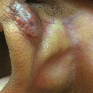
This patient received several intralesional injections of triamcinolone acetonide once monthly for treatment of the keloid scar on the left ear at an outside institution. There was improvement in the size of the keloid over time. On physical examination during the most recent visit there was a prominent streak of hypopigmentation and atrophy near the corticosteroid injection site with extension to the postauricular region. There also was telangiectasia noted within the area of hypopigmentation. Intralesional triamcinolone injections were discontinued and the patient was advised to return for monitoring.
Intra-articular and intralesional corticosteroid injections frequently are used by clinicians. Cutaneous complications associated with these injections include atrophy, pigmentary changes, hypersensitivity reactions, flushing, cellulitis, and necrotizing fasciitis. Tendon rupture also has been reported.1
There are several case reports in the literature describing hypopigmentation and/or subcutaneous atrophy after intralesional or intra-articular corticosteroid injections. A variety of underlying conditions were treated including alopecia areata, keloids, rheumatoid arthritis, de Quervain tendonitis, and psoriasis.2-6 The lesions typically are described as linear rays of atrophy and hypopigmentation at or near the injection site, with some cases noting extension along lymph channels and proximal veins.4,6 There usually is no associated pruritus or pain.3 This phenomenon can be seen after single or multiple injections.4,6
Extension of hypopigmentation from the site of injection has been postulated to be due to venous or lymphatic uptake.2,4-6 The mechanism of hypopigmentation is not known. Biopsy of a previously described case showed intact melanocytes along the dermoepidermal junction.2 Biopsy from another case revealed a decrease in melanin staining, which suggests a decrease in number or activity of melanocytes.4 It was proposed that hypopigmentation was secondary to loss of melanocyte function instead of loss of melanocytes.2 Spontaneous improvement or resolution of the hypopigmentation were noted in some cases ranging from 1 month to 1 year after initial presentation, but the hypopigmentation also can be persistent.3-6
Hypopigmented sarcoidosis and hypopigmented mycosis fungoides, both often present on dark-skinned individuals, are included in the differential diagnosis. Hypopigmented sarcoidosis presents with hypopigmented macules or patches, some with central papules, and hypopigmented mycosis fungoides presents with hypopigmented patches or plaques with fine scale and onset often in childhood or adolescence.7,8 Morphea can present with an initial inflammatory stage that develops into a sclerotic firm plaque or nodule with hyperpigmentation or hypopigmentation.9 Vitiligo usually presents with depigmented macules or patches and depigmented hair within the lesion.10
- Brinks A, Koes BW, Volkers AC, et al. Adverse effects of extra-articular corticosteroid injections: a systematic review. BMC Musculoskelet Disord. 2010;11:206.
- Venkatesan P, Fangman WL. Linear hypopigmentation and cutaneous atrophy following intra-articular steroid injections for de Quervain's tendonitis. J Drugs Dermatol. 2009;8:492-493.
- Evans AV, McGibbon DH. Symmetrical hypopigmentation following triamcinolone injection for de Quervain's tenosynovitis. Clin Exp Dermatol. 2002;27:247-251.
- Friedman SJ, Butler DF, Pittelkow MR. Perilesional linear atrophy and hypopigmentation after intralesional corticosteroid therapy. report of two cases and review of the literature. J Am Acad Dermatol. 1988;19:537-541.
- van Vendeloo SN, Ettema HB. Skin depigmentation along lymph vessels of the lower leg following local corticosteroid injection for interdigital neuroma. Foot Ankle Surg. 2016;22:139-141.
- Kumar P, Adolph S. Hypopigmentation along subcutaneous veins following intrakeloid triamcinolone injection: a case report and review of literature. Burns. 1998;24:487-488.
- Elgart ML. Cutaneous sarcoidosis: definitions and types of lesions. Clin Dermatol. 1986;4:35-45.
- El-Shabrawi-Caelen L, Cerroni L, Medeiros LJ, et al. Hypopigmented mycosis fungoides: frequent expression of a CD8+ T-cell phenotype. Am J Surg Pathol. 2002;26:450-457.
- Marzano AV, Menni S, Parodi A, et al. Localized scleroderma in adults and children. clinical and laboratory investigations on 239 cases. Eur J Dermatol. 2003;13:171-176.
- Yaghoobi R, Omidian M, Bagherani N. Vitiligo: a review of the published work. J Dermatol. 2011;38:419-431.
The Diagnosis: Corticosteroid-Induced Hypopigmentation
This patient received several intralesional injections of triamcinolone acetonide once monthly for treatment of the keloid scar on the left ear at an outside institution. There was improvement in the size of the keloid over time. On physical examination during the most recent visit there was a prominent streak of hypopigmentation and atrophy near the corticosteroid injection site with extension to the postauricular region. There also was telangiectasia noted within the area of hypopigmentation. Intralesional triamcinolone injections were discontinued and the patient was advised to return for monitoring.
Intra-articular and intralesional corticosteroid injections frequently are used by clinicians. Cutaneous complications associated with these injections include atrophy, pigmentary changes, hypersensitivity reactions, flushing, cellulitis, and necrotizing fasciitis. Tendon rupture also has been reported.1
There are several case reports in the literature describing hypopigmentation and/or subcutaneous atrophy after intralesional or intra-articular corticosteroid injections. A variety of underlying conditions were treated including alopecia areata, keloids, rheumatoid arthritis, de Quervain tendonitis, and psoriasis.2-6 The lesions typically are described as linear rays of atrophy and hypopigmentation at or near the injection site, with some cases noting extension along lymph channels and proximal veins.4,6 There usually is no associated pruritus or pain.3 This phenomenon can be seen after single or multiple injections.4,6
Extension of hypopigmentation from the site of injection has been postulated to be due to venous or lymphatic uptake.2,4-6 The mechanism of hypopigmentation is not known. Biopsy of a previously described case showed intact melanocytes along the dermoepidermal junction.2 Biopsy from another case revealed a decrease in melanin staining, which suggests a decrease in number or activity of melanocytes.4 It was proposed that hypopigmentation was secondary to loss of melanocyte function instead of loss of melanocytes.2 Spontaneous improvement or resolution of the hypopigmentation were noted in some cases ranging from 1 month to 1 year after initial presentation, but the hypopigmentation also can be persistent.3-6
Hypopigmented sarcoidosis and hypopigmented mycosis fungoides, both often present on dark-skinned individuals, are included in the differential diagnosis. Hypopigmented sarcoidosis presents with hypopigmented macules or patches, some with central papules, and hypopigmented mycosis fungoides presents with hypopigmented patches or plaques with fine scale and onset often in childhood or adolescence.7,8 Morphea can present with an initial inflammatory stage that develops into a sclerotic firm plaque or nodule with hyperpigmentation or hypopigmentation.9 Vitiligo usually presents with depigmented macules or patches and depigmented hair within the lesion.10
- Brinks A, Koes BW, Volkers AC, et al. Adverse effects of extra-articular corticosteroid injections: a systematic review. BMC Musculoskelet Disord. 2010;11:206.
- Venkatesan P, Fangman WL. Linear hypopigmentation and cutaneous atrophy following intra-articular steroid injections for de Quervain's tendonitis. J Drugs Dermatol. 2009;8:492-493.
- Evans AV, McGibbon DH. Symmetrical hypopigmentation following triamcinolone injection for de Quervain's tenosynovitis. Clin Exp Dermatol. 2002;27:247-251.
- Friedman SJ, Butler DF, Pittelkow MR. Perilesional linear atrophy and hypopigmentation after intralesional corticosteroid therapy. report of two cases and review of the literature. J Am Acad Dermatol. 1988;19:537-541.
- van Vendeloo SN, Ettema HB. Skin depigmentation along lymph vessels of the lower leg following local corticosteroid injection for interdigital neuroma. Foot Ankle Surg. 2016;22:139-141.
- Kumar P, Adolph S. Hypopigmentation along subcutaneous veins following intrakeloid triamcinolone injection: a case report and review of literature. Burns. 1998;24:487-488.
- Elgart ML. Cutaneous sarcoidosis: definitions and types of lesions. Clin Dermatol. 1986;4:35-45.
- El-Shabrawi-Caelen L, Cerroni L, Medeiros LJ, et al. Hypopigmented mycosis fungoides: frequent expression of a CD8+ T-cell phenotype. Am J Surg Pathol. 2002;26:450-457.
- Marzano AV, Menni S, Parodi A, et al. Localized scleroderma in adults and children. clinical and laboratory investigations on 239 cases. Eur J Dermatol. 2003;13:171-176.
- Yaghoobi R, Omidian M, Bagherani N. Vitiligo: a review of the published work. J Dermatol. 2011;38:419-431.
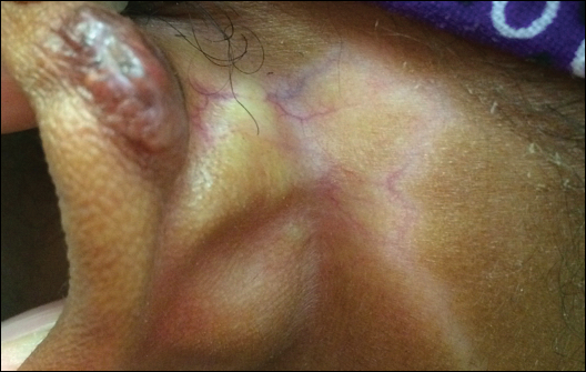
A 20-year-old black woman underwent multiple intralesional corticosteroid injections for treatment of a keloid on the superior aspect of the left helix and subsequently presented with a streak of atrophy and hypopigmentation in the postauricular region of unknown duration due to the lesion location.
FDA: Krintafel approved as ‘radical cure’ for preventing malaria relapse
The Food and Drug Administration has approved, under priority review, single-dose tafenoquine (to be marketed as Krintafel) for the prevention of relapse in patients aged 16 years and older who are receiving appropriate antimalarial therapy for acute Plasmodium vivax infection, according to an announcement by research partners GSK and the nonprofit Medicines for Malaria Venture.
Clinical efficacy and safety of the 300-mg single-dose tablet was provided by three randomized, double-blind studies: DETECTIVE Part 1 and Part 2 (TAF112582) and GATHER (TAF116564). The results of the two phase III studies were announced in June 2017.
Tafenoquine is referred to as a “radical cure,” because it targets the dormant liver forms of P. vivax and is coadministered with currently available antimalarials such as chloroquine or artemisinin-based combination therapies.
“The world has waited decades for a new medicine to counter P. vivax malaria relapse. Today, we can say the wait is over. Moreover, as the first ever single-dose for this indication, Krintafel will help improve patient compliance,” David Reddy, MD, CEO of MMV stated.
The FDA approval letter and the Krintafel label information are available online.
Gates Foundation, Lauder family launch $30 million Alzheimer’s biomarker search
Two billionaires have joined forces in a $30 million effort to develop peripheral biomarkers for the early detection of Alzheimer’s disease and other dementias.
Bill Gates and Leonard Lauder, cofounder of the Alzheimer’s Drug Discovery Foundation (ADDF) and chairman emeritus of Estée Lauder, provided initial funding for the project, dubbed Diagnostics Accelerator. Support from other philanthropists, including the Dolby family and the Charles and Helen Schwab Foundation, will bring the initiative up to its full funding capacity, according to an ADDF press statement.
“Over the next 3 years, we will provide more than $30 million in grants to researchers who are working on the most promising and innovative ideas to help diagnose Alzheimer’s disease [AD] early before the more devastating symptoms occur,” Mr. Lauder said in the statement.
Researchers at academic centers and nonprofit organizations are eligible to apply for the program; biotech companies and startups are also invited. Letters of intent for the first round of funding are due by Sept. 14, and the final proposals are due Nov. 16. Conducted under the auspices of the ADDF, the program will employ strict scientific review of all proposals and grant priority to blood and other peripheral markers, including saliva, urine, and ocular biomarkers. Neuroimaging and cerebrospinal fluid biomarkers won’t be considered, although researchers may use these as validation tests for their investigational markers.
Target areas include, but aren’t limited to:
- Neuroprotection
- Neurodegeneration
- Protein misfolding
- Synaptic integrity and/or activity
- Vascular injury and blood-brain barrier integrity
- Mitochondria and metabolic function
- Oxidative stress
- White matter changes
The initiative reflects recent emphasis on the critical role of biomarkers in drug development; this is especially important because emerging data paint a clear picture of a long AD prodrome during which the disease might be more amenable to treatment, ADDF’s Howard Fillit, MD, said in the press statement.
“The significance of biomarkers in Alzheimer’s disease research is underscored by recent FDA [Food and Drug Administration] guidelines that recognize the critical role of biomarkers in drug development and shift the research definition of the early stages of the disease to include biomarkers, even before clinical symptoms become apparent. ... Like in cancer today, using the biomarker-specific model of precision medicine, we will be able to predict more accurately which treatment and prevention strategies will work in different at-risk populations of people who have Alzheimer’s disease or other forms of dementia.”
Diagnostics Accelerator will award two general types of grants. Proof-of-principle awards of up to $500,000 will support exploratory analyses of biomarkers in smaller human sample sizes of 50-100. Proposals must be supported by human data that demonstrate that the candidate markers correspond with disease pathophysiology. Preliminary assay validation data for the proposed studies should be included. Successful proof-of principle projects may be eligible for follow-on funding in the form of a validation award.
Validation awards will support exploring biomarkers that need to be tested at a larger scale (500-1,000 samples). These must be supported by a significant extant body of human data that demonstrate that the biomarker corresponds to disease pathophysiology. Validation studies should compare peripheral analytes to quantitative measurements using PET imaging and/or cerebrospinal fluid testing, and not cognition alone. Award amounts will be based on stage and scope of research.
The announcement drew praise from researchers and clinicians.
“I think this is a terrific initiative,” said Michael S. Wolfe, PhD, the Mathias P. Mertes Professor of Medicinal Chemistry at the University of Kansas, Lawrence. “It’s so important to identify those at high risk of developing AD well before the onset of symptoms. All clinical trials for disease-modifying therapies have failed because these have enrolled subjects who already have AD or MCI. Preventing or delaying disease onset [i.e., prophylaxis] is the way forward, but this will require specific predictive biomarkers.”
Richard J. Caselli, MD, a professor of neurology at the Mayo Clinic Arizona in Scottsdale and clinical core director of the Arizona Alzheimer’s Disease Center, agreed.
“I would add that we are still trying to understand how various ‘experiences’ might push us toward Alzheimer’s disease; head injury, diabetes, and air pollution, for example. We know already, based on existing genomic and acquired risk factors, that AD is a complex disease and that many disparate things can influence our risk. Are each of these equally important in each of us or do we each have our own unique vulnerability profile? New biomarkers can help us determine that, and if we are in fact each unique, then maybe prevention strategies need to be personalized.”
Simplified formula validated for MRI Crohn’s disease assessment
WASHINGTON – The same Spanish research team who introduced an MRI-based formula for scoring the activity of luminal Crohn’s disease in 2011 have developed and validated a new, simplified version of their MRI score that speeds assessment.
“The simplified version of the MaRIA [Magnetic Resonance Index of Activity] score allows a faster and easier assessment of inflammation and quantification of severity in Crohn’s disease by keeping high accuracy for diagnosis and therapeutic response,” Ingrid Ordás, MD, said at the annual Digestive Disease Week®. The main advantage of the simplified MaRIA is that it is a “less time-consuming calculation that is not confounded by missing segments,” said Dr. Ordás, a gastroenterologist at the Hospital Clinic of Barcelona.
Although the data reported by Dr. Ordás included the derivation results, which used 98 patients enrolled in two separate prospective studies, and a separate prospective validation cohort of 37 patients, all these patients were evaluated by clinicians at the Hospital Clinic of Barcelona, and hence further validation with patients enrolled at other sites is now needed, Dr. Ordás said in an interview. Further accumulation of evidence for high sensitivity and specificity of Crohn’s disease assessment using the simplified MaRIA could allow it to replace endoscopy as the standard tool for assessing disease activity and severity in patients with luminal Crohn’s disease.
The derivation phase of the study identified four features that significantly correlated with disease activity and severity: bowel wall thickening to more than 3 mm, mural edema, perienteric fat stranding, and mucosal ulcerations. Limiting assessment to these four features cut in half the elements in the original MaRIA (Inflamm Bowel Dis. 2011 Aug;17[8];1759-68). Fat stranding – loss of the usual sharp interface between the wall and mesentery because of fluid – is a new parameter in the simplified MaRIA. The other three elements had been in the original index, but several other elements are now gone, including relative contrast enhancement wall signal intensity and consideration of lymph nodes.
In the validation phase, the researchers compared the MaRIA findings of the validation cohort with endoscopy findings both at baseline and then after they had received treatment. The sensitivity and specificity of the simplified MaRIA depended on the cutoff used, but as an example, a patient with a simplified MaRIA of 1 or greater as having active disease had a sensitivity of 90%, specificity of 81%, and an area under the receiver operator characteristic curve of 0.91. Using a simplified MaRIA of at least 2 as indicative of severe disease had a sensitivity of 85%, a specificity of 92%, and an AUROC of 0.94, Dr. Ordás reported.
Further assessment in patients who underwent treatment showed that reductions in the simplified MaRIA significantly correlated with treatment responses and remained essentially unchanged in patients who did not have clinical response to treatment. The analysis also showed a strong, positive correlation coefficient of 0.83 when the simplified MaRIA of an individual patient, compared with the patient’s Crohn’s disease endoscopy index of severity, and a correlation coefficient of 0.94 when a patient’s simplified MaRIA determined by one clinician, compared with the index score calculated by a second clinician.
Dr. Ordás had no disclosures to report.
SOURCE: Ordás I et al. DDW 2018, Presentation 437.
REPORTING FROM DDW 2018
Key clinical point: Researchers devised a simplified way to use MRI to noninvasively assess Crohn’s disease activity.
Major finding: The simplified, MRI-based formula identified Crohn’s disease activity with 90% sensitivity and 81% specificity.
Study details: The validation study included 37 patients with luminal Crohn’s disease at a single center in Barcelona.
Disclosures: Dr. Ordás had no disclosures to report.
Source: Ordás I et al. DDW 2018, Presentation 437.