User login
Patients with intellectual disability require nuanced care
SAN FRANCISCO – Some physicians are uncomfortable providing mental health care to patients with intellectual disability (ID) because many of the patients’ communications skills are limited. But many resources are available that can help.
In this video, Nita V. Bhatt, MD, MPH, interviews Julie P. Gentile, MD, about some of those resources and discusses how to approach psychiatric treatment interventions for patients with ID.
In addition to the DSM-5, Dr. Gentile said the National Association for the Dually Diagnosed has published the Diagnostic Manual – Intellectual Disability. Another resource is a practical reference manual originally proposed by one of Dr. Gentile’s residents.
“He came into my office for supervision one day and said, ‘You know, there’s all these nuances for psychiatric treatment in this patient population. So we should write a practice, quick reference manual to help clinicians who aren’t able to spend as much time concentrate on this patient population.’ ”
As a result, several residents and faculty members formed a team to produce an 18-chapter book published this year by Springer called the Guide to Intellectual Disabilities: A Clinical Handbook.
Dr. Bhatt is a staff psychiatrist at Twin Valley Behavioral Healthcare, the state psychiatric hospital in Columbus, Ohio. Dr. Gentile is professor and chair of the department of psychiatry at Wright State in Dayton. She is also serves as project director of Ohio’s Telepsychiatry Project for Intellectual Disability and has been awarded more than $7 million in grant funding to support her projects in the field of ID.
Dr. Gentile’s work has been funded by the Ohio Department of Developmental Disabilities and the Ohio Department of Mental Health and Addiction Services.
REPORTING FROM APA 2019

The great sunscreen ingredient debate
In a commentary issued on May 6, the Food and Drug Administration stated that “with sunscreens now being used with greater frequency, in larger amounts, and by broader populations, it is more important than ever to ensure that sunscreens are safe and effective for daily, lifelong use.” The statement coincided with the publication of the randomized study, “Effect of sunscreen application under maximal use conditions on plasma concentrations of sunscreen active ingredients,” by Matta et al. of the FDA and others in JAMA (2019 May 6. doi: 10.1001/jama.2019.5586). A maximal usage trial examines the systemic absorption of a topical drug when used according to the guidelines given for the product’s maximum usage. In this study, adult participants were randomized to one of four commercially available sunscreen products: spray 1 (n = 6), spray 2 (n = 6), a lotion (n = 6), and a cream (n = 6). Two mg of sunscreen per 1 cm2 was applied to 75% of body surface area four times per day for 4 days, and blood samples were collected from each individual over 7 days.

The FDA’s guidance for industry and proposed rule on OTC sunscreens state that active ingredients with systemic absorption at 0.5 ng/mL or higher or with possible safety concerns need to undergo further nonclinical toxicology assessment to evaluate risk of systemic carcinogenicity, developmental/reproductive abnormalities, or other adverse effects.
Absorption of some sunscreen ingredients has been detected in other studies. Despite systemic absorption, two active ingredients – zinc oxide and titanium dioxide – have been found by the FDA to be generally recognized as safe and effective. But for 12 other active ingredients (cinoxate, dioxybenzone, ensulizole, homosalate, meradimate, octinoxate, octisalate, octocrylene, padimate O, sulisobenzone, oxybenzone, and avobenzone), there are insufficient data to make a “generally recognized as safe and effective” determination; thus, more data have been requested from the manufacturers. While physical blocking sunscreens have improved in their UV-blocking ability without compromising cosmesis over the past several years, some sunscreens containing chemical blockers are able to achieve higher SPFs with good cosmesis when applied to the skin.
Our skin acts as the ultimate barrier between ourselves and the environment, and it is not uncommon for substances to be blocked, absorbed, or excreted from the skin. Absorption of an ingredient through the skin and into the body does not indicate that the ingredient is unsafe. Rather, findings such as these call for further testing and research to determine the safety of that ingredient with repeated use. Per the FDA, such testing is part of the standard premarket safety evaluation of most chronically administered drugs with appreciable systemic absorption.
In February 2019, the FDA’s proposed rule was issued to “update regulatory requirements for most sunscreen products in the United States,” with the goal of bringing OTC sunscreens “up to date with the latest scientific standards,” according to the FDA May 6 commentary. “As part of this rule, the FDA is asking industry and other interested parties for additional safety data on the 12 active sunscreen ingredients currently available in marketed products” mentioned previously. These rules are being put into place to address the “key data gap” for these 12 ingredients, which is “understanding whether, and to what extent the ingredient is absorbed into the body after topical application.”
In other previously published studies, oxybenzone, along with some other sunscreen active ingredients including octocrylene, have been found in human breast milk. In addition, oxybenzone has been detected in amniotic fluid, urine, and blood. Whether these findings have any clinical implications needs to be further assessed. Some studies in the literature have raised questions about the potential for oxybenzone to affect endocrine activity.
Another issue that has been raised is the potential impact of sunscreen on the environment, specifically, coral reefs. In July 2018, Hawaii Governor David Ige (D) signed a bill (SB 2571) that bans the sale of sunscreens containing oxybenzone and octinoxate beginning in 2021, making Hawaii the first state to ban the sale of sunscreens containing these two chemicals. Shortly afterward, the Republic of Palau and city of Key West, Fla., also took action to ban sunscreens containing chemicals potentially harmful to marine life. In Hawaii, what’s know as “reef safe” sunscreen is sold.

More research in this area is needed, but studies have linked these ingredients to harming coral by bleaching, disease, and damage to DNA, and also to decreasing fertility in fish, impairing algae growth, inducing defects in mussel and sea urchin young, and accumulating in the tissues of dolphins. According to NASA, as much as 27% of monitored reef formation have already been lost and over the following 32 years, 32% more are at risk. Reefs cover a mere 0.2% of the ocean’s floor, but it is estimated that reefs are home to and protect nearly 1 million species of fish, invertebrates, and algae.
In early May, Rep. Tulsi Gabbard (D-Hawaii) and Sen. Tim Ryan (D-Ohio) introduced legislation known as the Oxybenzone and Octinoxate Impact Study Act of 2019 (H.R. 2588) to require the Environmental Protection Agency to study the impact of those two chemicals on human health and the environment and to provide findings to Congress and the public within 18 months.
The importance of sun protection and prevention of sunburns is paramount. We know that multiple sunburn events during childhood double a child’s risk of developing skin cancer later in life, and skin cancer is the most common cancer diagnosed in the United States, with 5 million cases treated every year. One in five Americans will develop skin cancer by age 70 years.
As a Mohs and a cosmetic dermatologic surgeon, I appreciate the unquestionable protective effects of sunscreen products with regards to skin cancer, dyspigmentation, solar elastosis, and rhytids associated with photoaging. We can applaud the FDA for improving testing and regulation of OTC ingredients, including those in sunscreen. These types of studies are important and monumental in ensuring that we are utilizing the right type of ingredients to protect our patients, our oceans, and our reefs.
Dr. Wesley and Dr. Talakoub are co-contributors to this column. Dr. Wesley practices dermatology in Beverly Hills, Calif. Dr. Talakoub is in private practice in McLean, Va. This month’s column is by Dr. Wesley. Write to them at [email protected]. They had no relevant disclosures.
References
- Matta MK et al. JAMA. 2019 May 6. doi: 10.1001/jama.2019.5586.
- Shedding new light on sunscreen absorption, by Janet Woodcock, MD, director, Center for Drug Evaluation and Research, and Theresa M. Michele, MD, director, CDER’s Division of Nonprescription Drug Products, Office of New Drugs
- Food and Drug Administration. Sunscreen drug products for over-the-counter human use: Proposed rule. Fed Regist. 2019;84(38):6204-75.
- Schlumpf M et al. Chemosphere. 2010 Nov;81(10):1171-83.
- Krause M et al. Int J Androl. 2012 Jun;35(3):424-36.
In a commentary issued on May 6, the Food and Drug Administration stated that “with sunscreens now being used with greater frequency, in larger amounts, and by broader populations, it is more important than ever to ensure that sunscreens are safe and effective for daily, lifelong use.” The statement coincided with the publication of the randomized study, “Effect of sunscreen application under maximal use conditions on plasma concentrations of sunscreen active ingredients,” by Matta et al. of the FDA and others in JAMA (2019 May 6. doi: 10.1001/jama.2019.5586). A maximal usage trial examines the systemic absorption of a topical drug when used according to the guidelines given for the product’s maximum usage. In this study, adult participants were randomized to one of four commercially available sunscreen products: spray 1 (n = 6), spray 2 (n = 6), a lotion (n = 6), and a cream (n = 6). Two mg of sunscreen per 1 cm2 was applied to 75% of body surface area four times per day for 4 days, and blood samples were collected from each individual over 7 days.
The FDA’s guidance for industry and proposed rule on OTC sunscreens state that active ingredients with systemic absorption at 0.5 ng/mL or higher or with possible safety concerns need to undergo further nonclinical toxicology assessment to evaluate risk of systemic carcinogenicity, developmental/reproductive abnormalities, or other adverse effects.
Absorption of some sunscreen ingredients has been detected in other studies. Despite systemic absorption, two active ingredients – zinc oxide and titanium dioxide – have been found by the FDA to be generally recognized as safe and effective. But for 12 other active ingredients (cinoxate, dioxybenzone, ensulizole, homosalate, meradimate, octinoxate, octisalate, octocrylene, padimate O, sulisobenzone, oxybenzone, and avobenzone), there are insufficient data to make a “generally recognized as safe and effective” determination; thus, more data have been requested from the manufacturers. While physical blocking sunscreens have improved in their UV-blocking ability without compromising cosmesis over the past several years, some sunscreens containing chemical blockers are able to achieve higher SPFs with good cosmesis when applied to the skin.
Our skin acts as the ultimate barrier between ourselves and the environment, and it is not uncommon for substances to be blocked, absorbed, or excreted from the skin. Absorption of an ingredient through the skin and into the body does not indicate that the ingredient is unsafe. Rather, findings such as these call for further testing and research to determine the safety of that ingredient with repeated use. Per the FDA, such testing is part of the standard premarket safety evaluation of most chronically administered drugs with appreciable systemic absorption.
In February 2019, the FDA’s proposed rule was issued to “update regulatory requirements for most sunscreen products in the United States,” with the goal of bringing OTC sunscreens “up to date with the latest scientific standards,” according to the FDA May 6 commentary. “As part of this rule, the FDA is asking industry and other interested parties for additional safety data on the 12 active sunscreen ingredients currently available in marketed products” mentioned previously. These rules are being put into place to address the “key data gap” for these 12 ingredients, which is “understanding whether, and to what extent the ingredient is absorbed into the body after topical application.”
In other previously published studies, oxybenzone, along with some other sunscreen active ingredients including octocrylene, have been found in human breast milk. In addition, oxybenzone has been detected in amniotic fluid, urine, and blood. Whether these findings have any clinical implications needs to be further assessed. Some studies in the literature have raised questions about the potential for oxybenzone to affect endocrine activity.
Another issue that has been raised is the potential impact of sunscreen on the environment, specifically, coral reefs. In July 2018, Hawaii Governor David Ige (D) signed a bill (SB 2571) that bans the sale of sunscreens containing oxybenzone and octinoxate beginning in 2021, making Hawaii the first state to ban the sale of sunscreens containing these two chemicals. Shortly afterward, the Republic of Palau and city of Key West, Fla., also took action to ban sunscreens containing chemicals potentially harmful to marine life. In Hawaii, what’s know as “reef safe” sunscreen is sold.
More research in this area is needed, but studies have linked these ingredients to harming coral by bleaching, disease, and damage to DNA, and also to decreasing fertility in fish, impairing algae growth, inducing defects in mussel and sea urchin young, and accumulating in the tissues of dolphins. According to NASA, as much as 27% of monitored reef formation have already been lost and over the following 32 years, 32% more are at risk. Reefs cover a mere 0.2% of the ocean’s floor, but it is estimated that reefs are home to and protect nearly 1 million species of fish, invertebrates, and algae.
In early May, Rep. Tulsi Gabbard (D-Hawaii) and Sen. Tim Ryan (D-Ohio) introduced legislation known as the Oxybenzone and Octinoxate Impact Study Act of 2019 (H.R. 2588) to require the Environmental Protection Agency to study the impact of those two chemicals on human health and the environment and to provide findings to Congress and the public within 18 months.
The importance of sun protection and prevention of sunburns is paramount. We know that multiple sunburn events during childhood double a child’s risk of developing skin cancer later in life, and skin cancer is the most common cancer diagnosed in the United States, with 5 million cases treated every year. One in five Americans will develop skin cancer by age 70 years.
As a Mohs and a cosmetic dermatologic surgeon, I appreciate the unquestionable protective effects of sunscreen products with regards to skin cancer, dyspigmentation, solar elastosis, and rhytids associated with photoaging. We can applaud the FDA for improving testing and regulation of OTC ingredients, including those in sunscreen. These types of studies are important and monumental in ensuring that we are utilizing the right type of ingredients to protect our patients, our oceans, and our reefs.
Dr. Wesley and Dr. Talakoub are co-contributors to this column. Dr. Wesley practices dermatology in Beverly Hills, Calif. Dr. Talakoub is in private practice in McLean, Va. This month’s column is by Dr. Wesley. Write to them at [email protected]. They had no relevant disclosures.
References
- Matta MK et al. JAMA. 2019 May 6. doi: 10.1001/jama.2019.5586.
- Shedding new light on sunscreen absorption, by Janet Woodcock, MD, director, Center for Drug Evaluation and Research, and Theresa M. Michele, MD, director, CDER’s Division of Nonprescription Drug Products, Office of New Drugs
- Food and Drug Administration. Sunscreen drug products for over-the-counter human use: Proposed rule. Fed Regist. 2019;84(38):6204-75.
- Schlumpf M et al. Chemosphere. 2010 Nov;81(10):1171-83.
- Krause M et al. Int J Androl. 2012 Jun;35(3):424-36.
p-TIPS improves outcomes for high-risk variceal bleeding
Background: Acute variceal bleeding remains the most severe and life-threatening complication of portal hypertension in cirrhotic patients. Several small studies have shown improved outcomes with p-TIPS without worsening of hepatic encephalopathy or other adverse events.

Study design: Multicenter, international, observational study.
Setting: One Canadian and 33 European referral centers.
Synopsis: 2,138 patients were registered for analysis, of which 671 were identified as high risk based on Child-Pugh score (either Child class C of less than 14 or Child class B with active bleeding seen on endoscopy). Multiple exclusion criteria were used including Child-Pugh score of 14 or more, renal failure, occlusive portal vein thrombosis, sepsis, heart failure, or hepatocellular carcinoma outside Milan criteria. Each patient underwent initial management with vasoactive medications, antibiotics, and endoscopy with subsequent intervention (p-TIPS vs. standard care) based on provider decision. p-TIPS was defined as TIPS within 72 hours of initial bleed. 31.4% of the cohort was lost to follow-up at 1 year. p-TIPS improved 1-year mortality significantly (78% vs. 62%; P = .014) and did not confer an increased risk of hepatic encephalopathy or other complication. Additionally, the authors found that the effect was significantly greater in the Child-Pugh Class C group (1-year mortality rate of 78% vs. 53%; P = .002). The authors then compared observed mortality with MELD-predicted mortality and found that with standard care, MELD scores matched with predicted mortality, but with p-TIPS, MELD scores predicted a greater mortality than the observed mortality. The authors calculated that the number needed to treat to save one life for 1 year with p-TIPS is 4.2. The major limitation of this study is the observational design and the inherent risk of selection bias. Additionally, almost one-third of patients were lost to follow-up.
Bottom line: Significant improvements in mortality are observed when high-risk patients undergo p-TIPS procedures as opposed to usual care with medications and endoscopy.
Citation: Hernández Gea V et al. Preemptive TIPS improves outcome in high risk variceal bleeding: An observational study. Hepatology. 2018 Jul 16. doi: 10.1002/hep.30182.
Dr. Imber is an assistant professor in the division of hospital medicine, University of New Mexico.
Background: Acute variceal bleeding remains the most severe and life-threatening complication of portal hypertension in cirrhotic patients. Several small studies have shown improved outcomes with p-TIPS without worsening of hepatic encephalopathy or other adverse events.
Study design: Multicenter, international, observational study.
Setting: One Canadian and 33 European referral centers.
Synopsis: 2,138 patients were registered for analysis, of which 671 were identified as high risk based on Child-Pugh score (either Child class C of less than 14 or Child class B with active bleeding seen on endoscopy). Multiple exclusion criteria were used including Child-Pugh score of 14 or more, renal failure, occlusive portal vein thrombosis, sepsis, heart failure, or hepatocellular carcinoma outside Milan criteria. Each patient underwent initial management with vasoactive medications, antibiotics, and endoscopy with subsequent intervention (p-TIPS vs. standard care) based on provider decision. p-TIPS was defined as TIPS within 72 hours of initial bleed. 31.4% of the cohort was lost to follow-up at 1 year. p-TIPS improved 1-year mortality significantly (78% vs. 62%; P = .014) and did not confer an increased risk of hepatic encephalopathy or other complication. Additionally, the authors found that the effect was significantly greater in the Child-Pugh Class C group (1-year mortality rate of 78% vs. 53%; P = .002). The authors then compared observed mortality with MELD-predicted mortality and found that with standard care, MELD scores matched with predicted mortality, but with p-TIPS, MELD scores predicted a greater mortality than the observed mortality. The authors calculated that the number needed to treat to save one life for 1 year with p-TIPS is 4.2. The major limitation of this study is the observational design and the inherent risk of selection bias. Additionally, almost one-third of patients were lost to follow-up.
Bottom line: Significant improvements in mortality are observed when high-risk patients undergo p-TIPS procedures as opposed to usual care with medications and endoscopy.
Citation: Hernández Gea V et al. Preemptive TIPS improves outcome in high risk variceal bleeding: An observational study. Hepatology. 2018 Jul 16. doi: 10.1002/hep.30182.
Dr. Imber is an assistant professor in the division of hospital medicine, University of New Mexico.
Novel chromogenic assay looks accurate in hemophilia A diagnosis
, according to recent study findings.
“The original one‐stage clotting assay is still the most widely used method for measuring FVIII activity in these patients, although the chromogenic assay is recognized to be less prone to the variability related to the use of different reagents and to the presence of interferences,” Cristina Novembrino, MD, of the Fondazione IRCCS Ca’ Granda Ospedale Maggiore Policlinico, in Milan, and colleagues wrote in Haemophilia. “The choice of the proper assay is a crucial point in the frame of diagnosis, particularly in moderate or mild [hemophilia A] patients.”
The BIOPHEN FVIII:C assay, used on the Sysmex CS‐2400 analyzer, is a novel chromogenic diagnostic tool used to analyze FVIII clotting activity in patients with hemophilia A of all severity levels. The researchers evaluated the diagnostic and clinical capabilities of the assay in 60 patients with hemophilia A and 120 healthy controls.
Dr. Novembrino and colleagues used samples of FVIII deficient plasma and Actin FS to compare the novel tool to a one-stage assay and another chromogenic assay.
After analysis, the researchers found that the inter‐assay and intra‐assay coefficient of variation were less than 6%. The mean recovery and limit of detection were 91.7% (range, 79.8%-98.6%) and 0.2%, respectively.
The linearity test revealed positive results of up to 1/128 dilution (r = 0.99).
“The K coefficient was 0.91 when BIOPHEN FVIII:C was compared with the historical classification of the patients, demonstrating an optimal diagnostic accuracy in hemophilia A,” the researchers wrote.
The novel assay may be an appropriate laboratory tool for the diagnosis and therapeutic monitoring of patients with hemophilia A, they added.
Sysmex Corporation and Hyphen BioMed provided the instrument and reagents for the study. One of the authors is an employee of Sysmex Corporation. The authors reported having no other conflicts of interest.
SOURCE: Novembrino C et al. Haemophilia. 2019 May 2. doi: 10.1111/hae.13746.
FROM HAEMOPHILIA
Zoster vaccination is underused but looks effective in IBD
For men with inflammatory bowel disease, herpes zoster vaccination was associated with about a 46% decrease in risk of associated infection, according to the results of a retrospective study from the national Veterans Affairs Healthcare System.
Crude rates of herpes zoster infection were 4.09 cases per 1,000 person-years among vaccinated patients versus 6.97 cases per 1,000 person-years among unvaccinated patients, for an adjusted hazard ratio of 0.54 (95% confidence interval, 0.44-0.68), reported Nabeel Khan, MD, of the University of Pennsylvania, Philadelphia, and associates. “This vaccine is therefore effective in patients with IBD, but underused,” they wrote in Clinical Gastroenterology and Hepatology.
Studies have linked IBD with a 1.2- to 1.8-fold increased risk of herpes zoster infection, the researchers noted. Relevant risk factors include older age, disease flare, recent use or high cumulative use of prednisone, and use of thiopurines, either alone or in combination with a tumor necrosis factor (TNF) inhibitor. Although the American College of Gastroenterology recommends that all patients with IBD receive the herpes zoster vaccine by age 50 years, the efficacy of the vaccine in these patients remains unclear.
For their study, Dr. Khan and associates analyzed International Classification of Diseases (ICD) codes and other medical record data from 39,983 veterans with IBD who had not received the herpes zoster vaccine by age 60 years. In all, 97% of patients were male, and 94% were white. Most patients had high rates of health care utilization: Approximately half visited VA clinics or hospitals at least 13 times per year, and another third made 6-12 annual visits.
Despite their many contacts with VA health care systems, only 7,170 (17.9%) patients received the herpes zoster vaccine during 2000-2016, the researchers found. Vaccination rates varied substantially by region – they were highest in the Midwest (35%) and North Atlantic states (29%) but reached only 9% in Montana, Utah, Wyoming, Colorado, Oklahoma, Texas, Arkansas, and Louisiana, collectively.
The crude rate of herpes zoster infection among unvaccinated patients with IBD resembled the incidence reported in prior studies, the researchers said. After researchers accounted for differences in geography, demographics, and health care utilization between vaccinated and unvaccinated veterans with IBD, they found that vaccination was associated with an approximately 46% decrease in the risk of herpes zoster infection.
Very few patients were vaccinated for herpes zoster while on a TNF inhibitor, precluding the ability to study this subgroup. However, the vaccine showed a protective effect (adjusted HR, 0.63) among patients who received thiopurines without a TNF inhibitor. This effect did not reach statistical significance, perhaps because of lack of power, the researchers noted. “Among the 315 patients who were [vaccinated while] on thiopurines, none developed a documented painful or painless vesicular rash within 42 days of herpes zoster vaccination,” they added. One patient developed a painful blister 20 days post vaccination without vesicles or long-term sequelae.
Pfizer provided funding. Dr. Khan disclosed research funding from Pfizer, Luitpold, and Takeda Pharmaceuticals. One coinvestigator disclosed ties to Pfizer, Gilead, Merck, AbbVie, Lilly, Janssen, Johnson & Johnson, UCB, and Nestle Health Science. The remaining researchers disclosed no conflicts.
SOURCE: Khan N et al. Clin Gastroenterol Hepatol. 2018 Oct 13. doi: 10.1016/j.cgh.2018.10.016.
Preventive care is an underemphasized component of IBD management because the primary focus tends to be control of active symptoms. However, as patients are treated with immunosuppression, particularly combinations of therapies and newer mechanisms of action such as the Janus kinase inhibitors, the risk of infections increases, including those that are vaccine preventable including shingles and its related complications.
This study by Khan et al. highlights several important messages for patients and providers. First, in this large older IBD cohort, the vaccination rates were very low at 18% even though more than 80% of patients had more than six annual visits to the VA Health Systems during the study period. These represent multiple missed opportunities to discuss and administer vaccinations. Second, the authors highlighted the vaccine’s efficacy: Persons receiving herpes zoster vaccination had a clearly decreased risk of subsequent infection. While the number of vaccinated patients on immunosuppression was too small to draw conclusions about efficacy, the live attenuated vaccination is contraindicated for immunosuppressed patients. However, the newer recombinant shingles vaccine offers the opportunity to extend the reach of shingles vaccination to include those on immunosuppression. As utilization of the newer vaccine series increases, we will be able to evaluate the efficacy for immunosuppressed IBD patients, although studies from other disease states suggest efficacy. However, vaccinations will never work if they aren’t administered. Counseling patients and providers regarding the importance of vaccinations is a low-risk, efficacious means to decrease infection and associated morbidity.
Christina Ha, MD, AGAF, associate professor of medicine, Inflammatory Bowel Disease Center, division of digestive diseases, Cedars-Sinai Medical Center, Los Angeles. She is a speaker, consultant, or on the advisory board for AbbVie, Janssen, Genentech, Samsung Bioepis, and Takeda. She received grant funding from Pfizer.
For men with inflammatory bowel disease, herpes zoster vaccination was associated with about a 46% decrease in risk of associated infection, according to the results of a retrospective study from the national Veterans Affairs Healthcare System.
Crude rates of herpes zoster infection were 4.09 cases per 1,000 person-years among vaccinated patients versus 6.97 cases per 1,000 person-years among unvaccinated patients, for an adjusted hazard ratio of 0.54 (95% confidence interval, 0.44-0.68), reported Nabeel Khan, MD, of the University of Pennsylvania, Philadelphia, and associates. “This vaccine is therefore effective in patients with IBD, but underused,” they wrote in Clinical Gastroenterology and Hepatology.
Studies have linked IBD with a 1.2- to 1.8-fold increased risk of herpes zoster infection, the researchers noted. Relevant risk factors include older age, disease flare, recent use or high cumulative use of prednisone, and use of thiopurines, either alone or in combination with a tumor necrosis factor (TNF) inhibitor. Although the American College of Gastroenterology recommends that all patients with IBD receive the herpes zoster vaccine by age 50 years, the efficacy of the vaccine in these patients remains unclear.
For their study, Dr. Khan and associates analyzed International Classification of Diseases (ICD) codes and other medical record data from 39,983 veterans with IBD who had not received the herpes zoster vaccine by age 60 years. In all, 97% of patients were male, and 94% were white. Most patients had high rates of health care utilization: Approximately half visited VA clinics or hospitals at least 13 times per year, and another third made 6-12 annual visits.
Despite their many contacts with VA health care systems, only 7,170 (17.9%) patients received the herpes zoster vaccine during 2000-2016, the researchers found. Vaccination rates varied substantially by region – they were highest in the Midwest (35%) and North Atlantic states (29%) but reached only 9% in Montana, Utah, Wyoming, Colorado, Oklahoma, Texas, Arkansas, and Louisiana, collectively.
The crude rate of herpes zoster infection among unvaccinated patients with IBD resembled the incidence reported in prior studies, the researchers said. After researchers accounted for differences in geography, demographics, and health care utilization between vaccinated and unvaccinated veterans with IBD, they found that vaccination was associated with an approximately 46% decrease in the risk of herpes zoster infection.
Very few patients were vaccinated for herpes zoster while on a TNF inhibitor, precluding the ability to study this subgroup. However, the vaccine showed a protective effect (adjusted HR, 0.63) among patients who received thiopurines without a TNF inhibitor. This effect did not reach statistical significance, perhaps because of lack of power, the researchers noted. “Among the 315 patients who were [vaccinated while] on thiopurines, none developed a documented painful or painless vesicular rash within 42 days of herpes zoster vaccination,” they added. One patient developed a painful blister 20 days post vaccination without vesicles or long-term sequelae.
Pfizer provided funding. Dr. Khan disclosed research funding from Pfizer, Luitpold, and Takeda Pharmaceuticals. One coinvestigator disclosed ties to Pfizer, Gilead, Merck, AbbVie, Lilly, Janssen, Johnson & Johnson, UCB, and Nestle Health Science. The remaining researchers disclosed no conflicts.
SOURCE: Khan N et al. Clin Gastroenterol Hepatol. 2018 Oct 13. doi: 10.1016/j.cgh.2018.10.016.
FROM CLINICAL GASTROENTEROLOGY AND HEPATOLOGY
Prepare for deluge of JAK inhibitors for RA
MAUI, HAWAII – As it grows increasingly likely that oral Janus kinase inhibitors will constitute a major development in the treatment of rheumatoid arthritis, with a bevy of these agents becoming available for that indication, rheumatologists are asking questions about the coming revolution. Like, when should these agents be used? What are the major safety and efficacy differences, if any, within the class? How clinically relevant is JAK selectivity? And which JAK inhibitor is the best choice?
on these and other related questions at the 2019 Rheumatology Winter Clinical Symposium.
These issues take on growing relevance for clinicians and their RA patients because two oral small molecule JAK inhibitors – tofacitinib (Xeljanz) and baricitinib (Olumiant) – are already approved for RA, and three more – upadacitinib, filgotinib, and peficitinib – are on the horizon. Indeed, AbbVie has already filed for marketing approval of once-daily upadacitinib for RA on the basis of an impressive development program featuring six phase 3 trials, with a priority review decision from the Food and Drug Administration anticipated this fall. Filgotinib is the focus of three phase 3 studies, one of which is viewed as a home run, with the other two yet to report results. Peficitinib is backed by two positive phase 3 trials, although its manufacturer will at least initially seek marketing approval only in Japan and South Korea. And numerous other JAK inhibitors are in development for a variety of indications.
When should a JAK inhibitor be used?
That’s easy, according to Roy M. Fleischmann, MD: If the cost proves comparable, it makes sense to turn to a JAK inhibitor ahead of a tumor necrosis factor inhibitor or other biologic.
He noted that in the double-blind, phase 3 SELECT-COMPARE head-to-head comparison of upadacitinib at 15 mg/day, adalimumab (Humira) at 40 mg every other week, versus placebo, all on top of background methotrexate, upadacitinib proved superior to the market-leading tumor necrosis factor inhibitor in terms of both the American College of Rheumatology–defined 20% level of response (ACR 20) and 28-joint Disease Activity Score based on C-reactive protein (DAS28-CRP).
“The results were very dramatic,” noted Dr. Fleischmann, who presented the SELECT-COMPARE findings at the 2018 annual meeting of the American College of Rheumatology.
Moreover, other major trials have shown that baricitinib at 4 mg/day was superior in efficacy to adalimumab, and tofacitinib and peficitinib were “at least equal” to anti-TNF therapy, he added.
“These numbers are clinically meaningful – not so much for the difference in ACR 20, but in the depth of response: the ACR 50 and 70, the CDAI. I think these drugs are better than adalimumab,” declared Dr. Fleischmann, codirector of the division of rheumatology at Texas Health Presbyterian Medical Center, Dallas.
Mark Genovese, MD, concurred.
“I think that for most patients who don’t have a lot of other comorbidities, they would certainly prefer to take a pill over a shot. And if you have a drug that’s more effective than the standard of care and it comes at a reasonable price point – and ‘reasonable’ is in the eye of the beholder – but if I can get access to it on the formulary, I’d have no qualms about putting them on a JAK inhibitor before I’d move to a TNF inhibitor,” said Dr. Genovese, professor of medicine and director of the rheumatology clinic at Stanford (Calif.) University.
Upadacitinib elicited a better response at 30 mg than at 15 mg once daily in the phase 3 program; however, both speakers indicated they’d be happy with access to the 15-mg dose, should the FDA go that route, since it has a better safety profile.
Dr. Genovese was principal investigator in the previously reported multicenter FINCH2 trial of filgotinib at 100 or 200 mg/day in RA patients with a prior inadequate response to one or more biologic disease-modifying antirheumatic drugs.
“Impressive results in a refractory population,” he said. “I don’t see a big difference in safety between 100 and 200 mg, so I’d opt for the 200 because it worked really well in patients who had refractory disease.”
Other advantages of JAK inhibitors
Speed of onset is another advantage in addition to oral administration and efficacy greater than or equivalent to anti-TNF therapy, according to Dr. Genovese.
“As a class, JAK inhibitors have a faster onset than methotrexate in terms of improvement in disease activity and pain. So in a few weeks you can have a sense of whether folks are going to be responders,” the rheumatologist said.
Does JAK isoform selectivity really make a difference in terms of efficacy and safety?
It’s doubtful, the rheumatologists agreed. All of these oral small molecules target JAK1, and that’s what’s key.
Tofacitinib is relatively selective for JAK1 and JAK3, baricitinib for JAK1 and JAK2, upadacitinib and fibotinib for JAK1, and peficitinib is a pan-JAK inhibitor.
What are the safety concerns with this class of medications?
The risk of herpes zoster is higher than with TNF inhibitors, reinforcing the importance of varicella vaccination in JAK inhibitor candidates. Anemia occurs in a small percentage of patients. As for the risk of venous thromboembolism as a potential side effect of JAK inhibitors, a topic of great concern to the FDA, Dr. Fleischmann dismissed it as vastly overblown.
“I think VTEs are an RA effect. You see it with all the drugs, including methotrexate,” he said.
Idiosyncratic self-limited increases in creatine kinase have been seen in 2%-4% of patients on JAK inhibitors in pretty much all of the clinical trials. “I’m not aware of any cases of myositis, though,” Dr. Fleischmann noted.
As for the teratogenicity potential of JAK inhibitors, Dr. Genovese said that, as is true for most medications, it hasn’t been well studied.
“We don’t know. But I would not choose to use a JAK inhibitor in a woman who is going to conceive, has conceived, or is breastfeeding. I just don’t think that would be a good decision,” according to the rheumatologist.
Which JAK inhibitor is the best choice for treatment of RA?
It’s impossible to say because of the hazards in trying to draw meaningful conclusions from cross-study comparisons, the experts agreed.
“It’s a challenge. I think at the end of the day there will probably be one agent that looks like it might be best in class predicated on having the most number of indications, and that will probably become a preferred agent. The question is, does that happen before tofacitinib goes generic? And I don’t know the answer to that,” Dr. Genovese said.
Notably, upadacitinib is the subject of a plethora of ongoing phase 3 trials in atopic dermatitis, psoriatic arthritis, Crohn’s disease, and ulcerative colitis. It is also in earlier-phase investigation for the treatment of ankylosing spondylitis.
Do we really need all these JAK inhibitors?
“How many TNF inhibitors do you need?” Dr. Genovese retorted. “I think the reality is there’s probably a finite number and additional members add to the class, but there probably will always be one or two that are going to be best in class.”
Both rheumatologists indicated they serve as consultants to more than a dozen pharmaceutical companies and receive research grants from numerous firms.
REPORTING FROM RWCS 2019
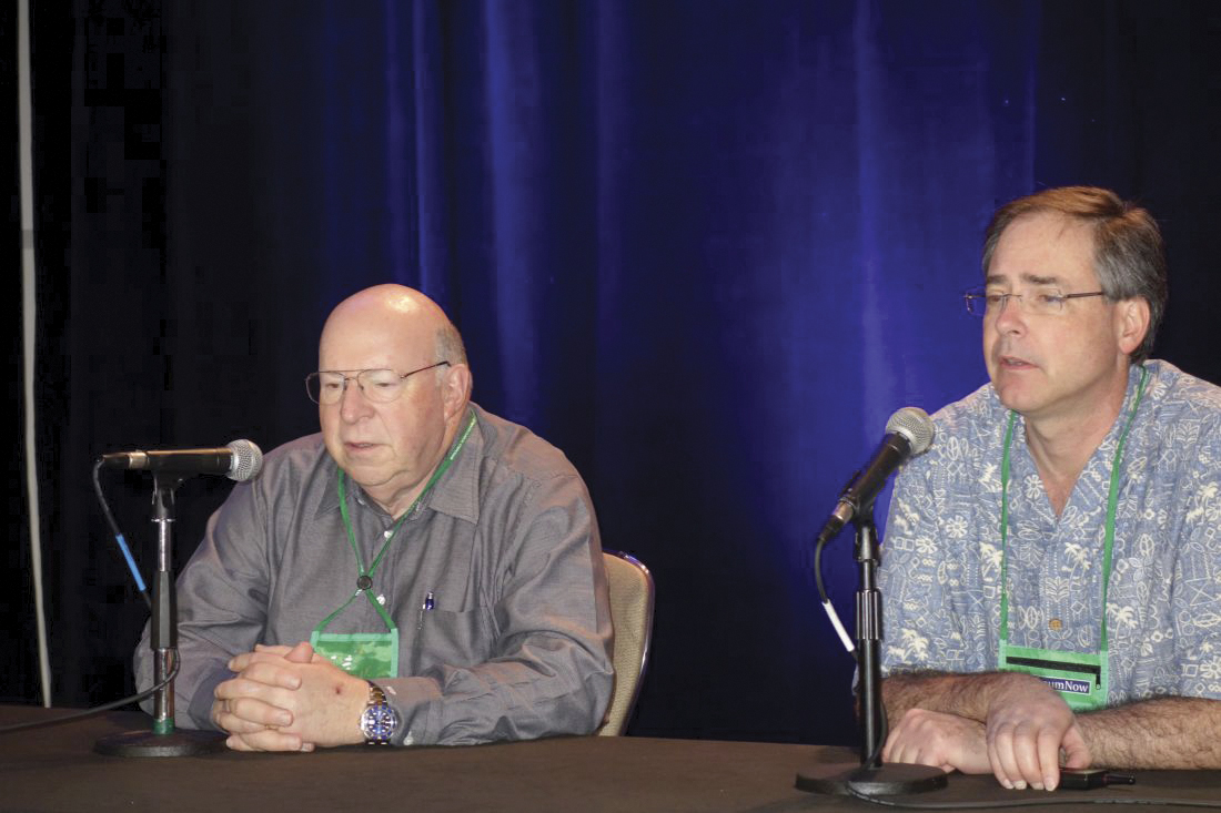
How to counsel patients interested in trying marijuana
SAN FRANCISCO – It’s a common scenario, and it’s going to become more common: Psychiatric patients will be coming into the office not quite satisfied with their current treatment and wanting to try cannabis.
With laws varying from state to state and a limited but growing body of evidence, the situation “presents a complicated, often difficult clinical situation,” said David A. Gorelick, MD, PhD, professor of psychiatry at the University of Maryland, Baltimore.
There is some evidence supporting cannabis for chronic pain, neuropathic pain, spasticity in multiple sclerosis, and chemotherapy-induced nausea and vomiting. Cannabidiol is also Food and Drug Administration approved for two severe forms of pediatric epilepsy.
When it comes to psychiatric disorders, however, evidence is largely lacking. On the other hand, some patients swear by marijuana for psychiatric symptom relief.
So what to do? Dr. Gorelick, editor of the Journal of Cannabis Research, had some advice in a video interview at the annual meeting of the American Psychiatric Association.
Where it’s legal, a cannabis recommendation for pain and sleep problems is reasonable, but it’s important to figure out what’s lacking in the patients’ treatment regimen that makes them want to try cannabis, and to first try to maximize treatment with approved, evidence-based options. As with any medication, a talk about risks and benefits is also essential. There’s an increased risk of motor vehicle accidents, acute panic attacks, and other issues with cannabis. Also, keep in mind that cannabinoids, particularly tetrahydrocannabinol, are metabolized in the liver and can interfere with concentrations of other liver-metabolized drugs, including some antidepressants and anticonvulsants. It’s not clear how significant the clinical interactions are, “but if patients are on warfarin, I would be very careful,” he said.
Dr. Gorelick disclosed speaking fees from hospitals and organizations for presentations on medical cannabis and royalties from Up-to-Date for cannabis topics. He also disclosed receiving honoraria from Colorado State University, Pueblo, and from Springer for his work on the Journal of Cannabis Research.
REPORTING FROM APA 2019
Low-dose FVIII prophylaxis reduced bleeding in hemophilia A
The findings have implications for low resource countries, the researchers reported.
Novie A. Chozie, MD, of Universitas Indonesia in Jakarta, along with colleagues, conducted a parallel-group, randomized controlled study of 50 children with severe hemophilia A. Study participants were randomized to receive either low‐dose FVIII prophylaxis (n = 25) or on‐demand treatment (n = 25) for a total of 12 months. The findings were published in Haemophilia.
Participants in the prophylaxis arm received FVIII at a dose of 10 IU/kg, infused twice weekly. If patients experienced an episode of acute bleeding, prophylaxis was delayed until the episode resolved.
The primary outcome was the number of total and joint bleeding events from the start of therapy to 12 months. Secondary outcomes included evidence of FVIII inhibitor, Hemophilia Joint Health Score (HJHS), and Hemophilia Early Arthropathy Detection Ultrasound (HEAD‐US) score.
After analysis, the team found that the number of joint and total bleeding events was significantly lower in the prophylaxis group. For total bleeding events, there was a median of 8 events with the prophylaxis group, compared with 25 in the on-demand treatment group (P less than .001). There was a median of three joint bleeding events in the prophylaxis group versus nine in the on-demand group (P less than .001).
Patients in the prophylaxis arm also showed improved joint function (P = .004), while those in the on‐demand arm showed evidence of deterioration (P = .001).
Two key limitations of the study were short duration of follow-up and single‐center design, which could have limited the generalizability of the results.
“In countries with limited resources, low‐dose prophylaxis is strongly recommended as a therapeutic option for severe haemophilia A [in] children,” the researchers wrote.
The study was funded by Grifols. The authors reported having no conflicts of interest.
SOURCE: Chozie NA et al. Haemophilia. 2019 May 2. doi: 10.1111/hae.13770.
FROM HAEMOPHILIA
Navigating racism while in medical training
SAN FRANCISCO – When Jessica Isom, MD, MPH, and her colleagues noticed the absence of good role modeling about how to address race and racism in interactions with supervisors at their long-term care clinic, they conducted a research study.

“Now, we’re working on rolling out what those solutions might be for addressing this gap in the education,” said Dr. Isom, a PGY-4 resident at Yale University, New Haven, Conn. Many of the issues Dr. Isom and her colleagues found are reminiscent of those identified almost 50 years ago by Billy E. Jones, MD, and his colleagues. Their article (1970 Dec; 127[6]:798-803) focusing on the problems of black residents in predominantly white institutions was published in the American Journal of Psychiatry, Constance E. Dunlap, MD, said at the annual meeting of the American Psychiatric Association.
In this video, Dr. Dunlap interviews Dr. Isom about her experiences with both supervisors and patients on issues tied to race and racism – and examines possible solutions.
In one case, Dr. Isom said she created an opening for discussion with a supervisor by advising him to read the book White Fragility (Beacon Press, 2018). “He read the book,” Dr. Isom said. “And And he’s thinking about these things now as a white man and his relationship to racism. So that’s sort of a positive outcome from having that conversation in the first place.”
In another case involving a patient who declined treatment from Dr. Isom and an Asian male medical student, Dr. Isom said she respected the patient’s wishes and walked out of the room. When she shared the experience with her team, the incident was not addressed.
Dr. Isom’s experiences reflects that of African Americans and other ethnic minorities in the United States, Dr. Dunlap said. “I think about being in junior high school and reading ... and doing my own study outside of school and that has carried through college, medical school, and even in residency – and definitely psychoanalytic training – where psychoanalysts like Frantz Fanon, Frances Cress Welsing, even Dorothy Holmes ... were not included in the mainstream curriculum.” However, those thinkers are included in some curricula now.
“This is an example of how medicine and psychoanalysis have been functioning in parallel with the way the country functions,” Dr. Dunlap said. “This is a huge blind spot.”
Dr. Isom worked on the research study with fellow residents* Myra Mathis, MD; Flavia DeSouza, MHS, MD; and Natalie Lastra, MD. Dr. Isom is member of the American Psychiatric Association Assembly as the area 1 representative for resident fellow members. She also is one of the chief residents of diversity and inclusion, and a codirector of the social justice and health equity curriculum at the department of psychiatry at Yale. Dr. Dunlap is a psychiatrist and psychoanalyst with more than 25 years’ experience in clinical care, psychiatric education, patient advocacy, and community outreach. She is a member of the APA assembly representing the Washington Psychiatric Society. Dr. Isom and Dr. Dunlap had no disclosures.
*CLARIFICATION, 5/22/2019
SAN FRANCISCO – When Jessica Isom, MD, MPH, and her colleagues noticed the absence of good role modeling about how to address race and racism in interactions with supervisors at their long-term care clinic, they conducted a research study.
“Now, we’re working on rolling out what those solutions might be for addressing this gap in the education,” said Dr. Isom, a PGY-4 resident at Yale University, New Haven, Conn. Many of the issues Dr. Isom and her colleagues found are reminiscent of those identified almost 50 years ago by Billy E. Jones, MD, and his colleagues. Their article (1970 Dec; 127[6]:798-803) focusing on the problems of black residents in predominantly white institutions was published in the American Journal of Psychiatry, Constance E. Dunlap, MD, said at the annual meeting of the American Psychiatric Association.
In this video, Dr. Dunlap interviews Dr. Isom about her experiences with both supervisors and patients on issues tied to race and racism – and examines possible solutions.
In one case, Dr. Isom said she created an opening for discussion with a supervisor by advising him to read the book White Fragility (Beacon Press, 2018). “He read the book,” Dr. Isom said. “And And he’s thinking about these things now as a white man and his relationship to racism. So that’s sort of a positive outcome from having that conversation in the first place.”
In another case involving a patient who declined treatment from Dr. Isom and an Asian male medical student, Dr. Isom said she respected the patient’s wishes and walked out of the room. When she shared the experience with her team, the incident was not addressed.
Dr. Isom’s experiences reflects that of African Americans and other ethnic minorities in the United States, Dr. Dunlap said. “I think about being in junior high school and reading ... and doing my own study outside of school and that has carried through college, medical school, and even in residency – and definitely psychoanalytic training – where psychoanalysts like Frantz Fanon, Frances Cress Welsing, even Dorothy Holmes ... were not included in the mainstream curriculum.” However, those thinkers are included in some curricula now.
“This is an example of how medicine and psychoanalysis have been functioning in parallel with the way the country functions,” Dr. Dunlap said. “This is a huge blind spot.”
Dr. Isom worked on the research study with fellow residents* Myra Mathis, MD; Flavia DeSouza, MHS, MD; and Natalie Lastra, MD. Dr. Isom is member of the American Psychiatric Association Assembly as the area 1 representative for resident fellow members. She also is one of the chief residents of diversity and inclusion, and a codirector of the social justice and health equity curriculum at the department of psychiatry at Yale. Dr. Dunlap is a psychiatrist and psychoanalyst with more than 25 years’ experience in clinical care, psychiatric education, patient advocacy, and community outreach. She is a member of the APA assembly representing the Washington Psychiatric Society. Dr. Isom and Dr. Dunlap had no disclosures.
*CLARIFICATION, 5/22/2019
REPORTING FROM APA 2019

Acting up at APA
SAN FRANCISCO – A number of years ago, Anne Hanson, Steve Daviss, and I worked together on a psychiatry podcast called “My Three Shrinks.” In the course of making the podcast, Dr. Daviss suggested the three of us should take an improv class together – he felt it would help us blend together better as we interacted to create these dialogues.

We met on Sunday afternoons around one of our dining room tables, often with chili and beer, sometimes with guest psychiatrists, and over the course of a few years, we produced 70 episodes. But we never did take that improv class together.
Steve conveyed that in improv, it’s bad to say, “No, but ...” and instead, one should say, “Yes, and ...” to build upon a theme while working in concert with others. With this limited background, I decided that at this year’s American Psychiatric Association meeting in San Francisco I would report on a session called “You Are Human: Addressing Burnout Through Improv,” organized by Tristan Gorrindo, MD, the director of education and deputy medical director for the APA, and Ashley Whitehurst, a program manager in continuing medical education and faculty at the Second City Training Center, a Chicago-based comedy institution. The session was held on Tuesday morning and was attended by psychiatrists of all training levels. Name badges revealed that attendees were from across the United States and from Canada, Mexico, and one psychiatrist from South Africa.
Before I write about the session on using improv to address physician burnout, I’d like to back up a day, as this was not the first session I found at APA where people were acting out! On Monday, I had gone to a workshop called “Inside OCD: I Am Not My Illness.” I went with the hope of learning something about obsessive-compulsive disorder that I could use to help my patients who suffer from this disorder, with no intention of writing about the session. I was running quite late and chose the session based solely on the title. I stumbled into a rather unusual venue: Patients with OCD were putting on a performance where they discussed how it was to live with the symptoms of this disabling illness, stretched into a humorous storytelling adventure.
The performance group, a joint venture of the Center for Arts in Medicine at the University of Florida in partnership with University of Florida Center for OCD, Anxiety, & Related Disorders consisted of patients who came together in a 10-week course with an acting coach, a resident psychiatrist who participated with the group, and the oversight of their attending psychiatrist, Carol Mathews, MD, to create this collaborative and moving theatrical performance. The group performed, then talked about how this endeavor had helped them to share their stories, to grow in their self-acceptance and self-confidence, and to enjoy a sense of community, and escape from shame and loneliness.
On Monday, I went from the OCD theater to a session called “Unscripting: Using Improvisational Theatre to Move Beyond Personal Limitations.” This workshop was led by Jeffrey Katzman, MD, a psychiatrist who practices in Albuquerque, N.M., and coauthor of “Life Unscripted: Using Improv Principles to Get Unstuck, Boost Confidence, and Transform Your Life.” Dr. Katzman referenced how improv requires the participants to collaborate and respond to one another in ways that are not unlike what occurs in psychotherapy.
“It’s about two people listening to each other, reacting to each other, and ultimately regulating one another.”
A second speaker, Peter Felsman, PhD, LMSW, presented his doctoral dissertation work looking at how improv classes might impact teens with anxiety disorders.
It was at this session that I had my first experiences actually doing some improv exercises. “Improv involves increasing your uncertainty tolerance,” Dr. Katzman noted. “The available scripts are much broader than what you are used to, and they increase the sense of autonomy.” Participants were challenged to work at mirroring the actions of a partner, of switching who was leading in these exercises, and of telling stories where we built upon what the last person gave to the tale by adding unpredictable paths of plot development.
The Tuesday morning session was longer; it lasted from 8 a.m. to 11 a.m. I arrived early, and the first name tag I saw was that of Steven Reidbord, MD, a psychiatrist in San Francisco whom I had never met, but with whom I had interacted many times over the years as we both have had psychiatry blogs. I was delighted as I started the session. Dr. Gorrindo started the symposium by defining physician burnout and discussing how our current system fuels burnout. He discussed his own interest in improv and its use to foster more creative, flexible, and collaborative responses.
Ashley Whitehurst then led the participants in a series of exercises. We walked around the room taking unscripted turns yelling out “I am a star” while the rest of the participants clustered around to frame each individual star! We took partners and discussed a toy each of us had longed for and never gotten in childhood, then created a fictional toy as conglomerate of those toys never received. This light exercise included conversations about the sadness of the toys longed for and the disappointments we’d suffered. One psychiatrist felt gratitude: She had received most of the toys she’d wanted. There were others who’d wanted a real pony or a real typewriter, only to be gifted with disappointing plastic versions. One gentleman longed for a sibling who had died before he had even been born; there were no toys for this space.
Our circle conversations moved into tales we created by interrupting one another with our associations about what we loved and hated; there was the annoyance of having sand in your underwear and superheros who deliver ice cream. We all talked about what it involved to let go of our own agendas and fold into what was going on in the moment, to sway with a plot that changed as soon as it was formed, to function with rules so different from what we were used to. We discussed how improv might be useful in teaching trainees.
“This was interesting and different,” said Sergio Lobato, MD, a psychiatrist from Tijuana, Mexico, who retired after more than 30 years of working in a government hospital. “I saw 20 to 30 patients a day, and there was some burnout. I’m here at the meeting and trying to learn things to help my daughter, who is in her third year of psychiatry training.”
Ms. Whitehurst, our improv instructor, has done many of these workshops with people of all ages and with other groups of physicians. “When people sign up for improv classes, they usually have some idea what they are getting into. With doctors at a conference, it takes just a little longer for them to let their guard down. Improv is an art form and a way to create, it’s a great equalizer and I’ve noticed an evolution in myself as it has changed how I interact. ”
Veronica Samet, a PGY-4 resident from Emory University, Atlanta, added: “In psychiatry, we are taught to leave space in the room for the other person. You get used to compressing yourself into something neutral and it’s hard not to bring that state home. This experience was revitalizing!”
I was delighted to find a friend when I walked into the morning symposium and by the time I left, I felt like I’d made a roomful of friends. We’d played games and I was completely consumed by the tasks at hand. We talked about how each game made us feel, and in some ways this was not all that far off from work as a psychiatrist – the humor and fun were on the surface, but ... or rather, “ ... yes, and” the stories that went along with what we did made for a moment of connection in a whole new way.
Dr. Miller is coauthor with Annette Hanson, MD, of “Committed: The Battle Over Involuntary Psychiatric Care,” (Baltimore: Johns Hopkins University Press, 2016). She has a private practice in Baltimore.
SAN FRANCISCO – A number of years ago, Anne Hanson, Steve Daviss, and I worked together on a psychiatry podcast called “My Three Shrinks.” In the course of making the podcast, Dr. Daviss suggested the three of us should take an improv class together – he felt it would help us blend together better as we interacted to create these dialogues.
We met on Sunday afternoons around one of our dining room tables, often with chili and beer, sometimes with guest psychiatrists, and over the course of a few years, we produced 70 episodes. But we never did take that improv class together.
Steve conveyed that in improv, it’s bad to say, “No, but ...” and instead, one should say, “Yes, and ...” to build upon a theme while working in concert with others. With this limited background, I decided that at this year’s American Psychiatric Association meeting in San Francisco I would report on a session called “You Are Human: Addressing Burnout Through Improv,” organized by Tristan Gorrindo, MD, the director of education and deputy medical director for the APA, and Ashley Whitehurst, a program manager in continuing medical education and faculty at the Second City Training Center, a Chicago-based comedy institution. The session was held on Tuesday morning and was attended by psychiatrists of all training levels. Name badges revealed that attendees were from across the United States and from Canada, Mexico, and one psychiatrist from South Africa.
Before I write about the session on using improv to address physician burnout, I’d like to back up a day, as this was not the first session I found at APA where people were acting out! On Monday, I had gone to a workshop called “Inside OCD: I Am Not My Illness.” I went with the hope of learning something about obsessive-compulsive disorder that I could use to help my patients who suffer from this disorder, with no intention of writing about the session. I was running quite late and chose the session based solely on the title. I stumbled into a rather unusual venue: Patients with OCD were putting on a performance where they discussed how it was to live with the symptoms of this disabling illness, stretched into a humorous storytelling adventure.
The performance group, a joint venture of the Center for Arts in Medicine at the University of Florida in partnership with University of Florida Center for OCD, Anxiety, & Related Disorders consisted of patients who came together in a 10-week course with an acting coach, a resident psychiatrist who participated with the group, and the oversight of their attending psychiatrist, Carol Mathews, MD, to create this collaborative and moving theatrical performance. The group performed, then talked about how this endeavor had helped them to share their stories, to grow in their self-acceptance and self-confidence, and to enjoy a sense of community, and escape from shame and loneliness.
On Monday, I went from the OCD theater to a session called “Unscripting: Using Improvisational Theatre to Move Beyond Personal Limitations.” This workshop was led by Jeffrey Katzman, MD, a psychiatrist who practices in Albuquerque, N.M., and coauthor of “Life Unscripted: Using Improv Principles to Get Unstuck, Boost Confidence, and Transform Your Life.” Dr. Katzman referenced how improv requires the participants to collaborate and respond to one another in ways that are not unlike what occurs in psychotherapy.
“It’s about two people listening to each other, reacting to each other, and ultimately regulating one another.”
A second speaker, Peter Felsman, PhD, LMSW, presented his doctoral dissertation work looking at how improv classes might impact teens with anxiety disorders.
It was at this session that I had my first experiences actually doing some improv exercises. “Improv involves increasing your uncertainty tolerance,” Dr. Katzman noted. “The available scripts are much broader than what you are used to, and they increase the sense of autonomy.” Participants were challenged to work at mirroring the actions of a partner, of switching who was leading in these exercises, and of telling stories where we built upon what the last person gave to the tale by adding unpredictable paths of plot development.
The Tuesday morning session was longer; it lasted from 8 a.m. to 11 a.m. I arrived early, and the first name tag I saw was that of Steven Reidbord, MD, a psychiatrist in San Francisco whom I had never met, but with whom I had interacted many times over the years as we both have had psychiatry blogs. I was delighted as I started the session. Dr. Gorrindo started the symposium by defining physician burnout and discussing how our current system fuels burnout. He discussed his own interest in improv and its use to foster more creative, flexible, and collaborative responses.
Ashley Whitehurst then led the participants in a series of exercises. We walked around the room taking unscripted turns yelling out “I am a star” while the rest of the participants clustered around to frame each individual star! We took partners and discussed a toy each of us had longed for and never gotten in childhood, then created a fictional toy as conglomerate of those toys never received. This light exercise included conversations about the sadness of the toys longed for and the disappointments we’d suffered. One psychiatrist felt gratitude: She had received most of the toys she’d wanted. There were others who’d wanted a real pony or a real typewriter, only to be gifted with disappointing plastic versions. One gentleman longed for a sibling who had died before he had even been born; there were no toys for this space.
Our circle conversations moved into tales we created by interrupting one another with our associations about what we loved and hated; there was the annoyance of having sand in your underwear and superheros who deliver ice cream. We all talked about what it involved to let go of our own agendas and fold into what was going on in the moment, to sway with a plot that changed as soon as it was formed, to function with rules so different from what we were used to. We discussed how improv might be useful in teaching trainees.
“This was interesting and different,” said Sergio Lobato, MD, a psychiatrist from Tijuana, Mexico, who retired after more than 30 years of working in a government hospital. “I saw 20 to 30 patients a day, and there was some burnout. I’m here at the meeting and trying to learn things to help my daughter, who is in her third year of psychiatry training.”
Ms. Whitehurst, our improv instructor, has done many of these workshops with people of all ages and with other groups of physicians. “When people sign up for improv classes, they usually have some idea what they are getting into. With doctors at a conference, it takes just a little longer for them to let their guard down. Improv is an art form and a way to create, it’s a great equalizer and I’ve noticed an evolution in myself as it has changed how I interact. ”
Veronica Samet, a PGY-4 resident from Emory University, Atlanta, added: “In psychiatry, we are taught to leave space in the room for the other person. You get used to compressing yourself into something neutral and it’s hard not to bring that state home. This experience was revitalizing!”
I was delighted to find a friend when I walked into the morning symposium and by the time I left, I felt like I’d made a roomful of friends. We’d played games and I was completely consumed by the tasks at hand. We talked about how each game made us feel, and in some ways this was not all that far off from work as a psychiatrist – the humor and fun were on the surface, but ... or rather, “ ... yes, and” the stories that went along with what we did made for a moment of connection in a whole new way.
Dr. Miller is coauthor with Annette Hanson, MD, of “Committed: The Battle Over Involuntary Psychiatric Care,” (Baltimore: Johns Hopkins University Press, 2016). She has a private practice in Baltimore.