User login
FIB-4 Index Misclassifies Many Patients
, potentially impacting clinical decisions, according to investigators.
These findings call for a cautious interpretation of low-risk FIB-4 results among patients at greatest risk of misclassification, and/or use of alternative assessment strategies, reported Mazen Noureddin, MD, MHSc, of Houston Methodist Hospital, and coauthors.

“Currently, the AGA/AASLD Pathways recommends identifying patients at risk for metabolic dysfunction-associated steatotic liver disease (MASLD), then using sequential testing with FIB-4 followed by FibroScan to risk-stratify patients,” the investigators wrote in Clinical Gastroenterology and Hepatology.
Yet the performance of the FIB-4 index in this context remains unclear.
“Previous studies have shown FIB-4 to have low accuracy for screening liver fibrosis, especially among obese and diabetic patients,” the investigators wrote. “Thus, there is a concern that classifying patients with FIB-4 can lead to misclassification and missed diagnosis.”
To explore this concern, Dr. Noureddin and colleagues turned to data from the 2017-2020 National Health and Nutrition Examination Surveys, including 5285 subjects at risk for MASLD. Exclusions were made for those with excessive alcohol intake or other liver diseases, resulting in a final cohort of 3741 individuals.
All subjects were classified as low-, indeterminate-, or high-risk for advanced liver fibrosis based on FIB-4 scores. These scores were then compared with liver stiffness measurements (LSM) obtained through transient elastography (FibroScan).
Out of 2776 subjects classified as low-risk by FIB-4, 277 (10%) were reclassified as higher risk by LSM, including 75 (2.7%) who were found to be at high risk. Out of 879 subjects with indeterminate FIB-4 scores, 37 (4.2%) were at high risk according to LSM. Finally, among the 86 subjects classified as high risk by FIB-4, 68 (79.1%) were reclassified as lower risk by LSM, including 54 (62.8%) who were deemed low risk.
Subjects misclassified as low risk by FIB-4 were typically older and had higher waist circumferences, body mass indices, glycohemoglobin A1c levels, fasting glucose levels, liver enzyme levels, diastolic blood pressures, controlled attenuation parameter scores, white blood cell counts, and alkaline phosphatase levels, but lower high-density lipoprotein and albumin levels (all P less than .05). They were also more likely to have prediabetes or diabetes.
“[I]t is important to acknowledge that 10% of the subjects were misclassified as low risk by FIB-4,” Dr. Noureddin and colleagues wrote, including 2.7% of patients who were actually high risk. “This misclassification of high-risk patients can lead to missed diagnoses, delaying crucial medical treatments or lifestyle interventions.”
They therefore suggested cautious interpretation of low-risk FIB-4 results among patients with factors predicting misclassification, or even use of alternative diagnostic strategies.
“Some possible alternatives to FIB-4 include new serum tests such NIS-2+, MASEF, SAFE score, and machine learning methods,” Dr. Noureddin and colleagues wrote. “However, additional confirmatory and cost-effective studies are required to validate the effectiveness of these tests, including studies conducted on the general population.”
The investigators disclosed relationships with AbbVie, Corcept, Galectin, and others.
, potentially impacting clinical decisions, according to investigators.
These findings call for a cautious interpretation of low-risk FIB-4 results among patients at greatest risk of misclassification, and/or use of alternative assessment strategies, reported Mazen Noureddin, MD, MHSc, of Houston Methodist Hospital, and coauthors.
“Currently, the AGA/AASLD Pathways recommends identifying patients at risk for metabolic dysfunction-associated steatotic liver disease (MASLD), then using sequential testing with FIB-4 followed by FibroScan to risk-stratify patients,” the investigators wrote in Clinical Gastroenterology and Hepatology.
Yet the performance of the FIB-4 index in this context remains unclear.
“Previous studies have shown FIB-4 to have low accuracy for screening liver fibrosis, especially among obese and diabetic patients,” the investigators wrote. “Thus, there is a concern that classifying patients with FIB-4 can lead to misclassification and missed diagnosis.”
To explore this concern, Dr. Noureddin and colleagues turned to data from the 2017-2020 National Health and Nutrition Examination Surveys, including 5285 subjects at risk for MASLD. Exclusions were made for those with excessive alcohol intake or other liver diseases, resulting in a final cohort of 3741 individuals.
All subjects were classified as low-, indeterminate-, or high-risk for advanced liver fibrosis based on FIB-4 scores. These scores were then compared with liver stiffness measurements (LSM) obtained through transient elastography (FibroScan).
Out of 2776 subjects classified as low-risk by FIB-4, 277 (10%) were reclassified as higher risk by LSM, including 75 (2.7%) who were found to be at high risk. Out of 879 subjects with indeterminate FIB-4 scores, 37 (4.2%) were at high risk according to LSM. Finally, among the 86 subjects classified as high risk by FIB-4, 68 (79.1%) were reclassified as lower risk by LSM, including 54 (62.8%) who were deemed low risk.
Subjects misclassified as low risk by FIB-4 were typically older and had higher waist circumferences, body mass indices, glycohemoglobin A1c levels, fasting glucose levels, liver enzyme levels, diastolic blood pressures, controlled attenuation parameter scores, white blood cell counts, and alkaline phosphatase levels, but lower high-density lipoprotein and albumin levels (all P less than .05). They were also more likely to have prediabetes or diabetes.
“[I]t is important to acknowledge that 10% of the subjects were misclassified as low risk by FIB-4,” Dr. Noureddin and colleagues wrote, including 2.7% of patients who were actually high risk. “This misclassification of high-risk patients can lead to missed diagnoses, delaying crucial medical treatments or lifestyle interventions.”
They therefore suggested cautious interpretation of low-risk FIB-4 results among patients with factors predicting misclassification, or even use of alternative diagnostic strategies.
“Some possible alternatives to FIB-4 include new serum tests such NIS-2+, MASEF, SAFE score, and machine learning methods,” Dr. Noureddin and colleagues wrote. “However, additional confirmatory and cost-effective studies are required to validate the effectiveness of these tests, including studies conducted on the general population.”
The investigators disclosed relationships with AbbVie, Corcept, Galectin, and others.
FROM CLINICAL GASTROENTEROLOGY AND HEPATOLOGY
Introducing the 119th AGA President: Dr. Maria T. Abreu
She currently serves as the Martin Kalser Endowed Chair of Gastroenterology; professor of medicine, microbiology, and immunology; and director of the Crohn’s and Colitis Center at the University of Miami. Dr. Abreu is the fifth woman to lead AGA as president.
Born in New York and raised in New Jersey, Dr. Abreu grew up surrounded by a strong, tight-knit Cuban community. Her family moved to Miami when she was in the ninth grade. She later entered the 6-year medical program at the University of Miami, which was the beginning of her unparalleled academic and professional excellence in medicine.
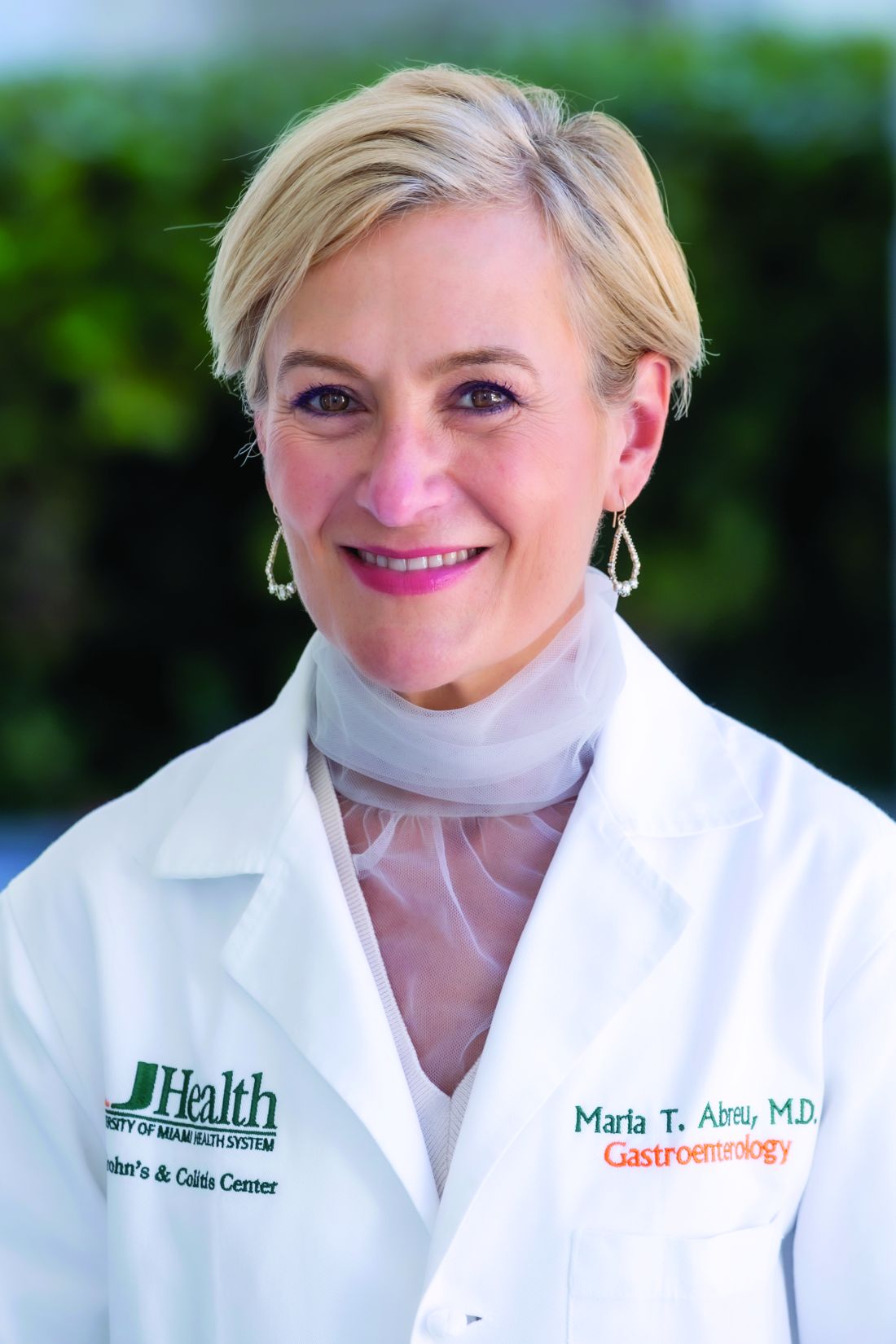
Dr. Abreu is a leader in inflammatory bowel disease patient care, and she was honored by the prestigious Sherman Prize in 2019. Her service to AGA is lengthy and begins when she took on the role of fellow representative for the research grant committee. She has since sat on both the government advocacy and diversity committees. She also served as the chair of the Immunology, Microbiology and Inflammatory Bowel Diseases Section of the AGA Council, and later as chair of the full AGA Council. While chair she developed a more streamlined in-person planning committee meeting to better organize DDW.
When asked about goals for her presidency, Dr. Abreu wants to make DDW a better experience for the modern gastroenterologist. This includes finding that perfect balance between digesting the latest education and science with networking and socializing. She plans to collaborate with the presidents of the other societies to make this come to fruition.
Perhaps the area that Dr. Abreu is most passionate about is welcoming and fostering the growth of women in gastroenterology. She wants to support women who want to succeed in academics and in practice, who want ergonomics to match their work needs, and who want to have families.
“Maria is the ultimate ‘triple threat’: master scientist, master clinician, and devoted mentor. She has not only been a major player advancing knowledge in IBD, but also motivating and pushing others to develop successful careers,” said Andres Yarur, MD, AGAF, associate professor of medicine at Cedars-Sinai Medical Center. “Her work, brilliance, passion, and charm inspire all of us and will continue to inspire many generations to come.”
She currently serves as the Martin Kalser Endowed Chair of Gastroenterology; professor of medicine, microbiology, and immunology; and director of the Crohn’s and Colitis Center at the University of Miami. Dr. Abreu is the fifth woman to lead AGA as president.
Born in New York and raised in New Jersey, Dr. Abreu grew up surrounded by a strong, tight-knit Cuban community. Her family moved to Miami when she was in the ninth grade. She later entered the 6-year medical program at the University of Miami, which was the beginning of her unparalleled academic and professional excellence in medicine.
Dr. Abreu is a leader in inflammatory bowel disease patient care, and she was honored by the prestigious Sherman Prize in 2019. Her service to AGA is lengthy and begins when she took on the role of fellow representative for the research grant committee. She has since sat on both the government advocacy and diversity committees. She also served as the chair of the Immunology, Microbiology and Inflammatory Bowel Diseases Section of the AGA Council, and later as chair of the full AGA Council. While chair she developed a more streamlined in-person planning committee meeting to better organize DDW.
When asked about goals for her presidency, Dr. Abreu wants to make DDW a better experience for the modern gastroenterologist. This includes finding that perfect balance between digesting the latest education and science with networking and socializing. She plans to collaborate with the presidents of the other societies to make this come to fruition.
Perhaps the area that Dr. Abreu is most passionate about is welcoming and fostering the growth of women in gastroenterology. She wants to support women who want to succeed in academics and in practice, who want ergonomics to match their work needs, and who want to have families.
“Maria is the ultimate ‘triple threat’: master scientist, master clinician, and devoted mentor. She has not only been a major player advancing knowledge in IBD, but also motivating and pushing others to develop successful careers,” said Andres Yarur, MD, AGAF, associate professor of medicine at Cedars-Sinai Medical Center. “Her work, brilliance, passion, and charm inspire all of us and will continue to inspire many generations to come.”
Chemo May Benefit Some Older Patients With Metastatic Pancreatic Cancer
TOPLINE:
METHODOLOGY:
Pancreatic cancer is most often diagnosed in adults aged 65 years or older. Providing cancer treatment for this older, often vulnerable, population comes with significant challenges and can lead to worse survival.
To examine real-world outcomes of older adults with untreated metastatic pancreatic cancer, researchers recruited patients aged 70 years or older and performed a geriatric assessment to identify comorbidities, cognitive issues, and other geriatric abnormalities.
Those who were deemed “fit” (ie, with no geriatric abnormalities) were assigned to receive off-study standard-of-care treatment, whereas those classified as “frail” (ie, with severe abnormalities) received off-study supportive care.
The remaining 176 “vulnerable” patients with mild to moderate geriatric abnormalities completed a geriatric and quality-of-life assessment and were then randomly assigned to receive either dose-reduced 5-fluorouracil (5-FU), leucovorin plus liposomal irinotecan (n = 88) or modified gemcitabine plus nab-paclitaxel (n = 88) every 2 weeks. Ultimately, 79 patients started the 5-FU combination and 75 received gemcitabine plus nab-paclitaxel. Patients were assessed every 8 weeks until disease progression or intolerance.
Overall, patients had a median age of 77 years; 61.9% were aged 75 years or older. About half were female, and 81.5% were White. The majority (87.5%) had a performance status of 0 or 1.
TAKEAWAY:
- Median overall survival was 4.7 months in the gemcitabine plus nab-paclitaxel arm and 4.4 months in the 5-FU combination group, with no significant survival difference observed between the two arms (P = .72).
- When the overall survival analysis was restricted to patients who received at least 4 weeks, or two cycles, of treatment (about 62% of patients), the median overall survival across the two treatment arms reached 8.0 months, in line with expectations for these regimens.
- Patient stratification revealed that those with a performance status of 2 had significantly worse overall survival than those with a status of 0: 1.4 months vs 6.9 months, respectively (hazard ratio [HR], 2.77; P < .001). A similar divide was seen when patients were stratified by physical/functional status and well-being. Age, however, did not significantly influence the results.
- Overall, more than half of patients experienced grade 3 or higher adverse events. Just over 38% of patients received only one to three cycles of therapy, whereas 26% remained on treatment for 12 or more cycles. The adverse event rates were similar between the two regimens, but the toxicity profile was slightly different — the researchers, for instance, observed more peripheral neuropathy with gemcitabine plus nab-paclitaxel and more diarrhea in the 5-FU combination arm.
IN PRACTICE:
- Overall, the “survival outcomes among vulnerable older patients were lower than expected, with high percentage of patients not able to start treatment, or complete one month of therapy due to clinical deterioration,” said study presenter Efrat Dotan, MD, chief, Division of Gastrointestinal Medical Oncology, Fox Chase Cancer Center, Philadelphia.
- “For vulnerable older adults who can tolerate treatment, these two regimens provide clinicians with options for tailoring therapy based on toxicity profile,” Dr. Dotan added. But “tools are needed to better identify patients who can benefit from treatment.”
- The results underline the need to perform geriatric assessments, as opposed to merely looking at performance status, commented David F. Chang, PhD, MS, MBBS, professor of Surgical Oncology, University of Glasgow, Scotland, who was not involved in the study.
SOURCE:
The research, presented at the 2024 annual meeting of the American Society of Clinical Oncology, was funded by the National Cancer Institute and the Eastern Cooperative Oncology Group.
LIMITATIONS:
Dr. Chang noted that the study did not reveal which treatment regimen was more effective.
DISCLOSURES:
Dr. Dotan declared relationships with Agenus, Amgen, G1 Therapeutics, Incyte, Olympus, and Taiho Pharmaceutical and institutional relationships with Dragonfly Therapeutics, Gilead Sciences, Ipsen, Kinnate Biopharma, Leap Therapeutics, Lilly, Lutris, NGM Biopharmaceuticals, Relay Therapeutics, and Zymeworks. Dr. Chang declared relationships with Immodulon Therapeutics and Mylan and institutional relationships with AstraZeneca, BMS GmbH & Co. KG, Immodulon Therapeutics, and Merck.
A version of this article appeared on Medscape.com.
HPV Vaccine Offers Cancer Protection Beyond Cervical Cancer
The analysis, featured at a press briefing ahead of the presentation at the American Society of Clinical Oncology (ASCO) 2024 annual meeting, notably found that men who received the HPV vaccine had a 56% lower risk for head and neck cancers.
“We’ve known for a long time that having the HPV vaccine can prevent the development of HPV infection, yes, but importantly, cancer,” primarily cervical cancer, said briefing moderator and ASCO president Lynn Schuchter, MD, Abramson Cancer Center, University of Pennsylvania, Philadelphia. “This is a really important study that extends the information about the impact.”
Using the US TriNetX database, lead investigator Jefferson DeKloe, BS, a research fellow with Thomas Jefferson University, Philadelphia, and colleagues created a matched cohort of 760,540 HPV-vaccinated and unvaccinated men and 945,999 HPV-vaccinated and unvaccinated women.
HPV-vaccinated men had a 54% lower risk for all HPV-related cancers (odds ratio [OR], 0.46; P < .001) and a 56% lower risk for head and neck cancers (OR, 0.44; P < .001) than unvaccinated men. There were not enough cases of anal and penile cancers for analysis.
HPV-vaccinated women had a 27% lower risk for all HPV-related cancers (OR, 0.73; P < .05), a 54% lower risk for cervical cancer (OR, 0.46; P < .05), and a 33% lower risk for head and neck cancers (OR, 0.67; 95% CI, 0.42-1.08) than HPV-unvaccinated women, but this finding was not significant. There were not enough cases of anal cancers for analysis, and the odds of developing vulvar or vaginal cancer was not significantly different in HPV-vaccinated vs unvaccinated women.
Vaccinated women, however, were less likely than unvaccinated women to develop high-grade squamous intraepithelial lesions (OR, 0.44), cervical carcinoma in situ (OR, 0.42), or abnormal Pap findings (OR, 0.87), and were less likely to undergo cone biopsy and loop electrosurgical excision (OR, 0.45).
“This study really highlights the importance of getting the HPV vaccine,” Dr. Schuchter said at the briefing.
“HPV vaccination is cancer prevention,” Glenn Hanna, MD, with Dana-Farber Cancer Institute, Boston, said in an ASCO statement.
Still, HPV vaccination rates in the United States remain relatively low. According to the National Cancer Institute, in 2022, only about 58% of adolescents aged 13-15 years had received two or three doses of HPV vaccine as recommended.
“The goal,” Dr. Schuchter said at the briefing, “is that younger girls and young boys get vaccinated to prevent development of HPV infection, and that should decrease the risk of cancer, which is what we’ve seen.”
Mr. DeKloe agreed and highlighted the importance of improving vaccination rates. “Identifying effective interventions that increase HPV vaccination rates is critical in reducing undue cancer burden in the United States,” Mr. DeKloe said in a statement.
The study had no funding source. Mr. DeKloe had no relevant disclosures. Dr. Hanna has disclosed relationships with Bicara Therapeutics, Bristol Myers Squibb, Coherus BioSciences, and others. Dr. Schuchter had no relevant disclosures.
A version of this article appeared on Medscape.com .
FROM ASCO 2024
EULAR 2024 Preview: Therapeutics in Development Take Center Stage
The European Alliance of Associations for Rheumatology (EULAR) 2024 European Congress of Rheumatology annual meeting is about to take place in Vienna, Austria. From June 12 to 15, some of the world’s leading researchers and clinicians will convene to present and learn about data on some of the new and innovative treatments for people with rheumatic and musculoskeletal diseases (RMDs) as well as to discuss how to use and optimize existing approaches.
Ahead of the Congress, this news organization asked the Congress Committee’s Scientific Programme Chair Caroline Ospelt, MD, PhD, and Abstract Chair Christian Dejaco, MD, PhD, MBA, to discuss some of their highlights of this year’s meeting.
From Bench to Bedside
“For me, the beauty at EULAR is really that you have the latest on basic research, how this can be translated in clinical trials, and then the last step would be how EULAR recommends it to be used in clinical practice,” Dr. Ospelt, professor of experimental rheumatology at University Hospital Zurich, said in an interview.

“So, if you go to EULAR continuously, you can actually follow the whole story of how novelty comes into clinical practice,” she added.
In a separate interview, Dr. Dejaco, a consultant rheumatologist and associate professor at the Medical University of Graz in Austria, said: “There are several new drug trials that are going to be presented.”
One of his highlights on the use of new drugs for the treatment of giant cell arteritis will be the phase 3 SELECT-GCA trial of the Janus kinase (JAK) inhibitor upadacitinib (LBA0001).
“It’s a trial that hopefully will lead to the approval of this drug in this indication,” Dr. Dejaco said.

Late-Breaking Abstracts
Dr. Ospelt noted: “We had a lot of good late-breaking abstracts this year.”
Some of these include:
- Real-world data on the comparative effectiveness of five different classes of drugs used to treat psoriatic arthritis (PsA; LBA0002)
- The 16-week results of a phase 2b/3 study with the novel interleukin (IL)–17A inhibitor izokibep in people with PsA (LBA0005)
- Data from the COSPIRIT-JIA trial on the efficacy and safety of ixekizumab (Taltz) in juvenile idiopathic arthritis (LBA0009)
- Phase 2 data on the safety and efficacy of the CD38-targeting monoclonal antibody daratumumab in systemic lupus erythematosus (LBA0007)
- Results of the phase 2 DAHLIAS study of the anti–neonatal Fc receptor monoclonal antibody nipocalimab in people with primary Sjögren disease (LBA0010)
- Safety and immunogenicity data from a phase 1 study of an active anti–IL-6 immunotherapy in people with knee osteoarthritis (LBA0011)
The latter is “really interesting,” Dr. Ospelt said. As of now, there is no approved treatment for osteoarthritis, and there is no immunotherapy, “so this would be the first.”
But it’s not just the late-breaker abstracts to look out for. Dr. Dejaco highlighted two abstracts that will be presented during the Abstract Plenary:
- A phase 3 study of a new selective JAK1 inhibitor, SHR0302, in rheumatoid arthritis (OP0037)
- A multi-omics analysis and targeted gene-editing study in people with , which causes inflammatory and hematologic changes (OP0073)
Of the latter, he said, “this disease is still incompletely understood, and this abstract really helps to better understand the mechanisms underlying this disease.”
One to Watch: CAR T-Cell Therapy
Dr. Ospelt said that the scientific program is about 80% clinical and 20% basic science overall. However, more sessions are being held jointly because data are starting to move from the bench to bedside.
One of the basic science areas that has had “a real buzz” around it and is now producing results in the clinic is the use of chimeric antigen receptor (CAR) T cells. In one of the first, and perhaps aptly titled What Is New, or WIN, sessions of the congress, Georg Schett, MD, vice president of research at Friedrich-Alexander-Universität Erlangen-Nüremberg in Germany, will discuss the use of CAR T-cell therapy for inflammatory RMDs. There are also multiple abstract presentations on this topic.
In-depth tissue analysis and prediction of treatment response is another interesting approach, Dr. Ospelt said. “I think that’s the way to go, that we come from the blood, we go into the tissue.” A “very nice” example of this approach will be presented during the Abstract Plenary session on Wednesday, June 12, looking at how synovial tissue macrophages may be able to give information on likely treatment response in treatment-naive rheumatoid arthritis (OP0062). There are also some further findings related to the tissue biopsy–driven treatment trial R4RA that are being presented at the meeting (OP0218, OP0242, and POS0351).
EULAR Highlighted Sessions
Among the highlighted sessions on the EULAR 2024 website is one on axial involvement in PsA and spondyloarthritis (SpA).
“Axial involvement in psoriatic arthritis and peripheral involvement in axial spondyloarthritis is quite a hot topic at the moment,” Dr. Ospelt said. There are lots of questions: “How connected are they? How different are they? Do we need different treatment for axial involvement compared to peripheral involvement?”
Another EULAR highlighted session is the 75th anniversary of glucocorticoid treatment, during which Past President of EULAR and Emeritus Professor of Rheumatology Josef S. Smolen, MD, will overview the “past, present, and future” of glucocorticoids in RMDs. Consultant rheumatologist Frank Buttgereit, MD, from the German Rheumatism Research Center in Berlin, will discuss the practicalities of using these drugs in clinical practice.
Dr. Dejaco noted: “Glucocorticoids have been one of the most important treatments for a very long time, and they’re still the most important treatment for the acute treatment of systemic inflammatory diseases.”
For a long time, there was no alternative to using steroids, he added, but steroid-sparing options now exist, and there will be data presented on a new type of drug that could potentially be used to control cortisol levels in the body (OP0335).
Recommendations and More
Dr. Ospelt and Dr. Dejaco both pointed out other sessions that are likely to be very popular, such as the first and second EULAR Recommendations sessions, a session on rheumatoid arthritis prevention, as well as the many presentations and sessions on digital health and nonpharmacologic interventions such as exercise.
With over 5242 submitted abstracts, there is going to be no shortage of data being presented at EULAR 2024. Alongside the traditional abstract submission categories, this year there is a new clinical case reports category.
“We had about 578 submissions for that category,” Dr. Dejaco said. There were 3315 abstracts submitted for the clinical research category, 812 for the basic and translational research category, 283 from health professionals in rheumatology, 152 from patient groups, and 102 in the field of pediatric rheumatology.
Join in On-Site, Watch on Demand
EULAR 2024 reverts to an on-site–only meeting this year. Some of the more lighthearted yet educational elements of the program for those attending include the second edition of the EMEUNET Rheumatology Quiz and, new for this year, two escape rooms. These rooms will provide an interactive experience where small teams will have to solve rheumatologic conundrums in order to escape the room within the hour, Dr. Dejaco explained. There will also be a morning run on Friday, June 14. “It’s not a race, it’s simply to meet and run together,” Dr. Dejaco said.
But if you cannot make the congress in person, the EULAR 2024 Livestream will be broadcasting throughout the congress. Anyone registered by June 30 will have on-demand access to the recorded content from June 17 until December 31, 2024.
Abstracts for the meeting will be published as a supplement to Annals of the Rheumatic Diseases, the official journal of EULAR.
Dr. Ospelt reported no relevant financial relationships. Dr. Dejaco has received consulting/speaker fees from AbbVie, Eli Lilly, Janssen, Sparrow, Novartis, Pfizer, Roche, Galapagos, and Sanofi.
A version of this article appeared on Medscape.com.
The European Alliance of Associations for Rheumatology (EULAR) 2024 European Congress of Rheumatology annual meeting is about to take place in Vienna, Austria. From June 12 to 15, some of the world’s leading researchers and clinicians will convene to present and learn about data on some of the new and innovative treatments for people with rheumatic and musculoskeletal diseases (RMDs) as well as to discuss how to use and optimize existing approaches.
Ahead of the Congress, this news organization asked the Congress Committee’s Scientific Programme Chair Caroline Ospelt, MD, PhD, and Abstract Chair Christian Dejaco, MD, PhD, MBA, to discuss some of their highlights of this year’s meeting.
From Bench to Bedside
“For me, the beauty at EULAR is really that you have the latest on basic research, how this can be translated in clinical trials, and then the last step would be how EULAR recommends it to be used in clinical practice,” Dr. Ospelt, professor of experimental rheumatology at University Hospital Zurich, said in an interview.
“So, if you go to EULAR continuously, you can actually follow the whole story of how novelty comes into clinical practice,” she added.
In a separate interview, Dr. Dejaco, a consultant rheumatologist and associate professor at the Medical University of Graz in Austria, said: “There are several new drug trials that are going to be presented.”
One of his highlights on the use of new drugs for the treatment of giant cell arteritis will be the phase 3 SELECT-GCA trial of the Janus kinase (JAK) inhibitor upadacitinib (LBA0001).
“It’s a trial that hopefully will lead to the approval of this drug in this indication,” Dr. Dejaco said.
Late-Breaking Abstracts
Dr. Ospelt noted: “We had a lot of good late-breaking abstracts this year.”
Some of these include:
- Real-world data on the comparative effectiveness of five different classes of drugs used to treat psoriatic arthritis (PsA; LBA0002)
- The 16-week results of a phase 2b/3 study with the novel interleukin (IL)–17A inhibitor izokibep in people with PsA (LBA0005)
- Data from the COSPIRIT-JIA trial on the efficacy and safety of ixekizumab (Taltz) in juvenile idiopathic arthritis (LBA0009)
- Phase 2 data on the safety and efficacy of the CD38-targeting monoclonal antibody daratumumab in systemic lupus erythematosus (LBA0007)
- Results of the phase 2 DAHLIAS study of the anti–neonatal Fc receptor monoclonal antibody nipocalimab in people with primary Sjögren disease (LBA0010)
- Safety and immunogenicity data from a phase 1 study of an active anti–IL-6 immunotherapy in people with knee osteoarthritis (LBA0011)
The latter is “really interesting,” Dr. Ospelt said. As of now, there is no approved treatment for osteoarthritis, and there is no immunotherapy, “so this would be the first.”
But it’s not just the late-breaker abstracts to look out for. Dr. Dejaco highlighted two abstracts that will be presented during the Abstract Plenary:
- A phase 3 study of a new selective JAK1 inhibitor, SHR0302, in rheumatoid arthritis (OP0037)
- A multi-omics analysis and targeted gene-editing study in people with , which causes inflammatory and hematologic changes (OP0073)
Of the latter, he said, “this disease is still incompletely understood, and this abstract really helps to better understand the mechanisms underlying this disease.”
One to Watch: CAR T-Cell Therapy
Dr. Ospelt said that the scientific program is about 80% clinical and 20% basic science overall. However, more sessions are being held jointly because data are starting to move from the bench to bedside.
One of the basic science areas that has had “a real buzz” around it and is now producing results in the clinic is the use of chimeric antigen receptor (CAR) T cells. In one of the first, and perhaps aptly titled What Is New, or WIN, sessions of the congress, Georg Schett, MD, vice president of research at Friedrich-Alexander-Universität Erlangen-Nüremberg in Germany, will discuss the use of CAR T-cell therapy for inflammatory RMDs. There are also multiple abstract presentations on this topic.
In-depth tissue analysis and prediction of treatment response is another interesting approach, Dr. Ospelt said. “I think that’s the way to go, that we come from the blood, we go into the tissue.” A “very nice” example of this approach will be presented during the Abstract Plenary session on Wednesday, June 12, looking at how synovial tissue macrophages may be able to give information on likely treatment response in treatment-naive rheumatoid arthritis (OP0062). There are also some further findings related to the tissue biopsy–driven treatment trial R4RA that are being presented at the meeting (OP0218, OP0242, and POS0351).
EULAR Highlighted Sessions
Among the highlighted sessions on the EULAR 2024 website is one on axial involvement in PsA and spondyloarthritis (SpA).
“Axial involvement in psoriatic arthritis and peripheral involvement in axial spondyloarthritis is quite a hot topic at the moment,” Dr. Ospelt said. There are lots of questions: “How connected are they? How different are they? Do we need different treatment for axial involvement compared to peripheral involvement?”
Another EULAR highlighted session is the 75th anniversary of glucocorticoid treatment, during which Past President of EULAR and Emeritus Professor of Rheumatology Josef S. Smolen, MD, will overview the “past, present, and future” of glucocorticoids in RMDs. Consultant rheumatologist Frank Buttgereit, MD, from the German Rheumatism Research Center in Berlin, will discuss the practicalities of using these drugs in clinical practice.
Dr. Dejaco noted: “Glucocorticoids have been one of the most important treatments for a very long time, and they’re still the most important treatment for the acute treatment of systemic inflammatory diseases.”
For a long time, there was no alternative to using steroids, he added, but steroid-sparing options now exist, and there will be data presented on a new type of drug that could potentially be used to control cortisol levels in the body (OP0335).
Recommendations and More
Dr. Ospelt and Dr. Dejaco both pointed out other sessions that are likely to be very popular, such as the first and second EULAR Recommendations sessions, a session on rheumatoid arthritis prevention, as well as the many presentations and sessions on digital health and nonpharmacologic interventions such as exercise.
With over 5242 submitted abstracts, there is going to be no shortage of data being presented at EULAR 2024. Alongside the traditional abstract submission categories, this year there is a new clinical case reports category.
“We had about 578 submissions for that category,” Dr. Dejaco said. There were 3315 abstracts submitted for the clinical research category, 812 for the basic and translational research category, 283 from health professionals in rheumatology, 152 from patient groups, and 102 in the field of pediatric rheumatology.
Join in On-Site, Watch on Demand
EULAR 2024 reverts to an on-site–only meeting this year. Some of the more lighthearted yet educational elements of the program for those attending include the second edition of the EMEUNET Rheumatology Quiz and, new for this year, two escape rooms. These rooms will provide an interactive experience where small teams will have to solve rheumatologic conundrums in order to escape the room within the hour, Dr. Dejaco explained. There will also be a morning run on Friday, June 14. “It’s not a race, it’s simply to meet and run together,” Dr. Dejaco said.
But if you cannot make the congress in person, the EULAR 2024 Livestream will be broadcasting throughout the congress. Anyone registered by June 30 will have on-demand access to the recorded content from June 17 until December 31, 2024.
Abstracts for the meeting will be published as a supplement to Annals of the Rheumatic Diseases, the official journal of EULAR.
Dr. Ospelt reported no relevant financial relationships. Dr. Dejaco has received consulting/speaker fees from AbbVie, Eli Lilly, Janssen, Sparrow, Novartis, Pfizer, Roche, Galapagos, and Sanofi.
A version of this article appeared on Medscape.com.
Autonomous AI Outperforms Humans in Optical Diagnosis of Colorectal Polyps
, while providing greater alignment with pathology-based surveillance intervals, based on a randomized controlled trial.
These findings suggest that autonomous AI may one day replace histologic assessment of diminutive polyps, reported lead author Roupen Djinbachian, MD, of the Montreal University Hospital Research Center, Montreal, Quebec, Canada, and colleagues.Optical diagnosis of diminutive colorectal polyps has been proposed as a cost-effective alternative to histologic diagnosis, but its implementation in general clinical practice has been hindered by endoscopists’ concerns about incorrect diagnoses, the investigators wrote in Gastroenterology.“AI-based systems (CADx) have been proposed as a solution to these barriers to implementation, with studies showing high adherence to Preservation and Incorporation of Valuable Endoscopic Innovations (PIVI) thresholds when using AI-H,” they wrote. “However, the efficacy and safety of autonomous AI-based diagnostic platforms have not yet been evaluated.”
To address this knowledge gap, Dr. Djinbachian and colleagues conducted a randomized controlled noninferiority trial involving 467 patients, all of whom underwent elective colonoscopies at a single academic institution.
Participants were randomly assigned to one of two groups. The first group received an optical diagnosis of diminutive (1-5 mm) colorectal polyps using an autonomous AI-based CADx system without any human input. The second group had diagnoses performed by endoscopists who used AI-H to make their optical diagnoses.
The primary outcome was the accuracy of optical diagnosis compared with the gold standard of histologic evaluation. Secondarily, the investigators explored associations between pathology-based surveillance intervals and various measures of accuracy, including sensitivity, specificity, positive predictive value (PPV), and negative predictive value (NPV).
The results showed that the accuracy of optical diagnosis for diminutive polyps was similar between the two groups, supporting noninferiority. Autonomous AI achieved an accuracy rate of 77.2%, while the AI-H group had an accuracy of 72.1%, which was not statistically significant (P = .86).
But when it came to pathology-based surveillance intervals, autonomous AI showed a clear advantage; the autonomous AI system achieved a 91.5% agreement rate, compared with 82.1% for the AI-H group (P = .016).
“These findings indicate that autonomous AI not only matches but also surpasses AI-H in accuracy for determining surveillance intervals,” the investigators wrote, noting that this finding highlights the “complexities of human interaction with AI modules where human intervention could lead to worse outcomes.”
Further analysis revealed that the sensitivity of autonomous AI for identifying adenomas was 84.8%, slightly higher than the 83.6% sensitivity of the AI-H group. Specificity was 64.4% for autonomous AI vs 63.8% for AI-H. While PPV was higher in the autonomous AI group (85.6%), compared with the AI-H group (78.6%), NPV was lower for autonomous AI than AI-H (63.0% vs 71.0%).
Dr. Djinbachian and colleagues suggested that future research should focus on larger, multicenter trials to validate these findings and further explore the integration of autonomous AI systems in clinical practice. They also noted that improving AI algorithms to accurately diagnose sessile serrated lesions could enhance the overall effectiveness of AI-based optical diagnosis.
“The performance of autonomous AI in accurately diagnosing diminutive polyps and determining appropriate surveillance intervals suggests that it could play a crucial role in streamlining colorectal cancer screening processes, reducing the burden on pathologists, and potentially lowering healthcare costs,” the investigators concluded.The study was supported by Fujifilm, which had no role in the study design or data analysis. Dr. von Renteln reported additional research funding from Vantage and Fujifilm.
In the era of computer vision for endoscopy and colonoscopy, current paradigms rely on AI as a co-pilot or second observer, with the physician serving as the final arbiter in procedure-related decision-making. This study by Djinbachian and Haumesser et al brings up the interesting wrinkle of autonomous AI as a potentially superior (or noninferior) option in narrow, task-specific use cases.
In this study, human input from the endoscopist after CADx diagnosis led to lower agreement between the AI-predicted diagnosis and corresponding surveillance intervals; human oversight more often incorrectly changed the resultant diagnosis and led to shorter than recommended surveillance intervals.
This study offers a small but very important update to the growing body of literature on CADx in colonoscopy. So far, prospective validation of CADx compared with the human eye for in-situ diagnosis of polyps has provided mixed results. This study is one of the first to examine the potential role of “automatic” CADx without additional human input and sheds light on the importance of the AI-human hybrid in medical care. How do the ways in which humans interact with the user interface and output of AI lead to changes in outcome? How can we optimize the AI-human interaction in order to provide optimal results?
Jeremy R. Glissen Brown is an assistant professor in the Department of Internal Medicine and Division of Gastroenterology at Duke University Medical Center, Durham, North Carolina. He has served as a consultant for Medtronic and Olympus, and on the advisory board for Odin Vision.
, while providing greater alignment with pathology-based surveillance intervals, based on a randomized controlled trial.
These findings suggest that autonomous AI may one day replace histologic assessment of diminutive polyps, reported lead author Roupen Djinbachian, MD, of the Montreal University Hospital Research Center, Montreal, Quebec, Canada, and colleagues.Optical diagnosis of diminutive colorectal polyps has been proposed as a cost-effective alternative to histologic diagnosis, but its implementation in general clinical practice has been hindered by endoscopists’ concerns about incorrect diagnoses, the investigators wrote in Gastroenterology.“AI-based systems (CADx) have been proposed as a solution to these barriers to implementation, with studies showing high adherence to Preservation and Incorporation of Valuable Endoscopic Innovations (PIVI) thresholds when using AI-H,” they wrote. “However, the efficacy and safety of autonomous AI-based diagnostic platforms have not yet been evaluated.”
To address this knowledge gap, Dr. Djinbachian and colleagues conducted a randomized controlled noninferiority trial involving 467 patients, all of whom underwent elective colonoscopies at a single academic institution.
Participants were randomly assigned to one of two groups. The first group received an optical diagnosis of diminutive (1-5 mm) colorectal polyps using an autonomous AI-based CADx system without any human input. The second group had diagnoses performed by endoscopists who used AI-H to make their optical diagnoses.
The primary outcome was the accuracy of optical diagnosis compared with the gold standard of histologic evaluation. Secondarily, the investigators explored associations between pathology-based surveillance intervals and various measures of accuracy, including sensitivity, specificity, positive predictive value (PPV), and negative predictive value (NPV).
The results showed that the accuracy of optical diagnosis for diminutive polyps was similar between the two groups, supporting noninferiority. Autonomous AI achieved an accuracy rate of 77.2%, while the AI-H group had an accuracy of 72.1%, which was not statistically significant (P = .86).
But when it came to pathology-based surveillance intervals, autonomous AI showed a clear advantage; the autonomous AI system achieved a 91.5% agreement rate, compared with 82.1% for the AI-H group (P = .016).
“These findings indicate that autonomous AI not only matches but also surpasses AI-H in accuracy for determining surveillance intervals,” the investigators wrote, noting that this finding highlights the “complexities of human interaction with AI modules where human intervention could lead to worse outcomes.”
Further analysis revealed that the sensitivity of autonomous AI for identifying adenomas was 84.8%, slightly higher than the 83.6% sensitivity of the AI-H group. Specificity was 64.4% for autonomous AI vs 63.8% for AI-H. While PPV was higher in the autonomous AI group (85.6%), compared with the AI-H group (78.6%), NPV was lower for autonomous AI than AI-H (63.0% vs 71.0%).
Dr. Djinbachian and colleagues suggested that future research should focus on larger, multicenter trials to validate these findings and further explore the integration of autonomous AI systems in clinical practice. They also noted that improving AI algorithms to accurately diagnose sessile serrated lesions could enhance the overall effectiveness of AI-based optical diagnosis.
“The performance of autonomous AI in accurately diagnosing diminutive polyps and determining appropriate surveillance intervals suggests that it could play a crucial role in streamlining colorectal cancer screening processes, reducing the burden on pathologists, and potentially lowering healthcare costs,” the investigators concluded.The study was supported by Fujifilm, which had no role in the study design or data analysis. Dr. von Renteln reported additional research funding from Vantage and Fujifilm.
FROM GASTROENTEROLOGY
Inflammatory Bowel Disease Highlights From Digestive Disease Week 2024
Highlights in ulcerative colitis (UC) and Crohn's disease (CD) from Digestive Disease Week® (DDW) 2024 are reported on by Dr. Andres Yarur from Cedars Sinai Medical Center in Los Angeles.
Dr. Yarur opens by discussing two phase 3 studies focused on risankizumab (RZB), which is currently approved for treatment of CD and has shown efficacy in UC. The first showed an induction period extended from 12 to 24 weeks resulted in clinical response in more than half of patients with UC.
The second study compared maintenance therapy with RZB to ustekinumab in patients with CD and found that RZB resulted in a higher rate of remission.
Dr. Yarur next looks at a study that explored use of darvadstrocel, an allogeneic stem cell therapy, in a subset of patients with CD and complex perianal fistulas. The disappointing results of the ADMIRE-CD II trial showed no benefit over placebo.
Patients hospitalized with UC, a population with few therapeutic options, were the focus of the next study. The TRIUMPH study explored use of the Janus kinase inhibitor tofacitinib for these patients and found that clinical response was achieved by 58.3% of them by day 7.
The final study addressed a clinical challenge: devising the optimal vaccination strategy for patients on immunosuppressive or anti–tumor necrosis factor therapies. Dr. Yarur reports that the study found an intensified pneumococcal vaccine regimen was more immunogenic and provided immunity for a longer duration than did the standard regimen.
--
Andres J. Yarur, MD, Associate Professor of Medicine, Cedars Sinai Medical Center, Los Angeles, California
Andres J. Yarur, MD, has disclosed the following relevant financial relationships:
Serve(d) as a consultant for: Takeda; Pfizer; Arena; AbbVie; Bristol Myers Squibb; Boehringer Ingelheim; Celltrion

COPD Highlights From ATS 2024
The latest research on treatment of patients with COPD presented at the American Thoracic Society (ATS) 2024 annual meeting is reported on by Diego J. Maselli, MD, FCCP, CHEST Physician Editorial Board Member, from UT Health San Antonio in Texas.
Dr. Maselli discusses the phase 2a COURSE study, which looked at patients with moderate to severe COPD to determine whether novel tezepelumab would help reduce exacerbations over 52 weeks. The study reached a nonsignificant numerical reduction in the annual rate vs placebo, but Dr. Maselli suggests that outcomes in patients. with high blood eosinophil counts merit further study.
Next, Dr. Maselli discusses the phase 3 NOTUS trial, looking at the efficacy and safety of the monoclonal antibody dupilumab in patients with moderate to severe COPD. The researchers found a 34% reduction in exacerbations in the dupilumab group vs placebo after 52 weeks.
He then details a 272-patient study looking at nebulized ensifentrine, a dual inhibitor of PDE3 and PDE4. The study demonstrated improved lung function as well as a reduction in exacerbation rate to patients with moderate to severe COPD treated with ensifentrined added to long-acting beta agonists-inhaled corticosteroid maintenance therapy.
Finally, Dr. Maselli highlights the MAZI study, a large retrospective analysis comparing the mortality rate in patients with COPD taking single-inhaler triple therapy (SITT) vs multiple-inhaler triple therapy (MITT). The researchers found that SITT was superior to MITT.
--
Diego J. Maselli, MD, FCCP, Professor, Chief, Division of Pulmonary Diseases & Critical Care, UT Health San Antonio, Texas
Diego J. Maselli, MD, FCCP has disclosed the following relevant financial relationships:
Serve(d) as a speaker or a member of a speakers bureau for: GSK; AstraZeneca; Sanofi/Regeneron; Amgen
Received research grant from: Gates Foundation; COPD Foundation; NIH

Narcolepsy an Independent Cardiovascular Disease Risk Factor
HOUSTON — Narcolepsy is associated with a significantly increased risk for cardiovascular disease (CVD) and major adverse cardiac events (MACEs), independent of common comorbid conditions and medications commonly used to treat the chronic sleep disorder, according to two new studies.
A nationwide analysis revealed that people with narcolepsy had a 77% higher risk for CVD and an 82% higher risk for MACE than those without the disorder.
“These findings indicate that it is important for clinicians to regularly monitor patients for cardiovascular disease and take this into consideration when recommending specific treatments for narcolepsy,” study investigators Christopher Kaufmann, PhD; Munaza Riaz, PharmD, MPhil; and Rakesh Bhattacharjee, MD, told this news organization.
“Additionally, physicians should consider monitoring the presence of other health conditions as contributing factors to the risk of CVD,” they said. Dr. Kaufmann and Dr. Riaz are with the University of Florida, Gainesville, Florida, and Dr. Bhattacharjee is with the University of California, San Diego.
They presented their research at SLEEP 2024: 38th Annual Meeting of the Associated Professional Sleep Societies.
Independent Risk Factor
The National Institute of Neurological Disorders and Stroke reports an estimated 125,000 to 200,000 people in the United States live with narcolepsy. The condition often coexists with other common health conditions including obstructive sleep apnea (OSA), diabetes, and other comorbidities, which can all contribute to the risk for CVD.
This raises doubt as to whether narcolepsy itself directly leads to CVD or if it is the result of these comorbid health conditions. Additionally, some medications used to treat narcolepsy carry their own cardiovascular risks.
Using the IBM MarketScan Commercial and Medicare supplemental databases, the researchers identified 34,562 adults with a diagnosis of narcolepsy and a propensity-matched comparison cohort of 100,405 adults without narcolepsy. The patients had a mean age of 40 years, and 62% were women.
Compared with adults without narcolepsy, those with the chronic sleep disorder that causes overwhelming daytime drowsiness had a 77% increased risk for any CVD (hazard ratio [HR], 1.77) and an 82% increased risk for MACE (HR, 1.82).
They also had an increased risk for stroke (HR, 2.04), heart failure or myocardial infarction (MI; HR, 1.64), and atrial fibrillation (HR, 1.58).
The results were similar in a separate analysis of the same population that also controlled for baseline use of stimulants, oxybates, and wake-promoting agents — medications commonly used to treat excessive daytime sleepiness associated with narcolepsy.
In this analysis, narcolepsy was associated with an 89% higher risk for CVD (HR, 1.89) and a 95% increased risk for MACE (HR, 1.95). The risk for any stroke (HR, 2.06), heart failure (HR, 1.90), atrial fibrillation (HR, 1.66), and MI (HR, 1.93) was also higher in those with narcolepsy.
“Our study found that even after considering the presence of health conditions like obstructive sleep apnea, diabetes, hypertension, hyperlipidemia, and even depression, as well as medication use, there still appears to be an independent relationship between narcolepsy and CVD,” the investigators said.
They cautioned that the mechanisms explaining the link between CVD and narcolepsy are unclear and warrant further study.
“Sleep fragmentation is a hallmark of narcolepsy, and it is speculated that this fragmentation, which may trigger disturbances in autonomic function, predisposes individuals to CVD. In rodent models, a possible link has been observed between hypocretin — a central neurotransmitter that is reduced or deficient in patients with narcolepsy — and atherosclerosis.
“However, it remains uncertain whether this is the primary mechanism related to CVD,” they commented.
Compelling Evidence for Higher CVD
Commenting on the findings for this news organization, Shaheen Lakhan, MD, a neurologist and researcher based in Miami, Florida, called for narcolepsy to be recognized as a significant contributor to higher CVD risk.
“Given the compelling evidence linking narcolepsy to a higher incidence of cardiovascular disease, it is crucial that narcolepsy be included in clinical guidelines and risk assessment tools alongside other known risk factors,” said Dr. Lakhan, who was not involved in this research.
“Physicians and health care providers should proactively address the increased cardiovascular risk associated with narcolepsy by incorporating preventive strategies and interventions into the management of patients with this condition,” Dr. Lakhan suggested.
Regular CVD screening, a healthier lifestyle, and targeted therapies could all decrease cardiac risk, Dr. Lakhan added.
“Ultimately, novel disease-modifying therapies for narcolepsy should target the core mechanisms driving the increased cardiovascular risk associated with this condition. By elucidating the specific biological pathways and developing targeted therapies that address the unique challenges faced by narcolepsy patients, we can effectively mitigate the risk,” Dr. Lakhan said.
The studies were funded by the Sleep Research Society Foundation. The authors and Dr. Lakhan had no relevant disclosures.
A version of this article appeared on Medscape.com.
FROM SLEEP 2024
Helping Patients Cut Down on Sodium: Useful Substitutes and Strategies
Humans have used salt for centuries, to preserve or cure food before refrigeration was readily available, and even as currency in some cultures. Though modern food preservation efforts have decreased our reliance on salt, we still heavily incorporate it as a flavor enhancer.
It’s only relatively recently that we’ve begun limiting salt in our diets, as research has linked high sodium intake with chronic, preventable conditions like hypertension, heart disease, and kidney disease.
How to Recommend Restriction in a Helpful Way
The US Department of Agriculture’s Dietary Guidelines for Americans recommends intake of no more than 2300 mg of sodium daily for adults and children aged 14 years or older. This echoes similar recommendations for people at risk for heart disease, kidney disease, and hypertension. However, the sodium intake of the average American still sits at a whopping 3400 mg daily.
High sodium intake is primarily the result of modern commercial food processing. Food prepared outside the home accounts for up to 70% of sodium intake in the United States, whereas only about 10% comes from salt that is added to food either during or after cooking. For this reason, I hesitate to recommend withholding salt as a primary focus when counseling on a low-sodium diet.
To many people, certain foods just taste better with salt. Many of my patients in the southern United States simply will not eat foods like eggs and tomatoes if they cannot salt them. We can spend every moment of patient interaction time explaining why excess sodium is unhealthy, but the fact remains that humans prefer food that tastes good. This is why I try to avoid counseling a “no-added-salt” diet; instead, I recommend a low-sodium diet with a focus on fresh, whole foods and limiting salt to only a few food items.
Patients should be counseled to slowly restrict their salt intake and be made aware that doing so may increase the time it takes for their sensitivity to the taste of less salty foods to return. But it is also important for them to know that it will return. The surest way to kill progress is for an unprepared patient to believe that their food will taste bland forever. A prepared patient understands that their food may taste different for a couple of weeks, but that the change will not last forever.
Types of Salt
I have often worked with patients who insist that their sodium intake is acceptable because they are using sea salt instead of table salt. This is the result of exceptional marketing and misinformation.
Specialty salts like sea salt and Himalayan pink salt contain about 560 mg and 590 mg of sodium, respectively, per quarter teaspoon. These products do have a slightly different mineral content, with sea salt typically having a negligible amount of calcium, magnesium, or potassium. The very small amount of these minerals offers no obvious health benefits compared with more affordable table salt.
The sodium content of iodized table salt is comparable to these products, with about 590 mg of sodium per quarter teaspoon. Though its high sodium content will put some practitioners off, it is also an excellent source of iodine, at about 75 mg per serving. It has been estimated that upward of 35% of the US population has iodine deficiency, most commonly due to pregnancy, avoidance of dairy products, increasing rates of vegetarianism, intake of highly processed foods, and avoidance of added salt. For this reason, and its relative affordability, I find table salt to be far more appropriate for the average American than specialty salts.
Salt Substitutes
Monosodium glutamate (MSG). MSG was previously at the center of public health concern owing to reports of “Chinese restaurant syndrome” that have since been debunked. I often recommend MSG to people trying to decrease sodium intake because the US Food and Drug Administration has designated it as GRAS (“generally recognized as safe”), and it has about one quarter of the sodium content of table salt at 125 mg per quarter teaspoon. Its crystalline structure makes it a lower-sodium salt substitute in savory applications like soups, stews, and gravies.
Hot sauce. These sauces are generally composed of peppers, vinegar, salt, and sugar. There may be some variation and occasionally added ingredients depending upon the brand. However, I find most hot sauces to be a low-sodium seasoning option that works especially well on proteins like eggs, chicken, and pork.
Potassium-based substitutes. Salt alternatives such as Nu-Salt and Morton Salt Substitute are sodium-free options with a significant amount of potassium, at 525 mg per quarter-teaspoon serving. These alternatives may not be ideal for patients with kidney problems, but they can be very helpful for those with potassium deficiency.
Herb-based seasonings. Garlic and onion powder are both sodium-free seasonings that many of my patients have found help to increase palatability while decreasing salt use. Black pepper; lemon and lime juice; salt-free herb mixes like Mrs. Dash; and spices like cumin, paprika, dill, chili powder, and ginger are also sodium-free or low-sodium alternatives that can help to alleviate blandness for someone new to a low-sodium diet. I recommend them often and use them in my own cooking at home.
Plant-based diet. If the goal of care is to improve cardiovascular or kidney health, then I find that working with patients to increase intake of plant foods to be a helpful option. This way of eating encourages replacing highly processed foods that may be high in sodium and sugar with plants that tend to be higher in potassium and calcium. The Dietary Approaches to Stop Hypertension (DASH), Mediterranean, and other plant-based diets have been shown to increase cardiovascular and metabolic health by significantly decreasing serum lipids, blood pressure, and hemoglobin A1c and promoting weight loss. They have also been shown to increase the gut microbiome and promote increased cognitive function.
I rarely encourage the use of added salt. However, research shows that putting down the salt shaker is probably not the most effective option to restrict sodium intake. For those who can cut back, these options can help keep food flavorful and patients compliant.
Ms. Winfree is a renal dietitian in private practice in Mary Esther, Florida. She has disclosed no relevant financial relationships.
A version of this article appeared on Medscape.com.