User login
Optimal JAK Inhibition for Severe Alopecia Areata
Alopecia areata (AA) is an autoimmune condition that affects children, adolescents, and adults. Severe AA often causes significant burdens, physical discomfort, and psychological stress. Yet, response to therapy is often unpredictable, running the gamut from being refractory to treatment to spontaneous remission.
Dermatologists Raj Chovatiya from Chicago Medical School and Jason Hawkes from the Pacific Skin Institute discuss how to assess AA severity and appropriate therapies, particularly the evolving landscape of JAK inhibitors for patients with severe AA.
The panelists begin by defining severe AA on the basis of the Severity of Alopecia Tool (SALT) score, which assesses AA severity by percentage of hair loss. A patient who has lost over 50% of scalp hair is considered to have severe AA.
Until recently, traditional therapy for severe AA has relied on injectable and systemic corticosteroids, both of which have drawbacks for patients with severe disease. The emergence of Janus kinase (JAK) inhibitors has offered another option for these patients. The panelists discuss recent clinical trials that have shown promising results from JAK inhibitors for treatment of severe AA.
--
Raj Chovatiya, MD, PhD, MSCI, Clinical Associate Professor, Chicago Medical School, Rosalind Franklin University of Medicine and Science, North Chicago, Illinois; Founder and Director, Center for Medical Dermatology and Immunology Research, Chicago, Illinois
Raj Chovatiya, MD, PhD, MSCI, has disclosed the following relevant financial relationships:
Serve(d) as a director, officer, partner, employee, advisor, consultant, or trustee for: AbbVie; Amgen; Apogee Therapeutics; Arcutis; Argenx; ASLAN Pharmaceuticals; Beiersdorf; Boehringer Ingelheim; Bristol Myers Squibb; Cara Therapeutics; Dermavant; Eli Lilly
Serve(d) as a speaker or a member of a speakers bureau for: AbbVie; Arcutis; Beiersdorf; Boehringer Ingelheim; Bristol Myers Squibb; Dermavant; Eli Lilly and Company; Incyte; LEO Pharma

Confronting Healthcare Disinformation on Social Media
More than 90% of internet users are active on social media, which had 4.76 billion users worldwide in January 2023. The digital revolution has reshaped the news landscape and changed how users interact with information. Social media has fostered an active relationship with the media, including the ability to interact directly with the content presented. It also has augmented media’s ability to reach a large audience with tight deadlines.
These developments suggest that social media can be a useful tool in everyday medical practice for professionals and patients. But social media also can spread misinformation, as happened during the COVID-19 pandemic.
This characteristic is the focus of the latest research by Fabiana Zollo, a computer science professor at Ca’ Foscari University of Venice, Italy, and coordinator of the Data Science for Society laboratory. The research was published in The BMJ. Ms. Zollo’s research group aims to assess the effect of social media on misinformation and consequent behaviors related to health. “The study results focus primarily on two topics, the COVID-19 pandemic and vaccinations, but can also be applied to other health-related behaviors such as smoking and diet,” Ms. Zollo told Univadis Italy.
Social media has become an important tool for public health organizations to inform and educate citizens. Institutions can use it to monitor choices and understand which topics are being discussed most at a given time, thus comprehending how the topics evolve and take shape in public discourse. “This could lead to the emergence of people’s perceptions, allowing us to understand, among other things, what the population’s needs might be, including informational needs,” said Ms. Zollo.
Tenuous Causal Link
While social media offers public health organizations the opportunity to inform and engage the public, it also raises concerns about misinformation and the difficulty of measuring its effect on health behavior. Although some studies have observed correlations between exposure to misinformation on social media and levels of adherence to vaccination campaigns, establishing a causal link is complex. As the authors emphasize, “despite the importance of the effect of social media and misinformation on people’s behavior and the broad hypotheses within public and political debates, the current state of the art cannot provide definitive conclusions on a clear causal association between social media and health behaviors.” Establishing a clear causal link between information obtained from social media and offline behavior is challenging due to methodologic limitations and the complexity of connections between online and offline behaviors. Studies often rely on self-reported data, which may not accurately reflect real behaviors, and struggle to isolate the effect of social media from other external influences. Moreover, many studies primarily focus on Western countries, limiting the generalizability of the results to other cultural and geographical conditions.
Another issue highlighted by Ms. Zollo and colleagues is the lack of complete and representative data. Studies often lack detailed information about participants, such as demographic or geolocation data, and rely on limited samples. This lack makes it difficult to assess the effect of misinformation on different segments of the population and in different geographic areas.
“The main methodologic difficulty concerns behavior, which is difficult to measure because it would require tracking a person’s actions over time and having a shared methodology to do so. We need to understand whether online stated intentions do or do not translate into actual behaviors,” said Ms. Zollo. Therefore, despite the recognized importance of the effect of social media and misinformation on people’s general behavior and the broad hypotheses expressed within public and political debates, the current state of the art cannot provide definitive conclusions on a causal association between social media and health behaviors.
Institutions’ Role
Social media is a fertile ground for the formation of echo chambers (where users find themselves dialoguing with like-minded people, forming a distorted impression of the real prevalence of that opinion) and for reinforcing polarized positions around certain topics. “We know that on certain topics, especially those related to health, there is a lot of misinformation circulating precisely because it is easy to leverage factors such as fear and beliefs, even the difficulties in understanding the technical aspects of a message,” said Ms. Zollo. Moreover, institutions have not always provided timely information during the pandemic. “Often, when there is a gap in response to a specific informational need, people turn elsewhere, where those questions find answers. And even if the response is not of high quality, it sometimes confirms the idea that the user had already created in their mind.”
The article published in The BMJ aims primarily to provide information and evaluation insights to institutions rather than professionals or healthcare workers. “We would like to spark the interest of institutions and ministries that can analyze this type of data and integrate it into their monitoring system. Social monitoring (the observation of what happens on social media) is a practice that the World Health Organization is also evaluating and trying to integrate with more traditional tools, such as questionnaires. The aim is to understand as well as possible what a population thinks about a particular health measure, such as a vaccine: Through data obtained from social monitoring, a more realistic and comprehensive view of the problem could be achieved,” said Ms. Zollo.
A Doctor’s Role
And this is where the doctor comes in: All the information thus obtained allows for identifying the needs that the population expresses and that “could push a patient to turn elsewhere, toward sources that provide answers even if of dubious quality or extremely oversimplified.” The doctor can enter this landscape by trying to understand, even with the data provided by institutions, what needs the patients are trying to fill and what drives them to seek elsewhere and to look for a reference community that offers the relevant confirmations.
From the doctor’s perspective, therefore, it can be useful to understand how these dynamics arise and evolve because they could help improve interactions with patients. At the institutional level, social monitoring would be an excellent tool for providing services to doctors who, in turn, offer a service to patients. If it were possible to identify areas where a disinformation narrative is developing from the outset, both the doctor and the institutions would benefit.
Misinformation vs Disinformation
The rapid spread of false or misleading information on social media can undermine trust in healthcare institutions and negatively influence health-related behaviors. Ms. Zollo and colleagues, in fact, speak of misinformation in their discussion, not disinformation. “In English, a distinction is made between misinformation and disinformation, a distinction that we are also adopting in Italian. When we talk about misinformation, we mean information that is generally false, inaccurate, or misleading but has not been created with the intention to harm, an intention that is present in disinformation,” said Ms. Zollo.
The distinction is often not easy to define even at the operational level, but in her studies, Ms. Zollo is mainly interested in understanding how the end user interacts with content, not the purposes for which that content was created. “This allows us to focus on users and the relationships that are created on various social platforms, thus bypassing the author of that information and focusing on how misinformation arises and evolves so that it can be effectively combated before it translates into action (ie, into incorrect health choices),” said Ms. Zollo.
This story was translated from Univadis Italy, which is part of the Medscape Professional Network, using several editorial tools, including AI, as part of the process. Human editors reviewed this content before publication. A version of this article appeared on Medscape.com.
Depression Diagnosis
Editor's Note: This article was created using several editorial tools, including AI, as part of the process. Human editors reviewed this content before publication.
Editor's Note: This article was created using several editorial tools, including AI, as part of the process. Human editors reviewed this content before publication.
Editor's Note: This article was created using several editorial tools, including AI, as part of the process. Human editors reviewed this content before publication.
Should Cancer Trial Eligibility Become More Inclusive?
The study, published online in Clinical Cancer Research, highlighted the potential benefits of broadening eligibility criteria for clinical trials.
“It is well known that results in an ‘ideal’ population do not always translate to the real-world population,” senior author Hans Gelderblom, MD, chair of the Department of Medical Oncology at the Leiden University Medical Center, Leiden, the Netherlands, said in a press release. “Eligibility criteria are often too strict, and educated exemptions by experienced investigators can help individual patients, especially in a last-resort trial.”
Although experts have expressed interest in improving trial inclusivity, it’s unclear how doing so might impact treatment safety and efficacy.
In the Drug Rediscovery Protocol (DRUP), Dr. Gelderblom and colleagues examined the impact of broadening trial eligibility on patient outcomes. DRUP is an ongoing Dutch national, multicenter, pan-cancer, nonrandomized clinical trial in which patients are treated off-label with approved molecularly targeted or immunotherapies.
In the trial, 1019 patients with treatment-refractory disease were matched to one of the available study drugs based on their tumor molecular profile and enrolled in parallel cohorts. Cohorts were defined by tumor type, molecular profile, and study drug.
Among these patients, 82 patients — 8% of the cohort — were granted waivers to participate. Most waivers (45%) were granted as exceptions to general- or drug-related eligibility criteria, often because of out-of-range lab results. Other categories included treatment and testing exceptions, as well as out-of-window testing.
The researchers then compared safety and efficacy outcomes between the 82 participants granted waivers and the 937 who did not receive waivers.
Overall, Dr. Gelderblom’s team found that the rate of serious adverse events was similar between patients who received a waiver and those who did not: 39% vs 41%, respectively.
A relationship between waivers and serious adverse events was deemed “unlikely” for 86% of patients and “possible” for 14%. In two cases concerning a direct relationship, for instance, patients who received waivers for decreased hemoglobin levels developed anemia.
The rate of clinical benefit — defined as an objective response or stable disease for at least 16 weeks — was similar between the groups. Overall, 40% of patients who received a waiver (33 of 82) had a clinical benefit vs 33% of patients without a waiver (P = .43). Median overall survival for patients that received a waiver was also similar — 11 months in the waiver group and 8 months in the nonwaiver group (hazard ratio, 0.87; P = .33).
“Safety and clinical benefit were preserved in patients for whom a waiver was granted,” the authors concluded.
The study had several limitations. The diversity of cancer types, treatments, and reasons for protocol exemptions precluded subgroup analyses. In addition, because the decision to grant waivers depended in large part on the likelihood of clinical benefit, “it is possible that patients who received waivers were positively selected for clinical benefit compared with the general study population,” the authors wrote.
So, “although the clinical benefit rate of the patient group for whom a waiver was granted appears to be slightly higher, this difference might be explained by the selection process of the central study team, in which each waiver request was carefully considered, weighing the risks and potential benefits for the patient in question,” the authors explained.
Overall, “these findings advocate for a broader and more inclusive design when establishing novel trials, paving the way for a more effective and tailored application of cancer therapies in patients with advanced or refractory disease,” Dr. Gelderblom said.
Commenting on the study, Bishal Gyawali, MD, PhD, said that “relaxing eligibility criteria is important, and I support this. Trials should include patients that are more representative of the real-world, so that results are generalizable.”
However, “the paper overemphasized efficacy,” said Dr. Gyawali, from Queen’s University, Kingston, Ontario, Canada. The sample size of waiver-granted patients was small, plus “the clinical benefit rate is not a marker of efficacy.
“The response rate is somewhat better, but for a heterogeneous study with multiple targets and drugs, it is difficult to say much about treatment effects here,” Dr. Gyawali added. Overall, “we shouldn’t read too much into treatment benefits based on these numbers.”
Funding for the study was provided by the Stelvio for Life Foundation, the Dutch Cancer Society, Amgen, AstraZeneca, Bayer, Boehringer Ingelheim, Bristol Myers Squibb, pharma&, Eisai Co., Ipsen, Merck Sharp & Dohme, Novartis, Pfizer, and Roche. Dr. Gelderblom declared no conflicts of interest, and Dr. Gyawali declared no conflicts of interest related to his comment.
A version of this article appeared on Medscape.com.
Bowel Prep Quality Affects Long-Term Colonoscopy Outcomes
TOPLINE:
METHODOLOGY:
- Few large studies have investigated the degree of bowel preparation with long-term colorectal cancer (CRC) outcomes.
- Researchers analyzed data from 335,466 individuals aged 50 years and older who underwent screening colonoscopy in Austria over 10 years (2012-2022).
- Bowel preparation quality was assessed using the five-point Aronchick scale and categorized as excellent, good, fair, poor, or inadequate.
- Logistic regression and time-to-event analyses were used to assess the impact of bowel preparation quality on adenoma detection and PCCRC mortality.
TAKEAWAY:
- Bowel prep was excellent in 37% of procedures, good in 48%, fair in 11%, poor in 3%, and inadequate in 1%.
- With worsening degrees of bowel prep, the odds of detecting an adenoma, high-risk polyp, sessile serrated lesion (SSL), or traditional serrated adenoma (TSA) decreased significantly.
- For patients with inadequate bowel preparation, the odds ratio for detection was 0.44 for adenomas and 0.53 for SSL or TSA.
- The risk of dying from PCCRC was more than twofold higher with fair or poor bowel prep and more than fourfold higher with inadequate prep.
- Cumulative 10-year CRC mortality was 0.14% for excellent/good bowel preparation vs 0.41% for fair or worse preparation.
IN PRACTICE:
“Our findings further support the evidence that bowel preparation is a crucial element of high-quality colonoscopy that affects CRC outcomes in screening participants. Efforts should be made to increase bowel cleansing above fair scores,” the authors concluded.
SOURCE:
The study, led by Jasmin Zessner-Spitzenberg, MD, from the Division of Gastroenterology and Hepatology at the Medical University of Vienna, was published online in the American Journal of Gastroenterology.
LIMITATIONS:
The researchers lacked data on CRC risk factors and information on surveillance colonoscopies, which could bias the results. Bowel preparation solutions and preferences of endoscopists, or whether split dosing was applied, were unknown, which limits insights into variations in preparation effectiveness.
DISCLOSURES:
The study was supported by the Main Association of Statutory Insurance Institutions, the Austrian Society of Gastroenterology and Hepatology, and the Austrian Cancer Aid. Dr. Zessner-Spitzenberg had no relevant disclosures. Other participating authors disclosed competing interests in the form of advisory roles, grant/research support, and speaker fees received from industry and academic institutions.
A version of this article appeared on Medscape.com.
Opioids Post T&A
I recently encountered a study that reviewed return visits of pediatric patients after undergoing adenotonsillectomy. The investigators discovered that pain-related visits were higher for patients who had received prescriptions for opioids. After the Food and Drug Administration (FDA) issued a boxed warning about the use of codeine in postoperative pediatric tonsillectomy with adenoidectomy (T&A), patients pain-related return visits declined and steroid prescriptions increased.

On the surface, this inverse relationship between opioid prescriptions and pain-related visits seems counterintuitive. This is particularly true if you believe that opioids are effective pain medications. The relationship between pain-related visits, steroid use, and the boxed warning is a bit easier to understand and most likely points to the effectiveness of the steroids.
Keeping in mind this was a single-institution study that included more than 5000 patients and more than 700 return visits, we should be careful in reading too much into these results. However, I can’t resist the temptation to use it as a springboard from which to launch a short dissertation on pain management.
First, let’s consider whether there was something about the opioids that was causing more pain for the patients. I’m not aware of any studies that suggest pain as a side effect of codeine. Nausea and vomiting, yes. And, although the investigators were focusing on pain, it may have been that the general discomfort associated with the gastrointestinal effects of the drug were lowering the patients’ pain threshold. I certainly know of many adults who have said that they now avoid opioids postoperatively because of the general sense of unwellness they have experienced during previous surgical adventures.
However, my bias leads me to focus on this question: If the patients didn’t receive opioids postoperatively, were they receiving something else that was making them less likely to arrive at the hospital or clinic complaining of pain? I assume the researchers would have told us about some new alternative miracle painkiller that was being prescribed.
As a card-carrying nihilist in good standing, I am tempted to claim that this is another example of nothing is better than most well-intentioned somethings. However, I am going to posit that these patients were receiving something that lessened their need to seek help with their pain.
Most likely that something was a thoughtful preemptive dialogue postoperatively about what they (and in most cases their parents) might expect in the way of symptoms. And ... an easy-to-reach contact point preferably with a person with whom they were familiar. And ... were scheduled to receive follow up phone calls at intervals relevant to the details of their surgery.
I know many of you are going to say, “We are already doing those things.” And, if so, you are to be commended. And, I’m sure that every outpatient postoperative manual includes all of those common-sense ingredients of good follow-up care. However, you know as well as I do that not all postoperative instructions are delivered with same degree of thoroughness nor with sufficient pauses thoughtfully delivered to make it a real dialogue. Nor is the follow-up contact person as easy to reach as promised.
I’m not sure how much we can thank the FDA boxed warning about codeine for the decrease in postoperative pain-generated visits. However, it could be that when physicians were discouraged from prescribing postoperative opioids, they may have felt the need to lean more heavily on good old-fashioned postoperative follow-up care. Instructions presented more as a dialogue and preemptive follow-up calls made with an aura of caring are well known deterrents of middle-of-the-night calls for help.
Dr. Wilkoff practiced primary care pediatrics in Brunswick, Maine, for nearly 40 years. He has authored several books on behavioral pediatrics, including “How to Say No to Your Toddler.” Other than a Littman stethoscope he accepted as a first-year medical student in 1966, Dr. Wilkoff reports having nothing to disclose. Email him at [email protected].
I recently encountered a study that reviewed return visits of pediatric patients after undergoing adenotonsillectomy. The investigators discovered that pain-related visits were higher for patients who had received prescriptions for opioids. After the Food and Drug Administration (FDA) issued a boxed warning about the use of codeine in postoperative pediatric tonsillectomy with adenoidectomy (T&A), patients pain-related return visits declined and steroid prescriptions increased.
On the surface, this inverse relationship between opioid prescriptions and pain-related visits seems counterintuitive. This is particularly true if you believe that opioids are effective pain medications. The relationship between pain-related visits, steroid use, and the boxed warning is a bit easier to understand and most likely points to the effectiveness of the steroids.
Keeping in mind this was a single-institution study that included more than 5000 patients and more than 700 return visits, we should be careful in reading too much into these results. However, I can’t resist the temptation to use it as a springboard from which to launch a short dissertation on pain management.
First, let’s consider whether there was something about the opioids that was causing more pain for the patients. I’m not aware of any studies that suggest pain as a side effect of codeine. Nausea and vomiting, yes. And, although the investigators were focusing on pain, it may have been that the general discomfort associated with the gastrointestinal effects of the drug were lowering the patients’ pain threshold. I certainly know of many adults who have said that they now avoid opioids postoperatively because of the general sense of unwellness they have experienced during previous surgical adventures.
However, my bias leads me to focus on this question: If the patients didn’t receive opioids postoperatively, were they receiving something else that was making them less likely to arrive at the hospital or clinic complaining of pain? I assume the researchers would have told us about some new alternative miracle painkiller that was being prescribed.
As a card-carrying nihilist in good standing, I am tempted to claim that this is another example of nothing is better than most well-intentioned somethings. However, I am going to posit that these patients were receiving something that lessened their need to seek help with their pain.
Most likely that something was a thoughtful preemptive dialogue postoperatively about what they (and in most cases their parents) might expect in the way of symptoms. And ... an easy-to-reach contact point preferably with a person with whom they were familiar. And ... were scheduled to receive follow up phone calls at intervals relevant to the details of their surgery.
I know many of you are going to say, “We are already doing those things.” And, if so, you are to be commended. And, I’m sure that every outpatient postoperative manual includes all of those common-sense ingredients of good follow-up care. However, you know as well as I do that not all postoperative instructions are delivered with same degree of thoroughness nor with sufficient pauses thoughtfully delivered to make it a real dialogue. Nor is the follow-up contact person as easy to reach as promised.
I’m not sure how much we can thank the FDA boxed warning about codeine for the decrease in postoperative pain-generated visits. However, it could be that when physicians were discouraged from prescribing postoperative opioids, they may have felt the need to lean more heavily on good old-fashioned postoperative follow-up care. Instructions presented more as a dialogue and preemptive follow-up calls made with an aura of caring are well known deterrents of middle-of-the-night calls for help.
Dr. Wilkoff practiced primary care pediatrics in Brunswick, Maine, for nearly 40 years. He has authored several books on behavioral pediatrics, including “How to Say No to Your Toddler.” Other than a Littman stethoscope he accepted as a first-year medical student in 1966, Dr. Wilkoff reports having nothing to disclose. Email him at [email protected].
Seladelpar Shows Clinically Meaningful Improvements in PBC
MILAN — according to two interim analyses of the ASSURE long-term extension study.
The first analysis of 337 patients with PBC, with and without cirrhosis, showed that treatment with seladelpar had a durable effect up to 2 years on cholestasis and markers of liver injury, as well as a sustained reduction in pruritus, Palak Trivedi, MD, associate professor at the National Institute for Health Research Birmingham Biomedical Research Centre, University of Birmingham, Birmingham, England, reported in a poster presented at the European Association for the Study of the Liver (EASL) Congress 2024.
The 2-year analysis also showed that seladelpar, a first-in-class, orally active agent, was safe and well tolerated in this patient population, he added.
These “results are consistent with the pivotal phase 3 RESPONSE study,” Dr. Trivedi noted. The RESPONSE study showed that seladelpar significantly improved liver biomarkers of disease activity and symptoms of pruritus at 12 months in patients with PBC who had an inadequate response or intolerance to ursodeoxycholic acid (UDCA), the standard of care, and had no history of hepatic decompensation. Patients with cirrhosis were allowed to enroll.
A total of 158 patients from the RESPONSE trial, both from the placebo and from the active treatment arm, were rolled over into the ASSURE trial. Another subset of 179 patients were drawn from prior seladelpar placebo-controlled studies (referred to as “legacy studies”), including the ENHANCE study. All participants in the current analysis received 10 mg of seladelpar, once daily, for up to 155 weeks.
Of the participants from the legacy studies, 99 completed 24 months of treatment with seladelpar, and 164 completed 12 months of treatment. In the 24-month treatment group, 70% met the composite response endpoint, which included alkaline phosphatase (ALP) levels below 1.67 times the upper limit of normal, a decrease in ALP levels of at least 15%, and total bilirubin levels at or below the upper limit of normal, according to a press release of the study findings. In addition, 42% of these participants achieved ALP normalization at 24 months, a marker of liver disease progression. In the 12-month treatment group, 73% achieved the clinically meaningful composite response endpoint, with 42% experiencing ALP normalization.
For patients rolled over from RESPONSE, 102 received 18 months of treatment with seladelpar, and 29 received 24 months of treatment. A total of 62% of patients in the 18-month group achieved the composite endpoint, and 33% achieved ALP normalization, while 72% of the 24-month group reached the composite endpoint, and 17% had ALP normalization.
Of patients who had received a placebo in the RESPONSE trial and went on to receive treatment with seladelpar, 75% achieved the composite endpoint, 27% had ALP normalization at 6 months, and 94% achieved the composite endpoint and 50% reached ALP normalization at 12 months.
Key secondary endpoints included ALP normalization and changes in liver enzymes (ALP, total bilirubin, gamma-glutamyl transferase [GGT], alanine transaminase [ALT], and aspartate aminotransferase [AST]).
Pruritus Relief Important for Quality of Life
Among study participants who reported a four or more at baseline on the numerical rating scale (NRS) for pruritus, legacy patients at 12 months and 24 months of treatment reported a mean reduction of 3.8 and 3.1, respectively. Participants from RESPONSE also reported a mean reduction of 3.8.
This level of reduction in NRS is “considered clinically significant” and takes patients from a level of moderate to severe itching down to mild, said Carrie Frenette, MD, executive director, Global Medical Affairs, Liver Diseases, Gilead Sciences, Foster City, California, and a former hepatologist of 20 years with a special interest in liver transplantation.
This “is a huge benefit in quality of life for these patients,” Dr. Frenette said in an interview.
Dr. Frenette also noted that UDCA, the current first-line treatment for PBC, is inadequate in up to 40% of patients, and second-line treatments, notably obeticholic acid, can cause itching.

Eleonora De Martin, MD, transplant hepatologist at Centre Hépato-Biliaire, Paul Brousse Hospital, Paris, France, who comoderated the session, pointed out that PBC is a complex disease.
“We need both disease control and symptom control, and they’re not always compatible,” she said. “Sometimes you can control the disease but not the symptoms, and symptomatic control is so important,” especially with pruritus.
Patients With PBC and Cirrhosis
A separate analysis from ASSURE looked at a subset of 17 patients with PBC and cirrhosis who completed 24 months of treatment. The findings were presented by Stuart Gordon, MD, professor of medicine, Wayne State University School of Medicine, and hepatologist at Henry Ford Hospital, both in Detroit.
In this analysis, the mean patient age was 60.8 years, 91.4% were female, 88.6% were Child-Pugh A, and 22.9% had portal hypertension, while the mean baseline liver stiffness by FibroScan was 19.9 kPa.
Baseline biochemical measures were mean ALP of 245.4 U/L, mean total bilirubin of 0.995 mg/dL, mean GGT of 216.1 U/L, and mean ALT of 36.6 U/L.
A total of 11 participants (65%) met the composite endpoint at 24 months, with ALP normalization in 4 patients (24%). The overall mean percent change from baseline in ALP was approximately −30% and in total bilirubin was around −14%. Other changes in biochemical markers included reductions from baseline in GGT and ALT of approximately −30% and −10%, respectively. No change was observed in AST.
While 80% of patients with cirrhosis “had an adverse event of some form,” there were no treatment-related serious adverse events.
“It’s interesting to see results in these patients who have advanced disease and are cirrhotic because it might stabilize disease or even provide improvement,” Dr. De Martin commented. “However, the numbers in the study are very small, so it’s hard to draw firm conclusions yet, but it is a first step in showing that this drug is safe.”
Seladelpar is an “important step forward in PBC because we’ve been stuck with ursodeoxycholic acid for so many years,” Dr. De Martin added. “We’ve seen in liver disease with other etiologies that sometimes just one drug can make a difference, and you can change the natural history of the disease.”
Dr. Frenette is an employee and stockholder of Gilead Sciences. Dr. Gordon declared grants and support from AbbVie, Arbutus, CymaBay, Cour Pharmaceuticals, GlaxoSmithKline (GSK), Ipsen, and Mirum Pharmaceuticals; and advisory board activity from CymaBay, GSK, and Ipsen Pharmaceuticals. Dr. De Martin had no disclosures of relevance to seladelpar but has received speaker fees from other companies, including GSK, Ipsen, and Astellas. Dr. Trivedi reports institutional funding support from National Institute for Health Research Birmingham (UK); lecture fees from Advanz Pharma/Intercept Pharmaceuticals, Albireo/Ipsen, and Dr. Falk Pharma; advisory board/consulting fees from Advanz Pharma/Intercept Pharmaceuticals, Albireo/Ipsen, Chemomab Therapeutics, CymaBay, Dr. Falk Pharma, Gilead Sciences, Perspectum, and Pliant Therapeutics; and grant support from Advanz Pharma/Intercept Pharmaceuticals, Albireo/Ipsen, Bristol-Myers Squibb, Core (Guts UK), EASL, Gilead Sciences, GSK, LifeArc, NIHR, Mirum Pharma, PSC Support, The Wellcome Trust, The Medical Research Foundation (UK), and Regeneron.
A version of this article first appeared on Medscape.com.
MILAN — according to two interim analyses of the ASSURE long-term extension study.
The first analysis of 337 patients with PBC, with and without cirrhosis, showed that treatment with seladelpar had a durable effect up to 2 years on cholestasis and markers of liver injury, as well as a sustained reduction in pruritus, Palak Trivedi, MD, associate professor at the National Institute for Health Research Birmingham Biomedical Research Centre, University of Birmingham, Birmingham, England, reported in a poster presented at the European Association for the Study of the Liver (EASL) Congress 2024.
The 2-year analysis also showed that seladelpar, a first-in-class, orally active agent, was safe and well tolerated in this patient population, he added.
These “results are consistent with the pivotal phase 3 RESPONSE study,” Dr. Trivedi noted. The RESPONSE study showed that seladelpar significantly improved liver biomarkers of disease activity and symptoms of pruritus at 12 months in patients with PBC who had an inadequate response or intolerance to ursodeoxycholic acid (UDCA), the standard of care, and had no history of hepatic decompensation. Patients with cirrhosis were allowed to enroll.
A total of 158 patients from the RESPONSE trial, both from the placebo and from the active treatment arm, were rolled over into the ASSURE trial. Another subset of 179 patients were drawn from prior seladelpar placebo-controlled studies (referred to as “legacy studies”), including the ENHANCE study. All participants in the current analysis received 10 mg of seladelpar, once daily, for up to 155 weeks.
Of the participants from the legacy studies, 99 completed 24 months of treatment with seladelpar, and 164 completed 12 months of treatment. In the 24-month treatment group, 70% met the composite response endpoint, which included alkaline phosphatase (ALP) levels below 1.67 times the upper limit of normal, a decrease in ALP levels of at least 15%, and total bilirubin levels at or below the upper limit of normal, according to a press release of the study findings. In addition, 42% of these participants achieved ALP normalization at 24 months, a marker of liver disease progression. In the 12-month treatment group, 73% achieved the clinically meaningful composite response endpoint, with 42% experiencing ALP normalization.
For patients rolled over from RESPONSE, 102 received 18 months of treatment with seladelpar, and 29 received 24 months of treatment. A total of 62% of patients in the 18-month group achieved the composite endpoint, and 33% achieved ALP normalization, while 72% of the 24-month group reached the composite endpoint, and 17% had ALP normalization.
Of patients who had received a placebo in the RESPONSE trial and went on to receive treatment with seladelpar, 75% achieved the composite endpoint, 27% had ALP normalization at 6 months, and 94% achieved the composite endpoint and 50% reached ALP normalization at 12 months.
Key secondary endpoints included ALP normalization and changes in liver enzymes (ALP, total bilirubin, gamma-glutamyl transferase [GGT], alanine transaminase [ALT], and aspartate aminotransferase [AST]).
Pruritus Relief Important for Quality of Life
Among study participants who reported a four or more at baseline on the numerical rating scale (NRS) for pruritus, legacy patients at 12 months and 24 months of treatment reported a mean reduction of 3.8 and 3.1, respectively. Participants from RESPONSE also reported a mean reduction of 3.8.
This level of reduction in NRS is “considered clinically significant” and takes patients from a level of moderate to severe itching down to mild, said Carrie Frenette, MD, executive director, Global Medical Affairs, Liver Diseases, Gilead Sciences, Foster City, California, and a former hepatologist of 20 years with a special interest in liver transplantation.
This “is a huge benefit in quality of life for these patients,” Dr. Frenette said in an interview.
Dr. Frenette also noted that UDCA, the current first-line treatment for PBC, is inadequate in up to 40% of patients, and second-line treatments, notably obeticholic acid, can cause itching.
Eleonora De Martin, MD, transplant hepatologist at Centre Hépato-Biliaire, Paul Brousse Hospital, Paris, France, who comoderated the session, pointed out that PBC is a complex disease.
“We need both disease control and symptom control, and they’re not always compatible,” she said. “Sometimes you can control the disease but not the symptoms, and symptomatic control is so important,” especially with pruritus.
Patients With PBC and Cirrhosis
A separate analysis from ASSURE looked at a subset of 17 patients with PBC and cirrhosis who completed 24 months of treatment. The findings were presented by Stuart Gordon, MD, professor of medicine, Wayne State University School of Medicine, and hepatologist at Henry Ford Hospital, both in Detroit.
In this analysis, the mean patient age was 60.8 years, 91.4% were female, 88.6% were Child-Pugh A, and 22.9% had portal hypertension, while the mean baseline liver stiffness by FibroScan was 19.9 kPa.
Baseline biochemical measures were mean ALP of 245.4 U/L, mean total bilirubin of 0.995 mg/dL, mean GGT of 216.1 U/L, and mean ALT of 36.6 U/L.
A total of 11 participants (65%) met the composite endpoint at 24 months, with ALP normalization in 4 patients (24%). The overall mean percent change from baseline in ALP was approximately −30% and in total bilirubin was around −14%. Other changes in biochemical markers included reductions from baseline in GGT and ALT of approximately −30% and −10%, respectively. No change was observed in AST.
While 80% of patients with cirrhosis “had an adverse event of some form,” there were no treatment-related serious adverse events.
“It’s interesting to see results in these patients who have advanced disease and are cirrhotic because it might stabilize disease or even provide improvement,” Dr. De Martin commented. “However, the numbers in the study are very small, so it’s hard to draw firm conclusions yet, but it is a first step in showing that this drug is safe.”
Seladelpar is an “important step forward in PBC because we’ve been stuck with ursodeoxycholic acid for so many years,” Dr. De Martin added. “We’ve seen in liver disease with other etiologies that sometimes just one drug can make a difference, and you can change the natural history of the disease.”
Dr. Frenette is an employee and stockholder of Gilead Sciences. Dr. Gordon declared grants and support from AbbVie, Arbutus, CymaBay, Cour Pharmaceuticals, GlaxoSmithKline (GSK), Ipsen, and Mirum Pharmaceuticals; and advisory board activity from CymaBay, GSK, and Ipsen Pharmaceuticals. Dr. De Martin had no disclosures of relevance to seladelpar but has received speaker fees from other companies, including GSK, Ipsen, and Astellas. Dr. Trivedi reports institutional funding support from National Institute for Health Research Birmingham (UK); lecture fees from Advanz Pharma/Intercept Pharmaceuticals, Albireo/Ipsen, and Dr. Falk Pharma; advisory board/consulting fees from Advanz Pharma/Intercept Pharmaceuticals, Albireo/Ipsen, Chemomab Therapeutics, CymaBay, Dr. Falk Pharma, Gilead Sciences, Perspectum, and Pliant Therapeutics; and grant support from Advanz Pharma/Intercept Pharmaceuticals, Albireo/Ipsen, Bristol-Myers Squibb, Core (Guts UK), EASL, Gilead Sciences, GSK, LifeArc, NIHR, Mirum Pharma, PSC Support, The Wellcome Trust, The Medical Research Foundation (UK), and Regeneron.
A version of this article first appeared on Medscape.com.
FROM EASL 2024
Act Fast With Traction Alopecia to Avoid Permanent Hair Loss
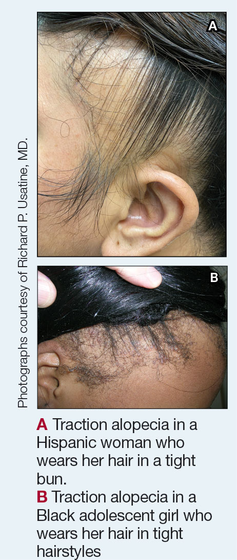
Traction alopecia (TA) is a common type of alopecia that ultimately can result in permanent hair loss. It often is caused or worsened by repetitive and prolonged hairstyling practices such as tight ponytails, braids, or locs, or use of wigs or weaves.1 Use of headwear, as in certain religious or ethnic groups, also can be contributory.2 Individuals participating in or training for occupations involving military service or ballet are at risk for TA due to hairstyling-specific policies. Early stages of TA are reversible with proper treatment and avoidance of exacerbating factors, emphasizing the importance of prompt recognition.3
Epidemiology
Data on the true prevalence of TA are lacking. It can occur in individuals of any race or any hair type. However, it is most common in women of African descent, affecting approximately one-third of this population.4 Other commonly affected groups include ballerinas and active-duty service members due to tight ponytails and buns, as well as the Sikh population due to the use of turbans as a part of their religious practice.2,5,6
Traction alopecia also impacts children, particularly those of African descent. A 2007 study of schoolchildren in South Africa determined that more than 17% of young African girls had evidence of TA—even some as young as 6 years of age.7
Traction alopecia can be caused or exacerbated by the use of hair clips and bobby pins that aid holding styles in place.8 Hair shaft morphology may contribute to the risk for TA, with more tightly coiled hair types being more susceptible.8 Variables such as use of chemical relaxers also increase the risk for disease, especially when combined with high-tension styling methods such as braids.9
Key clinical features
Patients with TA clinically present with hair loss and breakage in areas with tension, most commonly the marginal areas of the scalp as well as the frontal hairline and temporal scalp. Hair loss can result in a “fringe sign,” in which a patient may have preservation of a thin line of hairs at the frontal aspect of the hairline with a band of hair loss behind.10 This presentation may be used to differentiate TA from other forms of alopecia, including frontal fibrosing alopecia and female pattern hair loss. When the hair loss is not marginal, it may mimic other forms of patchy hair loss including alopecia areata and trichotillomania. Other clinical findings in TA may include broken hairs, pustules, and follicular papules.10 Patients also may describe symptoms such as scalp tenderness with specific hairstyles or headaches,11 or they may be completely asymptomatic.
Trichoscopy can be helpful in guiding diagnosis and treatment. Patients with TA often have perifollicular erythema and hair casts (cylindrical structures that encircle the proximal hair shafts) in the earlier stages of the disease, with eventual loss of follicular ostia in the later stages.10,12 Hair casts also may indicate ongoing traction.12 The flambeau sign—white tracks seen on trichoscopy in the direction the hair is pulled—resembles a lit torch.13
Worth noting
Early-stage TA can be reversed by avoiding hair tension. However, patients may not be amenable to this due to personal hairstyling preferences, job duties, or religious practices. Treatment with topical or intralesional steroids or even oral antibiotics such as doxycycline for its anti-inflammatory ability may result in regrowth of lost hair if the follicles are not permanently lost and exacerbating factors are avoided.3,14 Both topical and oral minoxidil have been used with success, with minoxidil thought to increase hair density by extending the anagen (growth) phase of hair follicles.3,15 Culturally sensitive patient counseling on the condition and potential exacerbating factors is critical.16
At later stages of the disease—after loss of follicular ostia has occurred—surgical interventions should be considered,17 such as hair transplantation, which can be successful but remains a technical challenge due to variability in hair shaft curvature.18 Additionally, the cost of the procedure can limit use, and some patients may not be optimal candidates due to the extent of their hair loss. Traction alopecia may not be the only hair loss condition present. Examining the scalp is important even if the chief area of concern is the marginal scalp.
Health disparity highlight
Prevention, early identification, and treatment initiated in a timely fashion are crucial to prevent permanent hair loss. There are added societal and cultural pressures that impact hairstyle and hair care practices, especially for those with tightly coiled hair.19 Historically, tightly coiled hair has been unfairly viewed as “unprofessional,” “unkempt,” and a challenge to “manage” by some. Thus, heat, chemical relaxers, and tight hairstyles holding hair in one position have been used to straighten the hair permanently or temporarily or to keep it maintained in a style that did not necessitate excessive manipulation—often contributing to further tension on the hair.
Military service branches have evaluated and changed some hair-related policies to reflect the diverse hair types of military personnel.20 The CROWN Act (www.thecrownact.com/about)—“Creating a Respectful and Open World for Natural Hair”—is a model law passed by 26 states that prohibits race-based hair discrimination, which is the denial of employment and educational opportunities because of hair texture. Although the law has not been passed in every state, it may help individuals with tightly coiled hair to embrace natural hairstyles. However, even hairstyles with one’s own natural curl pattern can contribute to tension and thus potential development of TA.
1. Larrondo J, McMichael AJ. Traction alopecia. JAMA Dermatol. 2023;159:676. doi:10.1001/jamadermatol.2022.6298
2. James J, Saladi RN, Fox JL. Traction alopecia in Sikh male patients. J Am Board Fam Med. 2007;20:497-498. doi:10.3122/jabfm.2007.05.070076
3. Callender VD, McMichael AJ, Cohen GF. Medical and surgical therapies for alopecias in black women. Dermatol Ther. 2004;17:164-176.
4. Loussouarn G, El Rawadi C, Genain G. Diversity of hair growth profiles. Int J Dermatol. 2005;44(suppl 1):6-9.
5. Samrao AChen CZedek Det al. Traction alopecia in a ballerina: clinicopathologic features. Arch Dermatol. 2010;146:918-935. doi:10.1001/archdermatol.2010.183
6. Korona-Bailey J, Banaag A, Nguyen DR, et al. Free the bun: prevalence of alopecia among active duty service women, fiscal years 2010-2019. Mil Med. 2023;188:e492-e496. doi:10.1093/milmed/usab274
7. Khumalo NP, Jessop S, Gumedze F, et al. Hairdressing is associated with scalp disease in African schoolchildren. Br J Dermatol. 2007;157:106-110. doi:10.1111/j.1365-2133.2007.07987.x
8. Billero V, Miteva M. Traction alopecia: the root of the problem. Clin Cosmet Investig Dermatol. 2018;11:149-159. doi:10.2147/CCID.S137296
9. Haskin A, Aguh C. All hairstyles are not created equal: what the dermatologist needs to know about black hairstyling practices and the risk of traction alopecia (TA). J Am Acad Dermatol. 2016;75:606-611. doi:10.1016/j.jaad.2016.02.1162
10. Samrao A, Price VH, Zedek D, et al. The “fringe sign”—a useful clinical finding in traction alopecia of the marginal hair line. Dermatol Online J. 2011;17:1.
11. Kararizou E, Bougea AM, Giotopoulou D, et al. An update on the less-known group of other primary headaches—a review. Eur Neurol Rev. 2014;9:71-77. doi:10.17925/ENR.2014.09.01.71
12. Tosti A, Miteva M, Torres F, et al. Hair casts are a dermoscopic clue for the diagnosis of traction alopecia. Br J Dermatol. 2010;163:1353-1355.
13. Agrawal S, Daruwalla SB, Dhurat RS. The flambeau sign—a new dermoscopy finding in a case of marginal traction alopecia. Australas J Dermatol. 2020;61:49-50. doi:10.1111/ajd.13187
14. Lawson CN, Hollinger J, Sethi S, et al. Updates in the understanding and treatments of skin & hair disorders in women of color. Int J Womens Dermatol. 2017;3:S21-S37.
15. Awad A, Chim I, Sharma P, et al. Low-dose oral minoxidil improves hair density in traction alopecia. J Am Acad Dermatol. 2023;89:157-159. doi:10.1016/j.jaad.2023.02.024
16. Grayson C, Heath CR. Counseling about traction alopecia: a “compliment, discuss, and suggest” method. Cutis. 2021;108:20-22.
17. Ozçelik D. Extensive traction alopecia attributable to ponytail hairstyle and its treatment with hair transplantation. Aesthetic Plast Surg. 2005;29:325-327. doi:10.1007/s00266-005-0004-5
18. Singh MK, Avram MR. Technical considerations for follicular unit extraction in African-American hair. Dermatol Surg. 2013;39:1282-1284. doi:10.1111/dsu.12229
19. Jones NL, Heath CR. Hair at the intersection of dermatology and anthropology: a conversation on race and relationships. Pediatr Dermatol. 2021;38(suppl 2):158-160.
20. Franklin JMM, Wohltmann WE, Wong EB. From buns to braids and ponytails: entering a new era of female military hair-grooming standards. Cutis. 2021;108:31-35. doi:10.12788/cutis.0296
Traction alopecia (TA) is a common type of alopecia that ultimately can result in permanent hair loss. It often is caused or worsened by repetitive and prolonged hairstyling practices such as tight ponytails, braids, or locs, or use of wigs or weaves.1 Use of headwear, as in certain religious or ethnic groups, also can be contributory.2 Individuals participating in or training for occupations involving military service or ballet are at risk for TA due to hairstyling-specific policies. Early stages of TA are reversible with proper treatment and avoidance of exacerbating factors, emphasizing the importance of prompt recognition.3
Epidemiology
Data on the true prevalence of TA are lacking. It can occur in individuals of any race or any hair type. However, it is most common in women of African descent, affecting approximately one-third of this population.4 Other commonly affected groups include ballerinas and active-duty service members due to tight ponytails and buns, as well as the Sikh population due to the use of turbans as a part of their religious practice.2,5,6
Traction alopecia also impacts children, particularly those of African descent. A 2007 study of schoolchildren in South Africa determined that more than 17% of young African girls had evidence of TA—even some as young as 6 years of age.7
Traction alopecia can be caused or exacerbated by the use of hair clips and bobby pins that aid holding styles in place.8 Hair shaft morphology may contribute to the risk for TA, with more tightly coiled hair types being more susceptible.8 Variables such as use of chemical relaxers also increase the risk for disease, especially when combined with high-tension styling methods such as braids.9
Key clinical features
Patients with TA clinically present with hair loss and breakage in areas with tension, most commonly the marginal areas of the scalp as well as the frontal hairline and temporal scalp. Hair loss can result in a “fringe sign,” in which a patient may have preservation of a thin line of hairs at the frontal aspect of the hairline with a band of hair loss behind.10 This presentation may be used to differentiate TA from other forms of alopecia, including frontal fibrosing alopecia and female pattern hair loss. When the hair loss is not marginal, it may mimic other forms of patchy hair loss including alopecia areata and trichotillomania. Other clinical findings in TA may include broken hairs, pustules, and follicular papules.10 Patients also may describe symptoms such as scalp tenderness with specific hairstyles or headaches,11 or they may be completely asymptomatic.
Trichoscopy can be helpful in guiding diagnosis and treatment. Patients with TA often have perifollicular erythema and hair casts (cylindrical structures that encircle the proximal hair shafts) in the earlier stages of the disease, with eventual loss of follicular ostia in the later stages.10,12 Hair casts also may indicate ongoing traction.12 The flambeau sign—white tracks seen on trichoscopy in the direction the hair is pulled—resembles a lit torch.13
Worth noting
Early-stage TA can be reversed by avoiding hair tension. However, patients may not be amenable to this due to personal hairstyling preferences, job duties, or religious practices. Treatment with topical or intralesional steroids or even oral antibiotics such as doxycycline for its anti-inflammatory ability may result in regrowth of lost hair if the follicles are not permanently lost and exacerbating factors are avoided.3,14 Both topical and oral minoxidil have been used with success, with minoxidil thought to increase hair density by extending the anagen (growth) phase of hair follicles.3,15 Culturally sensitive patient counseling on the condition and potential exacerbating factors is critical.16
At later stages of the disease—after loss of follicular ostia has occurred—surgical interventions should be considered,17 such as hair transplantation, which can be successful but remains a technical challenge due to variability in hair shaft curvature.18 Additionally, the cost of the procedure can limit use, and some patients may not be optimal candidates due to the extent of their hair loss. Traction alopecia may not be the only hair loss condition present. Examining the scalp is important even if the chief area of concern is the marginal scalp.
Health disparity highlight
Prevention, early identification, and treatment initiated in a timely fashion are crucial to prevent permanent hair loss. There are added societal and cultural pressures that impact hairstyle and hair care practices, especially for those with tightly coiled hair.19 Historically, tightly coiled hair has been unfairly viewed as “unprofessional,” “unkempt,” and a challenge to “manage” by some. Thus, heat, chemical relaxers, and tight hairstyles holding hair in one position have been used to straighten the hair permanently or temporarily or to keep it maintained in a style that did not necessitate excessive manipulation—often contributing to further tension on the hair.
Military service branches have evaluated and changed some hair-related policies to reflect the diverse hair types of military personnel.20 The CROWN Act (www.thecrownact.com/about)—“Creating a Respectful and Open World for Natural Hair”—is a model law passed by 26 states that prohibits race-based hair discrimination, which is the denial of employment and educational opportunities because of hair texture. Although the law has not been passed in every state, it may help individuals with tightly coiled hair to embrace natural hairstyles. However, even hairstyles with one’s own natural curl pattern can contribute to tension and thus potential development of TA.
1. Larrondo J, McMichael AJ. Traction alopecia. JAMA Dermatol. 2023;159:676. doi:10.1001/jamadermatol.2022.6298
2. James J, Saladi RN, Fox JL. Traction alopecia in Sikh male patients. J Am Board Fam Med. 2007;20:497-498. doi:10.3122/jabfm.2007.05.070076
3. Callender VD, McMichael AJ, Cohen GF. Medical and surgical therapies for alopecias in black women. Dermatol Ther. 2004;17:164-176.
4. Loussouarn G, El Rawadi C, Genain G. Diversity of hair growth profiles. Int J Dermatol. 2005;44(suppl 1):6-9.
5. Samrao AChen CZedek Det al. Traction alopecia in a ballerina: clinicopathologic features. Arch Dermatol. 2010;146:918-935. doi:10.1001/archdermatol.2010.183
6. Korona-Bailey J, Banaag A, Nguyen DR, et al. Free the bun: prevalence of alopecia among active duty service women, fiscal years 2010-2019. Mil Med. 2023;188:e492-e496. doi:10.1093/milmed/usab274
7. Khumalo NP, Jessop S, Gumedze F, et al. Hairdressing is associated with scalp disease in African schoolchildren. Br J Dermatol. 2007;157:106-110. doi:10.1111/j.1365-2133.2007.07987.x
8. Billero V, Miteva M. Traction alopecia: the root of the problem. Clin Cosmet Investig Dermatol. 2018;11:149-159. doi:10.2147/CCID.S137296
9. Haskin A, Aguh C. All hairstyles are not created equal: what the dermatologist needs to know about black hairstyling practices and the risk of traction alopecia (TA). J Am Acad Dermatol. 2016;75:606-611. doi:10.1016/j.jaad.2016.02.1162
10. Samrao A, Price VH, Zedek D, et al. The “fringe sign”—a useful clinical finding in traction alopecia of the marginal hair line. Dermatol Online J. 2011;17:1.
11. Kararizou E, Bougea AM, Giotopoulou D, et al. An update on the less-known group of other primary headaches—a review. Eur Neurol Rev. 2014;9:71-77. doi:10.17925/ENR.2014.09.01.71
12. Tosti A, Miteva M, Torres F, et al. Hair casts are a dermoscopic clue for the diagnosis of traction alopecia. Br J Dermatol. 2010;163:1353-1355.
13. Agrawal S, Daruwalla SB, Dhurat RS. The flambeau sign—a new dermoscopy finding in a case of marginal traction alopecia. Australas J Dermatol. 2020;61:49-50. doi:10.1111/ajd.13187
14. Lawson CN, Hollinger J, Sethi S, et al. Updates in the understanding and treatments of skin & hair disorders in women of color. Int J Womens Dermatol. 2017;3:S21-S37.
15. Awad A, Chim I, Sharma P, et al. Low-dose oral minoxidil improves hair density in traction alopecia. J Am Acad Dermatol. 2023;89:157-159. doi:10.1016/j.jaad.2023.02.024
16. Grayson C, Heath CR. Counseling about traction alopecia: a “compliment, discuss, and suggest” method. Cutis. 2021;108:20-22.
17. Ozçelik D. Extensive traction alopecia attributable to ponytail hairstyle and its treatment with hair transplantation. Aesthetic Plast Surg. 2005;29:325-327. doi:10.1007/s00266-005-0004-5
18. Singh MK, Avram MR. Technical considerations for follicular unit extraction in African-American hair. Dermatol Surg. 2013;39:1282-1284. doi:10.1111/dsu.12229
19. Jones NL, Heath CR. Hair at the intersection of dermatology and anthropology: a conversation on race and relationships. Pediatr Dermatol. 2021;38(suppl 2):158-160.
20. Franklin JMM, Wohltmann WE, Wong EB. From buns to braids and ponytails: entering a new era of female military hair-grooming standards. Cutis. 2021;108:31-35. doi:10.12788/cutis.0296
In Prostate Cancer, Most Roads Lead to VA Pathway
The newly updated US Department of Veterans Affairs (VA) prostate cancer clinical pathway looks like a set of guidelines, but it’s really something unique. As attendees learned at an Association of VA Hematology/Oncology (AVAHO) regional meeting in Detroit in June, the clinical pathways are designed to point the way toward a standard ideal treatment for the majority of cases, not just to suggest a number of possible options.
“Pathways will always offer one scenario. They try to get oncologists to practice in a similar fashion so things can be managed more uniformly,” Michael M. Goodman, MD, told Federal Practitioner prior to the AVAHO meeting that was focused on prostate cancer care. Goodman is an associate professor of medicine with Atrium Health Wake Forest Baptist Medical Center and helped develop the VA genitourinary oncology pathways.
“The overall goal is not just to standardize care as much as possible but also to synthesize the best and most cost-effective practices,” Goodman said. For example, “If you have 5 different therapies, and they all have about the same efficacy and safety, and 1 is less costly than the other 4, then it would make sense to choose that.”
The VA has offered pathways for multiple types of cancer since 2021, and the pathway for prostate cancer is among the most comprehensive. The VA system updated the pathway in March 2024, is available online both via SharePoint and externally.
“It goes through the entire gamut from screening, diagnosis, and management to end of life,” Goodman explained. Multiple disciplines, from primary care and surgery to genetics and imaging, can rely on the pathway to assist decision-making.
In terms of screening, the pathway offers a flow map guiding the screening choices. In patients aged ≤ 54 years, only certain high-risk groups, such as African Americans and those with a family history of prostate cancer, should be screened. From ages 54 to 69 years, patients should be consulted as part of a shared decision making process, while screening is not recommended for patients aged ≥ 70 years.
Pathway flow maps also provide information about diagnostic standards, evaluation of the newly diagnosed, risk stratification, molecular testing, and end-of-life care.
Goodman says the pathway is now integrated into the VA electronic health record system via a template so clinicians can easily document pathway use. This allows the VA to track the use of the pathways locally, regionally, and nationally track the use of the pathways.
Clinicians are not mandated to follow every step in the pathway, but Goodman said the goal is > 80% adherence. If clinicians follow the standards, he said, “you’re considering efficacy, safety, and cost for that veteran.”
Prospective data suggests that adherence to the pathway eliminates certain disparities. African American veterans, for example, are as well-represented or even better represented than White veterans in prostate cancer care when pathways are followed.
Why might clinicians veer from the pathway? “If you’re seeing a patient who was treated in the community with drug X, but drug Y is chosen by the pathway, you can carry on with the previous care.” Alternatively, in some cases, patients may not tolerate the pathway standard, Goodman noted.
Goodman reports that he consults the pathway every day. “It’s helped standardize the care I provide to ensure there’s no gaps in how I’m treating patients.”
Fit for Promotion: Navy Changes the Policy
Time was—recent time, that is—sailors had two chances to pass a physical fitness assessment (PFA). Failing the first meant no promotion. Failing the second: No career. They could neither be promoted nor reenlist.
That’s changed; as of this month, the Navy now allows the sailor’s commanding officer to decide whether the sailor gets to go on, even after failing a second test.
In an administrative letter, Vice Adm. Rick Cheeseman, chief of naval personnel, said, "Commanding officers can now evaluate a sailor's physical readiness progress or lack of progress in performance evaluations, giving them the ability to manage risk, recognize earnest effort, and best take care of their people.”
According to the new policy, sailors who fail any PFA no longer need to have it noted on their annual evaluation (although they still may not advance until they pass another test). Enlisted sailors who fail a second consecutive PFA are no longer required to receive the lowest possible score in the "Military Bearing/Professionalism" category and are not denied the ability to reenlist.
In assessing eligibility for enlisted members, the memo states that commanders should consider a sailor’s ability to perform the functions of their rate without physical or medical limitation at sea, shore or isolated duty; their overall ability to contribute to Navy missions; and the likelihood of improvement in meeting PFA standards within the next 12 months.
“Building the bodies of great people,” Cheeseman wrote, “is more than annual (or biannual) testing and includes ensuring healthy food, adequate sleep, opportunities to exercise (especially outside), and medical readiness.”
According to a report by Military.com, “critics have argued that many of the changes were the Navy relaxing its standards in the face of a challenging recruiting environment and an increasingly overweight population of Americans.” However, Navy data provided in November indicate that the number of sailors failing PFAs has remained very low. In 2017, nearly 98% of sailors passed the PFA, and 95.1% passed the first post-pandemic PFA in 2022.
The PFA policy changes are part of the Navy’s Culture of Excellence 2.0, initiated earlier this year, Cheeseman says. This initiative “charges our leaders to build great people, great leaders, and great teams: their minds, bodies, and spirits, eliminating barriers wherever possible. In response, we are modernizing our PFA policy to acknowledge our diverse population, increase sailor trust, and enhance quality of service.”