User login
AVAHO
div[contains(@class, 'header__large-screen')]
div[contains(@class, 'read-next-article')]
div[contains(@class, 'nav-primary')]
nav[contains(@class, 'nav-primary')]
section[contains(@class, 'footer-nav-section-wrapper')]
footer[@id='footer']
div[contains(@class, 'main-prefix')]
section[contains(@class, 'nav-hidden')]
div[contains(@class, 'ce-card-content')]
nav[contains(@class, 'nav-ce-stack')]
AVAHO Mtg: Germline Testing Key for Vets With High-Risk PC
Not too long ago, prostate-cancer genetics didn’t mean much to patient care. But in recent years, the landscape of therapy has transformed as researchers have discovered links between multiple genes and aggressive tumors.
Now, as a hematologist-oncologist explained to attendees at an Association of VA Hematology/Oncology regional meeting in Detroit, genetic tests can guide treatment for some—but not all—men with prostate cancer.
For patients with mutations, appropriate supplemental medications “can improve overall outcomes and have a long-standing impact on patients” said Scott J. Dawsey, MD, of the John D. Dingell Veterans Affairs Medical Center in Detroit in an interview following the AVAHO meeting, which focused on the management of prostate cancer.
As Dawsey explained, about 10% of patients with prostate cancer appear to have genetic mutations, although the exact percentage is unclear. The mutations are especially common in metastatic forms of prostate cancer. They’re estimated to be present in 11.8%-16.2% of those cases.
While these proportions are relatively small, the number of overall prostate-cancer cases with mutations is large due to the high burden of the disease, Dawsey said. Prostate cancer is by far the most common cancer in men, and estimated 299,010 cases will be diagnosed in the United States this year.
According to Dawsey, genetic mutations seem to boost the risk of more aggressive disease—and the risk of other malignancies—by disrupting DNA repair. This process can lead to even more mutations that may “make the cancer behave and grow more aggressively.”
But not all prostate cancer patients need to undergo genetic testing. Dawsey urged colleagues to figure out which patients should be tested by consulting National Comprehensive Cancer Network (NCCN) guidelines and the newly updated US Department of Veterans Affairs (VA) prostate cancer clinical pathway.
The two sets of recommendations agree on germline testing in patients with cases that are metastatic, very high risk, and high risk. Lower-risk cases should only be tested if patients meet family history criteria. The sets of guidelines also recommend somatic testing in patients with metastatic cancer.
In addition to providing guidance about treatment, genetic test results can have implications regarding other potential malignancies that may affect patients, Dawsey said. The results may also have implications for cancer risk in family members.
Several drugs are now available for patients with genetic mutations, including checkpoint inhibitors and PARP inhibitors. The drugs, which have unique mechanisms of action, are given in addition to standard prostate cancer treatments, he said.
“If a patient doesn’t have one of these genetic changes,” he said, “these drugs aren’t an option.”
A long list of drugs or combinations of drugs are in clinical trials, including the poly(adenosine diphosphate–ribose) polymerase (PARP) inhibitors olaparib, abiraterone, and niraparib and the checkpoint inhibitors nivolumab and cemiplimab.
The drugs generally improve response rates and progression-free survival, Dawsey said, and patients are generally able to tolerate them. In regard to which drugs to choose, he suggested consulting the and NCCN guidelines and the VA oncology clinical pathway for prostate cancer.
ESOPEC: FLOT Bests CROSS in Resectable Esophageal Cancer
The study results, presented as a late-breaking abstract at the annual meeting of the American Society of Clinical Oncology (ASCO), help settle a long-standing debate about whether chemotherapy with FLOT — 5-florouracil, leucovorin, oxaliplatin, and docetaxel — before and after surgery, or neoadjuvant radiation plus CROSS — carboplatin and paclitaxel — followed by surgery is the best approach.
There has been “considerable disagreement as to whether giving all adjuvant therapy upfront versus ‘sandwich’ adjuvant therapy before and after surgery is the better standard of care for locally advanced resectable esophageal cancer,” Jennifer Tseng, MD, of Boston Medical Center, Boston, said in an ASCO press release. This randomized clinical trial shows the sandwich approach “provides better outcomes.”
The practice-changing ESOPEC findings will have an important effect on the management of patients with resectable esophageal adenocarcinoma and gastroesophageal junction adenocarcinoma, but local and distant failures remain a challenge in this population, explained invited discussant Karyn A. Goodman, MD.
Advances since the initiation of ESOPEC — such as immunotherapy options and personalized strategies — suggest the esophageal adenocarcinoma story is still evolving, said Dr. Goodman, professor and vice chair of research and quality in the Department of Radiation Oncology at Icahn School of Medicine at Mount Sinai, New York.
The ESOPEC trial
Both the FLOT and CROSS regimens are established standards of care in resectable esophageal adenocarcinoma, and the choice of treatment has largely varied based on geographical location.
The current randomized, prospective, open-label ESOPEC trial, however, demonstrated that FLOT can prolong overall survival, first author Jens Hoeppner, MD, from the University of Bielefeld in Detmold, Germany, reported.
Overall, 438 patients with locally advanced, resectable esophageal adenocarcinoma recruited between February 2016 and April 2020 from 25 sites in Germany and randomized to either FLOT (n = 221) or CROSS (n = 217). The median age was 63 years, and most (89.3%) were men. Patients were followed until November 2023, and median follow-up was 55 months.
Patients in the FLOT arm received four cycles — one every 2 weeks for 8 weeks — followed by surgery 4-6 weeks later. FLOT cycles were reinitiated 4-6 weeks after surgery and given every 2 weeks for 8 weeks.
Those in the CROSS arm received one cycle per week of radiation therapy for 5 weeks plus carboplatin and paclitaxel followed by surgery 4-6 weeks after the last cycle.
Overall, 86% received both neoadjuvant therapy and surgery in the FLOT arm versus 82.9% in the CROSS group. Among these patients, 16.8% in the FLOT group achieved a pathological complete remission versus 10.0% in the CROSS arm.
In the intention-to-treat population, median overall survival was almost twice as long in the FLOT group — 66 months versus 37 months. At 3 years, those who received FLOT had a 30% lower risk of dying (hazard ratio [HR], 0.70), with 57.4% of patients alive at that point, compared with 50.7% patients in the CROSS arm.
The 5-year overall survival was 50.6% in the FLOT group versus 38.7% in the CROSS group.
Patients receiving FLOT also demonstrated improved progression-free survival (PFS), with a median PFS of 38 months versus 16 months. The 3-year PFS was 51.6% with FLOT versus 35.0% with CROSS (HR, 0.66). The exploratory subgroup analyses for sex, age, ECOG status, and clinical T and N stages also favored FLOT.
The 30-day postoperative mortality was 1.0% in the FLOT group and 1.7% in the CROSS group, and the 90-day postoperative mortality rate was 3.2% and 5.6%, respectively.
Based on these findings, perioperative chemotherapy with FLOT should be preferred over neoadjuvant chemoradiation with CROSS, Dr. Hoeppner concluded.
Dr. Goodman agreed, noting that, in the wake of ESOPEC, FLOT will likely be adopted as a more standard approach in the United States for patients who are fit. And, for patients who are not candidates for FLOT, CROSS is a reasonable option, she said.
But, she asked, does it really have to be an either/or situation?
Multiple studies, including Dr. Goodman’s 2021 Alliance/CALGB 80803 study, have demonstrated promising outcomes with combined modalities and adapting therapy based on treatment response. Several trials, for instance, are evaluating combining FLOT and CROSS, with some showing the approach is feasible and comes with manageable toxicity.
It’s also important to look outside of FLOT and CROSS. During ESOPEC, new approaches entered the treatment landscape, including the use of adjuvant immunotherapy following neoadjuvant chemoradiation and surgery for noncomplete response.
Take the CheckMate 577 study, which found that adjuvant nivolumab immunotherapy after preoperative CROSS and surgery significantly reduced metastatic recurrence and doubled disease-free survival in patients who did not achieve a complete response. This approach is now a standard of care for those patients.
FLOT plus neoadjuvant nivolumab may also be a viable option, Dr. Goodman noted, but we haven’t yet seen “any benefit in survival with the combo of chemotherapy and immunotherapy for resectable esophago-gastric cancer.”
Further studies are needed to evaluate the synergy of immunotherapy and radiotherapy. The next chapter of the esophageal adenocarcinoma story may feature a “best-of-both-worlds” approach that combines induction chemotherapy, followed by personalized chemoradiation, surgery, and potentially adjuvant immunotherapy, Dr. Goodman explained.
While the ESOPEC findings are impressive, the 5-year overall survival of only 50% is still suboptimal, she noted. “Given the poor prognosis with this disease, we need to continue to develop clinical trials to identify better targets, novel treatment combinations, and select patients that will respond best to specific treatment.”
ESOPEC was funded by the Deutsche Forschungsgemeinschaft (German Research Foundation). Dr. Hoeppner reported receiving travel, accommodations, and expenses from Intuitive Surgical. Dr. Goodman reported a relationship with the National Cancer Institute and consulting or advisory roles for Novartis, Philips Healthcare, RenovoRX, and Roche/Genentech.
A version of this article first appeared on Medscape.com.
FROM ASCO 2024
New Trials in Lung Cancer: Could Your Patients Benefit?
Resected stage II, IIIA, or IIIB with nodal involvement non–small cell lung cancer (NSCLC). Adult patients with this type of cancer can join a randomized, controlled, phase 3 study assessing whether an investigational drug called V940 added to pembrolizumab (Keytruda) delays cancer recurrence better than pembrolizumab alone.
V940 is an individualized neoantigen therapy designed to generate T-cell antitumor responses targeted to a patient’s specific mutation profile.
V940 plus pembrolizumab showed a trend toward longer recurrence-free survival vs pembrolizumab alone in a recent phase 2 study in melanoma (hazard ratio, 0.561; P = .053).
In the current trial, one group of participants will receive intramuscular injections of V940 every 3 weeks plus intravenous (IV) pembrolizumab every 6 weeks for up to approximately 1 year or until disease recurrence or unacceptable toxicity, whichever happens first. The other people in the trial will be on the same schedule, with a placebo replacing V940.
Centers in Florida, Georgia, Kentucky, Montana, New Jersey, New York, North Dakota, and six other countries started recruiting for the trial’s 868 participants in December 2023. Disease-free survival is the primary endpoint. Overall survival over approximately 12 years and quality of life (QoL) are secondary endpoints. More details at clinicaltrials.gov.
Metastatic NSCLC with a programmed cell death ligand 1 (PD-L1)–tumor proportion score of > 50%. Adults in this clinical situation are eligible for a randomized, open-label, phase 3 trial to determine whether an experimental antibody-drug conjugate called MK-2870 added to standard pembrolizumab prolongs survival.
MK-2870 delivers a cytotoxin to cancer cells by binding to trophoblast cell-surface antigen 2, known to promote tumor cell growth and metastasis. For up to 2 years, half of participants will receive MK-2870 by IV every 2 weeks plus IV pembrolizumab every 6 weeks. The other group will receive only pembrolizumab.
In December 2023, study sites in Georgia, Minnesota, Mississippi, Nevada, Oregon, Australia, Denmark, Taiwan, and Turkey started seeking the trial’s 614 participants. Overall survival over approximately 4 years is the primary endpoint; QoL is a secondary endpoint. More details at clinicaltrials.gov.
Untreated locally advanced or metastatic NSCLC with KRAS G12C mutations. Individuals with this type of lung cancer may be interested in a randomized, controlled, phase 3 study examining whether an experimental oral KRAS G12C inhibitor called LY3537982 boosts the effectiveness of standard treatment and patients can tolerate the combination. Currently approved KRAS G12C inhibitors sotorasib (Lumakras, Lumykras) and adagrasib (Krazati) are indicated for second-line treatment; this trial may lead to a first-line approval for newcomer LY3537982.
The trial has three parts: dose optimization, safety, and efficacy. During dose optimization, each participant will take one of two oral doses of LY3537982 and receive IV pembrolizumab every 3 weeks. In the safety phase, all participants will receive oral LY3537982 at the chosen dose plus standard therapy of 3-times-weekly IV pembrolizumab, pemetrexed, and a platinum therapy (cisplatin or carboplatin). In the experimental phase, for up to about 1 year, participants will receive one of these four options: Pembrolizumab plus LY3537982, pembrolizumab plus a placebo, standard therapy plus LY3537982, or standard therapy plus a placebo.
The study, which is planning to recruit 1016 participants, opened across 16 US states and 12 countries worldwide in December 2023. Sites in 11 more US states, the District of Columbia, Brazil, Canada, China, India, and 11 more European countries are gearing up. Adverse events and progression-free survival are the primary endpoints. Overall survival over approximately 3 years and QoL are secondary endpoints. More details at clinicaltrials.gov.
Unresectable, untreated locally advanced or metastatic non-squamous NSCLC with human epidermal growth factor receptor 2 (HER2) mutations. People with this diagnosis who have HER2 mutations instead of KRAS G12C mutations can participate in a phase 3 study comparing an investigational oral first-line treatment with standard IV therapy. The drug in this study, zongertinib, is a HER2 tyrosine kinase inhibitor.
For up to approximately 4 years, one group of participants will take oral zongertinib only, and the other individuals will receive IV pembrolizumab, pemetrexed, and a platinum agent (cisplatin or carboplatin). Study sites in California, Missouri, South Carolina, Australia, China, Japan, South Korea, and Singapore opened in January ready to welcome 270 participants. Progression-free survival is the primary outcome. Overall survival over 53 months and QoL are secondary endpoints. More details at clinicaltrials.gov.
Completely resected stage IIB, IIIA, or select IIIB, PD-L1–positive NSCLC. Adults with this type of lung cancer who have received adjuvant platinum-based chemotherapy may be eligible for a randomized, controlled, phase 3 study to assess whether two immune checkpoint inhibitors are better than one at delaying cancer recurrence. In this trial, tiragolumab will be added to the approved PD-L1 inhibitor atezolizumab (Tecentriq).
A recent study, however, found that tiragolumab did not confer an additional benefit when added to atezolizumab, carboplatin, and etoposide in untreated extensive-stage small cell lung cancer.
In the current trial, one group of participants will receive IV atezolizumab and tiragolumab, while the other people will receive a placebo instead of tiragolumab. Centers in California, Georgia, Illinois, New Mexico, Australia, China, South Korea, and Taiwan started recruiting for the trial’s 1150 participants in March 2024. Disease-free survival is the primary endpoint. Overall survival over approximately 15 years and QoL are secondary outcomes. More details at clinicaltrials.gov.
Previously treated metastatic or non-operable non-squamous NSCLC. Adults in this position who have received no more than one platinum-based chemotherapy and one anti–PD-L1 drug are sought for a randomized, open-label, phase 3 trial comparing second-line standard docetaxel with experimental antibody-drug conjugate sigvotatug vedotin. Patients who have tumors with certain treatable genomic alterations must have received at least one drug targeted to that alteration, as well as a platinum-based agent.
Approximately half the participants will receive sigvotatug vedotin by IV every 2 weeks, and the other half will receive IV docetaxel every 3 weeks. The study opened in March across 13 US states, France, Hungary, Poland, and Spain seeking 600 people eligible to participate. The primary outcomes are overall survival over approximately 5 years and objective response rate. QoL is a secondary outcome. More details at clinicaltrials.gov.All trial information is from the National Institutes of Health US National Library of Medicine (online at clinicaltrials.gov).
A version of this article appeared on Medscape.com .
Is This Journal Legit? Predatory Publishers
This transcript has been edited for clarity.
Andrew N. Wilner, MD: My guest today is Dr. Jose Merino, editor in chief of the Neurology family of journals and professor of neurology and co-vice chair of education at Georgetown University in Washington, DC.
Our program today is a follow-up of Dr. Merino’s presentation at the recent American Academy of Neurology meeting in Denver, Colorado. Along with two other panelists, Dr. Merino discussed the role of open-access publication and the dangers of predatory journals.
Jose G. Merino, MD, MPhil: Thank you for having me here. It’s a pleasure.
Open Access Defined
Dr. Wilner: I remember when publication in neurology was pretty straightforward. It was either the green journal or the blue journal, but things have certainly changed. I think one topic that is not clear to everyone is this concept of open access. Could you define that for us?
Dr. Merino: Sure. Open access is a mode of publication that fosters more open or accessible science. The idea of open access is that it combines two main elements. One is that the papers that are published become immediately available to anybody with an internet connection anywhere in the world without any restrictions.
The second important element from open access, which makes it different from other models we can talk about, is the fact that the authors retain the copyright of their work, but they give the journal and readers a license to use, reproduce, and modify the content.
This is different, for example, from instances where we have funder mandates. For example, NIH papers have to become available 6 months after publication, so they’re available to everybody but not immediately.
Dr. Wilner: I remember that when a journal article was published, say, in Neurology, if you didn’t have a subscription to Neurology, you went to the library that hopefully had a subscription.
If they didn’t have it, you would write to the author and say, “Hey, I heard you have this great paper because the abstract was out there. Could you send me a reprint?” Has that whole universe evaporated?
Dr. Merino: It depends on how the paper is published. For example, in Neurology, some of the research we publish is open access. Basically, if you have an internet connection, you can access the paper.
That’s the case for papers published in our wholly open-access journals in the Neurology family like Neurology Neuroimmunology & Neuroinflammation, Neurology Genetics, or Neurology Education.
For other papers that are published in Neurology, not under open access, there is a paywall. For some of them, the paywall comes down after a few months based on funder mandates and so on. As I was mentioning, the NIH-funded papers are available 6 months later.
In the first 6 months, you may have to go to your library, and if your library has a subscription, you can download it directly. [This is also true for] those that always stay behind the paywall, where you have to have a subscription or your library has to have a subscription.
Is Pay to Publish a Red Flag?
Dr. Wilner: I’m a professional writer. With any luck, when I write something, I get paid to write it. There’s been a long tradition in academic medicine that when you submit an article to, say, Neurology, you don’t get paid as an author for the publication. Your reward is the honor of it being published.
Neurology supports itself in various ways, including advertising and so on. That’s been the contract: free publication for work that merits it, and the journal survives on its own.
With open access, one of the things that’s happened is that — and I’ve published open access myself — is that I get a notification that I need to pay to have my article that I’ve slaved over published. Explain that, please.
Dr. Merino: This is the issue with open access. As I mentioned, the paper gets published. You’re giving the journal a license to publish it. You’re retaining the copyright of your work. That means that the journal cannot make money or support itself by just publishing open access because they belong to you.
Typically, open-access journals are not in print and don’t have much in terms of advertising. The contract is you’re giving me a license to publish it, but it’s your journal, so you’re paying a fee for the journal expenses to basically produce your paper. That’s what’s happening with open access.
That’s been recognized with many funders, for example, with NIH funding or many of the European funders, they’re including open-access fees as part of their funding for research. Now, of course, this doesn’t help if you’re not a funded researcher or if you’re a fellow who’s doing work and so on.
Typically, most journals will have waived fees or lower fees for these situations. The reason for the open-access fee is the fact that you’re retaining the copyright. You’re not giving it to the journal who can then use it to generate its revenue for supporting itself, the editorial staff, and so on.
Dr. Wilner: This idea of charging for publication has created a satellite business of what are called predatory journals. How does one know if the open-access journal that I’m submitting to is really just in the business of wanting my $300 or my $900 to get published? How do I know if that’s a reasonable place to publish?
Predatory Journals
Dr. Merino: That’s a big challenge that has come with this whole idea of open access and the fact that now, many journals are online only, so you’re no longer seeing a physical copy. That has given rise to the predatory journals.
The predatory journal, by definition, is a journal that claims to be open access. They’ll take your paper and publish it, but they don’t provide all the other services that you would typically expect from the fact that you’re paying an open-access fee. This includes getting appropriate peer review, production of the manuscript, and long-term curation and storage of the manuscript.
Many predatory journals will take your open-access fee, accept any paper that you submit, regardless of the quality, because they’re charging the fees for that. They don’t send it to real peer review, and then in a few months, the journal disappears so there’s no way for anybody to actually find your paper anymore.
There are certain checklists. Dr. David Moher at the University of Toronto has produced some work trying to help us identify predatory journals.
One thing I typically suggest to people who ask me this question is: Have you ever heard of this journal before? Does the journal have a track record? How far back does the story of the journal go? Is it supported by a publisher that you know? Do you know anybody who has published there? Is it something you can easily access?
If in doubt, always ask your friendly medical librarian. There used to be lists that were kept in terms of predatory journals that were being constantly updated, but those had to be shut down. As far as I understand, there were legal issues in terms of how things got on that list.
I think that overall, if you’ve heard of it, if it’s relevant, if it’s known in your field, and if your librarian knows it, it’s probably a good legitimate open-access journal. There are many very good legitimate open-access journals.
I mentioned the two that we have in our family, but all the other major journals have their own open-access journal within their family. There are some, like BMC or PLOS, that are completely open-access and legitimate journals.
Impact Factor
Dr. Wilner: What about impact factor? Many journals boast about their impact factor. I’m not sure how to interpret that number.
Dr. Merino: Impact factor is very interesting. The impact factor was developed by medical librarians to try to identify the journals they should be subscribing to. It’s a measure of the average citations to an average paper in the journal.
It doesn’t tell you about specific papers. It tells you, on average, how many of the papers in this journal get cited so many times. It’s calculated by the number of articles that were cited divided by the number of articles that were published. Journals that publish many papers, like Neurology, have a hard time bringing up their impact factor beyond a certain level.
Similarly, very small journals with one or two very highly cited papers have a very high impact factor. It’s being used as a measure, perhaps inappropriately, of how good or how reputable a journal is. We all say we don’t care about journal impact factors, but we all know our journal impact factor and we used to know it to three decimals. Now, they changed the system, and there’s only one decimal point, which makes more sense.
This is more important, for example, for authors when deciding where to submit papers. I know that in some countries, particularly in Europe, the impact factor of the journal where you publish has an impact on your promotion decisions.
I would say what’s even more important than the impact factor, is to say, “Well, is this the journal that fits the scope of my paper? Is this the journal that reaches the audience that I want to reach when I write my paper?”
There are some papers, for example, that are very influential. The impact factor just captures citations. There are some papers that are very influential that may not get cited very often. There may be papers that change clinical practice.
If you read a paper that tells you that you should be changing how you treat your patients with myasthenia based on this paper, that may not get cited. It’s a very clinically focused paper, but it’s probably more impactful than one that gets cited very much in some respect, or they make it to public policy decisions, and so on.
I think it’s important to look more at the audience and the journal scope when you submit your papers.
Dr. Wilner: One other technical question. The journals also say they’re indexed in PubMed or Google Scholar. If I want to publish my paper and I want it indexed where the right people are going to find it, where does it need to be indexed?
Dr. Merino: I grew up using Index Medicus, MedlinePlus, and the Library of Science. I still do. If I need to find something, I go to PubMed. Ideally, papers are listed in MedlinePlus or can be found in PubMed. They’re not the same thing, but you can find them through them.
That would be an important thing. Nowadays, a lot more people are using Google Scholar or Google just to identify papers. It may be a little bit less relevant, but it’s still a measure of the quality of the journal before they get indexed in some of these. For example, if you get listed in MedlinePlus, it has gone through certain quality checks by the index itself to see whether they would accept the journal or not. That’s something you want to check.
Typically, most of the large journals or the journals you and I know about are listed in more than one place, right? They’re listed in Scopus and Web of Science. They’re listed in MedlinePlus and so on. Again, if you’re submitting your paper, go somewhere where you know the journal and you’ve heard about it.
Dr. Wilner: I’m not going to ask you about artificial intelligence. We can do that another time. I want to ask something closer to me, which is this question of publish or perish.
There seems to be, in academics, more emphasis on the number of papers that one has published rather than their quality. How does a younger academician or one who really needs to publish cope with that?
Dr. Merino: Many people are writing up research that may not be relevant or that may not be high quality just because you need to have a long list of papers to get promoted, for example, if you’re an academician.
Doug Altman, who was a very influential person in the field quality of not only medical statistics but also medical publishing, had the idea that we need less research, but we need better research.
We often receive papers where you say, well, what’s the rationale behind the question in this paper? It’s like they had a large amount of data and were trying to squeeze as much as they could out of that. I think, as a young academician, the important thing to think about is whether it is an important question that matters to you and to the field, from whatever perspective, whether it’s going to advance research, advance clinical care, or have public policy implications.
Is this one where the answer will be important no matter what the answer is? If you’re thinking of that, your work will be well recognized, people will know you, and you’ll get invited to collaborate. I think that’s the most important thing rather than just churning out a large number of papers.
The productivity will come from the fact that you start by saying, let me ask something that’s really meaningful to me and to the field, with a good question and using strong research methodology.
Dr. Wilner: Thanks for that, Dr. Merino. I think that’s very valuable for all of us. This has been a great discussion. Do you have any final comments before we wrap up?
Dr. Merino: I want to encourage people to continue reading medical journals all the time and submitting to us, again, good research and important questions with robust methodology. That’s what we’re looking for in Neurology and most serious medical journals.
Dr. Wilner is an associate professor of neurology at the University of Tennessee Health Science Center, Memphis. Dr. Merino is a professor in the department of neurology at Georgetown University Medical Center, Washington, DC. Dr. Wilner reported conflicts of interest with Accordant Health Services and Lulu Publishing. Dr. Merino reported no relevant conflicts of interest.
A version of this article first appeared on Medscape.com.
Platinum Add-On Improves Survival in Early TNBC
presented at the annual meeting of the American Society of Clinical Oncology (ASCO).
The outcomes of the South Korean study, dubbed PEARLY, provide strong evidence for incorporating carboplatin into both the neoadjuvant and adjuvant settings in patients with early-stage TNBC, said lead investigator and presenter Joohyuk Sohn, MD, PhD, a medical oncologist at Yonsei University, Seoul, South Korea.
In early-stage TNBC, carboplatin is already being incorporated into the neoadjuvant setting on the basis of trial results from KEYNOTE-522 that demonstrated improved pathologic complete response rates and event-free survival with the platinum alongside pembrolizumab.
However, the overall survival benefit of carboplatin in this setting remains unclear, as does the benefit of platinum add-on in the adjuvant setting, Dr. Sohn explained.
Dr. Sohn and colleagues randomized 868 patients evenly to either standard treatment — doxorubicin, anthracycline, and cyclophosphamide followed by a taxane — or an experimental arm that added carboplatin to the taxane phase of treatment.
About 30% of women were treated in the adjuvant setting, the rest in the neoadjuvant setting. The two arms of the study were generally well balanced — about 80% of patients had stage II disease, half were node negative, and 11% had deleterious germline mutations.
The primary endpoint, event-free survival, was broadly defined. Events included disease progression, local or distant recurrence, occurrence of a second primary cancer, inoperable status after neoadjuvant therapy, or death from any cause.
Adding carboplatin increased 5-year event-free survival rates from 75.1% to 82.3% (hazard ratio [HR], 0.67; P = .012) with the benefit holding across various subgroup analyses and particularly strong for adjuvant carboplatin (HR, 0.26).
Five-year overall survival was also better in the carboplatin arm — 90.7% vs 87% in the control arm (HR, 0.65; 95% CI, 0.42-1.02) — but that benefit did not reach statistical significance (P = .057)
Invasive disease-free survival (HR, 0.73) and distant recurrence-free survival (HR, 0.77) favored carboplatin, but the results also weren’t statistically significant.
Overall, 46% of patients had a pathologic complete response with carboplatin vs nearly 40% in the control arm. The pathologic complete response benefit from carboplatin add-on was consistent with past reports.
As expected, adding carboplatin to treatment increased hematologic toxicity and other adverse events, with three-quarters of patients experiencing grade 3 or worse adverse events vs 56.7% of control participants. There was one death in the carboplatin arm from pneumonia and two in the control arm — one from septic shock and the other from suicide.
Dr. Sohn and colleagues, however, did not observe a quality of life difference between the two groups.
“The PEARLY trial provides compelling evidence for including carboplatin in the treatment of early-stage TNBC,” Dr. Sohn concluded, adding that the results underscore the benefit in the neoadjuvant setting and suggest “potential applicability in the adjuvant setting post surgery.”
Study discussant Javier Cortes, MD, PhD, believes that the PEARLY provides a strong signal for adding carboplatin in the adjuvant setting.
“That’s something I would do in my clinical practice,” said Dr. Cortes, head of the International Breast Cancer Center in Barcelona, Spain. “After ASCO this year, I would offer taxanes plus carboplatin following anthracyclines.”
An audience member, William Sikov, MD, a breast cancer specialist at Brown University in Providence, Rhode Island, said he hopes “we’ve reached the end of a road that started many years ago in terms of incorporating carboplatin as part of neoadjuvant and adjuvant therapy for triple-negative breast cancer, where we finally [reach] consensus that this is necessary in our triple-negative patients.”
The work was funded by the government of South Korea and others. Dr. Sohn reported stock in Daiichi Sankyo and research funding from Daiichi and other companies. Dr. Cortes disclosed numerous industry ties, including honoraria, research funding, and/or travel expenses from AstraZeneca, Daiichi, and others.
A version of this article first appeared on Medscape.com.
FROM ASCO 2024
Should ctDNA guide clinical decisions in GI cancers?
CHICAGO – Circulating tumor DNA (ctDNA), or DNA shed from tumors that is detected in the bloodstream, has shown increasing promise as a prognostic tool in gastrointestinal cancers, allowing investigators to make real-time assessments of treatment response and the likelihood of recurrence.
Depending on the type of assay and analysis used, ctDNA can provide a wealth of information about cancer genetic variants. ctDNA assays can be used for primary screening, to track tumor burden, or to detect minimal residual disease (MRD) after cancer surgery.
However, ctDNA’s role in guiding clinical decisions is still being defined.
The same group also presented exploratory findings showing that positive ctDNA is a significant predictor of recurrence in people with early-stage pancreatic cancer following surgery. However, the investigators concluded, ctDNA status should not be used to inform treatment decisions concerning duration of adjuvant chemotherapy in these patients.
DYNAMIC Trial Results
Jeanne Tie, MD, of the Peter MacCallum Cancer Centre in Melbourne, presented 5-year survival results at ASCO from the DYNAMIC randomized controlled trial, whose 2-year findings had already shown ctDNA to be helpful in stratifying stage II colon cancer patients for adjuvant chemotherapy or no treatment.
Because surgery is curative in 80% of these patients, it is important to identify the minority that will need chemotherapy, Dr. Tie said.
At 5 years’ follow-up, Dr. Tie reported, patients randomized to a ctDNA-guided approach (negative ctDNA post surgery resulted in no treatment, and positive ctDNA led to adjuvant chemotherapy) did not see differences in overall survival compared with conventionally managed patients, who received chemotherapy at the clinician’s discretion.
Among ctDNA-guided patients in the study (n = 302), 5-year overall survival was 93.8%. For conventionally managed patients (n = 153), overall survival was 93.3% at 5 years (hazard ratio [HR], 1.05; 95% CI, 0.47-2.37; P = .887).
Further, the researchers found that a high ctDNA clearance rate was achieved with adjuvant chemotherapy in postoperative patients who were ctDNA positive. And 5-year recurrence rates were markedly lower in patients who achieved ctDNA clearance, compared with those who did not: 85.2% vs 20% (HR, 15.4; 95% CI, 3.91-61.0; P < .001).
“This approach of only treating patients with a positive ctDNA achieved excellent survival outcomes, including in patients with T4 disease. A high ctDNA clearance rate can be achieved with adjuvant chemotherapy, and this in turn was associated with favorable outcomes,” Dr. Tie said during the meeting. “And finally, the precision of the ctDNA approach may be further refined by increasing [the number of genetic variants] tracked and by incorporating ctDNA molecular burden. However, these findings will require further validation.”
DYNAMIC-Pancreas Study Results
In a separate presentation during the same session, Belinda Lee, MD, also of the Peter MacCallum Cancer Centre, showed results from the DYNAMIC-Pancreas study, which looked at ctDNA testing a median 5 weeks after surgery in 102 people with early-stage (Eastern Cooperative Oncology Group 0-1) pancreatic cancer. Patients who were ctDNA positive received 6 months of adjuvant chemotherapy of the physician’s choice (FOLFIRINOX or gemcitabine/capecitabine) while those who were ctDNA negative after surgery had the option to de-escalate to 3 months of chemotherapy treatment at the physician’s discretion.
At a median 3 years’ follow-up, Dr. Lee and colleagues found that the median recurrence-free survival was 13 months for patients who were ctDNA positive after surgery and 22 months for those who were ctDNA negative (HR, 0.52; P = .003), showing that positive ctDNA is prognostic of earlier recurrence independent of other factors.
Dr. Lee said that, given the high recurrence risk also seen in ctDNA-negative patients, reducing duration of chemotherapy was not recommended based on ctDNA-negative status.
In an interview, Stacey Cohen, MD, of Fred Hutch Cancer Center in Seattle, Washington, the discussant on the two presentations at ASCO, said that, until these results are further validated in stage II colon cancer patients,t it is unlikely that they will change clinical practice guidelines.
“They did an amazing job,” Dr. Cohen said of the researchers. “They’re at the forefront of the field of actually doing prospective analysis. And yet there are still some gaps that are missing in our understanding.”
The assays used in both studies, Dr. Cohen noted, are used only in research and are not available commercially in the United States. That, plus the fact that physicians were allowed to choose between chemotherapy regimens, made it harder to parse the results.
“Provider choice increases bias,” Dr. Cohen said. “And I think that’s the problem of having two chemo regimens to choose from, or in the case of the colon cancer trial, not selecting whether patients got a single chemotherapy agent or a doublet. These are pretty big differences.”
But the field is moving quickly, “and it is an exciting time to improve patient selection for chemotherapy treatment,” she continued.
Allowing physicians to choose chemotherapy regimens reflected real-world clinical practice, “especially given that this study is designed to test a strategy rather than a specific treatment, said Dr. Tie in an interview. “More work will need to be done to specifically address the question of which chemotherapy regimen is more effective to treat ctDNA-positive disease.”
Dr. Cohen noted that, while evidence is mounting to support the value of ctDNA in colon cancer, there is far less evidence for pancreatic cancer.
Dr. Lee and colleagues’ study “adds to the literature, and I think what it teaches us is that ctDNA remains a prognostic risk factor,” she said. “But we saw that even patients who are negative have a high recurrence risk. So we’re not ready to act on it yet. As with the colon cancer study, different chemotherapy regimens were used, and for different time lengths.”
Whether in colon cancer or pancreatic cancer, ctDNA results, “are highly tied to which assay you’re using and which scenario you’re testing them in,” Dr. Cohen said.
Dr. Tie and colleagues’ study was sponsored by her institution, with additional funding received from the Australian government, the National Institutes of Health, and other foundations. She disclosed speaking and/or consulting fees from Haystack Oncology, Amgen, Novartis, Bristol-Myers Squibb, Merck, AstraZeneca, and others. Dr. Lee’s study was sponsored by the Marcus Foundation. She disclosed receiving honoraria from Roche. Dr. Cohen reported no conflicts of interest.
FROM ASCO 2024
Rethinking Management of Skin Cancer in Older Patients
WASHINGTON — In 2013, Vishal A. Patel, MD, was completing a fellowship in Mohs surgery and cutaneous oncology at Columbia University Irving Medical Center, New York City, when a study was published showing that most nonmelanoma skin cancers (NMSCs) were treated with surgery, regardless of the patient’s life expectancy. Life expectancy “should enter into treatment decisions,” the authors concluded.
“ Dr. Patel recalled at the ElderDerm conference hosted by the Department of Dermatology at George Washington University, Washington, DC, and described as a first-of-its-kind meeting dedicated to improving dermatologic care for older adults.
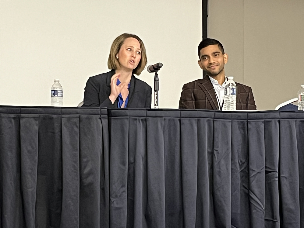
Today, however, more than a decade later, guidelines still promote surgical therapy as the gold standard across the board, and questions raised by the study are still unaddressed, Dr. Patel, associate professor of dermatology and medicine/oncology at George Washington University, said at the meeting. These questions are becoming increasingly urgent as the incidence of skin cancer, especially NMSC, rises in the older adult population, especially in patients older than 85 years. “It’s a function of our training and our treatment guidelines that we reach for the most definitive treatment, which happens to be the most aggressive, in these patients,” added Dr. Patel, who is also director of the cutaneous oncology program at the GW Cancer Center.
“Sometimes we lose track of what ... we need to do” to provide care that reflects the best interests of the older patient, he continued. “Surgery may be the gold standard for treating the majority of NMSCs ... but is it the [best option] for what our older patients and patients with limited life expectancy need?”
Learning about what truly matters to the patient is a key element of the “age-friendly, whole-person care” that dermatologists must embrace as older adults become an increasingly large subset of their patient population, Christina Prather, MD, director and associate professor of geriatrics and palliative medicine at George Washington University, said at the meeting.
By 2040, projections are that the number of adults aged 85 years and older in the United States will be nearly quadruple the number in 2000, according to one estimate.
“We know that there are less than 6000 practicing geriatricians in the country ... [so the healthcare system] needs more of you who know how to bring an age-friendly approach to care,” Dr. Prather said. Dermatology is among the specialties that need to be “geriatricized.”
NMSC Increasing in the Older Population
The incidence of skin cancer is rising faster than that of any other cancer, Dr. Patel said. One window into the epidemiology, he said, comes from recently published data showing that an average of 6.1 million adults were treated each year for skin cancer during 2016-2018 (5.2 million of them for NMSC) — an increase from an average of 5.8 million annually in the 2012-2015 period. The data come from the Medical Expenditure Panel Survey (MEPS), which is conducted by the US Public Health Service through the Agency for Healthcare Research and Quality and the Centers for Disease Control and Prevention.
As a frame of reference, the average number of adults treated each year for nonskin cancers during these periods rose from 10.8 to 11.9 million, according to the 2023 MEPS data. “Skin cancer is about one-third of all cancers combined,” Dr. Patel said.
Not only is the incidence of NMSC significantly higher than that of melanoma but it also shows a more prominent aging trend. This was documented recently in a long-term observational study from Japan, in which researchers looked at the change in the median age of patients with NMSC and melanoma, compared with cancers of other organs, from 1991 to 2020 and found that NMSC had by far the greatest rise in median age, to a median age of 80 years in 2021.
Even more notable, Dr. Patel said, was a particularly marked increase in the number of patients with skin cancer aged 90 years and older. In 2021, this group of older adults accounted for 17% of patients receiving treatment for skin cancer at the Japanese hospital where the data were collected.
The 2013 study that stirred Dr. Patel as a fellow was of 1536 consecutive patients diagnosed with NMSC at two dermatology clinics (a University of California San Francisco–based private clinic and a Veterans Affairs Medical Center clinic) and followed for 6 years. “What’s interesting and worth thinking about is that, regardless of patients’ life expectancy, NMSCs were treated aggressively and surgically, and the choice of surgery was not influenced by the patient’s poor prognosis in a multivariate model” adjusted for tumor and patient characteristics, he said at the meeting.
The researchers defined limited life expectancy as either 85 years or older or having a Charleston Comorbidity Index ≥ 3. Approximately half of the patients with limited life expectancy died within 5 years, none of NMSC. Most patients with limited life expectancy were not often bothered by their tumors, and approximately one in five reported a treatment complication within 2 years. The 5-year tumor recurrence rate was 3.7%.
A more recent study looked at 1181 patients older than 85 years with NMSC referred for Mohs surgery. Almost all patients in the multicenter, prospective cohort study (91.3%) were treated with Mohs.
Treated patients were more likely to have facial tumors and higher functional status than those not treated with Mohs surgery, and the most common reasons provided by surgeons for proceeding with the surgery were a patient desire for a high cure rate (66%), higher functional status for age (57%), and high-risk tumor type (40%). Almost 42% of the referred patients were 89 years or older.
“Granted, [the reasons] are justified indications for surgery,” Dr. Patel said. Yet the study brings up the question of “whether we need to do Mohs surgery this frequently in elderly patients?” In an email after the meeting, he added, “it’s a question we may need to reconsider as the elderly population continues to increase and median age of NMSC rises.”
Underutilized Management Options for NMSC
In his practice, discussions of treatment options are preceded by a thorough discussion of the disease itself. Many lesions are low risk, and helping patients understand risks, as well as understanding what is important to the patient — especially those with limited life expectancy — will guide shared decision-making to choose the best treatment, Dr. Patel said at the meeting.
The dermatologist’s risk assessment — both staging and stratifying risk as it relates to specific outcomes such as recurrence, metastases, or death — takes on added importance in the older patient, he emphasized. “I think we underutilize the risk assessment.”
Also underutilized is the option of shave removal for low-risk squamous cell carcinomas and basal cell carcinomas, Dr. Patel said, noting that, in the National Comprehensive Cancer Network guidelines, “there’s an option for shave removal and nothing more if you have clear margins.”
Alternatively, disc excision with the initial biopsy can often be considered. “Having that intent to treat at the time of biopsy may be all that needs to be done” in older patients with obvious or highly suspicious lesions, he said.
Systemic immunotherapy has joined the treatment armamentarium for advanced basal cell carcinoma and advanced cutaneous squamous cell carcinoma, and if early, ongoing research of intralesional programmed cell death protein 1 inhibitor treatment advances, this could be another option for older adults in the future, Dr. Patel said. Targeting drug delivery directly to the tumor would lower the total dose, decrease systemic exposure, and could be used to avoid surgery for some groups of patients, such as those with limited life expectancy.
A Personal Story, a Word on Melanoma
Dr. Prather recalled when her 97-year-old grandfather had a skin lesion on his forehead removed, and a conversation he had with her mother about whether he really needed to have the procedure because he had cognitive impairment and was on oral anticoagulants.
The clinician “said it absolutely had to go. ... I can’t tell you how much his doctors’ visits and wound care consumed my family’s life for the next few years — for this thing that never quite healed,” she said.
“Was it necessary? The more I’ve learned over time is that it wasn’t,” Dr. Prather added. “We have to take time [with our older patients] and think critically. What is feasible? What makes the most sense? What is the most important thing I need to know about the patient?”
Also important, Dr. Patel noted, is the big-picture consideration of skin cancer treatment costs. The MEPS survey data showing the rising prevalence of skin cancer treatment also documented the economic burden: A nearly 30% increase in the average annual cost of treating NMSC from $5 billion in 2012-2015 to $6.5 billion in 2016-2018. (The average annual costs of treating melanoma decreased slightly.) “Skin cancer is a big drain on our limited resources,” he said.
With melanoma as well, dermatologists must think critically and holistically about the individual patient — and not have “a single view lens of the disease and how we treat the disease,” said Dr. Patel, urging the audience to read a “Sounding Board” article published in The New England Journal of Medicine in 2021. The article argued that there is overdiagnosis of cutaneous melanoma stemming from increased screening, falling clinical thresholds for biopsy, and falling pathological thresholds for labeling morphologic changes as cancer.
“There’s a diagnostic disconnect and a problem of overdiagnosis ... because we’re afraid to miss or make a mistake,” he said. “It leads to the question, do all lesions denoted as skin cancers need aggressive treatment? What does it mean for the patient in front of you?”
Dr. Patel reported receiving honoraria from Regeneron, Almirall, Biofrontera, Sun Pharma, and SkylineDx and serving on the speaker bureau of Regeneron and Almirall. He is chief medical officer for Lazarus AI and is cofounder of the Skin Cancer Outcomes consortium. Dr. Prather disclosed relationships with the National Institutes of Health, AHRQ, The Washington Home Foundation, and the Alzheimer’s Association.
A version of this article appeared on Medscape.com.
WASHINGTON — In 2013, Vishal A. Patel, MD, was completing a fellowship in Mohs surgery and cutaneous oncology at Columbia University Irving Medical Center, New York City, when a study was published showing that most nonmelanoma skin cancers (NMSCs) were treated with surgery, regardless of the patient’s life expectancy. Life expectancy “should enter into treatment decisions,” the authors concluded.
“ Dr. Patel recalled at the ElderDerm conference hosted by the Department of Dermatology at George Washington University, Washington, DC, and described as a first-of-its-kind meeting dedicated to improving dermatologic care for older adults.
Today, however, more than a decade later, guidelines still promote surgical therapy as the gold standard across the board, and questions raised by the study are still unaddressed, Dr. Patel, associate professor of dermatology and medicine/oncology at George Washington University, said at the meeting. These questions are becoming increasingly urgent as the incidence of skin cancer, especially NMSC, rises in the older adult population, especially in patients older than 85 years. “It’s a function of our training and our treatment guidelines that we reach for the most definitive treatment, which happens to be the most aggressive, in these patients,” added Dr. Patel, who is also director of the cutaneous oncology program at the GW Cancer Center.
“Sometimes we lose track of what ... we need to do” to provide care that reflects the best interests of the older patient, he continued. “Surgery may be the gold standard for treating the majority of NMSCs ... but is it the [best option] for what our older patients and patients with limited life expectancy need?”
Learning about what truly matters to the patient is a key element of the “age-friendly, whole-person care” that dermatologists must embrace as older adults become an increasingly large subset of their patient population, Christina Prather, MD, director and associate professor of geriatrics and palliative medicine at George Washington University, said at the meeting.
By 2040, projections are that the number of adults aged 85 years and older in the United States will be nearly quadruple the number in 2000, according to one estimate.
“We know that there are less than 6000 practicing geriatricians in the country ... [so the healthcare system] needs more of you who know how to bring an age-friendly approach to care,” Dr. Prather said. Dermatology is among the specialties that need to be “geriatricized.”
NMSC Increasing in the Older Population
The incidence of skin cancer is rising faster than that of any other cancer, Dr. Patel said. One window into the epidemiology, he said, comes from recently published data showing that an average of 6.1 million adults were treated each year for skin cancer during 2016-2018 (5.2 million of them for NMSC) — an increase from an average of 5.8 million annually in the 2012-2015 period. The data come from the Medical Expenditure Panel Survey (MEPS), which is conducted by the US Public Health Service through the Agency for Healthcare Research and Quality and the Centers for Disease Control and Prevention.
As a frame of reference, the average number of adults treated each year for nonskin cancers during these periods rose from 10.8 to 11.9 million, according to the 2023 MEPS data. “Skin cancer is about one-third of all cancers combined,” Dr. Patel said.
Not only is the incidence of NMSC significantly higher than that of melanoma but it also shows a more prominent aging trend. This was documented recently in a long-term observational study from Japan, in which researchers looked at the change in the median age of patients with NMSC and melanoma, compared with cancers of other organs, from 1991 to 2020 and found that NMSC had by far the greatest rise in median age, to a median age of 80 years in 2021.
Even more notable, Dr. Patel said, was a particularly marked increase in the number of patients with skin cancer aged 90 years and older. In 2021, this group of older adults accounted for 17% of patients receiving treatment for skin cancer at the Japanese hospital where the data were collected.
The 2013 study that stirred Dr. Patel as a fellow was of 1536 consecutive patients diagnosed with NMSC at two dermatology clinics (a University of California San Francisco–based private clinic and a Veterans Affairs Medical Center clinic) and followed for 6 years. “What’s interesting and worth thinking about is that, regardless of patients’ life expectancy, NMSCs were treated aggressively and surgically, and the choice of surgery was not influenced by the patient’s poor prognosis in a multivariate model” adjusted for tumor and patient characteristics, he said at the meeting.
The researchers defined limited life expectancy as either 85 years or older or having a Charleston Comorbidity Index ≥ 3. Approximately half of the patients with limited life expectancy died within 5 years, none of NMSC. Most patients with limited life expectancy were not often bothered by their tumors, and approximately one in five reported a treatment complication within 2 years. The 5-year tumor recurrence rate was 3.7%.
A more recent study looked at 1181 patients older than 85 years with NMSC referred for Mohs surgery. Almost all patients in the multicenter, prospective cohort study (91.3%) were treated with Mohs.
Treated patients were more likely to have facial tumors and higher functional status than those not treated with Mohs surgery, and the most common reasons provided by surgeons for proceeding with the surgery were a patient desire for a high cure rate (66%), higher functional status for age (57%), and high-risk tumor type (40%). Almost 42% of the referred patients were 89 years or older.
“Granted, [the reasons] are justified indications for surgery,” Dr. Patel said. Yet the study brings up the question of “whether we need to do Mohs surgery this frequently in elderly patients?” In an email after the meeting, he added, “it’s a question we may need to reconsider as the elderly population continues to increase and median age of NMSC rises.”
Underutilized Management Options for NMSC
In his practice, discussions of treatment options are preceded by a thorough discussion of the disease itself. Many lesions are low risk, and helping patients understand risks, as well as understanding what is important to the patient — especially those with limited life expectancy — will guide shared decision-making to choose the best treatment, Dr. Patel said at the meeting.
The dermatologist’s risk assessment — both staging and stratifying risk as it relates to specific outcomes such as recurrence, metastases, or death — takes on added importance in the older patient, he emphasized. “I think we underutilize the risk assessment.”
Also underutilized is the option of shave removal for low-risk squamous cell carcinomas and basal cell carcinomas, Dr. Patel said, noting that, in the National Comprehensive Cancer Network guidelines, “there’s an option for shave removal and nothing more if you have clear margins.”
Alternatively, disc excision with the initial biopsy can often be considered. “Having that intent to treat at the time of biopsy may be all that needs to be done” in older patients with obvious or highly suspicious lesions, he said.
Systemic immunotherapy has joined the treatment armamentarium for advanced basal cell carcinoma and advanced cutaneous squamous cell carcinoma, and if early, ongoing research of intralesional programmed cell death protein 1 inhibitor treatment advances, this could be another option for older adults in the future, Dr. Patel said. Targeting drug delivery directly to the tumor would lower the total dose, decrease systemic exposure, and could be used to avoid surgery for some groups of patients, such as those with limited life expectancy.
A Personal Story, a Word on Melanoma
Dr. Prather recalled when her 97-year-old grandfather had a skin lesion on his forehead removed, and a conversation he had with her mother about whether he really needed to have the procedure because he had cognitive impairment and was on oral anticoagulants.
The clinician “said it absolutely had to go. ... I can’t tell you how much his doctors’ visits and wound care consumed my family’s life for the next few years — for this thing that never quite healed,” she said.
“Was it necessary? The more I’ve learned over time is that it wasn’t,” Dr. Prather added. “We have to take time [with our older patients] and think critically. What is feasible? What makes the most sense? What is the most important thing I need to know about the patient?”
Also important, Dr. Patel noted, is the big-picture consideration of skin cancer treatment costs. The MEPS survey data showing the rising prevalence of skin cancer treatment also documented the economic burden: A nearly 30% increase in the average annual cost of treating NMSC from $5 billion in 2012-2015 to $6.5 billion in 2016-2018. (The average annual costs of treating melanoma decreased slightly.) “Skin cancer is a big drain on our limited resources,” he said.
With melanoma as well, dermatologists must think critically and holistically about the individual patient — and not have “a single view lens of the disease and how we treat the disease,” said Dr. Patel, urging the audience to read a “Sounding Board” article published in The New England Journal of Medicine in 2021. The article argued that there is overdiagnosis of cutaneous melanoma stemming from increased screening, falling clinical thresholds for biopsy, and falling pathological thresholds for labeling morphologic changes as cancer.
“There’s a diagnostic disconnect and a problem of overdiagnosis ... because we’re afraid to miss or make a mistake,” he said. “It leads to the question, do all lesions denoted as skin cancers need aggressive treatment? What does it mean for the patient in front of you?”
Dr. Patel reported receiving honoraria from Regeneron, Almirall, Biofrontera, Sun Pharma, and SkylineDx and serving on the speaker bureau of Regeneron and Almirall. He is chief medical officer for Lazarus AI and is cofounder of the Skin Cancer Outcomes consortium. Dr. Prather disclosed relationships with the National Institutes of Health, AHRQ, The Washington Home Foundation, and the Alzheimer’s Association.
A version of this article appeared on Medscape.com.
Lung Cancer Expert at ASCO: From Fatal to ‘Chronic Disease’
After 20 years in Canada — 7 spent practicing community oncology near Toronto — Dr. Mok was visiting family in his native Hong Kong back in 1996 when a job offer there enabled him to revive his early dream of doing academic research. Dr. Mok and his family moved back home just before the former British colony was returned to China in 1997.

That leap of faith helped Dr. Mok play a role in the global paradigm shift on treating lung cancer. He chairs the department of clinical oncology at the Chinese University of Hong Kong. A leader in ushering in targeted therapies and personalized medicine in China and globally, he has helped advance the goal of transforming lung cancer from a death sentence to a chronic disease.
Among Dr. Mok’s other accomplishments, he has published eight books and more than 200 journal articles. Since 2006, he has been writing a twice-weekly column in the Hong Kong Economic Times. At the annual meeting of the American Society of Clinical Oncology (ASCO), Dr. Mok sat down with this news organization to discuss his latest findings, his career path, and China’s ever-growing presence in multinational clinical trials, pharmaceuticals, and cancer research in general.
Question: At ASCO 2024 in Chicago, you presented as first author of the KRYSTAL-12 study. Can you give a short “elevator speech” summarizing those findings?
Dr. Mok: KRYSTAL-12 is a randomized phase 3 study comparing adagrasib with docetaxel in patients with previously treated advanced/metastatic non–small cell lung cancer harboring a KRAS G12C-mutation. And the findings are positive, with a median progression free survival of 5.5 months vs 3.8 months, with a significant hazard ratio [of 0.58]. And then there are also differences in their response rates of 32% versus 9%, and that gives you an [odds] ratio of 4.86. So yes, it’s significant.
Question: Now that you’ve given this presentation and perhaps taken some good, meaningful questions about it, are there any further points you’d like to make — anything you’d like to add?
Dr. Mok: You have to understand that whatever I said has been scrutinized by the pharmaceutical company, but now I can say whatever I like. I think the key point is that we actually have made the first so-called achievement in the KRAS G12C space. But this is only the beginning.
I want to note that the median progression-free survival is different, but not the best. The median 5.5 months result is good, but not good enough. So, we still have to work hard to answer the question: How can we best deliver care to patients with KRAS G12C?
Question: Speaking more generally about the challenges of targeting KRAS, what issues arise in terms of biomarker testing for KRAS mutations in the clinic? Dr. Mok: In colorectal cancer, there has been testing for KRAS [mutations] for a long, long time. So, most of the laboratories, as long as they are well equipped, will be able to test for KRAS. Usually, the cheaper way is to buy PCR [polymerase chain reaction]. However, these days it’s getting trendier to use NGS [next-generation sequencing]. So, one way or another, specificity is very high. I don’t think we have too much of a problem. The only difference between colorectal cancer and lung cancer is that the tissue sample may not be as good for lung cancer with a small biopsy, but otherwise testing is not an issue.
Question: What clinical trials should oncologist be watching to come into this space?Dr. Mok: There are a lot. Right now, there is the so-called first-line study that’s coming up. So, I can cite you some examples for the KRYSTAL-7 trial, which is the combination of pembrolizumab together with adagrasib in the PD-L1 Tumor Proportion Score ≥ 50%.
That’s one example. And then there is the CodeBreaK 202 trial, which is actually the combination of chemotherapy with sotorasib versus chemotherapy and I-O [immune-oncology]. That is also an ongoing study.
Question: I also want to ask you some background questions about yourself. Back in the day, you lived in Canada and were a community oncologist. Then you made a very big change in your life and moved back home to Hong Kong in 1996, on the eve of its return to China the following year.
Dr. Mok: Well, I was born and raised in Hong Kong, but I left for Canada for education when I was 16 and kind of stayed there and got medical school oncology training and then started my practice. At that time, I never imagined myself going back. But 1996 was a big year. Incidentally, I went back to Hong Kong then to visit my friends and was offered a job at the Chinese University of Hong Kong. Then 1997 was coming. I found it very exciting that we could work with China. So that’s why I decided to return. And this was probably one of my best decisions I ever made in my life.
Question: And you went from being a community oncologist to academic research?
Dr. Mok: Here’s a personal thing that I can share with you: When I finished my oncology training at Princess Margaret Hospital in Toronto, I thought of going into research and becoming an academic. However, my son was born. Household costs went up, and I didn’t want to be a low-income, poor PhD student, so I decided that I may as well go into private practice. Returning to Hong Kong [in 1996] gave me a second chance. I went from being a community oncologist for seven years in Canada to a totally new environment in Hong Kong, where I started my academic work at age 36. It has been a good journey.
Question: Why do you say that was the best decision you ever made?Dr. Mok: At that time, it took me about 2 weeks to make this important decision. Basically: I had to give up my big house and my big car in Canada and move back to a small apartment in Hong Kong. That was a tough decision to make. However, it was a matter of certainty versus uncertainty.
In Canada, I actually had a very stable situation. I had a big practice in the Scarborough area [of Toronto], with a lot of Chinese patients, so I had a better, more comfortable life. It was predictable. But then I asked myself what I would be like in 10 years if I stayed in Canada versus Hong Kong. My answer is that I had no idea what would happen to me 10 years later in Hong Kong. In certain parts of life, you have to decide between certainty and uncertainty. And this time, uncertainty brought me great adventure. I definitely would not have done the things I’ve done if I’d stayed in Canada.
Question: At this ASCO, you’ve spoken primarily about your latest research on non–small cell lung cancer with KRAS G12C mutation.Dr. Mok: Actually, my research has been mostly on targeted therapy. My first break was on the EGFR [epidermal growth factor receptor] mutation. I was one of the first to help define personalized medicine according to the EGFR mutation in the IPASS study [2009]. That’s how I started my academic career.
Question: I read some quotes from your writing some years back about “living with imperfection,” and where you wrote about the whole continuum of cancer research. Years ago, you noted that lung cancer was moving from being a death sentence to becoming a chronic condition.
Dr. Mok: The objective is this: A lot of cancer patients, especially lung cancer patients, had a very short survival, but now we are able to identify a subgroup of patients with a driver oncogene.
And with that, we can use a tyrosine kinase inhibitor — which although it has toxicity, it’s manageable toxicity — such that you can take one pill a day and continue to live a normal life. So that would be not so different from diabetes or hypertension: You live with the disease. So that’s what we like to see: the conversion of a fatal disease into a chronic disease.
Question: So many countries now, including the United States and many others, are facing the challenges of cancer care in rural versus urban areas. Is this a topic you’d be willing to address? Dr. Mok: Well, in Hong Kong we don’t have rural areas! But in China, this is a major problem. There most of the cancer care is focused on the so-called three major cities [Shanghai, Beijing, Guangzhou]. And after that, there are second-tier cities that also have reasonably good care. But when you filter down to the third and fourth layer, the oncology care actually deteriorates. So that’s why we end up with a lot of people from the more rural areas moving and going to the city looking for care and consultation. So yes, the disparity is significant.
But China is a growing country. It takes time to change. Right now, we can see at ASCO this year, there are a lot of investigators from China sharing their new findings, which is a major development, compared to 10 years ago. Therefore, I think that when you have this type of proliferative development, eventually the good care, the high-quality care will filter down to more rural areas. So, at this moment, I think there is still a lot of work to do.
Question: You’ve talked about how oncologists from China are coming up in the field, and this year they have an even greater presence at ASCO, as well as oncologists from elsewhere in Asia, including South Korea, Japan, and Vietnam. You’ve been coming to ASCO for many years. Can you talk about the factors behind China’s increasing presence? Dr. Mok: I think it’s a combination of factors. First of all, I had the honor of working with lung cancer researchers from China from way back, 25 years ago. At that time, we all had nothing. Then with the development of multitargeted therapies, they managed to build up a very good infrastructure for clinical trials. And then, based on that good infrastructure, they were able to do international collaborative studies and provide a supply of patient resources and high-quality data. So, they’ve learned the trick, done a good job, but they cannot have so-called independence until there is a development of pharmaceuticals in China.
And then over the past 10 years, there’s been a proliferation — actually an explosion I would even say — of high-quality pharmaceutical companies in China. First, they’ve got the resources to build the companies. Second, they’ve got the talent resources returning from the United States. So, putting all that together, these were able to go from start-ups to full-fledged functional companies in a very short time.
And with that, they actually sponsored a lot of trials within China. And you can see that putting all the components together: you’ve got high-quality researchers, you’ve got the infrastructure, and now you’ve got your drugs and the money to do the trials. As a result, you’ve got a lot of good data coming from China.
Question: There’s also a population with these mutations.Dr. Mok: That for one, but most have multitargeted therapies, but they also have immunotherapies that have nothing to do with the high incidence. But I think in a sense, in the beginning, they were doing `me-too’ compounds, but now I think they are starting to do ‘me-better’ compounds.
Question: Is there anything you want to say about some of the other presentations that have your name on them at ASCO this year?Dr. Mok: I think the most important one I was engaged in is the CROWN study. The CROWN study is actually a phase 3 study that compares lorlatinib versus crizotinib in patients with advanced, ALK-positive non–small cell lung cancer.
This is a 5-year follow-up, and we were actually able to report an outrageously encouraging 5-year progression-free rate at 60%, meaning that the patient is walking in the door 5 years later when they are on the drug, and 60% of them actually do not have progression, not death, just not progression, just staying on the same pill—which is quite outrageously good for lung cancer.
After 20 years in Canada — 7 spent practicing community oncology near Toronto — Dr. Mok was visiting family in his native Hong Kong back in 1996 when a job offer there enabled him to revive his early dream of doing academic research. Dr. Mok and his family moved back home just before the former British colony was returned to China in 1997.
That leap of faith helped Dr. Mok play a role in the global paradigm shift on treating lung cancer. He chairs the department of clinical oncology at the Chinese University of Hong Kong. A leader in ushering in targeted therapies and personalized medicine in China and globally, he has helped advance the goal of transforming lung cancer from a death sentence to a chronic disease.
Among Dr. Mok’s other accomplishments, he has published eight books and more than 200 journal articles. Since 2006, he has been writing a twice-weekly column in the Hong Kong Economic Times. At the annual meeting of the American Society of Clinical Oncology (ASCO), Dr. Mok sat down with this news organization to discuss his latest findings, his career path, and China’s ever-growing presence in multinational clinical trials, pharmaceuticals, and cancer research in general.
Question: At ASCO 2024 in Chicago, you presented as first author of the KRYSTAL-12 study. Can you give a short “elevator speech” summarizing those findings?
Dr. Mok: KRYSTAL-12 is a randomized phase 3 study comparing adagrasib with docetaxel in patients with previously treated advanced/metastatic non–small cell lung cancer harboring a KRAS G12C-mutation. And the findings are positive, with a median progression free survival of 5.5 months vs 3.8 months, with a significant hazard ratio [of 0.58]. And then there are also differences in their response rates of 32% versus 9%, and that gives you an [odds] ratio of 4.86. So yes, it’s significant.
Question: Now that you’ve given this presentation and perhaps taken some good, meaningful questions about it, are there any further points you’d like to make — anything you’d like to add?
Dr. Mok: You have to understand that whatever I said has been scrutinized by the pharmaceutical company, but now I can say whatever I like. I think the key point is that we actually have made the first so-called achievement in the KRAS G12C space. But this is only the beginning.
I want to note that the median progression-free survival is different, but not the best. The median 5.5 months result is good, but not good enough. So, we still have to work hard to answer the question: How can we best deliver care to patients with KRAS G12C?
Question: Speaking more generally about the challenges of targeting KRAS, what issues arise in terms of biomarker testing for KRAS mutations in the clinic? Dr. Mok: In colorectal cancer, there has been testing for KRAS [mutations] for a long, long time. So, most of the laboratories, as long as they are well equipped, will be able to test for KRAS. Usually, the cheaper way is to buy PCR [polymerase chain reaction]. However, these days it’s getting trendier to use NGS [next-generation sequencing]. So, one way or another, specificity is very high. I don’t think we have too much of a problem. The only difference between colorectal cancer and lung cancer is that the tissue sample may not be as good for lung cancer with a small biopsy, but otherwise testing is not an issue.
Question: What clinical trials should oncologist be watching to come into this space?Dr. Mok: There are a lot. Right now, there is the so-called first-line study that’s coming up. So, I can cite you some examples for the KRYSTAL-7 trial, which is the combination of pembrolizumab together with adagrasib in the PD-L1 Tumor Proportion Score ≥ 50%.
That’s one example. And then there is the CodeBreaK 202 trial, which is actually the combination of chemotherapy with sotorasib versus chemotherapy and I-O [immune-oncology]. That is also an ongoing study.
Question: I also want to ask you some background questions about yourself. Back in the day, you lived in Canada and were a community oncologist. Then you made a very big change in your life and moved back home to Hong Kong in 1996, on the eve of its return to China the following year.
Dr. Mok: Well, I was born and raised in Hong Kong, but I left for Canada for education when I was 16 and kind of stayed there and got medical school oncology training and then started my practice. At that time, I never imagined myself going back. But 1996 was a big year. Incidentally, I went back to Hong Kong then to visit my friends and was offered a job at the Chinese University of Hong Kong. Then 1997 was coming. I found it very exciting that we could work with China. So that’s why I decided to return. And this was probably one of my best decisions I ever made in my life.
Question: And you went from being a community oncologist to academic research?
Dr. Mok: Here’s a personal thing that I can share with you: When I finished my oncology training at Princess Margaret Hospital in Toronto, I thought of going into research and becoming an academic. However, my son was born. Household costs went up, and I didn’t want to be a low-income, poor PhD student, so I decided that I may as well go into private practice. Returning to Hong Kong [in 1996] gave me a second chance. I went from being a community oncologist for seven years in Canada to a totally new environment in Hong Kong, where I started my academic work at age 36. It has been a good journey.
Question: Why do you say that was the best decision you ever made?Dr. Mok: At that time, it took me about 2 weeks to make this important decision. Basically: I had to give up my big house and my big car in Canada and move back to a small apartment in Hong Kong. That was a tough decision to make. However, it was a matter of certainty versus uncertainty.
In Canada, I actually had a very stable situation. I had a big practice in the Scarborough area [of Toronto], with a lot of Chinese patients, so I had a better, more comfortable life. It was predictable. But then I asked myself what I would be like in 10 years if I stayed in Canada versus Hong Kong. My answer is that I had no idea what would happen to me 10 years later in Hong Kong. In certain parts of life, you have to decide between certainty and uncertainty. And this time, uncertainty brought me great adventure. I definitely would not have done the things I’ve done if I’d stayed in Canada.
Question: At this ASCO, you’ve spoken primarily about your latest research on non–small cell lung cancer with KRAS G12C mutation.Dr. Mok: Actually, my research has been mostly on targeted therapy. My first break was on the EGFR [epidermal growth factor receptor] mutation. I was one of the first to help define personalized medicine according to the EGFR mutation in the IPASS study [2009]. That’s how I started my academic career.
Question: I read some quotes from your writing some years back about “living with imperfection,” and where you wrote about the whole continuum of cancer research. Years ago, you noted that lung cancer was moving from being a death sentence to becoming a chronic condition.
Dr. Mok: The objective is this: A lot of cancer patients, especially lung cancer patients, had a very short survival, but now we are able to identify a subgroup of patients with a driver oncogene.
And with that, we can use a tyrosine kinase inhibitor — which although it has toxicity, it’s manageable toxicity — such that you can take one pill a day and continue to live a normal life. So that would be not so different from diabetes or hypertension: You live with the disease. So that’s what we like to see: the conversion of a fatal disease into a chronic disease.
Question: So many countries now, including the United States and many others, are facing the challenges of cancer care in rural versus urban areas. Is this a topic you’d be willing to address? Dr. Mok: Well, in Hong Kong we don’t have rural areas! But in China, this is a major problem. There most of the cancer care is focused on the so-called three major cities [Shanghai, Beijing, Guangzhou]. And after that, there are second-tier cities that also have reasonably good care. But when you filter down to the third and fourth layer, the oncology care actually deteriorates. So that’s why we end up with a lot of people from the more rural areas moving and going to the city looking for care and consultation. So yes, the disparity is significant.
But China is a growing country. It takes time to change. Right now, we can see at ASCO this year, there are a lot of investigators from China sharing their new findings, which is a major development, compared to 10 years ago. Therefore, I think that when you have this type of proliferative development, eventually the good care, the high-quality care will filter down to more rural areas. So, at this moment, I think there is still a lot of work to do.
Question: You’ve talked about how oncologists from China are coming up in the field, and this year they have an even greater presence at ASCO, as well as oncologists from elsewhere in Asia, including South Korea, Japan, and Vietnam. You’ve been coming to ASCO for many years. Can you talk about the factors behind China’s increasing presence? Dr. Mok: I think it’s a combination of factors. First of all, I had the honor of working with lung cancer researchers from China from way back, 25 years ago. At that time, we all had nothing. Then with the development of multitargeted therapies, they managed to build up a very good infrastructure for clinical trials. And then, based on that good infrastructure, they were able to do international collaborative studies and provide a supply of patient resources and high-quality data. So, they’ve learned the trick, done a good job, but they cannot have so-called independence until there is a development of pharmaceuticals in China.
And then over the past 10 years, there’s been a proliferation — actually an explosion I would even say — of high-quality pharmaceutical companies in China. First, they’ve got the resources to build the companies. Second, they’ve got the talent resources returning from the United States. So, putting all that together, these were able to go from start-ups to full-fledged functional companies in a very short time.
And with that, they actually sponsored a lot of trials within China. And you can see that putting all the components together: you’ve got high-quality researchers, you’ve got the infrastructure, and now you’ve got your drugs and the money to do the trials. As a result, you’ve got a lot of good data coming from China.
Question: There’s also a population with these mutations.Dr. Mok: That for one, but most have multitargeted therapies, but they also have immunotherapies that have nothing to do with the high incidence. But I think in a sense, in the beginning, they were doing `me-too’ compounds, but now I think they are starting to do ‘me-better’ compounds.
Question: Is there anything you want to say about some of the other presentations that have your name on them at ASCO this year?Dr. Mok: I think the most important one I was engaged in is the CROWN study. The CROWN study is actually a phase 3 study that compares lorlatinib versus crizotinib in patients with advanced, ALK-positive non–small cell lung cancer.
This is a 5-year follow-up, and we were actually able to report an outrageously encouraging 5-year progression-free rate at 60%, meaning that the patient is walking in the door 5 years later when they are on the drug, and 60% of them actually do not have progression, not death, just not progression, just staying on the same pill—which is quite outrageously good for lung cancer.
FROM ASCO 2024
One Patient Changed This Oncologist’s View of Hope. Here’s How.
CHICAGO — Carlos, a 21-year-old, lay in a hospital bed, barely clinging to life. Following a stem cell transplant for leukemia, Carlos had developed a life-threatening case of graft-vs-host disease.
But Carlos’ mother had faith.
“I have hope things will get better,” she said, via interpreter, to Richard Leiter, MD, a palliative care doctor in training at that time.
“I hope they will,” Dr. Leiter told her.
“I should have stopped there,” said Dr. Leiter, recounting an early-career lesson on hope during the ASCO Voices session at the American Society of Clinical Oncology annual meeting. “But in my eagerness to show my attending and myself that I could handle this conversation, I kept going, mistakenly.”
“But none of us think they will,” Dr. Leiter continued.
Carlos’ mother looked Dr. Leiter in the eye. “You want him to die,” she said.
“I knew, even then, that she was right,” recalled Dr. Leiter, now a palliative care physician at Dana-Farber Cancer Institute and Brigham and Women’s Hospital and an assistant professor of medicine at Harvard Medical School, Boston.
Although there was nothing he could do to save Carlos, Dr. Leiter also couldn’t sit with the extreme suffering. “The pain was too great,” Dr. Leiter said. “I needed her to adopt our narrative that we had done everything we could to help him live, and now, we would do everything we could to help his death be a comfortable one.”
But looking back, Dr. Leiter realized, “How could we have asked her to accept what was fundamentally unacceptable, to comprehend the incomprehensible?”
The Importance of Hope
Alan B. Astrow, MD, said during an ASCO symposium on “The Art and Science of Hope.”
“How we think about hope directly influences patient care,” said Dr. Astrow, chief of hematology and medical oncology at NewYork-Presbyterian Brooklyn Methodist Hospital and a professor of clinical medicine at Weill Cornell Medicine in New York City.
Hope, whatever it turns out to be neurobiologically, is “very much a gift” that underlies human existence, he said.
Physicians have the capacity to restore or shatter a patient’s hopes, and those who come to understand the importance of hope will wish to extend the gift to others, Dr. Astrow said.
Asking patients about their hopes is the “golden question,” Steven Z. Pantilat, MD, said at the symposium. “When you think about the future, what do you hope for?”
Often, the answers reveal not only “things beyond a cure that matter tremendously to the patient but things that we can help with,” said Dr. Pantilat, professor and chief of the Division of Palliative Medicine at the University of California San Francisco.
Dr. Pantilat recalled a patient with advanced pancreatic cancer who wished to see her daughter’s wedding in 10 months. He knew that was unlikely, but the discussion led to another solution.
Her daughter moved the wedding to the ICU.
Hope can persist and uplift even in the darkest of times, and “as clinicians, we need to be in the true hope business,” he said.
While some patients may wish for a cure, others may want more time with family or comfort in the face of suffering. People can “hope for all the things that can still be, despite the fact that there’s a lot of things that can’t,” he said.
However, fear that a patient will hope for a cure, and that the difficult discussions to follow might destroy hope or lead to false hope, sometimes means physicians won’t begin the conversation.
“We want to be honest with our patients — compassionate and kind, but honest — when we talk about their hopes,” Dr. Pantilat explained. Sometimes that means he needs to tell patients, “I wish that could happen. I wish I had a treatment that could make your cancer go away, but unfortunately, I don’t. So let’s think about what else we can do to help you.”
Having these difficult discussions matters. The evidence, although limited, indicates that feeling hopeful can improve patients’ well-being and may even boost their cancer outcomes.
One recent study found, for instance, that patients who reported feeling more hopeful also had lower levels of depression and anxiety. Early research also suggests that greater levels of hope may have a hand in reducing inflammation in patients with ovarian cancer and could even improve survival in some patients with advanced cancer.
For Dr. Leiter, while these lessons came early in his career as a palliative care physician, they persist and influence his practice today.
“I know that I could not have prevented Carlos’ death. None of us could have, and none of us could have protected his mother from the unimaginable grief that will stay with her for the rest of her life,” he said. “But I could have made things just a little bit less difficult for her.
“I could have acted as her guide rather than her cross-examiner,” he continued, explaining that he now sees hope as “a generous collaborator” that can coexist with rising creatinine levels, failing livers, and fears about intubation.
“As clinicians, we can always find space to hope with our patients and their families,” he said. “So now, years later when I sit with a terrified and grieving family and they tell me they hope their loved one gets better, I remember Carlos’ mother’s eyes piercing mine ... and I know how to respond: ‘I hope so, too.’ And I do.”
A version of this article appeared on Medscape.com.
FROM ASCO 2024
Is Location a Risk Factor for Early-Onset Cancer?
Early-onset cancer—diagnosed in adults aged ≤ 50 years—is on the rise. Researchers have studied a variety of factors driving the trend, such as type of cancer. However, geographic locality might have as much, if not more, to do with it, according to a study by researchers at Fox Chase Cancer Center, a National Cancer Institute-designated Comprehensive Cancer Center research facility.
Using the US Cancer Statistics Public Use Research Database, the researchers collected data from adults aged 20 to 49 years with invasive cancer (excluding in situ cases) diagnosed from 2015 through 2020. They calculated the incidence for each state using the national rate as the reference. Then, they calculated a second set of rates, comparing each state to the US in terms of overall incidence and advanced-stage incidence for all early-onset cancers.
The resulting maps indicated that early-onset cancer cases are not evenly distributed. States with worse-than-national rates are frequently near each other geographically. For instance, the rate of early-onset female breast cancer was worse than the national rate in 17 states, 16 of which were located in the eastern half of the US (Hawaii was the 17th state). Similarly, most states with worse-than-national rates of digestive cancers were located in the eastern half of the US, with a concentration in the South. Rates of male genital cancers were worse than national rates in 18 states, primarily in the eastern half of the country (plus Montana, Nebraska, and Puerto Rico).
Three states in the Southeast, 7 in the Northeast, and Puerto Rico had the highest incidence of lymphohematopoietic cancers. Incidence rates of endocrine cancers were worse than national rates in 25 states, which the researchers found formed “a horizontal core of the country running from east to west,” plus Puerto Rico. Rates of urinary system cancers were worse than national rates in 17 contiguous states, from New Mexico to Pennsylvania.
Rates of female genital cancers were worse than national rates in 16 states, largely in the Midwest and South, plus California and Puerto Rico. Skin cancer, on the other hand, was a great leveler, with worse-than-national rates in 32 states, mostly in the northern portion of the country.
Kentucky and West Virginia had the highest overall and advanced-stage incidence rates of early-onset cancer for all cancer sites combined. They were followed by Arkansas, Connecticut, Florida, Georgia, Iowa, Louisiana, Maine, Missouri, New Jersey, New York, North Carolina, Ohio, and Pennsylvania.
According to the researchers, this study provides the first analysis of age-adjusted rates of early-onset cancer based on state-level population and case numbers. Geographic patterns in early-onset cancer, they suggest, indicate possible similarities that could relate to demographic, socioeconomic, behavioral, or environmental risks. “Focusing prevention efforts on the highest-incidence states for the most prevalent sites may reduce the rate of early-onset cancer nationally.”