User login
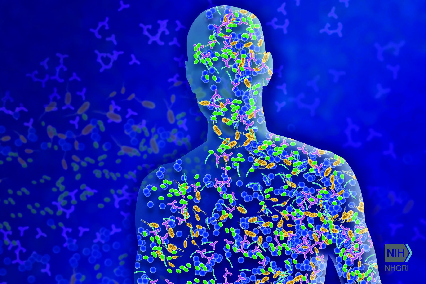
What is the ‘microbiome’ and how may it influence gynecologic cancers?
Bacteria are everywhere, good and bad alike! It is well known in the scientific community that microbes significantly outnumber the cells in the human body by at least 10 times. Joshua Lederberg, PhD, gave meaning to the term “microbiome” in 2001 as the “ecological community of commensal, symbiotic, and pathogenic microorganisms that literally share our body space.”1 This community of microorganisms comprises bacteria, fungi, viruses, archaea, and protists.

In 2007, the National Institutes of Health Human Microbiome Project was established to study the human microbiome starting with five specific sites – the gastrointestinal tract, the mouth, the vagina, the skin, and nasal cavity. The goal was not only to identify the microbes inhabiting a specific body site but also to establish a range of “normal” for resident microbes as well as sequence the genomes of these microbes.2 Much of the research predating this era focused on microorganisms in terms of disease potential rather than a focus on the benefits of resident microorganisms.
The richness – the number of microorganisms in an area – and diversity – the relative proportion of microorganisms in an environment – can vary regionally. The microbiota that contribute to the class of resident microorganisms in a specific body habitat can be described broadly as commensals or mutualistic. With commensal microorganisms, one partner benefits and the other is unaffected. On the other hand, mutualistic microorganisms allow both parties to derive benefit. For example, resident microorganisms in the gut aid in the absorption of nutrients and in the production of vitamin K. On mucosal surfaces and the skin, it is possible that these resident microorganisms prevent colonization of pathogenic microbes, which could aid in prevention of disease.3
The microbiota composition can be influenced by multiple factors such as age, diet, medications, environment, early microbial exposure, and host genetics. The gut microbiota, for example, can be significantly altered by dietary intake or antibiotic use. Alterations in the diversity of microbes in certain body habitats has been linked to several human diseases such as obesity, inflammatory bowel disease, and bacterial vaginosis.4

In women, there are differences noted in the composition of resident microorganisms soon after birth as well as at prepubertal, postpubertal, and postmenopausal transitions. At puberty, anaerobic and aerobic lactobacilli aid in maintaining vaginal pH. If the normal microbiota is suppressed, it allows for yeast and other bacteria to grow causing vaginitis, and dramatic shifts in the makeup of the vaginal microbiota can lead to bacterial vaginosis. Interestingly, research has shown that the pH and microbiome of the vagina differs by ethnicity. These differences in composition of the vaginal microbiome likely contribute to known differences in the acquisition of sexually transmitted infections and development of bacterial vaginosis. The microbiome is believed to have a complex role in regulating human health and disease, including cancer.
There is growing evidence to suggest the gut microbiome may play an important role in the pathogenesis of both obesity and cancer. Two divisions of bacteria predominate in the gut in humans and mice, Bacteroidetes and Firmicutes, and the relative ratio of these two divisions is dramatically affected by obesity, such that Bacteroidetes levels decrease and Firmicutes levels increase.5 The change in the microbial environment leads to a greater ability to harvest dietary energy, which would be conducive to cancer development.
The microbiome and gynecologic cancers

The presence and relative abundance of bacterial species in the vagina are affected by unique factors such as hormonal contraception, pregnancy, and menopause. There are researchers investigating alterations in the microbiome of the vagina and implications in persistence of high-risk human papillomavirus infections and HPV-induced carcinogenesis. There were significant differences found in the composition of the vaginal microbiota in healthy women, compared with women with low-grade squamous intraepithelial neoplasm and high-grade squamous intraepithelial neoplasm.6
Conceivably, the subsequent clinical questions are: Can we apply this data to diagnose women at risk for dysplasia or can we alter the vaginal microbiome to impact the clearance rate of the HPV virus in susceptible or infected women to decrease the long-term risk of cervical dysplasia or malignancy?
The upper reproductive tract in women – the uterus, fallopian tubes, and ovaries – had been presumed to be a sterile environment. However, we know that bacteria have been isolated in the pre- and postmenopausal uterus of healthy women. Therefore, there also are investigators seeking to establish the microbiome of normal uteri to accurately compare it with malignant uteri. Notably, there also is interest in how treatments for cancer – chemotherapy and radiation – ultimately can affect a woman’s vaginal and gut microbiome.
Currently, microbiome research has an expansive range. Women will greatly benefit from research seeking to define improved prevention, diagnosis, and treatment based on alterations of the microbiome for common gynecologic premalignant and malignant conditions.
Dr. Hawkins is a fellow of gynecologic oncology and Dr. Rossi is an assistant professor in the division of gynecologic oncology at the University of North Carolina at Chapel Hill. They had no conflicts of interest to disclose.
References
1. “ ’Ome Sweet ’Omics – a genealogical treasury of words,” by Joshua Lederberg, The Scientist, Apr 2, 2001.
2. Genome Res. 2009 Dec;19(12):2317-23.
3. “Normal Human Microbiota,” Jawetz, Melnick & Adelberg’s Medical Microbiology, 27th edition (New York, NY: McGraw-Hill, 2016).
4. Nature. 2012 Jun 13;486(7402):207-14.
5. Nature. 2006 Dec 21;444(7122):1027-31.
6. Oncol Lett. 2018 Dec; 16(6): 7035-47.
Bacteria are everywhere, good and bad alike! It is well known in the scientific community that microbes significantly outnumber the cells in the human body by at least 10 times. Joshua Lederberg, PhD, gave meaning to the term “microbiome” in 2001 as the “ecological community of commensal, symbiotic, and pathogenic microorganisms that literally share our body space.”1 This community of microorganisms comprises bacteria, fungi, viruses, archaea, and protists.
In 2007, the National Institutes of Health Human Microbiome Project was established to study the human microbiome starting with five specific sites – the gastrointestinal tract, the mouth, the vagina, the skin, and nasal cavity. The goal was not only to identify the microbes inhabiting a specific body site but also to establish a range of “normal” for resident microbes as well as sequence the genomes of these microbes.2 Much of the research predating this era focused on microorganisms in terms of disease potential rather than a focus on the benefits of resident microorganisms.
The richness – the number of microorganisms in an area – and diversity – the relative proportion of microorganisms in an environment – can vary regionally. The microbiota that contribute to the class of resident microorganisms in a specific body habitat can be described broadly as commensals or mutualistic. With commensal microorganisms, one partner benefits and the other is unaffected. On the other hand, mutualistic microorganisms allow both parties to derive benefit. For example, resident microorganisms in the gut aid in the absorption of nutrients and in the production of vitamin K. On mucosal surfaces and the skin, it is possible that these resident microorganisms prevent colonization of pathogenic microbes, which could aid in prevention of disease.3
The microbiota composition can be influenced by multiple factors such as age, diet, medications, environment, early microbial exposure, and host genetics. The gut microbiota, for example, can be significantly altered by dietary intake or antibiotic use. Alterations in the diversity of microbes in certain body habitats has been linked to several human diseases such as obesity, inflammatory bowel disease, and bacterial vaginosis.4
In women, there are differences noted in the composition of resident microorganisms soon after birth as well as at prepubertal, postpubertal, and postmenopausal transitions. At puberty, anaerobic and aerobic lactobacilli aid in maintaining vaginal pH. If the normal microbiota is suppressed, it allows for yeast and other bacteria to grow causing vaginitis, and dramatic shifts in the makeup of the vaginal microbiota can lead to bacterial vaginosis. Interestingly, research has shown that the pH and microbiome of the vagina differs by ethnicity. These differences in composition of the vaginal microbiome likely contribute to known differences in the acquisition of sexually transmitted infections and development of bacterial vaginosis. The microbiome is believed to have a complex role in regulating human health and disease, including cancer.
There is growing evidence to suggest the gut microbiome may play an important role in the pathogenesis of both obesity and cancer. Two divisions of bacteria predominate in the gut in humans and mice, Bacteroidetes and Firmicutes, and the relative ratio of these two divisions is dramatically affected by obesity, such that Bacteroidetes levels decrease and Firmicutes levels increase.5 The change in the microbial environment leads to a greater ability to harvest dietary energy, which would be conducive to cancer development.
The microbiome and gynecologic cancers
The presence and relative abundance of bacterial species in the vagina are affected by unique factors such as hormonal contraception, pregnancy, and menopause. There are researchers investigating alterations in the microbiome of the vagina and implications in persistence of high-risk human papillomavirus infections and HPV-induced carcinogenesis. There were significant differences found in the composition of the vaginal microbiota in healthy women, compared with women with low-grade squamous intraepithelial neoplasm and high-grade squamous intraepithelial neoplasm.6
Conceivably, the subsequent clinical questions are: Can we apply this data to diagnose women at risk for dysplasia or can we alter the vaginal microbiome to impact the clearance rate of the HPV virus in susceptible or infected women to decrease the long-term risk of cervical dysplasia or malignancy?
The upper reproductive tract in women – the uterus, fallopian tubes, and ovaries – had been presumed to be a sterile environment. However, we know that bacteria have been isolated in the pre- and postmenopausal uterus of healthy women. Therefore, there also are investigators seeking to establish the microbiome of normal uteri to accurately compare it with malignant uteri. Notably, there also is interest in how treatments for cancer – chemotherapy and radiation – ultimately can affect a woman’s vaginal and gut microbiome.
Currently, microbiome research has an expansive range. Women will greatly benefit from research seeking to define improved prevention, diagnosis, and treatment based on alterations of the microbiome for common gynecologic premalignant and malignant conditions.
Dr. Hawkins is a fellow of gynecologic oncology and Dr. Rossi is an assistant professor in the division of gynecologic oncology at the University of North Carolina at Chapel Hill. They had no conflicts of interest to disclose.
References
1. “ ’Ome Sweet ’Omics – a genealogical treasury of words,” by Joshua Lederberg, The Scientist, Apr 2, 2001.
2. Genome Res. 2009 Dec;19(12):2317-23.
3. “Normal Human Microbiota,” Jawetz, Melnick & Adelberg’s Medical Microbiology, 27th edition (New York, NY: McGraw-Hill, 2016).
4. Nature. 2012 Jun 13;486(7402):207-14.
5. Nature. 2006 Dec 21;444(7122):1027-31.
6. Oncol Lett. 2018 Dec; 16(6): 7035-47.
A.I. and U
There is a good chance that your car is equipped with a backup camera. It also may have sensors that alert you when there is another vehicle in one of your blind spots. These wonders of modern technology simply are vision enhancers much like an x-ray or an ultrasound. The sensors merely collect visual data, but the decision of what should be done with this additional information is up to you, just as you decide how to respond to your patient’s lab work and imaging studies.

If you have more disposable income than I do, you may have a vehicle that not only gathers information but also makes decisions based on what it senses by slowing down, applying the brakes, or adjusting the steering. My friends who own these semi-autonomous cars generally have given these control systems positive grades once they have experienced a few events in which the vehicle took over in what it considered a dangerous situation. However, even my friends who are fans of their semi-autonomous cars are uncomfortable about the widespread introduction of fully autonomous vehicles.
The practice of medicine is riding the crest of this same wave of artificial intelligence that promises, or some might say threatens, to remove humans from the driver’s seat (“A.I. Shows Promise Assisting Physicians,” by Cade Metz, The New York Times, Feb. 11, 2019). As reported in the New York Times, a team of physicians has created a system capable of making diagnoses based on a “neural network” that uses complex computer algorithms to learn by analyzing extremely large amounts of data. Once this system had been “taught” to identify certain medical conditions in EMRs, the team tasked the system with analyzing the records of nearly 600,000 patients at a women and children’s hospital in southern China. The investigators claim that the system was able to diagnose asthma with more than 90% accuracy, while physicians can diagnose with an accuracy of 80%-94%, and the system diagnosed gastrointestinal disease with 87% accuracy, well within the physicians’ accuracy range of 82%-90%.
Does this apparent success for A.I. mean that not only will you be vacating your place behind the wheel of your car, but also taking down your shingle and hanging up your stethoscope? Before you rush out and sign up for a federally-funded retraining program, you should remember that this study was done in China, where the privacy laws are somewhat skimpy and the data more voluminous by several scales of magnitude than here. Replicating their results and However, this report should serve as wake-up call to those of you who believe that making diagnoses is at the core of what makes you a physician. If sorting through pages of data to arrive at an explanation for your patients’ complaints is the intellectual challenge that keeps the practice of medicine fresh and exciting, you may want to start looking for other sources of mental stimulation.
A.I. isn’t going to replace the primary care physician. There still will need to be someone available at the initial point of contact who can do a physical exam, take, or at least review, the patient’s history, and then order the lab work and imaging studies that the A.I. system will use to make the diagnosis. In other words, the physician will be primarily responsible for data collection. You may feel that you are almost there already.

Will there be new roles for primary care physicians once A.I. systems are making the diagnoses? It is hard to imagine a fully autonomous health care system in which physicians completely disappear. But, now is the time to think seriously about how we are going to reinvent ourselves to adapt to the inevitable changes and continue as an (or could be "the") essential human element in an increasingly automated system.
Dr. Wilkoff practiced primary care pediatrics in Brunswick, Maine for nearly 40 years. He has authored several books on behavioral pediatrics, including “How to Say No to Your Toddler.” Email him at [email protected].
There is a good chance that your car is equipped with a backup camera. It also may have sensors that alert you when there is another vehicle in one of your blind spots. These wonders of modern technology simply are vision enhancers much like an x-ray or an ultrasound. The sensors merely collect visual data, but the decision of what should be done with this additional information is up to you, just as you decide how to respond to your patient’s lab work and imaging studies.
If you have more disposable income than I do, you may have a vehicle that not only gathers information but also makes decisions based on what it senses by slowing down, applying the brakes, or adjusting the steering. My friends who own these semi-autonomous cars generally have given these control systems positive grades once they have experienced a few events in which the vehicle took over in what it considered a dangerous situation. However, even my friends who are fans of their semi-autonomous cars are uncomfortable about the widespread introduction of fully autonomous vehicles.
The practice of medicine is riding the crest of this same wave of artificial intelligence that promises, or some might say threatens, to remove humans from the driver’s seat (“A.I. Shows Promise Assisting Physicians,” by Cade Metz, The New York Times, Feb. 11, 2019). As reported in the New York Times, a team of physicians has created a system capable of making diagnoses based on a “neural network” that uses complex computer algorithms to learn by analyzing extremely large amounts of data. Once this system had been “taught” to identify certain medical conditions in EMRs, the team tasked the system with analyzing the records of nearly 600,000 patients at a women and children’s hospital in southern China. The investigators claim that the system was able to diagnose asthma with more than 90% accuracy, while physicians can diagnose with an accuracy of 80%-94%, and the system diagnosed gastrointestinal disease with 87% accuracy, well within the physicians’ accuracy range of 82%-90%.
Does this apparent success for A.I. mean that not only will you be vacating your place behind the wheel of your car, but also taking down your shingle and hanging up your stethoscope? Before you rush out and sign up for a federally-funded retraining program, you should remember that this study was done in China, where the privacy laws are somewhat skimpy and the data more voluminous by several scales of magnitude than here. Replicating their results and However, this report should serve as wake-up call to those of you who believe that making diagnoses is at the core of what makes you a physician. If sorting through pages of data to arrive at an explanation for your patients’ complaints is the intellectual challenge that keeps the practice of medicine fresh and exciting, you may want to start looking for other sources of mental stimulation.
A.I. isn’t going to replace the primary care physician. There still will need to be someone available at the initial point of contact who can do a physical exam, take, or at least review, the patient’s history, and then order the lab work and imaging studies that the A.I. system will use to make the diagnosis. In other words, the physician will be primarily responsible for data collection. You may feel that you are almost there already.
Will there be new roles for primary care physicians once A.I. systems are making the diagnoses? It is hard to imagine a fully autonomous health care system in which physicians completely disappear. But, now is the time to think seriously about how we are going to reinvent ourselves to adapt to the inevitable changes and continue as an (or could be "the") essential human element in an increasingly automated system.
Dr. Wilkoff practiced primary care pediatrics in Brunswick, Maine for nearly 40 years. He has authored several books on behavioral pediatrics, including “How to Say No to Your Toddler.” Email him at [email protected].
Pseudoscience redux
My most recent column discussed the problem of pseudoscience that pervades some corners of the Internet. Personally, I respond to pseudoscience primarily by trying to provide accurate and less-biased information. I recognize that not everyone approaches decision making by seeking more information. When dealing a diverse public, a medical professional needs to have other approaches in the armamentarium.1 When dealing with other physicians, I am less flexible. Either the profession of medicine believes in science or it doesn’t.
![]()
Since that column was published, there have been major developments. There are measles outbreaks in the states of Washington and New York, and more than 100 deaths from a measles epidemic in the Philippines. The World Health Organization has made vaccine hesitancy one of its ten threats to global health in 2019.
Facebook has indicated that it might demote the priority and frequency with which it recommends articles that promulgate anti-vax information and conspiracy theories.2 Facebook isn’t doing this because it has had an epiphany; it has come under pressure for its role in the spread of misinformation. Current legislation was written before the rise of social media, when Internet Service Providers were primarily conduits to transfer bits and bytes between computers. Those ISPs were not liable for the content of the transmitted Web pages. Facebook, by producing what it called a newsfeed and by making personalized suggestions for other websites to browse, doesn’t fit the passive model of an ISP.
For alleged violations of user’s privacy, Facebook might be subject to billion dollar fines, according to a Washington Post article.3 Still, for a company whose revenue is $4 billion per month and whose stock market value is $400 billion, paying a billion dollar fine for years of alleged misbehaviors that have enabled it to become a giant empire is, “in the scheme of things ... a speeding ticket” in the parlance of the penultimate scene of the movie The Social Network. The real financial risk is people deciding they can’t trust the platform and going elsewhere.
Authorities in the United Kingdom in February 2019 released a highly critical, 108-page report about fake news, which said, “Facebook should not be allowed to behave like ‘digital gangsters’ in the online world.”4 The U.K. report urges new regulations to deal with privacy breaches and with fake news. It endeavors to create a duty for social media companies to combat the spread of misinformation.
Then the Wall Street Journal reported that Pinterest has stopped returning results for searches related to vaccination.5 Pinterest realized that most of the shared images on its platform cautioned against vaccination, which contradicts the recommendations of medical experts. Unable to otherwise combat the flow of misinformation, the company apparently has decided to eliminate returning results, pro or con, for any search terms related to vaccines.

While lamenting the public’s inability to distinguish misinformation on the Internet, I’ve also been observing the factors that lead physicians astray. I expect physicians, as trained scientists and as professionals, to be able to assimilate new information and change their practices accordingly. Those who do research on the translation of technology find that, this doesn’t happen with any regularity.
The February 2019 issue of Hospital Pediatrics has four items on the topic of treating bronchiolitis, including two research articles, a brief report, and a commentary. That is obviously a relevant topic this time of year. The impression after reading those four items is that hospitalists don’t really know how to best treat the most common illness they encounter. And even when they “know” how to do it, many factors distort the science. Those factors are highlighted in the article on barriers to minimizing viral testing.6
Dr. Powell is a pediatric hospitalist and clinical ethics consultant living in St. Louis. Email him at [email protected].
References
1. “Discussing immunization with vaccine-hesitant parents requires caring, individualized approach,” by Jeff Craven, Pediatric News, Nov. 7, 2018; “How do you get anti-vaxxers to vaccinate their kids? Talk to them – for hours,” by Nadine Gartner, Washington Post, Feb. 19, 2019.
2. “Facebook will consider removing or demoting anti-vaccination recommendations amid backlash,” by Taylor Telford, Washington Post, Feb. 15, 2019.
3. “U.S. regulators have met to discuss imposing a record-setting fine against Facebook for privacy violations,” by Tony Romm and Elizabeth Dwoskin, Washington Post, Jan. 18, 2019; “Report: Facebook, FTC discussing ‘multibillion dollar’ fine,” by Associated Press.
4. “Disinformation and ‘fake news’: Final Report,” House of Commons, Feb. 18, 2019, p. 42, item 139.
5. “Pinterest blocks vaccination searches in move to control the conversation,” by Robert McMillan and Daniela Hernandez, The Wall Street Journal, Feb. 20, 2019.
6. “Barriers to minimizing respiratory viral testing in bronchiolitis: Physician perceptions on testing practices,” by MZ Huang et al. Hospital Pediatrics 2019 Feb. doi: 10.1542/hpeds.2018-0108.
My most recent column discussed the problem of pseudoscience that pervades some corners of the Internet. Personally, I respond to pseudoscience primarily by trying to provide accurate and less-biased information. I recognize that not everyone approaches decision making by seeking more information. When dealing a diverse public, a medical professional needs to have other approaches in the armamentarium.1 When dealing with other physicians, I am less flexible. Either the profession of medicine believes in science or it doesn’t.
![]()
Since that column was published, there have been major developments. There are measles outbreaks in the states of Washington and New York, and more than 100 deaths from a measles epidemic in the Philippines. The World Health Organization has made vaccine hesitancy one of its ten threats to global health in 2019.
Facebook has indicated that it might demote the priority and frequency with which it recommends articles that promulgate anti-vax information and conspiracy theories.2 Facebook isn’t doing this because it has had an epiphany; it has come under pressure for its role in the spread of misinformation. Current legislation was written before the rise of social media, when Internet Service Providers were primarily conduits to transfer bits and bytes between computers. Those ISPs were not liable for the content of the transmitted Web pages. Facebook, by producing what it called a newsfeed and by making personalized suggestions for other websites to browse, doesn’t fit the passive model of an ISP.
For alleged violations of user’s privacy, Facebook might be subject to billion dollar fines, according to a Washington Post article.3 Still, for a company whose revenue is $4 billion per month and whose stock market value is $400 billion, paying a billion dollar fine for years of alleged misbehaviors that have enabled it to become a giant empire is, “in the scheme of things ... a speeding ticket” in the parlance of the penultimate scene of the movie The Social Network. The real financial risk is people deciding they can’t trust the platform and going elsewhere.
Authorities in the United Kingdom in February 2019 released a highly critical, 108-page report about fake news, which said, “Facebook should not be allowed to behave like ‘digital gangsters’ in the online world.”4 The U.K. report urges new regulations to deal with privacy breaches and with fake news. It endeavors to create a duty for social media companies to combat the spread of misinformation.
Then the Wall Street Journal reported that Pinterest has stopped returning results for searches related to vaccination.5 Pinterest realized that most of the shared images on its platform cautioned against vaccination, which contradicts the recommendations of medical experts. Unable to otherwise combat the flow of misinformation, the company apparently has decided to eliminate returning results, pro or con, for any search terms related to vaccines.
While lamenting the public’s inability to distinguish misinformation on the Internet, I’ve also been observing the factors that lead physicians astray. I expect physicians, as trained scientists and as professionals, to be able to assimilate new information and change their practices accordingly. Those who do research on the translation of technology find that, this doesn’t happen with any regularity.
The February 2019 issue of Hospital Pediatrics has four items on the topic of treating bronchiolitis, including two research articles, a brief report, and a commentary. That is obviously a relevant topic this time of year. The impression after reading those four items is that hospitalists don’t really know how to best treat the most common illness they encounter. And even when they “know” how to do it, many factors distort the science. Those factors are highlighted in the article on barriers to minimizing viral testing.6
Dr. Powell is a pediatric hospitalist and clinical ethics consultant living in St. Louis. Email him at [email protected].
References
1. “Discussing immunization with vaccine-hesitant parents requires caring, individualized approach,” by Jeff Craven, Pediatric News, Nov. 7, 2018; “How do you get anti-vaxxers to vaccinate their kids? Talk to them – for hours,” by Nadine Gartner, Washington Post, Feb. 19, 2019.
2. “Facebook will consider removing or demoting anti-vaccination recommendations amid backlash,” by Taylor Telford, Washington Post, Feb. 15, 2019.
3. “U.S. regulators have met to discuss imposing a record-setting fine against Facebook for privacy violations,” by Tony Romm and Elizabeth Dwoskin, Washington Post, Jan. 18, 2019; “Report: Facebook, FTC discussing ‘multibillion dollar’ fine,” by Associated Press.
4. “Disinformation and ‘fake news’: Final Report,” House of Commons, Feb. 18, 2019, p. 42, item 139.
5. “Pinterest blocks vaccination searches in move to control the conversation,” by Robert McMillan and Daniela Hernandez, The Wall Street Journal, Feb. 20, 2019.
6. “Barriers to minimizing respiratory viral testing in bronchiolitis: Physician perceptions on testing practices,” by MZ Huang et al. Hospital Pediatrics 2019 Feb. doi: 10.1542/hpeds.2018-0108.
Complementary and alternative medicine
Question: Which one of the following statements regarding complementary and alternative medicine (CAM) is correct?

A. CAM practitioners are just as likely as medical doctors to be sued.
B. Damages arising out of the use of CAM may be compensable if there is clear and convincing evidence of substandard care, and the plaintiff can prove legal causation.
C. An acupuncturist who treats an asthmatic patient will be sued if he/she fails to refer the patient to a medical specialist.
D. Obtaining informed consent after discussing all therapeutic options and material risks is especially important for those who practice CAM.
E. It is not a valid defense that the patient had fully and willingly assumed the risk of treatment.
Answer: D. Compared with medical doctors, non-MD practitioners of CAM pay lower malpractice insurance premiums, as they are much less likely to be sued, and patient injuries are usually less severe.
CAM covers a broad range of healing philosophies, approaches, and therapies that are typically outside mainstream Western medicine. It comprises modalities such as chiropractic, acupuncture, massage therapy, naturopathic medicine, nutritional therapy, and others.
More than half of the U.S. population uses some form of CAM, which is widely perceived as a natural and effective means of promoting overall well being in addition to treating a specific illness. The scope of practice for CAM providers is defined and limited by state rather than federal statutes, and enforced by regulatory bodies.
CAM treatments are generally noninvasive, and there are fewer than 50 indemnity insurers in the country for chiropractors, massage therapists, and acupuncturists, underwriting some 5% of the total medical malpractice insurance market.
In the event of an injury, damages are recoverable if there is a preponderance of evidence to indicate substandard care. “Clear and convincing” is a higher evidentiary level of legal proof, and it is not required in a negligence action. Whether an acupuncturist will be sued successfully for treating an asthmatic patient will depend on many factors, for example, whether it is an acceptable CAM practice in the jurisdiction, whether there is any statutory restriction on such treatment, whether there was a failure of a timely referral, etc.
Finally, assumption of risk is a valid defense in a negligence tort action under some circumstances.
For a negligence claim to prevail, the plaintiff must establish breach of duty, i.e., that the defendant deviated from the standard of care ordinarily exercised by a similarly situated practitioner. In addition to falling below that level of skill, practitioners may also be sued for having failed to refer to a medical doctor, for practicing outside the scope of CAM, or for venturing into traditional Western medical practice.
In one instance, a plaintiff alleged that he was led to believe that chiropractic manipulation would help his diabetes. The court found in favor of the plaintiff and awarded damages.1 In another, the plaintiff successfully sued a chiropractor for failing to take x-rays and refer to a medical doctor. The court held that the defendant fell below the standard of care, as the state licensing board required physician referral when the problem extended beyond the limits of chiropractic practice.2
However, an injured party does not always prevail. In Miyamoto v. Lazo, the plaintiff claimed that Dr. Lazo, a chiropractor, negligently treated his injuries from a car accident.3 The patient was taking Coumadin and developed a hematoma in his left shoulder following chiropractic treatment. This was complicated by neuropathy in his left hand when the hematoma expanded and required surgical drainage.
The jury, however, found Dr. Lazo not liable, because of evidence that a hematoma could spontaneously arise in someone on an anticoagulant.
Injured patients have also filed lawsuits against other CAM practitioners. Allegations against acupuncturists have included cases of pneumothorax, wrongful death in an adolescent girl with asthma, and burns from a heat lamp.4 And in Wallman v. Kelley,a plaintiff developed liver damage and filed negligence and breach of implied warranty claims against a seller of Chinese herbal medicine.5 The court held that the defendant was not liable, as the plaintiff failed to prove causation or give timely notice of suit.
Medical doctors are increasingly incorporating CAM into their practices, so they must adhere to CAM standards in addition to their own medical standards.
In Charell v. Gonzalez, a cancer patient refused conventional treatment by oncologists and opted instead for nutritional therapy by a physician.6 Her cancer metastasized, and she alleged negligence and failure to warn of risks. The jury found the physician 51% liable for departure from standard of care and lack of informed consent. The plaintiff was found to be 49% at fault for choosing to ignore the recommendations of her oncologists.
Even if it’s the patient’s choice, physicians must still exercise due care when implementing therapy.
In Gonzalez v. New York State Department of Health, Dr. Gonzales was charged with gross negligence and incompetence after he used nutritional therapies to treat six patients with incurable cancer who had failed or rejected conventional treatment.7
The hearing committee found that he missed signs of disease progression and failed to perform adequate assessments, testing, and follow-up evaluations. The court held that a patient’s consent to or even insistence upon a certain treatment does not relieve the physician from the obligation of treating the patient with the usual standard of care.
In general, a physician may employ several legal defenses to avert liability following an adverse event.
One defense is to assert the “respectable minority” standard of care, if it can be shown that a respectable minority in the medical community also accepts the treatment in question.
A second defense is assumption of risk. In Schneider v. Revici, a physician delivered nutritional (selenium and dietary restrictions) and other nonsurgical treatment for breast cancer after the patient refused conventional treatments offered by other physicians.8 The patient signed a detailed consent form releasing the physician from liability and acknowledging that the defendant’s treatments lacked Food and Drug Administration approval, and that no results could be guaranteed.
The cancer spread, and the patient sued for common law fraud, medical malpractice, and lack of informed consent. The court of appeals held that assumption of risk is a complete defense to malpractice. The same court also held in another case that a patient’s failure to sign a consent form did not preclude the jury from considering the assumption of risk defense.
A third defense, rarely successful, is to invoke “clinical innovation” when CAM is used to alleviate a desperate situation, for example, if the patient is terminal or has failed conventional therapy. The defendant-physician may plead involvement in a clinical trial study, or show that the unorthodox treatment is based on extensive personal experience or some newly discovered developments in the field.
When discussing CAM, the physician should first fully inform the patient about conventional treatments and their limitations. Next, the physician should explain why the “novel” rather than the recognized conventional therapy is being considered. Finally, whether the physician intends to carry out CAM therapy or refer to another practitioner, the patient must be warned about the potential risks associated with such therapy.
Dr. Tan is emeritus professor of medicine and former adjunct professor of law at the University of Hawaii, Honolulu. This article is modified from a chapter in the author’s book, “Medical Malpractice: Understanding the Law, Managing the Risk.” It is meant to be educational and does not constitute medical, ethical, or legal advice. For additional information, readers may contact the author at [email protected].
References
1. Wengel v. Herfert, 473 N.W.2d 741 (1991).
2. Salazar v. Ehmann, 505 P.2d 387 (1972).
3. Miyamoto v. Lum and Lazo, 84 P.3d 509 (2004).
4. Rosenberg v. Jing Jiang, 153 A.D.3d 744 (2017).
5. Wallman v. Kelley, 976 P.2d 330 (1998).
6. Charell v. Gonzales, 673 N.Y.S. 2d 685 (1998).
7. Gonzales v. NYS DOH, 232 A.D.2d 886 (1996).
8. Schneider v. Revici, 817 F. 2d 987 (1987).
Question: Which one of the following statements regarding complementary and alternative medicine (CAM) is correct?
A. CAM practitioners are just as likely as medical doctors to be sued.
B. Damages arising out of the use of CAM may be compensable if there is clear and convincing evidence of substandard care, and the plaintiff can prove legal causation.
C. An acupuncturist who treats an asthmatic patient will be sued if he/she fails to refer the patient to a medical specialist.
D. Obtaining informed consent after discussing all therapeutic options and material risks is especially important for those who practice CAM.
E. It is not a valid defense that the patient had fully and willingly assumed the risk of treatment.
Answer: D. Compared with medical doctors, non-MD practitioners of CAM pay lower malpractice insurance premiums, as they are much less likely to be sued, and patient injuries are usually less severe.
CAM covers a broad range of healing philosophies, approaches, and therapies that are typically outside mainstream Western medicine. It comprises modalities such as chiropractic, acupuncture, massage therapy, naturopathic medicine, nutritional therapy, and others.
More than half of the U.S. population uses some form of CAM, which is widely perceived as a natural and effective means of promoting overall well being in addition to treating a specific illness. The scope of practice for CAM providers is defined and limited by state rather than federal statutes, and enforced by regulatory bodies.
CAM treatments are generally noninvasive, and there are fewer than 50 indemnity insurers in the country for chiropractors, massage therapists, and acupuncturists, underwriting some 5% of the total medical malpractice insurance market.
In the event of an injury, damages are recoverable if there is a preponderance of evidence to indicate substandard care. “Clear and convincing” is a higher evidentiary level of legal proof, and it is not required in a negligence action. Whether an acupuncturist will be sued successfully for treating an asthmatic patient will depend on many factors, for example, whether it is an acceptable CAM practice in the jurisdiction, whether there is any statutory restriction on such treatment, whether there was a failure of a timely referral, etc.
Finally, assumption of risk is a valid defense in a negligence tort action under some circumstances.
For a negligence claim to prevail, the plaintiff must establish breach of duty, i.e., that the defendant deviated from the standard of care ordinarily exercised by a similarly situated practitioner. In addition to falling below that level of skill, practitioners may also be sued for having failed to refer to a medical doctor, for practicing outside the scope of CAM, or for venturing into traditional Western medical practice.
In one instance, a plaintiff alleged that he was led to believe that chiropractic manipulation would help his diabetes. The court found in favor of the plaintiff and awarded damages.1 In another, the plaintiff successfully sued a chiropractor for failing to take x-rays and refer to a medical doctor. The court held that the defendant fell below the standard of care, as the state licensing board required physician referral when the problem extended beyond the limits of chiropractic practice.2
However, an injured party does not always prevail. In Miyamoto v. Lazo, the plaintiff claimed that Dr. Lazo, a chiropractor, negligently treated his injuries from a car accident.3 The patient was taking Coumadin and developed a hematoma in his left shoulder following chiropractic treatment. This was complicated by neuropathy in his left hand when the hematoma expanded and required surgical drainage.
The jury, however, found Dr. Lazo not liable, because of evidence that a hematoma could spontaneously arise in someone on an anticoagulant.
Injured patients have also filed lawsuits against other CAM practitioners. Allegations against acupuncturists have included cases of pneumothorax, wrongful death in an adolescent girl with asthma, and burns from a heat lamp.4 And in Wallman v. Kelley,a plaintiff developed liver damage and filed negligence and breach of implied warranty claims against a seller of Chinese herbal medicine.5 The court held that the defendant was not liable, as the plaintiff failed to prove causation or give timely notice of suit.
Medical doctors are increasingly incorporating CAM into their practices, so they must adhere to CAM standards in addition to their own medical standards.
In Charell v. Gonzalez, a cancer patient refused conventional treatment by oncologists and opted instead for nutritional therapy by a physician.6 Her cancer metastasized, and she alleged negligence and failure to warn of risks. The jury found the physician 51% liable for departure from standard of care and lack of informed consent. The plaintiff was found to be 49% at fault for choosing to ignore the recommendations of her oncologists.
Even if it’s the patient’s choice, physicians must still exercise due care when implementing therapy.
In Gonzalez v. New York State Department of Health, Dr. Gonzales was charged with gross negligence and incompetence after he used nutritional therapies to treat six patients with incurable cancer who had failed or rejected conventional treatment.7
The hearing committee found that he missed signs of disease progression and failed to perform adequate assessments, testing, and follow-up evaluations. The court held that a patient’s consent to or even insistence upon a certain treatment does not relieve the physician from the obligation of treating the patient with the usual standard of care.
In general, a physician may employ several legal defenses to avert liability following an adverse event.
One defense is to assert the “respectable minority” standard of care, if it can be shown that a respectable minority in the medical community also accepts the treatment in question.
A second defense is assumption of risk. In Schneider v. Revici, a physician delivered nutritional (selenium and dietary restrictions) and other nonsurgical treatment for breast cancer after the patient refused conventional treatments offered by other physicians.8 The patient signed a detailed consent form releasing the physician from liability and acknowledging that the defendant’s treatments lacked Food and Drug Administration approval, and that no results could be guaranteed.
The cancer spread, and the patient sued for common law fraud, medical malpractice, and lack of informed consent. The court of appeals held that assumption of risk is a complete defense to malpractice. The same court also held in another case that a patient’s failure to sign a consent form did not preclude the jury from considering the assumption of risk defense.
A third defense, rarely successful, is to invoke “clinical innovation” when CAM is used to alleviate a desperate situation, for example, if the patient is terminal or has failed conventional therapy. The defendant-physician may plead involvement in a clinical trial study, or show that the unorthodox treatment is based on extensive personal experience or some newly discovered developments in the field.
When discussing CAM, the physician should first fully inform the patient about conventional treatments and their limitations. Next, the physician should explain why the “novel” rather than the recognized conventional therapy is being considered. Finally, whether the physician intends to carry out CAM therapy or refer to another practitioner, the patient must be warned about the potential risks associated with such therapy.
Dr. Tan is emeritus professor of medicine and former adjunct professor of law at the University of Hawaii, Honolulu. This article is modified from a chapter in the author’s book, “Medical Malpractice: Understanding the Law, Managing the Risk.” It is meant to be educational and does not constitute medical, ethical, or legal advice. For additional information, readers may contact the author at [email protected].
References
1. Wengel v. Herfert, 473 N.W.2d 741 (1991).
2. Salazar v. Ehmann, 505 P.2d 387 (1972).
3. Miyamoto v. Lum and Lazo, 84 P.3d 509 (2004).
4. Rosenberg v. Jing Jiang, 153 A.D.3d 744 (2017).
5. Wallman v. Kelley, 976 P.2d 330 (1998).
6. Charell v. Gonzales, 673 N.Y.S. 2d 685 (1998).
7. Gonzales v. NYS DOH, 232 A.D.2d 886 (1996).
8. Schneider v. Revici, 817 F. 2d 987 (1987).
Commentary: Physician burnout: It’s good to complain
Burnout among vascular surgeons and other physicians is a serious national epidemic that needs immediate attention by senior policy makers and health care leaders. Not only is maintaining an appropriate supply of fully qualified surgeons important to the medical demands of our country, the underlying causes of physician burnout clearly point to increased personal pain and suffering within the physician community.
While it is quite clear that a serious response to physician burnout requires immediate action, the most pressing and urgent question for senior leadership is exactly what can be done to best address the causes of this epidemic.
This commentary reflects an approach and strategy for building an effective response to physician burnout deeply rooted in the broad discipline of health care management theory and research. Our understanding of the problem starts with the simple and common observation that our thoughts about our job are deeply embedded in the conditions and “lived reality” of doing our job. We can see this link in everyday conversations when they quickly turn to detailed complaints about all things work related.
Listening to people complain about their jobs can sometimes sound like unfounded “whining.” But if we dig deeper into such complaints, we can start to see some common elements giving credence to such grievances. For example, if we step back a little from our current preoccupations and look at the history of work over the last 100 years or so, we can see the outline of a long and generally progressive arc of change aimed at improving the conditions for making a living.
This arc of change has allowed us to stop complaining so much about the risk of losing life and limb from industrial accidents because those complaints helped to create new laws that imposed strict regulations, making the conditions of working with big machines much safer. From the 40-hour week, paid vacations, and tenure to workplace discrimination, harassment, and abuse, there are many examples of how complaining about the conditions of one’s job has led to major changes in how people work together in an organization.
Coming back to the present, the big, clamoring machines that caused many to complain years ago have now been replaced by the clicking and hum of computers used by knowledge-based workers. But while the tools, physical environment, workforce, and other key characteristics of what people do for a living change over time, serious complaints about job conditions remain important sources of information about how to make those conditions job safer and healthier.
The importance of complaining
One of the primary goals of every health care organization should be to consciously create safe and healthy working conditions for physicians and everyone else involved in the daily production of health care services.
At present, there is considerable interest in developing new programs for addressing physician burnout by using therapeutic interventions. This approach is focused on mediating the severity of an unhealthy workplace by helping physicians better cope with personal frustrations and other psychological difficulties related to their job.
Personal counseling, yoga at noon, and other tools for building personal resilience can certainly improve coping skills but fundamentally miss the point for addressing the underlying causes for burnout.
The problem here is that a reliance on therapeutic interventions alone can mask and reflect the cause of the problem from their source in the conditions of the workplace back onto the physicians who must do their job under those conditions. This is roughly equivalent to providing therapeutic counseling to a factory worker who loses an arm to a machine in an industrial accident with no mention or effort to fix the dangerous machine that workers were loudly complaining about before the accident.
In order to develop an effective response to burnout, attention needs to be given to the specific content of what physicians are complaining about as existential threats to their personal health and safety in the environment in which they do their work as physicians.
A clear-eyed assessment of the real-life structures and processes that define how the work of physicians is routinely carried out every day is needed in every modern health care organization. Such an assessment is not a call for simply “whining” about everyday annoyances and bothers that are encountered as part of most people’s jobs. Rather, a thoughtful cataloging of what physicians are complaining about is required.
This examination needs to carefully listen to complaints to better understand two highly related factors. First: What do vascular surgeons and other physicians “want to do” in order to be personally “satisfied” with their job? And second: How does the organization (structure) and established “flow” (processes) of their given work environment encourage, help, hinder, or prevent them from being satisfied as a regular part of being a physician?
Such an assessment of complains will not be easy. Important methodological considerations will need to be made to make conceptual and measurable distinctions between complaints about major threats to physician health that are part of the current work environment and ongoing and rapid changes affecting the overall profession of medicine. For example, new and ongoing developments in medical technology, health informatics, generational shifts in the attributes of the workforce, evolution of state and federal policy, shifting patient and epidemiological profiles, and other major trends will continue to affect the workplace of physicians. Such changes are part of the current dynamics of the workplace of physicians and may be major components of the conditions of work that are generating complaints and contributing to burnout.
Viewing physician complaints as important tools for improving the working conditions of physician does not mean that such changes can be stopped. More directly, it means that physician complaints can become a critical part in the policy debate and management discussion about what changes in the physician workplace need to change to eliminate burnout.
From a health care management perspective, physicians should take the lead and keep complaining. It is an essential window for senior leadership to see exactly what needs to be done to create a safer and healthier workplace for physicians to be physicians.
Dr. Zimmerman is a professor of health care management at the University of New Orleans.
What I learned from Navy SEALs about resilience
In 2017, the National Academy of Medicine recognized the urgent need to address burnout, wellness, and resilience in physicians. A consortium was subsequently put together comprising many cosponsoring organizations, including the Accreditation Council for Graduate Medical Education (ACGME), and the American Board of Medical Specialties (ABMS). One of many outputs of this consortium was a discussion paper, “A Journey to Construct an All-Encompassing Conceptual Model of Factors Affecting Clinician Well-Being and Resilience.”
The authors conceptually divided wellness and resilience drivers into external and individual factors. It turns out that a large portion of clinician well-being and resilience is related to individual factors that include personal factors, skills, and abilities. Taking personal responsibility and ownership of developing these individual factors is important, but many do not know where to begin.
My journey in this area began 5 years ago. This was a time when organizational resources were sparse and there was little local or national attention to addressing physician wellness. My life was horribly out of balance. While this should have been obvious, the “hit-on-the-head” moment was weighing myself one day and realizing that I was 30 pounds overweight. This was the ultimate sign to me that there was a problem because throughout my entire life, I was always very athletic, even during residency and fellowship training. I was using food as a reward system for several years which, in combination with a dramatic decrease in physical activity due to prioritizing everything related to work, led to this problem. A slowing metabolism that we all face as we age certainly accentuated it.
I was taking care of everybody else, but not myself. Many family members, friends, and even patients told me this over the years, which I conveniently ignored. For several years, my patients were asking me, “How are you doing?” at the end of their office visits. As a surgeon with a busy cancer practice, this should have been a signal for me – my cancer patients asking me how I am doing!
I started to think more about why this was happening. I realized that I was a victim of my own passions. In terms of my clinical practice, I cherished and absolutely loved every aspect of my practice and taking care of patients. I loved educating our next generation and thrived on conducting research, presenting at meetings, and publishing papers. And as I was accumulating more administrative roles and responsibilities at the department, hospital, and medical school levels, I realized I had a growing passion for administrative work. I found that the administrative work was uniquely challenging and allowed me to meaningfully serve others in a very special way.
In all of these areas for which I had a deep passion, I was committed to nothing short of excellence in everything I did. That is what I expected of myself. Self-compassion was almost absent. In addition, I have a people-pleasing personality and find it difficult to say no to people. As I have come to realize, this characteristic can be self-destructive.
I began to recognize that I fell into an acceptance (and almost expectation) that every 6-8 months I’d experience an episode of burnout that lasted 3-4 days. My burnout trigger was feeling a sense of helplessness. Everything seemed to come down all at once, and I felt helpless to dig out of it.
I realized I wanted to change, but I had no idea what resources were available or how to go about making a change. One day, I was talking to a colleague about these issues, and he asked, “Have you read the book, ‘Lone Survivor?’ ” I hadn’t heard of it, but I picked it up and started reading. Looking back, this was one of the most important decisions I made in my effort to help myself. “Lone Survivor” tells the the story of Marcus Luttrell, a retired U.S. Navy SEAL who received the Navy Cross for his actions facing Taliban fighters during Operation Red Wings.
When I finished reading this book, I realized that this was a remarkable story of resilience. The entirety of his story really connected with me. I then began to think there might be something I could learn from the Navy SEAL community that I could apply to my own civilian life.
Candidates who enter training for Navy SEALs are physically fit to succeed, but only approximately 20% make it through Basic Underwater Demolition/SEAL (BUD/S). Many drop out on request, largely because they don’t have the mental toughness and emotional resilience to tolerate intense stress continuously over a prolonged period of time. The ones who succeed have a deep meaning to their “Why” to become a SEAL.
I then learned about a retired Navy SEAL Commander, Mark Devine, who had a program intended to train civilians in physical fitness, mental toughness, emotional resilience, intuitional awareness, and spiritual consciousness in a manner similar to that of preparing prospective candidates for BUD/S training. The website stated that the defining attribute for enrollees was “a burning desire to better oneself.” I connected with that. After resolving my self-doubts and uncomfortable feelings about doing this, I signed up for the 3-day Fundamentals immersion program.
My 3 days with Coach Divine and his team were truly transformative. This was definitely not a “Navy SEAL Fantasy Camp,” and perhaps were the 3 most difficult days of my life in many regards.
When I got back from this program, I had a framework and toolbox for developing resilience to avoid burnout and improve my personal wellness. I immediately changed several things in my life, in an enduring way for the past 5 years. I started to train regularly. While I could not find a predictable time to do this during the week, I prioritized training during weekends. I improved my nutrition, stopped using food as a reward system, and started getting more sleep. Within 6 months after completing the program, I dropped the 30 pounds by being disciplined, not motivated, to make these changes. I also developed a morning ritual upon awakening. This consists of drinking a glass of water, doing box-breathing exercises, positive self-talk, thinking through my day, prioritizing what needs to be done, doing an ethos check-in to make sure that the priorities of the day correlate with my “Why,” engaging in further positive self-talk, and then engaging in positive visualization. I think this mindfulness activity has been critically important.
With the enduring changes I made, my regular schedule of burnout episodes every 6-8 months stopped, despite some very stressful events in my life. Go figure. My productivity was not affected, and my happiness was certainly improved. I had a definite sense that the changes I made were real and effective. One day a few years later while rounding with an intern, one of my patients said to the resident, “I remember Dr. Nussenbaum when he was fat.” The intern looked at me with a puzzled expression.
Based on my own journey, what advice can I give you to improve your own personal wellness and resilience? Most importantly, know your “Why” and your “3 Ps” (passion, purpose, and principles in life). What’s your personal ethos? Make sure that the job you do and the activities you perform tie into your ethos as much and as often as possible. Engage in mindfulness activities. There are many possibilities. For me, the mindfulness activity is my morning ritual. Talking about failures with trusted friends and colleagues rather than hiding them can also increase your resilience.
Developing and maintaining resilience is still an evolving and ongoing process for me. I consider this a lifelong learning process, rather than a one-time deal. Most difficult has been becoming disciplined and patient to learn new things and incorporate them into my life, and along the way becoming comfortable with being uncomfortable. And taking the necessary time to define a personal ethos, which took much longer than I thought it would.
I’ve continued to learn from several resources available from the Harvard Business Review, and from reading several widely available books. I have taken an academic approach to supplement what I learned from Coach Divine and his team, which is not surprising to those that know me well. Societies also now have many resources, such as The American Medical Association’s Burnout Tip-of-the Week, as one example.
One of the four guiding principles from the recent article, “Charter on Physician Well-Being,” states that physician well-being is a shared responsibility. It’s shared among the organizations we work in, society and its regulatory agencies, and individuals. It’s important to remind ourselves that taking individual responsibility for your wellness and developing resilience will still be a key component even as resources from our organizations and society continue to expand and become more available. Improving physician well-being needs to be a team sport.
Dr. Brian Nussenbaum is executive director of the American Board of Otolaryngology–Head and Neck Surgery. He lives in Houston. These remarks were adapted from a presentation that Dr. Nussenbaum gave at the Triological Society’s Combined Sections Meeting in Coronado, Calif., which was jointly sponsored by the Triological Society and the American College of Surgeons.
The amazing work we get to do
Serving people, connecting, and improving care
Stories are told of the first meeting, 20 years ago, where a hat was passed to collect donations to develop a fledgling organization of inpatient physicians. Today, 90% of hospitals with 200+ beds operate with a hospitalist model. Today, we are the Society of Hospital Medicine.

In the early 1900s, health care in many ways was simple. It was a doctor with a shingle hung. It was house calls. Remedies were limited. In the 1940s, companies developed insurance benefits to lure workers during World War II; this third party, the payer, added complexity. Meanwhile, treatment options began to diversify. Then, in the 1960s, Medicare was passed, and the government came into the mix, further increasing this complexity. Diagnostic and treatment options continued to diversify, seemingly exponentially. Some would say it took 30 years for our country to recognize that it had created the most advanced and expensive, as well as one of the least quality-controlled, health systems in the world. Thus, as hospital medicine was conceived in the 1990s, our national health system was awakening to the need – the creative niche – that hospitalists would fill.
When I began my career, I was unaware that I was a hospitalist. The title didn’t exist. Yes, I was working solely in the hospital. I was developing programs to improve care delivery. I was rounding, teaching, collaborating, connecting – everything that we now call hospital medicine. That first job has evolved into my career, one that I find both honorable and enjoyable.
As health care changes with the passing years, being a hospitalist continues to be about serving people, connecting, and improving care. Being a hospitalist is being innovative, willing, and even daring. Dare to try, dare to fail, dare to redesign and try again. Hospital medicine is thinking outside of the box while knowing how to color between the lines. In the coming decades, health care will continue to evolve, and hospital medicine will too. We now encompass surgical comanagement and perioperative care, palliative care, postacute care, and transitions of care services. In corners, hospital medicine is already experimenting with telecare, virtual health, and hospital at home. Our hospital medicine workforce is innovative, diverse, tech savvy and poised for leading.
We are ready and willing to face the pressures affecting health care in the United States today: the recognition of an overwhelming expense to society, the relative underperformance with regard to quality, the increasing complexity of illness and treatment options, the worsening health of the average American citizen, the aging population, the role of medical error in patient harm, the increasing engagement of people in their own care, and the desire to make care better. What our country is facing is actually a phenomenal opportunity, no matter what side of the political aisle you live on. Being in hospital medicine today is being at the center front of this evolutionary stage.
Since joining the SHM board of directors, I continue to find examples of the stellar work of our staff and our members across the country. Having the privilege as a board member to join several Chapter meetings, I have witnessed firsthand the camaraderie, the compassion, the team that makes our Society work. From Houston to San Francisco and from St. Louis to Seattle, I have been honored to work with SHM members that create and nurture local and regional networks with the support of SHM’s Chapters program – a program that now houses more than 50 chapters and has launched regional districts to further support networking and growth. SHM’s chapter venues allow our larger hospital medicine team – yes, the national one – to connect and collaborate.
Take the Pacific Northwest Chapter. In its early years hospitalists from various and competing health systems would convene at a restaurant and just talk. They spoke of how to staff, how to pay, and how to negotiate with hospital leadership. As I have joined chapter meetings in recent years, meetings continue to be the place to share ideas – how to develop new programs; what is the most recent approach to glycemic control, sepsis care, or antibiotic stewardship; how best to approach diagnosis without “anchoring”; and even how to care for each other in the time of loss of a colleague to suicide. It is here in our community that we share experiences, knowledge, new ideas – and this sharing makes us all stronger.
Our hospital medicine community also comes together through areas of shared interest. There are 18 Special Interest Groups (SIGs), focused on specific topic areas. I have been privileged as a board member to work with our Perioperative/Comanagement SIG as it launched in 2017 and has grown rapidly. Currently, the community hosts a “case of the month” hospital medicine discussion as well as a regular journal club webinar that allows participants to review recent literature and interact directly with the authors. As this SIG has grown, shared resources and ideas have allowed for diffusion of knowledge, providing our nation with infrastructure for improving perioperative care. It is networks like this that support our national hospital medicine team to build strength through sharing.
It is our society, our people, that have taken us from the passing of a hat to developing our national community and network. This March, we get to celebrate our field in a new way – Thursday, March 7, 2019, marks the inaugural National Hospitalist Day. Then, March 24-27, our annual conference, Hospital Medicine 2019, will bring thousands of our national team to National Harbor, Maryland. Join your colleagues. Find your niche and your community. Be a part of the change you want to see. While you are there, come introduce yourself to me and let me thank you for the amazing work you are doing.
We are all a part of this movement transforming patient care both on a local and national level. As we move to the future, our innovative, diverse, and connected network of hospital medicine will continue to create and guide health care advances in our country.
Dr. Thompson is professor and chief of the section of hospital medicine at University of Nebraska Medical Center, and medical director of clinical care transitions at Nebraska Medicine, Omaha.
Serving people, connecting, and improving care
Serving people, connecting, and improving care
Stories are told of the first meeting, 20 years ago, where a hat was passed to collect donations to develop a fledgling organization of inpatient physicians. Today, 90% of hospitals with 200+ beds operate with a hospitalist model. Today, we are the Society of Hospital Medicine.
In the early 1900s, health care in many ways was simple. It was a doctor with a shingle hung. It was house calls. Remedies were limited. In the 1940s, companies developed insurance benefits to lure workers during World War II; this third party, the payer, added complexity. Meanwhile, treatment options began to diversify. Then, in the 1960s, Medicare was passed, and the government came into the mix, further increasing this complexity. Diagnostic and treatment options continued to diversify, seemingly exponentially. Some would say it took 30 years for our country to recognize that it had created the most advanced and expensive, as well as one of the least quality-controlled, health systems in the world. Thus, as hospital medicine was conceived in the 1990s, our national health system was awakening to the need – the creative niche – that hospitalists would fill.
When I began my career, I was unaware that I was a hospitalist. The title didn’t exist. Yes, I was working solely in the hospital. I was developing programs to improve care delivery. I was rounding, teaching, collaborating, connecting – everything that we now call hospital medicine. That first job has evolved into my career, one that I find both honorable and enjoyable.
As health care changes with the passing years, being a hospitalist continues to be about serving people, connecting, and improving care. Being a hospitalist is being innovative, willing, and even daring. Dare to try, dare to fail, dare to redesign and try again. Hospital medicine is thinking outside of the box while knowing how to color between the lines. In the coming decades, health care will continue to evolve, and hospital medicine will too. We now encompass surgical comanagement and perioperative care, palliative care, postacute care, and transitions of care services. In corners, hospital medicine is already experimenting with telecare, virtual health, and hospital at home. Our hospital medicine workforce is innovative, diverse, tech savvy and poised for leading.
We are ready and willing to face the pressures affecting health care in the United States today: the recognition of an overwhelming expense to society, the relative underperformance with regard to quality, the increasing complexity of illness and treatment options, the worsening health of the average American citizen, the aging population, the role of medical error in patient harm, the increasing engagement of people in their own care, and the desire to make care better. What our country is facing is actually a phenomenal opportunity, no matter what side of the political aisle you live on. Being in hospital medicine today is being at the center front of this evolutionary stage.
Since joining the SHM board of directors, I continue to find examples of the stellar work of our staff and our members across the country. Having the privilege as a board member to join several Chapter meetings, I have witnessed firsthand the camaraderie, the compassion, the team that makes our Society work. From Houston to San Francisco and from St. Louis to Seattle, I have been honored to work with SHM members that create and nurture local and regional networks with the support of SHM’s Chapters program – a program that now houses more than 50 chapters and has launched regional districts to further support networking and growth. SHM’s chapter venues allow our larger hospital medicine team – yes, the national one – to connect and collaborate.
Take the Pacific Northwest Chapter. In its early years hospitalists from various and competing health systems would convene at a restaurant and just talk. They spoke of how to staff, how to pay, and how to negotiate with hospital leadership. As I have joined chapter meetings in recent years, meetings continue to be the place to share ideas – how to develop new programs; what is the most recent approach to glycemic control, sepsis care, or antibiotic stewardship; how best to approach diagnosis without “anchoring”; and even how to care for each other in the time of loss of a colleague to suicide. It is here in our community that we share experiences, knowledge, new ideas – and this sharing makes us all stronger.
Our hospital medicine community also comes together through areas of shared interest. There are 18 Special Interest Groups (SIGs), focused on specific topic areas. I have been privileged as a board member to work with our Perioperative/Comanagement SIG as it launched in 2017 and has grown rapidly. Currently, the community hosts a “case of the month” hospital medicine discussion as well as a regular journal club webinar that allows participants to review recent literature and interact directly with the authors. As this SIG has grown, shared resources and ideas have allowed for diffusion of knowledge, providing our nation with infrastructure for improving perioperative care. It is networks like this that support our national hospital medicine team to build strength through sharing.
It is our society, our people, that have taken us from the passing of a hat to developing our national community and network. This March, we get to celebrate our field in a new way – Thursday, March 7, 2019, marks the inaugural National Hospitalist Day. Then, March 24-27, our annual conference, Hospital Medicine 2019, will bring thousands of our national team to National Harbor, Maryland. Join your colleagues. Find your niche and your community. Be a part of the change you want to see. While you are there, come introduce yourself to me and let me thank you for the amazing work you are doing.
We are all a part of this movement transforming patient care both on a local and national level. As we move to the future, our innovative, diverse, and connected network of hospital medicine will continue to create and guide health care advances in our country.
Dr. Thompson is professor and chief of the section of hospital medicine at University of Nebraska Medical Center, and medical director of clinical care transitions at Nebraska Medicine, Omaha.
How are you at coping with transparency?
As reported in the Wall Street Journal, the current administration has proposed a suite of initiatives that could improve patients’ access to their health data, including doctors’ and hospitals’ electronic records as well as insurance claim information (“Rules to Ease Patient Access to Health Data Are Proposed,” by Anna Wilde Mathews, Feb. 11, 2019). One of the draft rules would mandate new technology standards that allow health information data to flow seamlessly between providers and hospitals using different electronic systems, a step that should have been taken well before the federal government began cajoling physicians into adopting not-ready-for-prime-time EMR systems and rewarding their “meaningful use.” Other rules are aimed at discouraging the patient-unfriendly practice of delaying and charging for the transfer of medical records.
Apple already has begun research and development on systems and tools that would allow patients to receive and store their health information on their smart phones and tablets. Arriving at the ED or a consulting physician, the patient would need only unlock his or her device to share his or her medical record.
These proposals are long overdue and in the long run should save providers and patients time and expense. As long as they also include rules mandating true transparency in hospital billing, these initiatives appear to be heading us in the right direction.
Do you create your office notes with the assumption that your patient will be reading them? Seventy-five years ago, physicians, many of whom were in solo practice, scrawled their notes as simple mnemonics. They could barely decipher their own scribbles. If they needed to share information with a consultant, it was with a phone call or dictated letter. You probably are more aware of creating a readable note because you rely on covering physicians ... and you know that the folks who pay you will be auditing your charts.
Depending on your patient mix, most of the notes you generate probably don’t contain many observations that you are hesitant to share with the patient. If you haven’t already discussed his body mass index with the patient you have described as “obese,” you aren’t doing your job. However, occasionally there are topics that have arisen in the family and social history that may not be pertinent to the patient’s current problem, but provide a more nuanced picture of her and serve as a mnemonic at a later visit. Will the patient mind if you include these tidbits in an electronic record that may be shared by a wide audience outside the confines of your exam room?
How do you deal with situations like this when the threat of transparency could interfere with our relationship with our patients? You could ask the patient, “ Do you mind if I include that event you just told me, in your EMR?” You could create a “shadow record” that includes information the patient prefers not to be shared and your own observations that you don’t feel comfortable sharing with the patient. Is this “shadow record” something electronic that could be redacted by simply toggling a clickable box? Or is it an old-fashioned paper note you keep in a separate file in a locked drawer in a file cabinet (if you even have a file cabinet)? I fear the lawyers would have something to say about both those options. The best solution may simply be to rely on your memory. If you have so many patients that you can’t remember those occasional sensitive issues that have been shared with you, then maybe you have too many patients.
Dr. Wilkoff practiced primary care pediatrics in Brunswick, Maine for nearly 40 years. He has authored several books on behavioral pediatrics, including “Coping with a Picky Eater.” Email him at [email protected].
Getting a good night’s sleep
For most things, the harder you work at it, the more successful you’ll be. Except when it comes to sleep. Nothing frightens sleep away faster than an all-out effort to find it. And yet, it should be the easiest of all health habits to cultivate. Sleep should be a hardwired, physiologic, default condition (sort of like eating and sex, all are which are evolutionary imperatives). And yet, lack of sleep is a common and grave problem even in our safe and comfortable modern environment.

Lack of sleep depletes your willpower, making it less likely you’ll actually go to the gym or be able to resist that bear claw pastry calling you back to the break room. Poor sleep impairs your ability to lose and keep off weight. It can lead to mistakes of inattention – a problem if you’re flying a plane or screening for melanoma.
As a recovering insomniac, I’ve scouted out the territory for you and have taken a few notes as a Baedeker on your journey to better sleep. Tracking sleep is easy; most any fitness tracker or smart watch outfitted with the right app will do the work for you. I’ve used my Apple Watch and Pillow for years. (I’ve no conflict of interest). I’ve found that the quality score it provides each night is interesting, but not all that important. Using pad and paper you could just as easily quantify your sleep: How many hours were you in bed, asleep, and how did you feel the next day.
Here is something important I learned about myself: I don’t need 8 hours. You might not either. Most articles say that we adults need 7-8 hours of sleep. I wasted a lot of effort trying to keep it above the 7-hour mark. Then I realized that even on nights when I got 6-7, I felt fine the next day! Don’t assume you need 8 hours. It could be 6 or it could be 9. It might in fact change depending on how you slept recently, what is happening in your life, or which season it is. If you feel alert and well rested, then you’ve likely found all the sleep you need.
Let’s assume you aren’t well rested. Now what? Like most of good health, a behavioral approach is needed to get you on the right path. You’ve likely heard that bright, particularly blue, light is harmful to falling asleep. Good news! Most devices will let you filter blue light out if you must continue that “Better Call Saul” binge. Better options: Leave your tablet in the living room and plug in your phone on the opposite side of the room (with a short cord). Invest instead in a book light and actual books. There is something about the patina of paper that can encourage sleep to come find you.
Keep the room comfortably cool. What’s important here is the temperature drop. That is, going from warm to cool. This is why a warm shower or bath before getting into bed can help you. Your temperature will drop, a signal for sleep.

So now you’re asleep. But wait, you say you’re awake again and it’s 3:00 a.m.? This is sleep maintenance insomnia. You lie there, patiently waiting, like anticipating your waiter’s return when you’re eating in Rome – ah, you could be there all night. Nothing you do seems to bring sleep back around. The best advice is to try to retrain yourself that when you are up, you’re up, and when in bed, you’re asleep. You can try getting up, moving to a different room. Try meditation or reading. Wait until you feel the urge to sleep sneak back on you, then head back to bed. Although sometimes difficult, you might consider riding it out. If you can’t fall back, then get on with your day (although I don’t recommend sending emails at 3:45 a.m., it freaks people out, I’ve learned). The following night, you will likely be sleep deprived and might find you can fall asleep easier and for longer.
Be forgiving. Unlike your diet or exercise, sleep isn’t as much in your control. You can work a little harder in spin, or double your effort to keep to your plant/keto diet. But for sleep, you must just be patient. It will come. When it is good and ready.
Dr. Benabio is director of Healthcare Transformation and chief of dermatology at Kaiser Permanente San Diego. The opinions expressed in this column are his own and do not represent those of Kaiser Permanente. Dr. Benabio is @Dermdoc on Twitter. Write to him at [email protected].
For most things, the harder you work at it, the more successful you’ll be. Except when it comes to sleep. Nothing frightens sleep away faster than an all-out effort to find it. And yet, it should be the easiest of all health habits to cultivate. Sleep should be a hardwired, physiologic, default condition (sort of like eating and sex, all are which are evolutionary imperatives). And yet, lack of sleep is a common and grave problem even in our safe and comfortable modern environment.
Lack of sleep depletes your willpower, making it less likely you’ll actually go to the gym or be able to resist that bear claw pastry calling you back to the break room. Poor sleep impairs your ability to lose and keep off weight. It can lead to mistakes of inattention – a problem if you’re flying a plane or screening for melanoma.
As a recovering insomniac, I’ve scouted out the territory for you and have taken a few notes as a Baedeker on your journey to better sleep. Tracking sleep is easy; most any fitness tracker or smart watch outfitted with the right app will do the work for you. I’ve used my Apple Watch and Pillow for years. (I’ve no conflict of interest). I’ve found that the quality score it provides each night is interesting, but not all that important. Using pad and paper you could just as easily quantify your sleep: How many hours were you in bed, asleep, and how did you feel the next day.
Here is something important I learned about myself: I don’t need 8 hours. You might not either. Most articles say that we adults need 7-8 hours of sleep. I wasted a lot of effort trying to keep it above the 7-hour mark. Then I realized that even on nights when I got 6-7, I felt fine the next day! Don’t assume you need 8 hours. It could be 6 or it could be 9. It might in fact change depending on how you slept recently, what is happening in your life, or which season it is. If you feel alert and well rested, then you’ve likely found all the sleep you need.
Let’s assume you aren’t well rested. Now what? Like most of good health, a behavioral approach is needed to get you on the right path. You’ve likely heard that bright, particularly blue, light is harmful to falling asleep. Good news! Most devices will let you filter blue light out if you must continue that “Better Call Saul” binge. Better options: Leave your tablet in the living room and plug in your phone on the opposite side of the room (with a short cord). Invest instead in a book light and actual books. There is something about the patina of paper that can encourage sleep to come find you.
Keep the room comfortably cool. What’s important here is the temperature drop. That is, going from warm to cool. This is why a warm shower or bath before getting into bed can help you. Your temperature will drop, a signal for sleep.
So now you’re asleep. But wait, you say you’re awake again and it’s 3:00 a.m.? This is sleep maintenance insomnia. You lie there, patiently waiting, like anticipating your waiter’s return when you’re eating in Rome – ah, you could be there all night. Nothing you do seems to bring sleep back around. The best advice is to try to retrain yourself that when you are up, you’re up, and when in bed, you’re asleep. You can try getting up, moving to a different room. Try meditation or reading. Wait until you feel the urge to sleep sneak back on you, then head back to bed. Although sometimes difficult, you might consider riding it out. If you can’t fall back, then get on with your day (although I don’t recommend sending emails at 3:45 a.m., it freaks people out, I’ve learned). The following night, you will likely be sleep deprived and might find you can fall asleep easier and for longer.
Be forgiving. Unlike your diet or exercise, sleep isn’t as much in your control. You can work a little harder in spin, or double your effort to keep to your plant/keto diet. But for sleep, you must just be patient. It will come. When it is good and ready.
Dr. Benabio is director of Healthcare Transformation and chief of dermatology at Kaiser Permanente San Diego. The opinions expressed in this column are his own and do not represent those of Kaiser Permanente. Dr. Benabio is @Dermdoc on Twitter. Write to him at [email protected].

Probiotic, prebiotic, and postbiotic skin care
A decade ago, I gave my senior expert talk at the University of California, San Francisco, department of dermatology on skin care and brought up the controversial topic that sterile or clean skin is bad. At the time, I initiated the conversation on the Today, I not only preach this message to my patients, but I also practice the “less-is-more” philosophy every day. It is my hope that this brief summary of the skin microbiome and the importance of skin bacteria will affect the development of the next generation of skin care products.

The normal human skin is a microbiome colonized by 10,000-1,000,000 bacteria units/cm2 that prevent the growth of pathogenic organisms and maintain the immunity of the skin. The diversity and type of skin bacteria (that is, Staphylococcus or Propionibacterium acnes), as well as their concentration, varies by person, body location, and environment. Symbiotic with bacteria on the skin are yeasts, such as Malassezia, and parasites, such as Demodex. When the composition and diversity of microorganisms are disrupted, the skin can no longer protect its barrier functions, leading to pathogenic bacterial infections, altered skin pH, decreased production of antimicrobial peptides, and increased inflammation. The microbiome also serves to shield the skin from environmental stressors, such as free radicals, UV radiation, and pollution.
What can lead to disruption of our skin is hygiene. Over-washing; stripping of the skin with lathering cleaner; overexfoliation; long, hot showers; and the use of products with antibacterial properties have increased over the last 50 years, and so has skin disease. The removal of these microorganisms, either by overcleansing or with antibiotic use, disrupts the microflora and leads to pH-imbalanced and inflamed skin. Our microflora contains prebiotics, probiotics, and postbiotics. Prebiotics are the “fertilizer” or “food,” so to speak, that encourages these essential microorganisms to grow; probiotics are the microorganisms themselves; and postbiotics are the chemical byproducts of bacteria, such as antimicrobial peptides and fragments of dead bacterial cells that remain on the skin.

Skin care tailored to our unique microbiome is in its infancy. On the frontier of microflora-rich skin care are organisms like Bifidobacterium longum, which increases the skin’s resistance to temperature and product-related irritation. Streptococcus thermophilus has been shown to increase the production of ceramides in the skin, which could help atopic dermatitis. Lactobacillus paracasei has been shown to inhibit the neuropeptide substance P, which increases inflammation and oil production. Enterococcus faecalis, Streptococcus salivarius, and Lactobacillus plantarum have all been shown to decrease Propionibacterium acnes. Bacillus coagulans and Bifidobacterium breve have been shown to decrease free radicals and protect against UV rays.
Probiotic, prebiotic, and postbiotic skin care, however, does have its challenges. Probiotics are live bacteria, and thus need refrigeration. These products are also not intended for use in anyone who is immunosuppressed or neutropenic. Another complexity in the development of probiotic, prebiotic, and postbiotic skin care is that each person may have a different need in terms of their skin microflora and that microflora is inherently different in different body parts. Furthermore, people with skin inflammation may require a different concentration or population of that flora.
In 2007, the National Institutes of Health initiated the Human Microbiome Project, and in 2016, the White House announced the creation of a new National Microbiome Initiative (NMI). Through this research, the identification and importance of our gut bacteria has led to a vast increase in development and near obsession with probiotic supplements, foods, and drinks (examples include Kombucha tea, kimchi, miso, and Kefir). Although oral consumption of prebiotics and probiotics may prove to be helpful, the skin does have its own unique flora and will benefit from targeted skin care. In the meantime, fostering the skins’s microflora is as important or more important than the replacement of it. My recommendations include using “microflora friendly” products that are lather-free, cream- or oil-based cleansers with acidic pH’s, and moisturizing heavily and consistently. I recommend staying away from antibacterial wipes, antibacterial soaps, and sanitizers.
Fostering this bacterial rich environment will help maintain your skin integrity. Squeaky clean skin is damaged skin.
Dr. Wesley and Dr. Talakoub are cocontributors to this column. Dr. Wesley practices dermatology in Beverly Hills, Calif. Dr. Talakoub is in private practice in McLean, Va. This month’s column is by Dr. Talakoub. Write to them at [email protected]. They had no relevant disclosures.
References
Al-Ghazzewi F et al. Benef Microbes. 2014 Jun 1;5(2):99-107.
Baquerizo Nole K et al. J Am Acad Dermatol. 2014 Oct;71(4):814-21.
Chen Y et al. J Am Acad Dermatol. 2013 Jul;69(1):143-55.e3.
Grice E et al. Nat Rev Microbiol. 2011 Apr;9(4):244-53.
Kong H et al. J Invest Dermatol. 2012 Mar;132(3, part 2):933-9.
Hutkins R et al. Curr Opin Biotechnol. 2016 Feb;37:1-7.
Kober MM et al. Int J Womens Dermatol. 2015 Apr 6;1(2):85-9.
Maquire M. et al. Arch Dermatol Res. 2017 Aug;309(6):411-421.
Sugimoto S. et al. Photodermatol. Photoimmunol. Photomed. 2012 Dec;28(6): 312-9.
A decade ago, I gave my senior expert talk at the University of California, San Francisco, department of dermatology on skin care and brought up the controversial topic that sterile or clean skin is bad. At the time, I initiated the conversation on the Today, I not only preach this message to my patients, but I also practice the “less-is-more” philosophy every day. It is my hope that this brief summary of the skin microbiome and the importance of skin bacteria will affect the development of the next generation of skin care products.
The normal human skin is a microbiome colonized by 10,000-1,000,000 bacteria units/cm2 that prevent the growth of pathogenic organisms and maintain the immunity of the skin. The diversity and type of skin bacteria (that is, Staphylococcus or Propionibacterium acnes), as well as their concentration, varies by person, body location, and environment. Symbiotic with bacteria on the skin are yeasts, such as Malassezia, and parasites, such as Demodex. When the composition and diversity of microorganisms are disrupted, the skin can no longer protect its barrier functions, leading to pathogenic bacterial infections, altered skin pH, decreased production of antimicrobial peptides, and increased inflammation. The microbiome also serves to shield the skin from environmental stressors, such as free radicals, UV radiation, and pollution.
What can lead to disruption of our skin is hygiene. Over-washing; stripping of the skin with lathering cleaner; overexfoliation; long, hot showers; and the use of products with antibacterial properties have increased over the last 50 years, and so has skin disease. The removal of these microorganisms, either by overcleansing or with antibiotic use, disrupts the microflora and leads to pH-imbalanced and inflamed skin. Our microflora contains prebiotics, probiotics, and postbiotics. Prebiotics are the “fertilizer” or “food,” so to speak, that encourages these essential microorganisms to grow; probiotics are the microorganisms themselves; and postbiotics are the chemical byproducts of bacteria, such as antimicrobial peptides and fragments of dead bacterial cells that remain on the skin.
Skin care tailored to our unique microbiome is in its infancy. On the frontier of microflora-rich skin care are organisms like Bifidobacterium longum, which increases the skin’s resistance to temperature and product-related irritation. Streptococcus thermophilus has been shown to increase the production of ceramides in the skin, which could help atopic dermatitis. Lactobacillus paracasei has been shown to inhibit the neuropeptide substance P, which increases inflammation and oil production. Enterococcus faecalis, Streptococcus salivarius, and Lactobacillus plantarum have all been shown to decrease Propionibacterium acnes. Bacillus coagulans and Bifidobacterium breve have been shown to decrease free radicals and protect against UV rays.
Probiotic, prebiotic, and postbiotic skin care, however, does have its challenges. Probiotics are live bacteria, and thus need refrigeration. These products are also not intended for use in anyone who is immunosuppressed or neutropenic. Another complexity in the development of probiotic, prebiotic, and postbiotic skin care is that each person may have a different need in terms of their skin microflora and that microflora is inherently different in different body parts. Furthermore, people with skin inflammation may require a different concentration or population of that flora.
In 2007, the National Institutes of Health initiated the Human Microbiome Project, and in 2016, the White House announced the creation of a new National Microbiome Initiative (NMI). Through this research, the identification and importance of our gut bacteria has led to a vast increase in development and near obsession with probiotic supplements, foods, and drinks (examples include Kombucha tea, kimchi, miso, and Kefir). Although oral consumption of prebiotics and probiotics may prove to be helpful, the skin does have its own unique flora and will benefit from targeted skin care. In the meantime, fostering the skins’s microflora is as important or more important than the replacement of it. My recommendations include using “microflora friendly” products that are lather-free, cream- or oil-based cleansers with acidic pH’s, and moisturizing heavily and consistently. I recommend staying away from antibacterial wipes, antibacterial soaps, and sanitizers.
Fostering this bacterial rich environment will help maintain your skin integrity. Squeaky clean skin is damaged skin.
Dr. Wesley and Dr. Talakoub are cocontributors to this column. Dr. Wesley practices dermatology in Beverly Hills, Calif. Dr. Talakoub is in private practice in McLean, Va. This month’s column is by Dr. Talakoub. Write to them at [email protected]. They had no relevant disclosures.
References
Al-Ghazzewi F et al. Benef Microbes. 2014 Jun 1;5(2):99-107.
Baquerizo Nole K et al. J Am Acad Dermatol. 2014 Oct;71(4):814-21.
Chen Y et al. J Am Acad Dermatol. 2013 Jul;69(1):143-55.e3.
Grice E et al. Nat Rev Microbiol. 2011 Apr;9(4):244-53.
Kong H et al. J Invest Dermatol. 2012 Mar;132(3, part 2):933-9.
Hutkins R et al. Curr Opin Biotechnol. 2016 Feb;37:1-7.
Kober MM et al. Int J Womens Dermatol. 2015 Apr 6;1(2):85-9.
Maquire M. et al. Arch Dermatol Res. 2017 Aug;309(6):411-421.
Sugimoto S. et al. Photodermatol. Photoimmunol. Photomed. 2012 Dec;28(6): 312-9.