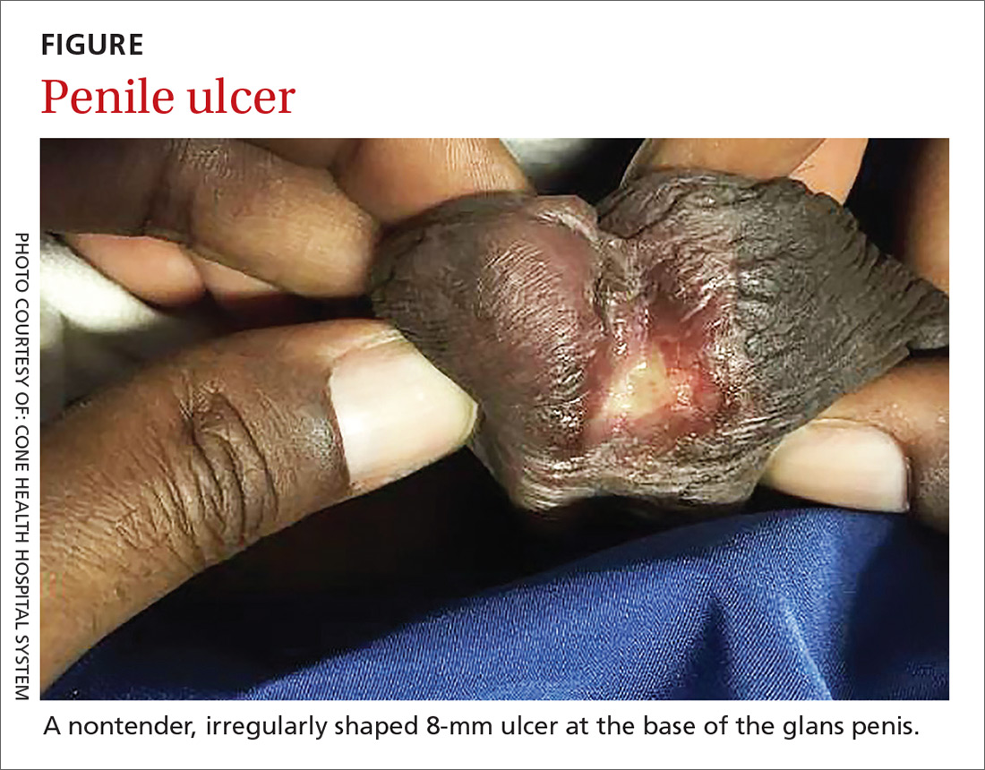
History of posttraumatic stress disorder • priapism • Dx?

Article Type
Changed
Fri, 01/18/2019 - 08:56
Display Headline
History of posttraumatic stress disorder • priapism • Dx?

THE CASE

A 35-year-old African-American man, who was an active duty service member, presented to the Troop Medical Clinic with a 4-hour history of priapism. He had been taking sertraline 100 mg and prazosin 10 mg nightly for 4 months to treat his posttraumatic stress disorder (PTSD) with no reported adverse effects. These doses were titrated 2 months prior to presentation. The patient reported that he took his usual medication doses before bed and awoke at 3 am with a penile erection. At 7 am, he presented to the clinic because of pain from the continued erection.

THE DIAGNOSIS

A penile erection was present on physical exam. All medications were reviewed for adverse effects. A work-up for anemia, sickle cell disease, thalassemia, and platelet abnormalities was negative. A blood gas analysis performed on blood aspirated from the corpus cavernosum showed hypoxemia, hypercarbia, and acidosis, confirming a diagnosis of ischemic priapism.

DISCUSSION

Priapism is a prolonged erection of the penis that is usually not associated with sexual activity or stimulation. It is considered a urologic emergency and requires prompt treatment to prevent long-term complications, such as permanent erectile dysfunction.


Priapism is classified as one of 2 types: ischemic (“low flow”) or nonischemic (“high flow”).

Ischemic priapism is the most common type. It is caused by dysfunctional cavernosal smooth muscle, which creates a compartment-like syndrome in the cavernous tissue that leads to hypoxia and acidosis.1 Nonischemic priapism is often caused by a fistula between the cavernosal artery and corpus cavernosum and is common with traumatic injuries. Nonischemic priapism has a lower risk for long-term complications (due to the blood being well-oxygenated) and often resolves spontaneously without treatment.2,3

Certain medications can cause priapism

Our patient’s ischemic priapism was most likely caused by the combined antagonistic properties of prazosin and sertraline on alpha-1 adrenergic receptors.3,4 Adrenergic alpha-blockers block the sympathetic system, which can in turn inhibit penile detumescence and cause priapism.4

An increasingly common Tx combination. Selective serotonin reuptake inhibitors (SSRIs) such as sertraline are considered first-line treatment for the symptoms of PTSD, and prazosin has been found to be effective in the treatment of nightmares associated with PTSD. (Treatment of PTSD-related nightmares with prazosin is an off-label but frequent use of the medication.) This combination of medications is becoming increasingly common for the treatment of PTSD and its associated symptoms.5-7

Continue to: Cases to date provide interesting insight into this adverse effect


Cases to date provide interesting insight into this adverse effect

In our literature review, no documented cases of priapism were attributed to prazosin when it was used for the treatment of nightmares, but there are multiple case reports of priapism linked to the drug’s use for hypertension.

In the majority of these case reports, the dosage exceeded 10 mg/d and was much higher than the dosage typically used to treat nightmares.7 Many of the affected patients also had associated comorbidities such as diabetes or chronic kidney disease.4

Sertraline has been associated with priapism when used as monotherapy and in combination therapy with antipsychotics. All SSRIs have antagonistic properties to alpha-1 adrenergic receptors, but sertraline appears to have more than a 10-fold increase in affinity when compared to other SSRIs.3


Treatment: An injection and aspiration

Our patient was treated with phenylephrine injection and aspiration, which resolved the priapism. Prazosin was stopped, and the patient was weaned off of sertraline. He continued to follow up closely with Behavioral Health for further management of his PTSD and associated symptoms.

Continue to: THE TAKEAWAY


THE TAKEAWAY

PTSD is being diagnosed more frequently, especially in active duty soldiers, veterans, members of the National Guard, and reservists.8 Because nightmares are a common symptom of PTSD and SSRIs are first-line treatment for PTSD, the combination of prazosin and an SSRI for the treatment of PTSD is frequently encountered.5-7 Providers who prescribe and/or care for patients treated with these medications need to counsel patients on the risk of priapism and the risks associated with a delay in seeking medical care.

All SSRIs have antagonistic properties to alpha-1adrenergic receptors, but sertraline appears to have more than a 10-fold increase in affinity when compared to other SSRIs.

If a patient who is taking these medications presents with priapism, contact Urology immediately for acute management. Both medications must be stopped to prevent future episodes; prazosin can be stopped immediately, but patients must be weaned off of sertraline to avoid experiencing withdrawal symptoms. Patients will need to follow up with a behavioral health team for continued management of their PTSD symptoms.

CORRESPONDENCE
Caleb Dickison, DO, Fort Belvoir Community Hospital, 9300 Dewitt Loop, Fort Belvoir, VA 22060; [email protected].

References

1. Pryor J, Akkus E, Alter G, et al. Priapism. J Sex Med. 2004;1:116-120.

2. Broderick GA, Gordon D, Hypolite J, et al. Anoxia and corporal smooth muscle dysfunction: a model for ischemic priapism. J Urol. 1994;151:259-262.

3. Choua, R, Lee HC, Castro J, et al. Priapism associated with multiple psychotropics: a case report and review of the literature. 2007. Available at: http://primarypsychiatry.com/priapism-associated-with-multiple-psychotropics-a-case-report-and-review-of-the-literature/. Accessed on May 7, 2018.

4. Spagnul SJ, Cabral PH, Verndl DO, et al. Adrenergic alpha-blockers: an infrequent and overlooked cause of priapism. Int J Impot Res. 2011;23:95-98.

5. Stein DJ, Ipser JC, Seedat S. Pharmacotherapy for posttraumatic stress disorder (PTSD). Cochrane Database Syst Rev. 2006;CD002795.

6. Taylor FB, Martin P, Thompson C, et al. Prazosin effects on objective sleep measures and clinical symptoms in civilian trauma PTSD: a placebo-controlled study. Biol Psychiatry. 2008;63:629-632.

7. Raskind MA, Peskind ER, Hoff DJ, et al. A parallel group placebo controlled study of prazosin for trauma nightmares and sleep disturbance in combat veterans with post-traumatic stress disorder. Biol Psychiatry. 2007;61:928-934.

8. Grieger TA, Cozza SJ, Ursano RJ, et al. Posttraumatic stress disorder and depression in battle-injured soldiers. Am J Psychiatry. 2006;163:1777-1783.

Article PDF
Author and Disclosure Information

Fort Belvoir Community Hospital, Va (Dr. Dickison); University of Louisiana Lafayette US Army Reserves (Mr. Venable)
[email protected] 

The authors reported no potential conflict of interest relevant to this article.

The opinions and assertions contained herein are those of the authors and are not to be construed as official or reflecting the views of the US Army or the Department of Defense.

Issue
The Journal of Family Practice - 67(6)
Publications
Topics
Page Number
E10-E11
Sections
Author and Disclosure Information

Fort Belvoir Community Hospital, Va (Dr. Dickison); University of Louisiana Lafayette US Army Reserves (Mr. Venable)
[email protected] 

The authors reported no potential conflict of interest relevant to this article.

The opinions and assertions contained herein are those of the authors and are not to be construed as official or reflecting the views of the US Army or the Department of Defense.

Article PDF
Article PDF

THE CASE

A 35-year-old African-American man, who was an active duty service member, presented to the Troop Medical Clinic with a 4-hour history of priapism. He had been taking sertraline 100 mg and prazosin 10 mg nightly for 4 months to treat his posttraumatic stress disorder (PTSD) with no reported adverse effects. These doses were titrated 2 months prior to presentation. The patient reported that he took his usual medication doses before bed and awoke at 3 am with a penile erection. At 7 am, he presented to the clinic because of pain from the continued erection.

THE DIAGNOSIS

A penile erection was present on physical exam. All medications were reviewed for adverse effects. A work-up for anemia, sickle cell disease, thalassemia, and platelet abnormalities was negative. A blood gas analysis performed on blood aspirated from the corpus cavernosum showed hypoxemia, hypercarbia, and acidosis, confirming a diagnosis of ischemic priapism.

DISCUSSION

Priapism is a prolonged erection of the penis that is usually not associated with sexual activity or stimulation. It is considered a urologic emergency and requires prompt treatment to prevent long-term complications, such as permanent erectile dysfunction.


Priapism is classified as one of 2 types: ischemic (“low flow”) or nonischemic (“high flow”).

Ischemic priapism is the most common type. It is caused by dysfunctional cavernosal smooth muscle, which creates a compartment-like syndrome in the cavernous tissue that leads to hypoxia and acidosis.1 Nonischemic priapism is often caused by a fistula between the cavernosal artery and corpus cavernosum and is common with traumatic injuries. Nonischemic priapism has a lower risk for long-term complications (due to the blood being well-oxygenated) and often resolves spontaneously without treatment.2,3

Certain medications can cause priapism

Our patient’s ischemic priapism was most likely caused by the combined antagonistic properties of prazosin and sertraline on alpha-1 adrenergic receptors.3,4 Adrenergic alpha-blockers block the sympathetic system, which can in turn inhibit penile detumescence and cause priapism.4

An increasingly common Tx combination. Selective serotonin reuptake inhibitors (SSRIs) such as sertraline are considered first-line treatment for the symptoms of PTSD, and prazosin has been found to be effective in the treatment of nightmares associated with PTSD. (Treatment of PTSD-related nightmares with prazosin is an off-label but frequent use of the medication.) This combination of medications is becoming increasingly common for the treatment of PTSD and its associated symptoms.5-7

Continue to: Cases to date provide interesting insight into this adverse effect


Cases to date provide interesting insight into this adverse effect

In our literature review, no documented cases of priapism were attributed to prazosin when it was used for the treatment of nightmares, but there are multiple case reports of priapism linked to the drug’s use for hypertension.

In the majority of these case reports, the dosage exceeded 10 mg/d and was much higher than the dosage typically used to treat nightmares.7 Many of the affected patients also had associated comorbidities such as diabetes or chronic kidney disease.4

Sertraline has been associated with priapism when used as monotherapy and in combination therapy with antipsychotics. All SSRIs have antagonistic properties to alpha-1 adrenergic receptors, but sertraline appears to have more than a 10-fold increase in affinity when compared to other SSRIs.3


Treatment: An injection and aspiration

Our patient was treated with phenylephrine injection and aspiration, which resolved the priapism. Prazosin was stopped, and the patient was weaned off of sertraline. He continued to follow up closely with Behavioral Health for further management of his PTSD and associated symptoms.

Continue to: THE TAKEAWAY


THE TAKEAWAY

PTSD is being diagnosed more frequently, especially in active duty soldiers, veterans, members of the National Guard, and reservists.8 Because nightmares are a common symptom of PTSD and SSRIs are first-line treatment for PTSD, the combination of prazosin and an SSRI for the treatment of PTSD is frequently encountered.5-7 Providers who prescribe and/or care for patients treated with these medications need to counsel patients on the risk of priapism and the risks associated with a delay in seeking medical care.

All SSRIs have antagonistic properties to alpha-1adrenergic receptors, but sertraline appears to have more than a 10-fold increase in affinity when compared to other SSRIs.

If a patient who is taking these medications presents with priapism, contact Urology immediately for acute management. Both medications must be stopped to prevent future episodes; prazosin can be stopped immediately, but patients must be weaned off of sertraline to avoid experiencing withdrawal symptoms. Patients will need to follow up with a behavioral health team for continued management of their PTSD symptoms.

CORRESPONDENCE
Caleb Dickison, DO, Fort Belvoir Community Hospital, 9300 Dewitt Loop, Fort Belvoir, VA 22060; [email protected].

References

1. Pryor J, Akkus E, Alter G, et al. Priapism. J Sex Med. 2004;1:116-120.

2. Broderick GA, Gordon D, Hypolite J, et al. Anoxia and corporal smooth muscle dysfunction: a model for ischemic priapism. J Urol. 1994;151:259-262.

3. Choua, R, Lee HC, Castro J, et al. Priapism associated with multiple psychotropics: a case report and review of the literature. 2007. Available at: http://primarypsychiatry.com/priapism-associated-with-multiple-psychotropics-a-case-report-and-review-of-the-literature/. Accessed on May 7, 2018.

4. Spagnul SJ, Cabral PH, Verndl DO, et al. Adrenergic alpha-blockers: an infrequent and overlooked cause of priapism. Int J Impot Res. 2011;23:95-98.

5. Stein DJ, Ipser JC, Seedat S. Pharmacotherapy for posttraumatic stress disorder (PTSD). Cochrane Database Syst Rev. 2006;CD002795.

6. Taylor FB, Martin P, Thompson C, et al. Prazosin effects on objective sleep measures and clinical symptoms in civilian trauma PTSD: a placebo-controlled study. Biol Psychiatry. 2008;63:629-632.

7. Raskind MA, Peskind ER, Hoff DJ, et al. A parallel group placebo controlled study of prazosin for trauma nightmares and sleep disturbance in combat veterans with post-traumatic stress disorder. Biol Psychiatry. 2007;61:928-934.

8. Grieger TA, Cozza SJ, Ursano RJ, et al. Posttraumatic stress disorder and depression in battle-injured soldiers. Am J Psychiatry. 2006;163:1777-1783.

Issue
The Journal of Family Practice - 67(6)
Issue
The Journal of Family Practice - 67(6)
Page Number
E10-E11
Page Number
E10-E11
Publications
Publications
Topics
Article Type
Display Headline
History of posttraumatic stress disorder • priapism • Dx?
Display Headline
History of posttraumatic stress disorder • priapism • Dx?
Sections
Disallow All Ads
Content Gating
No Gating (article Unlocked/Free)
Alternative CME
PubMed ID
29879245
Disqus Comments
Default
Use ProPublica
Article PDF Media

Testosterone therapy tied to kidney stone risk

Article Type
Changed
Fri, 01/18/2019 - 17:41


Testosterone replacement therapy was associated with heightened risk for kidney stones, according to an analysis of more than 50,000 men with low testosterone.

When researchers compared hypogonadal men to age- and comorbidity-matched controls, they found a statistically significantly higher number of clinical diagnoses of a kidney stone, or of patients undergoing a kidney stone–related procedure.

Jim Kling/MDedge News
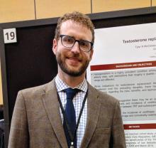
Dr. Tyler McClintock
Animal studies have suggested that testosterone replacement can increase oxalate and decrease citrate levels in the kidney, both of which may increase risk of stone formation.

The new study is the first large-scale analysis of the question in humans, according to Tyler McClintock, MD, who presented the findings at a poster session at the annual meeting of the American Urological Association. Dr. McClintock is a urology resident at Brigham and Women’s Hospital and Harvard Medical School in Boston.

Dr. McClintock and his colleagues analyzed data from the Military Health System Data Repository (MDR). The MDR includes beneficiaries of the TRICARE program for service members, retirees, and their families. They looked at 26,586 men aged 40-64 years who had been diagnosed with low testosterone and who had received continuous testosterone replacement therapy between April 2006 and March 2014. The researchers compared them to 26,586 controls with low testosterone who did not receive testosterone replacement therapy.

Stone events were significantly higher in the treatment group. There were 67 extracorporeal shock wave lithotripsy procedures in the treatment group, compared with 51 among controls. Similar trends were seen with ureteroscopy with lithotripsy (75 vs. 46) and clinical diagnoses of kidney stone (1,059 vs. 794).

The researchers also broke down stone events by type of testosterone replacement therapy. A total of 5.4% of patients who received pellets (9 of 167) experienced an event (P = .27), compared with 5.1% of those who received injections (218 of 4,259; P = .004) and 3.5% of those who received it topically (655 of 18,895; P less than .0001).

At 2 years, Dr. McClintock reported that there were significantly more kidney stone events in the testosterone-treated group than in the untreated group (659 and 482, respectively; P less than .001). Two years after starting testosterone replacement therapy, significantly more of the treatment group had experienced a stone episode, compared with the matched controls during the same time period (3.9% and 3%, respectively; P less than .001).


SOURCE: McClintock T. AUA Annual Meeting. Abstract MP13-19.

Meeting/Event
Publications
Topics
Sections
Meeting/Event
Meeting/Event


Testosterone replacement therapy was associated with heightened risk for kidney stones, according to an analysis of more than 50,000 men with low testosterone.

When researchers compared hypogonadal men to age- and comorbidity-matched controls, they found a statistically significantly higher number of clinical diagnoses of a kidney stone, or of patients undergoing a kidney stone–related procedure.

Jim Kling/MDedge News
Dr. Tyler McClintock
Animal studies have suggested that testosterone replacement can increase oxalate and decrease citrate levels in the kidney, both of which may increase risk of stone formation.

The new study is the first large-scale analysis of the question in humans, according to Tyler McClintock, MD, who presented the findings at a poster session at the annual meeting of the American Urological Association. Dr. McClintock is a urology resident at Brigham and Women’s Hospital and Harvard Medical School in Boston.

Dr. McClintock and his colleagues analyzed data from the Military Health System Data Repository (MDR). The MDR includes beneficiaries of the TRICARE program for service members, retirees, and their families. They looked at 26,586 men aged 40-64 years who had been diagnosed with low testosterone and who had received continuous testosterone replacement therapy between April 2006 and March 2014. The researchers compared them to 26,586 controls with low testosterone who did not receive testosterone replacement therapy.

Stone events were significantly higher in the treatment group. There were 67 extracorporeal shock wave lithotripsy procedures in the treatment group, compared with 51 among controls. Similar trends were seen with ureteroscopy with lithotripsy (75 vs. 46) and clinical diagnoses of kidney stone (1,059 vs. 794).

The researchers also broke down stone events by type of testosterone replacement therapy. A total of 5.4% of patients who received pellets (9 of 167) experienced an event (P = .27), compared with 5.1% of those who received injections (218 of 4,259; P = .004) and 3.5% of those who received it topically (655 of 18,895; P less than .0001).

At 2 years, Dr. McClintock reported that there were significantly more kidney stone events in the testosterone-treated group than in the untreated group (659 and 482, respectively; P less than .001). Two years after starting testosterone replacement therapy, significantly more of the treatment group had experienced a stone episode, compared with the matched controls during the same time period (3.9% and 3%, respectively; P less than .001).


SOURCE: McClintock T. AUA Annual Meeting. Abstract MP13-19.

Publications
Publications
Topics
Article Type
Sections
Article Source

REPORTING FROM THE AUA ANNUAL MEETING

Disallow All Ads
Content Gating
No Gating (article Unlocked/Free)
Alternative CME
Vitals


Key clinical point: Kidney stone risk may be a factor when considering testosterone replacement therapy.

Major finding: In untreated men, 482 kidney stone events occurred, compared with 659 in those receiving testosterone.

Study details: A case-control analysis of 26,586 treated men and 26,586 matched controls.

Disclosures: The Department of Defense funded the study. Dr. McClintock reported having no relevant financial disclosures.

Source: McClintock T. AUA Annual Meeting. Abstract MP13-19.

Disqus Comments
Default
Use ProPublica

Abstract: Divergent Responses to Mammography and Prostate-Specific Antigen Recommendations

Article Type
Changed
Fri, 01/18/2019 - 08:55
Display Headline
Abstract: Divergent Responses to Mammography and Prostate-Specific Antigen Recommendations
The video associated with this article is no longer available on this site. Please view all of our videos on the MDedge YouTube channel

Martinez, K.A., et al, Am J Prev Med 53(4):533, October 2017

The authors, from the Cleveland Clinic, explore the reasons for divergent responses to recent guideline recommendations against screening for breast cancer and prostate cancer. Both cancers are common (lifetime incidence of 12% and 16%, respectively), with indolent forms that are more prevalent with age. Screening with mammography and prostate-specific antigen (PSA) testing achieves earlier detection and relative risk reductions in cancer-specific mortality of 15% to 20%, but negligible effects on overall mortality and a risk of overtreatment that is estimated to be 30% to 80%. Benefits of screening and risks of overtreatment are unclear because of study limitations and evolution in diagnostic criteria, screening and management. Beginning in 2008, the US Preventive Services Task Force downgraded its recommendations for routine mammography (e.g., a rating of C for average-risk women aged 40-49) and for routine PSA testing (rating of D regardless of age). [EDITOR’S NOTE: USPSTF PSA screening recommendations have changed. See “USPSTF advises against widespread prostate cancer screening.”] While objections to the PSA rating were minimal, the public and professional backlash about mammography was profound. Reasons may include unique characteristics of breast (versus prostate) cancer, including mortality at a younger age, financial conflicts of interest (with mammography generating $8 billion per year in the US), bureaucratic incentives (e.g., quality measures, insurance reimbursement), and the influence of profit-generating advocacy groups. Many physicians do not understand the risks and harms of overtreatment or the inability of early detection to prevent metastasis, and patients want to be “better safe than sorry.” Rates of screening mammography may not decrease until truly informed decisions are possible, which will necessitate better algorithms to predict those cancers that are likely to spread.  20 references ([email protected] – no reprints)

Publications
Topics
Sections
The video associated with this article is no longer available on this site. Please view all of our videos on the MDedge YouTube channel

Martinez, K.A., et al, Am J Prev Med 53(4):533, October 2017

The authors, from the Cleveland Clinic, explore the reasons for divergent responses to recent guideline recommendations against screening for breast cancer and prostate cancer. Both cancers are common (lifetime incidence of 12% and 16%, respectively), with indolent forms that are more prevalent with age. Screening with mammography and prostate-specific antigen (PSA) testing achieves earlier detection and relative risk reductions in cancer-specific mortality of 15% to 20%, but negligible effects on overall mortality and a risk of overtreatment that is estimated to be 30% to 80%. Benefits of screening and risks of overtreatment are unclear because of study limitations and evolution in diagnostic criteria, screening and management. Beginning in 2008, the US Preventive Services Task Force downgraded its recommendations for routine mammography (e.g., a rating of C for average-risk women aged 40-49) and for routine PSA testing (rating of D regardless of age). [EDITOR’S NOTE: USPSTF PSA screening recommendations have changed. See “USPSTF advises against widespread prostate cancer screening.”] While objections to the PSA rating were minimal, the public and professional backlash about mammography was profound. Reasons may include unique characteristics of breast (versus prostate) cancer, including mortality at a younger age, financial conflicts of interest (with mammography generating $8 billion per year in the US), bureaucratic incentives (e.g., quality measures, insurance reimbursement), and the influence of profit-generating advocacy groups. Many physicians do not understand the risks and harms of overtreatment or the inability of early detection to prevent metastasis, and patients want to be “better safe than sorry.” Rates of screening mammography may not decrease until truly informed decisions are possible, which will necessitate better algorithms to predict those cancers that are likely to spread.  20 references ([email protected] – no reprints)

Publications
Publications
Topics
Article Type
Display Headline
Abstract: Divergent Responses to Mammography and Prostate-Specific Antigen Recommendations
Display Headline
Abstract: Divergent Responses to Mammography and Prostate-Specific Antigen Recommendations
Sections
Citation Override
Source: Primary Care Medical Abstracts, 05/18 - #23
PURLs Copyright

Learn more about the Primary Care Medical Abstracts and podcasts, for which you can earn up to 9 CME credits per month.

Copyright © The Center for Medical Education

Disallow All Ads
Content Gating
No Gating (article Unlocked/Free)
Alternative CME
Disqus Comments
Default
Gate On Date
Tue, 05/01/2018 - 14:00
Un-Gate On Date
Tue, 05/01/2018 - 14:00
Use ProPublica
CFC Schedule Remove Status
Tue, 05/01/2018 - 14:00

MDedge Daily News: Prostate screening should be by request

Article Type
Changed
Thu, 03/28/2019 - 09:14


The USPSTF has new recommendations against prostate cancer screening, increased water intake did not slow the decline of chronic kidney disease, the rate of uninsured Americans is on the rise, and when – and how – to suspect asthma misdiagnosis.

Listen to the MDedge Daily News podcast for all the details on today’s top news.


Publications
Topics
Sections


The USPSTF has new recommendations against prostate cancer screening, increased water intake did not slow the decline of chronic kidney disease, the rate of uninsured Americans is on the rise, and when – and how – to suspect asthma misdiagnosis.

Listen to the MDedge Daily News podcast for all the details on today’s top news.


Publications
Publications
Topics
Article Type
Sections
Disallow All Ads
Content Gating
No Gating (article Unlocked/Free)
Alternative CME
Disqus Comments
Default
Use ProPublica

USPSTF advises against widespread prostate cancer screening

Selectively screen those who could benefit
Article Type
Changed
Fri, 01/18/2019 - 17:38


The USPSTF recommends that, to reduce the risk of false positives and unnecessary complications from prostate cancer screening and treatment, physicians and their male patients aged 55-69 years should review together the pros and cons.

Clinicians should not conduct prostate cancer screening in men aged 55-69 years who do not ask for it (level C recommendation), according to the USPSTF recommendations, published in JAMA, which also recommend against any prostate cancer screening for men aged 70 years and older (level D recommendation). The recommendations replace those from 2012, and upgrade the statement against routine screening from a D to a C.

“The change in recommendation grade further reflects new evidence about and increased use of active surveillance of low-risk prostate cancer, which may reduce the risk of subsequent harms from screening,” according to the USPSTF.

The recommendations apply to asymptomatic adult men in the general United States population with no previous diagnosis of prostate cancer, as well as those whose ethnicity or family history put them at increased risk of death from prostate cancer.

In the evidence report published in JAMA, Joshua J. Fenton, MD, professor in the department of family and community medicine of the University of California, Davis, Sacramento, and his colleagues reviewed 63 studies comprising 1,904,950 individuals. The researchers examined the findings for information including the effectiveness of PSA screening and the potential harms associated with both screening and cancer treatment if disease was identified.

Overdiagnosis of prostate cancer ranged from 21% to 50% for cancers detected by screening, and one randomized trial of more than 1,000 men found no significant reduction in mortality for prostatectomy or radiation therapy compared with active monitoring.

Overall, men randomized to PSA screening had no significant reduction in risk of prostate cancer mortality in trials from the United States or the United Kingdom, although data from a European trial showed a significant reduction. Complications requiring hospitalization occurred in 0.5%-1.6% of men who had biopsies after screening showed abnormal results.


The evidence review was limited by several factors including a lack of data on newer treatments such as cryotherapy and high-intensity focused ultrasound, the researchers noted.

However, the data support an individualized approach to PSA screening for prostate cancer, in which each man can weigh the potential risks and benefits of screening, according to the USPSTF.

The research was funded by the Agency for Healthcare Research and Quality. The researchers had no financial conflicts to disclose.

SOURCE: Fenton J et al. JAMA. 2018;319(18):1914-31. and JAMA. 2018;319(18):1901-13.

Body


The new USPSTF guidelines take a thoughtful approach to assessing the pros and cons of PSA-based prostate cancer screening and highlight the importance of identifying subgroups who could most benefit from screening and treatment, H. Ballentine Carter, MD, wrote in an accompanying editorial.

“Patients, together with their physicians, should decide whether prostate cancer screening is right for the patient. In this regard, primary care physicians have an important role in reducing the harms associated with screening and could consider a number of factors in this decision process,” he said.

In particular, Dr. Carter noted that men aged 55-69 years without multiple comorbidities would reap the greatest benefits from screening, while those aged 70 years and older would be more susceptible to the harm associated with testing and treatment and should be screened rarely. He also endorsed a 2- to 4-year screening interval to help reduce false-positive test results and overdiagnosis.

“By virtue of their relationship with patients, primary care physicians are in a unique position to help ensure that men diagnosed with favorable-risk disease (Gleason score 6 cancer grade on biopsy, and PSA level less than 10 ng/mL) are presenting a balanced message regarding management options,” with active surveillance as the preferred choice, he said. (JAMA. 2018. May 8;319[18]:1866-8).
 

Dr. Carter is Bernard L. Schwartz distinguished professor of urologic oncology and professor of urology at Johns Hopkins University School of Medicine, Baltimore, and had no financial conflicts to disclose.

Publications
Topics
Sections
Body


The new USPSTF guidelines take a thoughtful approach to assessing the pros and cons of PSA-based prostate cancer screening and highlight the importance of identifying subgroups who could most benefit from screening and treatment, H. Ballentine Carter, MD, wrote in an accompanying editorial.

“Patients, together with their physicians, should decide whether prostate cancer screening is right for the patient. In this regard, primary care physicians have an important role in reducing the harms associated with screening and could consider a number of factors in this decision process,” he said.

In particular, Dr. Carter noted that men aged 55-69 years without multiple comorbidities would reap the greatest benefits from screening, while those aged 70 years and older would be more susceptible to the harm associated with testing and treatment and should be screened rarely. He also endorsed a 2- to 4-year screening interval to help reduce false-positive test results and overdiagnosis.

“By virtue of their relationship with patients, primary care physicians are in a unique position to help ensure that men diagnosed with favorable-risk disease (Gleason score 6 cancer grade on biopsy, and PSA level less than 10 ng/mL) are presenting a balanced message regarding management options,” with active surveillance as the preferred choice, he said. (JAMA. 2018. May 8;319[18]:1866-8).
 

Dr. Carter is Bernard L. Schwartz distinguished professor of urologic oncology and professor of urology at Johns Hopkins University School of Medicine, Baltimore, and had no financial conflicts to disclose.

Title
Selectively screen those who could benefit


The USPSTF recommends that, to reduce the risk of false positives and unnecessary complications from prostate cancer screening and treatment, physicians and their male patients aged 55-69 years should review together the pros and cons.

Clinicians should not conduct prostate cancer screening in men aged 55-69 years who do not ask for it (level C recommendation), according to the USPSTF recommendations, published in JAMA, which also recommend against any prostate cancer screening for men aged 70 years and older (level D recommendation). The recommendations replace those from 2012, and upgrade the statement against routine screening from a D to a C.

“The change in recommendation grade further reflects new evidence about and increased use of active surveillance of low-risk prostate cancer, which may reduce the risk of subsequent harms from screening,” according to the USPSTF.

The recommendations apply to asymptomatic adult men in the general United States population with no previous diagnosis of prostate cancer, as well as those whose ethnicity or family history put them at increased risk of death from prostate cancer.

In the evidence report published in JAMA, Joshua J. Fenton, MD, professor in the department of family and community medicine of the University of California, Davis, Sacramento, and his colleagues reviewed 63 studies comprising 1,904,950 individuals. The researchers examined the findings for information including the effectiveness of PSA screening and the potential harms associated with both screening and cancer treatment if disease was identified.

Overdiagnosis of prostate cancer ranged from 21% to 50% for cancers detected by screening, and one randomized trial of more than 1,000 men found no significant reduction in mortality for prostatectomy or radiation therapy compared with active monitoring.

Overall, men randomized to PSA screening had no significant reduction in risk of prostate cancer mortality in trials from the United States or the United Kingdom, although data from a European trial showed a significant reduction. Complications requiring hospitalization occurred in 0.5%-1.6% of men who had biopsies after screening showed abnormal results.


The evidence review was limited by several factors including a lack of data on newer treatments such as cryotherapy and high-intensity focused ultrasound, the researchers noted.

However, the data support an individualized approach to PSA screening for prostate cancer, in which each man can weigh the potential risks and benefits of screening, according to the USPSTF.

The research was funded by the Agency for Healthcare Research and Quality. The researchers had no financial conflicts to disclose.

SOURCE: Fenton J et al. JAMA. 2018;319(18):1914-31. and JAMA. 2018;319(18):1901-13.

Publications
Publications
Topics
Article Type
Click for Credit Status
Ready
Sections
Article Source
FROM JAMA
Disallow All Ads
Content Gating
No Gating (article Unlocked/Free)
Alternative CME
Vitals


Key clinical point: PSA-based screening for prostate cancer in men aged 55-69 years has limited benefits and significant risks.

Major finding: Overdiagnosis occurred in approximately 21%-50% of cancers identified during PSA screening.

Study details: The evidence report was based on 63 studies including 1.9 million men.

Disclosures: The research was funded by the Agency for Healthcare Research and Quality. The researchers had no financial conflicts to disclose.

Source: JAMA. 2018;319(18):1901-13. Fenton J et al. JAMA. 2018;319(18):1914-31.
 

Disqus Comments
Default
Use ProPublica

VIDEO: Few transgender patients desire care in a transgender-only clinic

Article Type
Changed
Mon, 06/17/2019 - 08:25

AUSTIN, TEX. – Transgender patients face many barriers to care, including a lack of necessary expertise among providers, but a large majority of those surveyed in a study in which they were asked whether they would want to go to a transgender-only clinic said they would not.

Lauren Abern, MD, of Atrius Health, Cambridge, Mass., discussed the aims and results of her survey at the annual clinical and scientific meeting of the American College of Obstetricians and Gynecologists.

The anonymous online survey consisted of 120 individuals, aged 18-64 years: 100 transgender men and 20 transgender women. Of these, 83 reported experiencing barriers to care. The most common problem cited was cost (68, 82%), and other barriers were access to care (47, 57%), stigma (33, 40%), and discrimination (23, 26%). Cost was a factor even though a large majority of the respondents had health insurance; a majority of respondents had an income of less than $24,000 per year.

The most common way respondents found transgender-competent health care was through word of mouth (79, 77%).

When asked whether they would want to go to a transgender-only clinic, a majority of both transgender women and transgender men respondents either answered, “no,” or that they were unsure (86, 77%). Some respondents cited a desire not to out themselves as transgender, and others considered the separate clinic medically unnecessary. One wrote: “You wouldn’t need a broken foot–only clinic.”

“Basic preventative services can be provided without specific expertise in transgender health. If providers are uncomfortable, they should refer [transgender patients] elsewhere.” said Dr. Abern.

The survey project was conducted in collaboration with the University of Miami and the YES Institute in Miami.

Dr. Abern also spoke about wider transgender health considerations for the ob.gyn. in a separate presentation at the meeting and in a video interview.

For example, transgender men on testosterone may have persistent bleeding and may be uncomfortable with pelvic exams.

Making more inclusive intake forms and fostering a respectful office environment (for example, having a nondiscrimination policy displayed in the waiting area) are measures beneficial to all patients, she said.

“My dream or goal would be that transgender people can be seen and accepted at any office and feel comfortable and not avoid seeking health care.”

Meeting/Event
Publications
Topics
Sections
Meeting/Event
Meeting/Event

AUSTIN, TEX. – Transgender patients face many barriers to care, including a lack of necessary expertise among providers, but a large majority of those surveyed in a study in which they were asked whether they would want to go to a transgender-only clinic said they would not.

Lauren Abern, MD, of Atrius Health, Cambridge, Mass., discussed the aims and results of her survey at the annual clinical and scientific meeting of the American College of Obstetricians and Gynecologists.

The anonymous online survey consisted of 120 individuals, aged 18-64 years: 100 transgender men and 20 transgender women. Of these, 83 reported experiencing barriers to care. The most common problem cited was cost (68, 82%), and other barriers were access to care (47, 57%), stigma (33, 40%), and discrimination (23, 26%). Cost was a factor even though a large majority of the respondents had health insurance; a majority of respondents had an income of less than $24,000 per year.

The most common way respondents found transgender-competent health care was through word of mouth (79, 77%).

When asked whether they would want to go to a transgender-only clinic, a majority of both transgender women and transgender men respondents either answered, “no,” or that they were unsure (86, 77%). Some respondents cited a desire not to out themselves as transgender, and others considered the separate clinic medically unnecessary. One wrote: “You wouldn’t need a broken foot–only clinic.”

“Basic preventative services can be provided without specific expertise in transgender health. If providers are uncomfortable, they should refer [transgender patients] elsewhere.” said Dr. Abern.

The survey project was conducted in collaboration with the University of Miami and the YES Institute in Miami.

Dr. Abern also spoke about wider transgender health considerations for the ob.gyn. in a separate presentation at the meeting and in a video interview.

For example, transgender men on testosterone may have persistent bleeding and may be uncomfortable with pelvic exams.

Making more inclusive intake forms and fostering a respectful office environment (for example, having a nondiscrimination policy displayed in the waiting area) are measures beneficial to all patients, she said.

“My dream or goal would be that transgender people can be seen and accepted at any office and feel comfortable and not avoid seeking health care.”

Publications
Publications
Topics
Article Type
Sections
Article Source

REPORTING FROM ACOG 2018

Disallow All Ads
Content Gating
No Gating (article Unlocked/Free)
Alternative CME
Disqus Comments
Default
Use ProPublica
Hide sidebar & use full width
render the right sidebar.

Idiopathic pulmonary fibrosis: What primary care physicians need to know

Article Type
Changed
Wed, 05/01/2019 - 07:35
Display Headline
Idiopathic pulmonary fibrosis: What primary care physicians need to know

Idiopathic pulmonary fibrosis (IPF) is a devastating and fatal lung disease that generally affects older adults. It is characterized by a radiographic and histopathologic pattern of usual interstitial pneumonia (UIP) that has no other known etiology.

See related editorial

Accurate diagnosis of IPF is crucial. We recommend early referral to a center specializing in interstitial lung disease to confirm the diagnosis, start appropriate therapy, advise the patient on prognosis and enrollment in disease registries and clinical trials, and determine candidacy for lung transplant.

Primary care physicians are uniquely positioned to encounter patients with IPF, whether because of a patient complaint or as an incidental finding on computed tomography. The goal of this article is to delineate the features of IPF so that it may be recognized early and thus expedite referral to a center with expertise in interstitial lung disease for a thorough evaluation and appropriate management.

WHAT IS IDIOPATHIC PULMONARY FIBROSIS?

Overview of interstitial lung diseases
IPF is 1 of more than 150 interstitial lung diseases, conditions that share nonspecific symptoms such as dysp­nea and a dry cough with insidious onset, certain radiologic findings, and a restrictive ventilatory defect.1 More specifically, IPF is categorized with the idiopathic interstitial pneumonias, which include idiopathic nonspecific interstitial pneumonia, respiratory bronchiolitis-associated interstitial lung disease, diffuse alveolar hemorrhage, cryptogenic organizing pneumonia, and acute interstitial pneumonia (Table 1).2 Identification of the pattern of UIP, the hallmark of IPF, is integral to establishing the diagnosis, as is exclusion of other causes of interstitial lung disease—eg, connective tissue disease, medication reaction, inhalational exposure, pneumoconioses, and granulomatous diseases (both infectious and noninfectious).

MORE COMMON THAN ONCE THOUGHT

The true incidence and prevalence of IPF are difficult to assess. IPF is generally considered a rare disease, but it is more common than once thought. In 2011, Raghu et al3 estimated the prevalence in Medicare beneficiaries to be 495 cases per 100,000. Based on this estimate and the current US population, up to 160,000 Americans could have IPF.4 Raghu et al also showed that IPF more often affects adults over age 65, which suggests that as the US population ages, the incidence of IPF may rise. Studies have also reported an increased incidence of IPF worldwide.5

Further, with the rising use of low-dose computed tomography to screen for lung cancer, more incidental cases of IPF will likely be found.6–8

Older data showed a lag from symptom onset to accurate diagnosis of 1 to 2 years.9 A more recent study found a lag in referral of patients with IPF to tertiary care centers, and this delay was associated with a higher rate of death independent of disease severity.10

TYPICALLY PROGRESSIVE, OFTEN FATAL

IPF is typically progressive and limited to the lungs, and it portends a poor prognosis. The median survival is commonly cited as 2 to 5 years from diagnosis, although this is based on older observations that may not reflect current best practice and newer therapies. More recent studies suggest higher survival rates if patients have preserved lung function.11

As the name indicates, the etiology of IPF is unknown, but studies have indicated genetic underpinnings in a notable proportion of cases.12 Regardless of the cause, the pathogenesis and progression of IPF are thought to be the result of an abnormal and persistent wound-repair response. The progressive deposition of scar tissue disrupts normal lung architecture and function, eventually causing clinical disease.13


SYMPTOMS AND KEY FEATURES

Patients with IPF typically present with the insidious onset of dyspnea on exertion, with or without chronic cough. Risk factors include male sex, increasing age, and a history of smoking. Patients with undiagnosed IPF who present with dyspnea and a history of smoking are often treated empirically for chronic obstructive pulmonary disease (COPD).

Rales are a common finding on auscultation in IPF, and this can lead to an exhaustive cardiac evaluation and empiric treatment for heart failure. Digital clubbing is also relatively common.14 Hypoxemia with exertion is another common feature that also often correlates with disease severity and prognosis. Resting hypoxemia is more common in advanced disease.

On spirometry, patients with IPF typically demonstrate restrictive physiology, suggested by a normal or elevated ratio of the forced expiratory volume in 1 second to the forced vital capacity (FEV1/FVC) (> 70% predicted or above the lower limit of normal) combined with a lower than normal FVC. Restrictive physiology is definitively demonstrated by a decreased total lung capacity (< 80% predicted or below the lower limit of normal) on plethysmography. Impaired gas exchange, manifested by a decreased diffusing capacity of the lungs for carbon monoxide (DLCO) on pulmonary function testing, is also common. Because pulmonary perfusion is higher in the lung bases, where IPF is also predominant, the DLCO is often reduced to a greater extent than the FVC.

PROGNOSTIC INDICATORS

Idiopathic pulmonary fibrosis progresses in several ways
Figure 1. Idiopathic pulmonary fibrosis progresses in several ways. Some patients have a rapid deterioration with a poor short-term prognosis. In some, the disease progresses slowly with possible occasional exacerbations that cause a marked decline in lung function with no subsequent return to prior baseline function. In some cases, these patients continue to have slow disease progression, while others continue to have a stepwise decline in lung function. In a small number of patients, the disease is stable over time or progresses so slowly that the patient dies of another condition.

Clinicians typically view IPF as a relentless and progressive process, but its course is variable and can be uncertain in an individual patient (Figure 1).15,16 Nevertheless, over time, most patients have a decline in lung function leading to respiratory failure. Respiratory failure, often preceded by a subacute deterioration (over weeks to months) or an acute deterioration (< 4 weeks), is the most common cause of death, but comorbid diseases such as lung cancer, infection, and heart failure are also common causes of death in these patients.17,18

Predictors of mortality include worsening FVC, DLCO, symptoms, and physiologic impairment, manifested by a decline in the 6-minute walking test or worsening exertional hypoxemia.19–22 Other common comorbidities linked with impaired quality of life and poor prognosis include obstructive sleep apnea, gastroesophageal reflux disease, and depression.16,23 Retrospective studies suggest that most IPF patients die 2 to 5 years after symptom onset. With the lag from symptom onset to final diagnosis, the average life expectancy is as little as 2 years from the time of diagnosis.9,18,24,25

Two staging systems have been developed to predict short-term and long-term mortality risk based on sex, age, and physiologic parameters.23,24 The GAP (gender, age, physiology) index provides an estimate of the risk of death for a cohort of patients: a score of 0 to 8 is calculated, and the score is then categorized as stage I, II, or III. Each stage is associated with 1-, 2-, and 3-year mortality rates, with stage III having the highest rates. The GAP calculator (www.acponline.org/journals/annals/extras/gap) provides an estimate of the risk of death for an individual patient. The application of these tools for the management of IPF is evolving; however, they may be helpful for counseling patients about disease prognosis.

CLUES TO DIAGNOSIS

Histologic patterns

Histopathologic appearance of definite pattern of usual interstitial pneumonia (UIP)
Figure 2. Histopathologic appearance of definite pattern of usual interstitial pneumonia (UIP), a key feature of idiopathic pulmonary fibrosis. A, dense fibrosis (plus sign) with a “honeycomb” change (asterisk) is prominent at the pleural surface (arrow) juxtaposed against normal lung tissue (star) toward the center of the lung parenchyma (hematoxylin and eosin, × 40). B, the same features appear at higher magnification, and fibroblastic foci (arrow) are seen at the leading edge of fibrosis (hematoxylin and eosin, × 100).
UIP is a histologic pattern observed on surgical lung biopsy specimens, characterized by fibrosis and a “honeycomb” pattern alternating with areas of normal lung parenchyma in a patchwork pattern (Figure 2). These abnormalities are usually subpleural and worse in the lower lobes. Inflammation is not typically seen.

UIP on histologic study is also seen in fibrotic lung diseases other than IPF, including connective tissue disease-associated interstitial lung disease, inhalational or occupational interstitial lung disease, and chronic hypersensitivity pneumonitis.26–29 Consequently, the diagnosis of IPF requires exclusion of other known causes of UIP.

According to the 2011 guidelines,16 the histology of interstitial lung disease can be categorized as definite UIP, probable UIP, or possible UIP, or as an atypical pattern suggesting another diagnosis. If no definite cause of the interstitial lung abnormality is found, the level of certainty of the histopathologic pattern of UIP helps formulate the clinical diagnosis and management plan.

Clues on computed tomography

The UIP nomenclature also describes patterns on high-resolution computed tomography (HRCT). HRCT is done without contrast and produces thin-sliced images (usually < 1.5 mm) in inspiratory, expiratory, and prone views; this allows detection of air trapping, which may indicate an airway-centric alternative diagnosis.

On HRCT, UIP appears as reticular opacities, often with traction bronchiectasis or bronchiolectasis, usually with a basilar and peripheral predominance. Honeycombing is a key feature and appears as clustered cystic spaces with well-defined walls in the periphery of the lung parenchyma. Ground-glass opacities are not a prominent feature of UIP, and although they do not exclude a UIP pattern, they should spur consideration of other diagnoses.16 Reactive mediastinal and hilar lymphadenopathy is another common feature of UIP.

Radiographic patterns of definite usual interstitial pneumonia
Figure 3. Radiographic patterns of definite usual interstitial pneumonia. Chest radiography (A) shows mildly decreased lung volumes with basilar-predominant coarse reticular (linear) opacities (arrow) and intervening areas of cystic lucencies, consistent with honeycombing (arrowhead). Axial high-resolution computed tomography (B, C, and D) shows coarse subpleural reticulation (arrow in B), traction bronchiectasis, and severe honeycombing, ie, rows of cysts stacked one on top of the other (arrowheads in C and D). These progressively worsen as the images move inferiorly.

When evaluating results of HRCT for UIP, the radiologist categorizes the pattern as definite UIP, possible UIP, or inconsistent. The definite pattern meets all the above features and has none of the features suggesting an alternative diagnosis (Figure 3). The possible pattern includes all the above features with the exception of honeycombing. If the predominant features on HRCT include any atypical finding listed above, then the study is considered inconsistent with UIP. If the pattern on HRCT is considered definite, evaluation of pathology is not necessary. If the pattern is categorized as possible or is inconsistent, then surgical lung biopsy-confirmed UIP is necessary for the definitive diagnosis of IPF.

However, evidence is emerging that in the correct clinical scenario, possible UIP behaves similarly to definite UIP and may be sufficient to make the clinical diagnosis of IPF even without surgical biopsy confirmation.30


A DIAGNOSTIC ALGORITHM FOR IPF

Given the multitude of interstitial lung diseases, their complexities, and the lack of a gold standard definitive diagnostic test, the diagnosis of IPF can be difficult, requiring the integration of clinical, radiologic, and, if necessary, pathologic findings.

A diagnostic algorithm for idiopathic pulmonary fibrosis (IPF)
Figure 4. A diagnostic algorithm for idiopathic pulmonary fibrosis (IPF). Patients with suspected IPF should be evaluated for causes of nonspecific dyspnea, and specifically for interstitial lung disease (ILD). If no cause is iden-tified, then high-resolution computed tomography (HRCT) is recommended to determine the pattern of usual interstitial pneumonia (UIP). If a definite UIP pattern is seen, then a diagnosis of IPF can be made. Otherwise, surgical lung biopsy is the next step. A final diagnosis of IPF can be made if the histologic pattern is definite, probable, or possible UIP, and if the multidiscplinary team (MDD) concurs that IPF is the most likely diagnosis.
Multiple pathologic processes can appear as UIP on imaging or lung biopsy, and these pathologic processes must be ruled out before diagnosing IPF. To aid in ruling out other conditions, the American Thoracic Society, European Respiratory Society, Japanese Respiratory Society, and Latin American Thoracic Association formulated joint evidence-based guidelines for the diagnosis and treatment of IPF.16 The guidelines include an algorithm as an aid to the systematic evaluation. We propose a new algorithm (Figure 4) that is slightly but not insignificantly different from the algorithm in the joint guidelines.

Demographic features

The patient’s demographic features and risk factors dictate the initial clinical suspicion of IPF compared with other interstitial lung diseases. The incidence of IPF increases with age, and IPF is more common in men. A history of smoking is another risk factor.31 A 45-year-old never-smoker is much less likely to have IPF than a 70-year-old former smoker, and a 70-year-old man is more likely to have IPF than a woman of the same age. Thus, the finding of interstitial lung disease in a patient with a demographic profile that is not typical (ie, a younger woman who never smoked) should prompt an exhaustive investigation for another diagnosis such as hypersensitivity pneumonitis or connective tissue disease.

Key elements of the history

After considering the demographic profile and risk factors, the next step in the evaluation is a thorough and accurate medical history. This should include assessment of the severity of dyspnea and cough, signs and symptoms of connective tissue disease (eg, arthralgias, sicca symptoms, Raynaud phenomenon, difficulty swallowing), and gastroesophageal reflux disease, which can be associated with connective tissue disease and, independently, with IPF.

It is also important to identify any environmental exposures that suggest pneumoconiosis or chronic hypersensitivity pneumonitis. The most common risk factors for hypersensitivity pneumonitis are birds and bird feathers, molds, fungi, hot tub use, and some industrial chemicals.32

A medication history is important. Many medications are associated with interstitial lung disease, but amiodarone, bleomycin, methotrexate, and nitrofurantoin are among the common offenders.33

A thorough family history is necessary, as there are familial forms of IPF.

Focus of the physical examination

The physical examination must include careful auscultation for rales. While rales are not specific for IPF, they are the most common pulmonary abnormality. Detailed skin, musculoskeletal, and cardiovascular examinations are also important to evaluate for rheumatologic signs, clubbing, or evidence of heart failure or pulmonary hypertension.

Laboratory tests

Laboratory testing should include a serologic autoantibody panel to evaluate for connective tissue diseases that can manifest as interstitial lung disease, including rheumatoid arthritis, dermatopolymyositis, scleroderma, Sjögren syndrome, and undifferentiated or mixed connective tissue disease. Typical preliminary laboratory tests are antinuclear antibody, rheumatoid factor, erythrocyte sedimentation rate, and C-reactive protein. Others may include anticyclic citrullinated peptide (anti-CCP), anti-Scl-70, anti-RNP, anti-SS-A, anti-SS-B, and anti-Jo-1.16 The breadth of the panel should depend on patient demographics and findings in the history or physical examination that increase or decrease the likelihood of a connective tissue disease.

Lung function testing

Assessing the patient’s pulmonary physiology should include spirometry, DLCO, and body plethysmography (lung volumes). In most cases, IPF manifests with restrictive physiology. Once restrictive physiology is confirmed with a low total lung capacity, FVC testing can be used as a longitudinal surrogate, as it is less expensive and easier for the patient to perform. In general, a lower total lung capacity or FVC indicates more severe impairment.

The DLCO serves as another marker of severity but is less reliable due to baseline variability and difficulties performing the maneuver.

A 6-minute walk test is another crucial physiologic assessment tool that can quantify exertional hypoxemia and functional status (ie, distance walked), and can assist in prognosis.

Imaging

Most patients undergo chest radiography in the workup for undiagnosed dyspnea. However, chest radiography is not adequate to formulate an accurate diagnosis in suspected interstitial lung disease, and a normal radiograph cannot exclude changes that might reflect early phases of the disease. As the disease progresses, the plain radiograph can show reticulonodular opacities and honeycombing in the peripheral and lower lung zones (Figure 3).34

The decision whether to order HRCT in the workup for a patient who has dyspnea and a normal chest radiograph is challenging. We recommend cross-sectional imaging when physiologic testing shows restriction or low DLCO, or when there is a high index of suspicion for underlying lung disease as the cause of symptoms.

Expert consultation can aid with this decision, especially when the underlying cause of dyspnea remains unclear after initial studies have been completed. Otherwise, HRCT is an essential test in the evaluation of interstitial lung disease.

Bronchoscopy’s role controversial

If the pattern on HRCT is nondiagnostic, then surgical biopsy is necessary, and the diagnosis of IPF requires a histologic pattern of UIP as described above.16,35

Although bronchoscopy can be valuable if an alternative diagnosis such as sarcoidosis or chronic hypersensitivity pneumonitis is suspected, the role of bronchoscopic biopsy in the workup of IPF is controversial. The patchy nature of UIP does not lend itself to the relatively small biopsy samples obtained through bronchoscopy.36,37

Surgical biopsy options

The favored biopsy approach is surgical, using either an open or a video-assisted thoracoscopic technique. The latter is preferred as it is less invasive, requires a shorter length of hospital stay, and allows a faster recovery.38 Bronchoscopic cryobiopsy, currently under investigation, is a potentially valuable tool whose role in diagnosing IPF is evolving.

Frequently, neither HRCT nor surgical lung biopsy demonstrates UIP, making the definitive diagnosis of IPF difficult. Moreover, some patients with nondiagnostic HRCT results are unable to tolerate surgical lung biopsy because of severely impaired lung function or other comorbidities.

The role of multidisciplinary discussions

When surgical lung biopsy is not possible, current practice at leading centers uses a multidisciplinary approach to allow for a confident diagnosis.30,39 Discussions take place between pulmonologists, pathologists, radiologists, and other specialists to collectively consider all aspects of a case before rendering a consensus opinion on the diagnosis and the management plan. If the discussion leads to a consensus diagnosis of IPF, then the patient’s clinician can move forward with treatment options. If not, the group can recommend further workup or alternative diagnoses and treatment regimens. The multidisciplinary group is also well positioned to consider the relative risks and benefits of moving forward with surgical lung biopsy for individual patients.

This approach illustrates the importance of referring these patients to centers of excellence in diagnosing and managing complex cases of interstitial lung disease, including IPF.40


TREATMENT OF IPF

Antifibrotic therapy

Antifibrotic therapy is a choice between pirfenidone and nintedanib.

Pirfenidone, which has an undefined molecular target, was approved based on the results of 3 trials.41,42 Pooled analyses from these trials showed a reduction in the decline from baseline in FVC percent predicted and improved progression-free survival.43 Pooled and meta-analyses of pirfenidone clinical trials have shown a mortality benefit, although no individual study has shown such an effect on mortality rates.44

The major adverse effects of pirfenidone are gastrointestinal distress and photosensitivity rash.

Nintedanib is a triple tyrosine kinase inhibitor that broadly targets fibroblast growth factor, vascular endothelial growth factor, and platelet-derived growth factor receptors. Combined analysis of 2 concurrent trials45 showed that nintedanib reduced the decline in FVC, similarly to pirfenidone. The major adverse event associated with nintedanib was diarrhea. Since it inhibits vascular endothelial growth factor, there is a risk of hematologic complications such as bleeding or clotting events.

Because pirfenidone and nintedanib can increase aminotransferase levels, regular monitoring is recommended.

To date, no trial has compared pirfenidone and nintedanib in terms of their efficacy and tolerability. Therefore, the choice of agent is based on the patient’s preference after a discussion of potential risks and expected benefits, a review of each drug’s side effects, and consideration of comorbid conditions and physician experience.

Patients need to understand that these drugs slow the rate of decline in FVC but have not been shown to improve symptoms or functional status.

Corticosteroids are not routine

Corticosteroids should not be used routinely in the treatment of IPF. Although steroids, alone or in combination with other immunosuppressive medications, were commonly used for IPF in the past, such use was not based on results of randomized controlled trials.46 Retrospective controlled studies have failed to show that corticosteroids improve mortality rates in IPF; indeed, they have shown that corticosteroids confer substantial morbidity.47,48 In addition, a randomized controlled trial combining corticosteroids with N-acetylcysteine and azathioprine was stopped early due to an increased risk of death and hospitalization.49 Collectively, these data suggest that corticosteroids confer no benefit and are potentially harmful. Their use in IPF is discouraged, and the joint international guidelines recommend against immunosuppression to treat IPF.16

Other treatments

The guidelines offer additional suggestions for the management of IPF.

Preliminary evidence suggests that microaspiration associated with abnormal gastroesophageal acid reflux is a risk factor for IPF. As such, there is a weak recommendation for aggressive treatment of reflux disease.50 However, because evidence suggests that proton-pump inhibitor therapy may be associated with adverse renal or central nervous system effects, this recommendation bears caution. It is hoped that ongoing studies will provide further insight into the role of acid-suppression in the management of IPF.51,52

Further treatment recommendations include best supportive management such as supplemental oxygen, pulmonary rehabilitation, and vaccinations.

Idiopathic pulmonary fibrosis: Checklist for evidence-based practice

Prompt referral for lung transplant is imperative. IPF is now the most common indication for lung transplant, and given the poor overall prognosis of advanced IPF, transplant confers a survival benefit in appropriately selected patients.53,54 Table 2 provides an evidence-based checklist for the workup and management of IPF.

ACUTE EXACERBATIONS OF IPF

The unpredictable nature of IPF can manifest in the form of acute exacerbations without an identifiable cause. The loosely defined diagnostic criteria for the diagnosis of acute exacerbations are a previous or new diagnosis of IPF, worsening or development of dyspnea in the last 30 days, and new bilateral ground-glass or consolidative changes with a background of UIP on HRCT.16

A new definition has been proposed55 to facilitate research in the characterization and treatment of acute exacerbations of IPF. The new definition includes all causes of respiratory deterioration except for heart failure and volume overload. It is less strict about the 30-day time frame. This newer definition is based on the lack of evidence differentiating outcomes when an acute deterioration is associated with known or unknown etiologies.55

The incidence of acute exacerbations is variable, with a 1- and 3-year incidence ranging between 8.6% and 23.9% depending on the criteria used.56 In general, acute exacerbations carry a grim prognosis, with a median life expectancy of 2.2 months.57

There is no approved therapy for exacerbations of IPF. Rather, treatment is mainly supportive with supplemental oxygen and mechanical ventilation. Current guidelines have a weak recommendation for the use of corticosteroids, but there are no recommendations regarding dose, route, or duration of therapy. Other treatments, primarily immunomodulatory agents, have been suggested but lack evidence of benefit.

Acknowledgments: Pathology images were provided by Carol Farver, MD, Pathology Institute, Cleveland Clinic. Radiology images were provided by Ruchi Yadav, MD, Imaging Institute, Cleveland Clinic.

References
  1. Brown KK, Raghu G. Medical treatment for pulmonary fibrosis: current trends, concepts, and prospects. Clin Chest Med 2004; 25(4):759–772, vii. doi:10.1016/j.ccm.2004.08.003
  2. Ryerson CJ, Collard HR. Update on the diagnosis and classification of ILD. Curr Opin Pulm Med 2013; 19(5):453–459. doi:10.1097/MCP.0b013e328363f48d
  3. Raghu G, Chen SY, Yeh WS, et al. Idiopathic pulmonary fibrosis in US Medicare beneficiaries aged 65 years and older: incidence, prevalence, and survival, 2001-11. Lancet Respir Med 2014; 2(7):566–572. doi:10.1016/S2213-2600(14)70101-8
  4. Nalysnyk L, Cid-Ruzafa J, Rotella P, Esser D. Incidence and prevalence of idiopathic pulmonary fibrosis: review of the literature. Eur Respir Rev 2012; 21(126):355–361. doi:10.1183/09059180.00002512
  5. Hutchinson J, Fogarty A, Hubbard R, McKeever T. Global incidence and mortality of idiopathic pulmonary fibrosis: a systematic review. Eur Respir J 2015; 46(3):795–806. doi:10.1183/09031936.00185114
  6. National Lung Screening Trial Research Team; Aberle DR, Adams AM, Berg CD, et al. Reduced lung-cancer mortality with low-dose computed tomographic screening. N Engl J Med 2011; 365(5):395–409. doi:10.1056/NEJMoa1102873
  7. Jin GY, Lynch D, Chawla A, et al. Interstitial lung abnormalities in a CT lung cancer screening population: prevalence and progression rate. Radiology 2013; 268(2):563–571. doi:10.1148/radiol.13120816
  8. Southern BD, Scheraga RG, Yadav R. Managing interstitial lung disease detected on CT during lung cancer screening. Cleve Clin J Med 2016; 83(1):55–65. doi:10.3949/ccjm.83a.14157
  9. King TE Jr, Schwarz MI, Brown K, et al. Idiopathic pulmonary fibrosis: relationship between histopathologic features and mortality. Am J Respir Crit Care Med 2001; 164(5):1025–1032. doi:10.1164/ajrccm.164.6.2001056
  10. Lamas DJ, Kawut SM, Bagiella E, et al. Delayed access and survival in idiopathic pulmonary fibrosis: a cohort study. Am J Respir Crit Care Med 2011; 184(7):842–847. doi:10.1164/rccm.201104-0668OC
  11. Jo HE, Glaspole I, Moodley Y, et al. Disease progression in idiopathic pulmonary fibrosis with mild physiological impairment: analysis from the Australian IPF registry. BMC Pulm Med 2018; 18(1):19. doi:10.1186/s12890-018-0575-y
  12. Yang IV, Schwartz DA. Epigenetics of idiopathic pulmonary fibrosis. Transl Res 2015; 165(1):48–60. doi:10.1016/j.trsl.2014.03.011
  13. King TE Jr, Pardo A, Selman M. Idiopathic pulmonary fibrosis. Lancet 2011; 378(9807):1949–1961. doi:10.1016/S0140-6736(11)60052-4
  14. Meltzer EB, Noble PW. Idiopathic pulmonary fibrosis. Orphanet J Rare Dis 2008; 3:8. doi:10.1186/1750-1172-3-8
  15. Raghu G. Idiopathic pulmonary fibrosis. A rational clinical approach. Chest 1987; 92(1):148–154. doi:10.1378/chest.92.1.148
  16. Raghu G, Collard HR, Egan JJ, et al; ATS/ERS/JRS/ALAT Committee on Idiopathic Pulmonary Fibrosis. An official ATS/ERS/JRS/ALAT statement: idiopathic pulmonary fibrosis: evidence-based guidelines for diagnosis and management. Am J Respir Crit Care Med 2011; 183(6):788–824. doi:10.1164/rccm.2009-040GL
  17. Panos RJ, Mortenson RL, Niccoli SA, King TE Jr. Clinical deterioration in patients with idiopathic pulmonary fibrosis: causes and assessment. Am J Med 1990; 88(4):396–404. doi:10.1016/0002-9343(90)90495-Y
  18. Ley B, Collard HR, King TE Jr. Clinical course and prediction of survival in idiopathic pulmonary fibrosis. Am J Respir Crit Care Med 2011; 183(4):431–440. doi:10.1164/rccm.201006-0894CI
  19. Collard HR, King TE Jr, Bartelson BB, Vourlekis JS, Schwarz MI, Brown KK. Changes in clinical and physiologic variables predict survival in idiopathic pulmonary fibrosis. Am J Respir Crit Care Med 2003; 168(5):538–542. doi:10.1164/rccm.200211-1311OC
  20. Flaherty KR, Andrei AC, Murray S, et al. Idiopathic pulmonary fibrosis: prognostic value of changes in physiology and six-minute-walk test. Am J Respir Crit Care Med 2006; 174(7):803–809. doi:10.1164/rccm.200604-488OC
  21. Jegal Y, Kim DS, Shim TS, et al. Physiology is a stronger predictor of survival than pathology in fibrotic interstitial pneumonia. Am J Respir Crit Care Med 2005; 171(6):639–644. doi:10.1164/rccm.200403-331OC
  22. Latsi PI, du Bois RM, Nicholson AG, et al. Fibrotic idiopathic interstitial pneumonia: the prognostic value of longitudinal functional trends. Am J Respir Crit Care Med 2003; 168(5):531–537. doi:10.1164/rccm.200210-1245OC
  23. King CS, Nathan SD. Idiopathic pulmonary fibrosis: effects and optimal management of comorbidities. Lancet Respir Med 2017; 5(1):72–84. doi:10.1016/S2213-2600(16)30222-3
  24. Ley B, Ryerson CJ, Vittinghoff E, et al. A multidimensional index and staging system for idiopathic pulmonary fibrosis. Ann Intern Med 2012; 156(1):684–691. doi:10.7326/0003-4819-156-10-201205150-00004
  25. Rudd RM, Prescott RJ, Chalmers JC, Johnston ID; Fibrosing Alveolitis Subcommittee of the Research Committee of the British Thoracic Society. British Thoracic Society study on cryptogenic fibrosing alveolitis: response to treatment and survival. Thorax 2007; 62(1):62–66. doi:10.1136/thx.2005.045591
  26. Gutsche M, Rosen GD, Swigris JJ. Connective tissue disease-associated interstitial lung disease: a review. Curr Respir Care Rep 2012; 1:224–232. doi:10.1007/s13665-012-0028-7
  27. Park JH, Kim DS, Park IN, et al. Prognosis of fibrotic interstitial pneumonia: idiopathic versus collagen vascular disease-related subtypes. Am J Respir Crit Care Med 2007; 175(7):705–711. doi:10.1164/rccm.200607-912OC
  28. Taskar VS, Coultas DB. Is idiopathic pulmonary fibrosis an environmental disease? Proc Am Thorac Soc 2006; 3(4):293–298. doi:10.1513/pats.200512-131TK
  29. Vourlekis JS, Schwarz MI, Cherniack RM, et al. The effect of pulmonary fibrosis on survival in patients with hypersensitivity pneumonitis. Am J Med 2004; 116(10):662–668. doi:10.1016/j.amjmed.2003.12.030
  30. Brownell R, Moua T, Henry TS, et al. The use of pretest probability increases the value of high-resolution CT in diagnosing usual interstitial pneumonia. Thorax 2017; 72(5):424–429. doi:10.1136/thoraxjnl-2016-209671
  31. Baumgartner KB, Samet JM, Stidley CA, Colby TV, Waldron JA. Cigarette smoking: a risk factor for idiopathic pulmonary fibrosis. Am J Respir Crit Care Med 1997; 155(1):242–248. doi:10.1164/ajrccm.155.1.9001319
  32. Selman M, Pardo A, King TE Jr. Hypersensitivity pneumonitis: insights in diagnosis and pathobiology. Am J Respir Crit Care Med 2012; 186(4):314–324. doi:10.1164/rccm.201203-0513CI
  33. Schwaiblmair M, Behr W, Haeckel T, Markl B, Foerg W, Berghaus T. Drug induced interstitial lung disease. Open Respir Med J 2012; 6:63–74. doi:10.2174/1874306401206010063
  34. Grenier P, Valeyre D, Cluzel P, Brauner MW, Lenoir S, Chastang C. Chronic diffuse interstitial lung disease: diagnostic value of chest radiography and high-resolution CT. Radiology 1991; 179(1):123–132. doi:10.1148/radiology.179.1.2006262
  35. Lynch JP 3rd, Huynh RH, Fishbein MC, Saggar R, Belperio JA, Weigt SS. Idiopathic pulmonary fibrosis: epidemiology, clinical features, prognosis, and management. Semin Respir Crit Care Med 2016; 37(3):331–357. doi:10.1055/s-0036-1582011
  36. Berbescu EA, Katzenstein AL, Snow JL, Zisman DA. Transbronchial biopsy in usual interstitial pneumonia. Chest 2006; 129(5):1126–1131. doi:10.1378/chest.129.5.1126
  37. Ohshimo S, Bonella F, Cui A, et al. Significance of bronchoalveolar lavage for the diagnosis of idiopathic pulmonary fibrosis. Am J Respir Crit Care Med 2009; 179(11):1043–1047. doi:10.1164/rccm.200808-1313OC
  38. Oparka J, Yan TD, Ryan E, Dunning J. Does video-assisted thoracic surgery provide a safe alternative to conventional techniques in patients with limited pulmonary function who are otherwise suitable for lung resection? Interact Cardiovasc Thorac Surg 2013; 17(1):159–162. doi:10.1093/icvts/ivt097
  39. Flaherty KR, King TE Jr, Raghu G, et al. Idiopathic interstitial pneumonia: what is the effect of a multidisciplinary approach to diagnosis? Am J Respir Crit Care Med 2004; 170(8):904–910. doi:10.1164/rccm.200402-147OC
  40. Walsh SL, Wells AU, Desai SR, et al. Multicentre evaluation of multidisciplinary team meeting agreement on diagnosis in diffuse parenchymal lung disease: a case-cohort study. Lancet Respir Med 2016; 4(7):557–565. doi:10.1016/S2213-2600(16)30033-9
  41. King TE Jr, Bradford WZ, Castro-Bernardini S, et al; ASCEND Study Group. A phase 3 trial of pirfenidone in patients with idiopathic pulmonary fibrosis. N Engl J Med 2014; 370(22):2083–2092. doi:10.1056/NEJMoa1402582
  42. Noble PW, Albera C, Bradford WZ, et al; CAPACITY Study Group. Pirfenidone in patients with idiopathic pulmonary fibrosis (CAPACITY): two randomised trials. Lancet 2011; 377(9779):1760–1769. doi:10.1016/S0140-6736(11)60405-4
  43. Noble PW, Albera C, Bradford WZ, et al. Pirfenidone for idiopathic pulmonary fibrosis: analysis of pooled data from three multinational phase 3 trials. Eur Respir J 2016; 47(1):243–253. doi:10.1183/13993003.00026-2015
  44. Nathan SD, Albera C, Bradford WZ, et al. Effect of pirfenidone on mortality: pooled analyses and meta-analyses of clinical trials in idiopathic pulmonary fibrosis. Lancet Respir Med 2017; 5(1):33–41. doi:10.1016/S2213-2600(16)30326-5
  45. Richeldi L, du Bois RM, Raghu G, et al; INPULSIS Trial Investigators. Efficacy and safety of nintedanib in idiopathic pulmonary fibrosis. N Engl J Med 2014; 370(22):2071–2082. doi:10.1056/NEJMoa1402584
  46. Richeldi L, Davies HR, Ferrara G, Franco F. Corticosteroids for idiopathic pulmonary fibrosis. Cochrane Database Syst Rev 2003: 3:CD002880. doi:10.1002/14651858.CD002880
  47. Douglas WW, Ryu JH, Schroeder DR. Idiopathic pulmonary fibrosis: impact of oxygen and colchicine, prednisone, or no therapy on survival. Am J Respir Crit Care Med 2000; 161(4 pt 1):1172–1178. doi:10.1164/ajrccm.161.4.9907002
  48. Gay SE, Kazerooni EA, Toews GB, et al. Idiopathic pulmonary fibrosis: predicting response to therapy and survival. Am J Respir Crit Care Med 1998; 157(4 pt 1):1063–1072. doi:10.1164/ajrccm.157.4.9703022
  49. Idiopathic Pulmonary Fibrosis Clinical Research Network; Raghu G, Anstrom KJ, King TE Jr, Lasky JA, Martinez FJ. Prednisone, azathioprine, and N-acetylcysteine for pulmonary fibrosis. N Engl J Med 2012; 366(21):1968–1977. doi:10.1056/NEJMoa1113354
  50. Raghu G, Freudenberger TD, Yang S, et al. High prevalence of abnormal acid gastro-oesophageal reflux in idiopathic pulmonary fibrosis. Eur Respir J 2006; 27(1):136–142. doi:10.1183/09031936.06.00037005
  51. Gomm W, von Holt K, Thome F, et al. Association of proton pump inhibitors with risk of dementia: a pharmacoepidemiological claims data analysis. JAMA Neurol 2016; 73(4):410–416. doi:10.1001/jamaneurol.2015.4791
  52. Xie Y, Bowe B, Li T, Xian H, Balasubramanian S, Al-Aly Z. Proton pump inhibitors and risk of incident CKD and progression to ESRD. J Am Soc Nephrol 2016; 27(10):3153–3163. doi:10.1681/ASN.2015121377
  53. Thabut G, Mal H, Castier Y, et al. Survival benefit of lung transplantation for patients with idiopathic pulmonary fibrosis. J Thorac Cardiovasc Surg 2003; 126(2):469–475. doi:10.1016/S0022-5223(03)00600-7
  54. Valapour M, Skeans MA, Smith JM, et al. Lung. Am J Transplant 2016; 16(suppl 2):141–168. doi:10.1111/ajt.13671
  55. Collard HR, Ryerson CJ, Corte TJ, et al. Acute exacerbation of idiopathic pulmonary fibrosis. An International Working Group Report. Am J Respir Crit Care Med 2016; 194(3):265–275. doi:10.1164/rccm.201604-0801CI
  56. Kondoh Y, Taniguchi H, Katsuta T, et al. Risk factors of acute exacerbation of idiopathic pulmonary fibrosis. Sarcoidosis Vasc Diffuse Lung Dis 2010; 27(2):103–110. doi:10.1016/j.resinv.2015.04.005
  57. Song JW, Hong SB, Lim CM, Koh Y, Kim DS. Acute exacerbation of idiopathic pulmonary fibrosis: incidence, risk factors and outcome. Eur Respir J 2011; 37(2):356–363. doi:10.1183/09031936.00159709
Article PDF
Author and Disclosure Information

Leslie B. Tolle, MD
Respiratory Institute, Cleveland Clinic; Assistant Professor, Cleveland Clinic Lerner College of Medicine of Case Western Reserve University, Cleveland, OH

Brian D. Southern, MD
Respiratory Institute, Cleveland Clinic; Assistant Professor, Cleveland Clinic Lerner College of Medicine of Case Western Reserve University, Cleveland, OH

Daniel A. Culver, DO
Director, Interstitial Lung Disease Program, Respiratory Institute, Cleveland Clinic

Jeffrey C. Horowitz, MD
Associate Professor of Medicine, Department of Internal Medicine, Division of Pulmonary and Critical Care Medicine, University of Michigan, Ann Arbor, MI

Address: Leslie B. Tolle, MD, Respiratory Institute, A90, Cleveland Clinic, 9500 Euclid Avenue, Cleveland, OH 44195; [email protected]

Dr. Tolle has disclosed working as an independent contractor, membership on advisory committees and review panels, and teaching and speaking for Boehringer Ingelheim, and teaching and speaking for Genentech.

Dr. Southern has disclosed teaching and speaking for Boehringer Ingelheim and consulting and membership on advisory committees and review panels for Genentech. Dr. Southern is supported by US National Institutes of Health/National Heart, Lung, and Blood Institute grant HL132079.

Dr. Culver has disclosed consulting and membership on advisory committees or review panels for Boehringer Ingelheim and teaching and speaking for Genentech.

Dr. Horowitz is supported by US National Institutes of Health/National Heart, Lung, and Blood Institute grant HL105489.

Issue
Cleveland Clinic Journal of Medicine - 85(5)
Publications
Topics
Page Number
377-386
Legacy Keywords
idiopathic pulmonary fibrosis, IPF, fibrosing interstitial pneumonia, lung disease, usual interstitial pneumonia, UIP, interstitial lung disease, reticulation, Leslie Tolle, Brian Southern, Daniel Culver, Jeffrey Horowitz
Sections
Author and Disclosure Information

Leslie B. Tolle, MD
Respiratory Institute, Cleveland Clinic; Assistant Professor, Cleveland Clinic Lerner College of Medicine of Case Western Reserve University, Cleveland, OH

Brian D. Southern, MD
Respiratory Institute, Cleveland Clinic; Assistant Professor, Cleveland Clinic Lerner College of Medicine of Case Western Reserve University, Cleveland, OH

Daniel A. Culver, DO
Director, Interstitial Lung Disease Program, Respiratory Institute, Cleveland Clinic

Jeffrey C. Horowitz, MD
Associate Professor of Medicine, Department of Internal Medicine, Division of Pulmonary and Critical Care Medicine, University of Michigan, Ann Arbor, MI

Address: Leslie B. Tolle, MD, Respiratory Institute, A90, Cleveland Clinic, 9500 Euclid Avenue, Cleveland, OH 44195; [email protected]

Dr. Tolle has disclosed working as an independent contractor, membership on advisory committees and review panels, and teaching and speaking for Boehringer Ingelheim, and teaching and speaking for Genentech.

Dr. Southern has disclosed teaching and speaking for Boehringer Ingelheim and consulting and membership on advisory committees and review panels for Genentech. Dr. Southern is supported by US National Institutes of Health/National Heart, Lung, and Blood Institute grant HL132079.

Dr. Culver has disclosed consulting and membership on advisory committees or review panels for Boehringer Ingelheim and teaching and speaking for Genentech.

Dr. Horowitz is supported by US National Institutes of Health/National Heart, Lung, and Blood Institute grant HL105489.

Article PDF
Article PDF
Related Articles

Idiopathic pulmonary fibrosis (IPF) is a devastating and fatal lung disease that generally affects older adults. It is characterized by a radiographic and histopathologic pattern of usual interstitial pneumonia (UIP) that has no other known etiology.

See related editorial

Accurate diagnosis of IPF is crucial. We recommend early referral to a center specializing in interstitial lung disease to confirm the diagnosis, start appropriate therapy, advise the patient on prognosis and enrollment in disease registries and clinical trials, and determine candidacy for lung transplant.

Primary care physicians are uniquely positioned to encounter patients with IPF, whether because of a patient complaint or as an incidental finding on computed tomography. The goal of this article is to delineate the features of IPF so that it may be recognized early and thus expedite referral to a center with expertise in interstitial lung disease for a thorough evaluation and appropriate management.

WHAT IS IDIOPATHIC PULMONARY FIBROSIS?

Overview of interstitial lung diseases
IPF is 1 of more than 150 interstitial lung diseases, conditions that share nonspecific symptoms such as dysp­nea and a dry cough with insidious onset, certain radiologic findings, and a restrictive ventilatory defect.1 More specifically, IPF is categorized with the idiopathic interstitial pneumonias, which include idiopathic nonspecific interstitial pneumonia, respiratory bronchiolitis-associated interstitial lung disease, diffuse alveolar hemorrhage, cryptogenic organizing pneumonia, and acute interstitial pneumonia (Table 1).2 Identification of the pattern of UIP, the hallmark of IPF, is integral to establishing the diagnosis, as is exclusion of other causes of interstitial lung disease—eg, connective tissue disease, medication reaction, inhalational exposure, pneumoconioses, and granulomatous diseases (both infectious and noninfectious).

MORE COMMON THAN ONCE THOUGHT

The true incidence and prevalence of IPF are difficult to assess. IPF is generally considered a rare disease, but it is more common than once thought. In 2011, Raghu et al3 estimated the prevalence in Medicare beneficiaries to be 495 cases per 100,000. Based on this estimate and the current US population, up to 160,000 Americans could have IPF.4 Raghu et al also showed that IPF more often affects adults over age 65, which suggests that as the US population ages, the incidence of IPF may rise. Studies have also reported an increased incidence of IPF worldwide.5

Further, with the rising use of low-dose computed tomography to screen for lung cancer, more incidental cases of IPF will likely be found.6–8

Older data showed a lag from symptom onset to accurate diagnosis of 1 to 2 years.9 A more recent study found a lag in referral of patients with IPF to tertiary care centers, and this delay was associated with a higher rate of death independent of disease severity.10

TYPICALLY PROGRESSIVE, OFTEN FATAL

IPF is typically progressive and limited to the lungs, and it portends a poor prognosis. The median survival is commonly cited as 2 to 5 years from diagnosis, although this is based on older observations that may not reflect current best practice and newer therapies. More recent studies suggest higher survival rates if patients have preserved lung function.11

As the name indicates, the etiology of IPF is unknown, but studies have indicated genetic underpinnings in a notable proportion of cases.12 Regardless of the cause, the pathogenesis and progression of IPF are thought to be the result of an abnormal and persistent wound-repair response. The progressive deposition of scar tissue disrupts normal lung architecture and function, eventually causing clinical disease.13


SYMPTOMS AND KEY FEATURES

Patients with IPF typically present with the insidious onset of dyspnea on exertion, with or without chronic cough. Risk factors include male sex, increasing age, and a history of smoking. Patients with undiagnosed IPF who present with dyspnea and a history of smoking are often treated empirically for chronic obstructive pulmonary disease (COPD).

Rales are a common finding on auscultation in IPF, and this can lead to an exhaustive cardiac evaluation and empiric treatment for heart failure. Digital clubbing is also relatively common.14 Hypoxemia with exertion is another common feature that also often correlates with disease severity and prognosis. Resting hypoxemia is more common in advanced disease.

On spirometry, patients with IPF typically demonstrate restrictive physiology, suggested by a normal or elevated ratio of the forced expiratory volume in 1 second to the forced vital capacity (FEV1/FVC) (> 70% predicted or above the lower limit of normal) combined with a lower than normal FVC. Restrictive physiology is definitively demonstrated by a decreased total lung capacity (< 80% predicted or below the lower limit of normal) on plethysmography. Impaired gas exchange, manifested by a decreased diffusing capacity of the lungs for carbon monoxide (DLCO) on pulmonary function testing, is also common. Because pulmonary perfusion is higher in the lung bases, where IPF is also predominant, the DLCO is often reduced to a greater extent than the FVC.

PROGNOSTIC INDICATORS

Idiopathic pulmonary fibrosis progresses in several ways
Figure 1. Idiopathic pulmonary fibrosis progresses in several ways. Some patients have a rapid deterioration with a poor short-term prognosis. In some, the disease progresses slowly with possible occasional exacerbations that cause a marked decline in lung function with no subsequent return to prior baseline function. In some cases, these patients continue to have slow disease progression, while others continue to have a stepwise decline in lung function. In a small number of patients, the disease is stable over time or progresses so slowly that the patient dies of another condition.

Clinicians typically view IPF as a relentless and progressive process, but its course is variable and can be uncertain in an individual patient (Figure 1).15,16 Nevertheless, over time, most patients have a decline in lung function leading to respiratory failure. Respiratory failure, often preceded by a subacute deterioration (over weeks to months) or an acute deterioration (< 4 weeks), is the most common cause of death, but comorbid diseases such as lung cancer, infection, and heart failure are also common causes of death in these patients.17,18

Predictors of mortality include worsening FVC, DLCO, symptoms, and physiologic impairment, manifested by a decline in the 6-minute walking test or worsening exertional hypoxemia.19–22 Other common comorbidities linked with impaired quality of life and poor prognosis include obstructive sleep apnea, gastroesophageal reflux disease, and depression.16,23 Retrospective studies suggest that most IPF patients die 2 to 5 years after symptom onset. With the lag from symptom onset to final diagnosis, the average life expectancy is as little as 2 years from the time of diagnosis.9,18,24,25

Two staging systems have been developed to predict short-term and long-term mortality risk based on sex, age, and physiologic parameters.23,24 The GAP (gender, age, physiology) index provides an estimate of the risk of death for a cohort of patients: a score of 0 to 8 is calculated, and the score is then categorized as stage I, II, or III. Each stage is associated with 1-, 2-, and 3-year mortality rates, with stage III having the highest rates. The GAP calculator (www.acponline.org/journals/annals/extras/gap) provides an estimate of the risk of death for an individual patient. The application of these tools for the management of IPF is evolving; however, they may be helpful for counseling patients about disease prognosis.

CLUES TO DIAGNOSIS

Histologic patterns

Histopathologic appearance of definite pattern of usual interstitial pneumonia (UIP)
Figure 2. Histopathologic appearance of definite pattern of usual interstitial pneumonia (UIP), a key feature of idiopathic pulmonary fibrosis. A, dense fibrosis (plus sign) with a “honeycomb” change (asterisk) is prominent at the pleural surface (arrow) juxtaposed against normal lung tissue (star) toward the center of the lung parenchyma (hematoxylin and eosin, × 40). B, the same features appear at higher magnification, and fibroblastic foci (arrow) are seen at the leading edge of fibrosis (hematoxylin and eosin, × 100).
UIP is a histologic pattern observed on surgical lung biopsy specimens, characterized by fibrosis and a “honeycomb” pattern alternating with areas of normal lung parenchyma in a patchwork pattern (Figure 2). These abnormalities are usually subpleural and worse in the lower lobes. Inflammation is not typically seen.

UIP on histologic study is also seen in fibrotic lung diseases other than IPF, including connective tissue disease-associated interstitial lung disease, inhalational or occupational interstitial lung disease, and chronic hypersensitivity pneumonitis.26–29 Consequently, the diagnosis of IPF requires exclusion of other known causes of UIP.

According to the 2011 guidelines,16 the histology of interstitial lung disease can be categorized as definite UIP, probable UIP, or possible UIP, or as an atypical pattern suggesting another diagnosis. If no definite cause of the interstitial lung abnormality is found, the level of certainty of the histopathologic pattern of UIP helps formulate the clinical diagnosis and management plan.

Clues on computed tomography

The UIP nomenclature also describes patterns on high-resolution computed tomography (HRCT). HRCT is done without contrast and produces thin-sliced images (usually < 1.5 mm) in inspiratory, expiratory, and prone views; this allows detection of air trapping, which may indicate an airway-centric alternative diagnosis.

On HRCT, UIP appears as reticular opacities, often with traction bronchiectasis or bronchiolectasis, usually with a basilar and peripheral predominance. Honeycombing is a key feature and appears as clustered cystic spaces with well-defined walls in the periphery of the lung parenchyma. Ground-glass opacities are not a prominent feature of UIP, and although they do not exclude a UIP pattern, they should spur consideration of other diagnoses.16 Reactive mediastinal and hilar lymphadenopathy is another common feature of UIP.

Radiographic patterns of definite usual interstitial pneumonia
Figure 3. Radiographic patterns of definite usual interstitial pneumonia. Chest radiography (A) shows mildly decreased lung volumes with basilar-predominant coarse reticular (linear) opacities (arrow) and intervening areas of cystic lucencies, consistent with honeycombing (arrowhead). Axial high-resolution computed tomography (B, C, and D) shows coarse subpleural reticulation (arrow in B), traction bronchiectasis, and severe honeycombing, ie, rows of cysts stacked one on top of the other (arrowheads in C and D). These progressively worsen as the images move inferiorly.

When evaluating results of HRCT for UIP, the radiologist categorizes the pattern as definite UIP, possible UIP, or inconsistent. The definite pattern meets all the above features and has none of the features suggesting an alternative diagnosis (Figure 3). The possible pattern includes all the above features with the exception of honeycombing. If the predominant features on HRCT include any atypical finding listed above, then the study is considered inconsistent with UIP. If the pattern on HRCT is considered definite, evaluation of pathology is not necessary. If the pattern is categorized as possible or is inconsistent, then surgical lung biopsy-confirmed UIP is necessary for the definitive diagnosis of IPF.

However, evidence is emerging that in the correct clinical scenario, possible UIP behaves similarly to definite UIP and may be sufficient to make the clinical diagnosis of IPF even without surgical biopsy confirmation.30


A DIAGNOSTIC ALGORITHM FOR IPF

Given the multitude of interstitial lung diseases, their complexities, and the lack of a gold standard definitive diagnostic test, the diagnosis of IPF can be difficult, requiring the integration of clinical, radiologic, and, if necessary, pathologic findings.

A diagnostic algorithm for idiopathic pulmonary fibrosis (IPF)
Figure 4. A diagnostic algorithm for idiopathic pulmonary fibrosis (IPF). Patients with suspected IPF should be evaluated for causes of nonspecific dyspnea, and specifically for interstitial lung disease (ILD). If no cause is iden-tified, then high-resolution computed tomography (HRCT) is recommended to determine the pattern of usual interstitial pneumonia (UIP). If a definite UIP pattern is seen, then a diagnosis of IPF can be made. Otherwise, surgical lung biopsy is the next step. A final diagnosis of IPF can be made if the histologic pattern is definite, probable, or possible UIP, and if the multidiscplinary team (MDD) concurs that IPF is the most likely diagnosis.
Multiple pathologic processes can appear as UIP on imaging or lung biopsy, and these pathologic processes must be ruled out before diagnosing IPF. To aid in ruling out other conditions, the American Thoracic Society, European Respiratory Society, Japanese Respiratory Society, and Latin American Thoracic Association formulated joint evidence-based guidelines for the diagnosis and treatment of IPF.16 The guidelines include an algorithm as an aid to the systematic evaluation. We propose a new algorithm (Figure 4) that is slightly but not insignificantly different from the algorithm in the joint guidelines.

Demographic features

The patient’s demographic features and risk factors dictate the initial clinical suspicion of IPF compared with other interstitial lung diseases. The incidence of IPF increases with age, and IPF is more common in men. A history of smoking is another risk factor.31 A 45-year-old never-smoker is much less likely to have IPF than a 70-year-old former smoker, and a 70-year-old man is more likely to have IPF than a woman of the same age. Thus, the finding of interstitial lung disease in a patient with a demographic profile that is not typical (ie, a younger woman who never smoked) should prompt an exhaustive investigation for another diagnosis such as hypersensitivity pneumonitis or connective tissue disease.

Key elements of the history

After considering the demographic profile and risk factors, the next step in the evaluation is a thorough and accurate medical history. This should include assessment of the severity of dyspnea and cough, signs and symptoms of connective tissue disease (eg, arthralgias, sicca symptoms, Raynaud phenomenon, difficulty swallowing), and gastroesophageal reflux disease, which can be associated with connective tissue disease and, independently, with IPF.

It is also important to identify any environmental exposures that suggest pneumoconiosis or chronic hypersensitivity pneumonitis. The most common risk factors for hypersensitivity pneumonitis are birds and bird feathers, molds, fungi, hot tub use, and some industrial chemicals.32

A medication history is important. Many medications are associated with interstitial lung disease, but amiodarone, bleomycin, methotrexate, and nitrofurantoin are among the common offenders.33

A thorough family history is necessary, as there are familial forms of IPF.

Focus of the physical examination

The physical examination must include careful auscultation for rales. While rales are not specific for IPF, they are the most common pulmonary abnormality. Detailed skin, musculoskeletal, and cardiovascular examinations are also important to evaluate for rheumatologic signs, clubbing, or evidence of heart failure or pulmonary hypertension.

Laboratory tests

Laboratory testing should include a serologic autoantibody panel to evaluate for connective tissue diseases that can manifest as interstitial lung disease, including rheumatoid arthritis, dermatopolymyositis, scleroderma, Sjögren syndrome, and undifferentiated or mixed connective tissue disease. Typical preliminary laboratory tests are antinuclear antibody, rheumatoid factor, erythrocyte sedimentation rate, and C-reactive protein. Others may include anticyclic citrullinated peptide (anti-CCP), anti-Scl-70, anti-RNP, anti-SS-A, anti-SS-B, and anti-Jo-1.16 The breadth of the panel should depend on patient demographics and findings in the history or physical examination that increase or decrease the likelihood of a connective tissue disease.

Lung function testing

Assessing the patient’s pulmonary physiology should include spirometry, DLCO, and body plethysmography (lung volumes). In most cases, IPF manifests with restrictive physiology. Once restrictive physiology is confirmed with a low total lung capacity, FVC testing can be used as a longitudinal surrogate, as it is less expensive and easier for the patient to perform. In general, a lower total lung capacity or FVC indicates more severe impairment.

The DLCO serves as another marker of severity but is less reliable due to baseline variability and difficulties performing the maneuver.

A 6-minute walk test is another crucial physiologic assessment tool that can quantify exertional hypoxemia and functional status (ie, distance walked), and can assist in prognosis.

Imaging

Most patients undergo chest radiography in the workup for undiagnosed dyspnea. However, chest radiography is not adequate to formulate an accurate diagnosis in suspected interstitial lung disease, and a normal radiograph cannot exclude changes that might reflect early phases of the disease. As the disease progresses, the plain radiograph can show reticulonodular opacities and honeycombing in the peripheral and lower lung zones (Figure 3).34

The decision whether to order HRCT in the workup for a patient who has dyspnea and a normal chest radiograph is challenging. We recommend cross-sectional imaging when physiologic testing shows restriction or low DLCO, or when there is a high index of suspicion for underlying lung disease as the cause of symptoms.

Expert consultation can aid with this decision, especially when the underlying cause of dyspnea remains unclear after initial studies have been completed. Otherwise, HRCT is an essential test in the evaluation of interstitial lung disease.

Bronchoscopy’s role controversial

If the pattern on HRCT is nondiagnostic, then surgical biopsy is necessary, and the diagnosis of IPF requires a histologic pattern of UIP as described above.16,35

Although bronchoscopy can be valuable if an alternative diagnosis such as sarcoidosis or chronic hypersensitivity pneumonitis is suspected, the role of bronchoscopic biopsy in the workup of IPF is controversial. The patchy nature of UIP does not lend itself to the relatively small biopsy samples obtained through bronchoscopy.36,37

Surgical biopsy options

The favored biopsy approach is surgical, using either an open or a video-assisted thoracoscopic technique. The latter is preferred as it is less invasive, requires a shorter length of hospital stay, and allows a faster recovery.38 Bronchoscopic cryobiopsy, currently under investigation, is a potentially valuable tool whose role in diagnosing IPF is evolving.

Frequently, neither HRCT nor surgical lung biopsy demonstrates UIP, making the definitive diagnosis of IPF difficult. Moreover, some patients with nondiagnostic HRCT results are unable to tolerate surgical lung biopsy because of severely impaired lung function or other comorbidities.

The role of multidisciplinary discussions

When surgical lung biopsy is not possible, current practice at leading centers uses a multidisciplinary approach to allow for a confident diagnosis.30,39 Discussions take place between pulmonologists, pathologists, radiologists, and other specialists to collectively consider all aspects of a case before rendering a consensus opinion on the diagnosis and the management plan. If the discussion leads to a consensus diagnosis of IPF, then the patient’s clinician can move forward with treatment options. If not, the group can recommend further workup or alternative diagnoses and treatment regimens. The multidisciplinary group is also well positioned to consider the relative risks and benefits of moving forward with surgical lung biopsy for individual patients.

This approach illustrates the importance of referring these patients to centers of excellence in diagnosing and managing complex cases of interstitial lung disease, including IPF.40


TREATMENT OF IPF

Antifibrotic therapy

Antifibrotic therapy is a choice between pirfenidone and nintedanib.

Pirfenidone, which has an undefined molecular target, was approved based on the results of 3 trials.41,42 Pooled analyses from these trials showed a reduction in the decline from baseline in FVC percent predicted and improved progression-free survival.43 Pooled and meta-analyses of pirfenidone clinical trials have shown a mortality benefit, although no individual study has shown such an effect on mortality rates.44

The major adverse effects of pirfenidone are gastrointestinal distress and photosensitivity rash.

Nintedanib is a triple tyrosine kinase inhibitor that broadly targets fibroblast growth factor, vascular endothelial growth factor, and platelet-derived growth factor receptors. Combined analysis of 2 concurrent trials45 showed that nintedanib reduced the decline in FVC, similarly to pirfenidone. The major adverse event associated with nintedanib was diarrhea. Since it inhibits vascular endothelial growth factor, there is a risk of hematologic complications such as bleeding or clotting events.

Because pirfenidone and nintedanib can increase aminotransferase levels, regular monitoring is recommended.

To date, no trial has compared pirfenidone and nintedanib in terms of their efficacy and tolerability. Therefore, the choice of agent is based on the patient’s preference after a discussion of potential risks and expected benefits, a review of each drug’s side effects, and consideration of comorbid conditions and physician experience.

Patients need to understand that these drugs slow the rate of decline in FVC but have not been shown to improve symptoms or functional status.

Corticosteroids are not routine

Corticosteroids should not be used routinely in the treatment of IPF. Although steroids, alone or in combination with other immunosuppressive medications, were commonly used for IPF in the past, such use was not based on results of randomized controlled trials.46 Retrospective controlled studies have failed to show that corticosteroids improve mortality rates in IPF; indeed, they have shown that corticosteroids confer substantial morbidity.47,48 In addition, a randomized controlled trial combining corticosteroids with N-acetylcysteine and azathioprine was stopped early due to an increased risk of death and hospitalization.49 Collectively, these data suggest that corticosteroids confer no benefit and are potentially harmful. Their use in IPF is discouraged, and the joint international guidelines recommend against immunosuppression to treat IPF.16

Other treatments

The guidelines offer additional suggestions for the management of IPF.

Preliminary evidence suggests that microaspiration associated with abnormal gastroesophageal acid reflux is a risk factor for IPF. As such, there is a weak recommendation for aggressive treatment of reflux disease.50 However, because evidence suggests that proton-pump inhibitor therapy may be associated with adverse renal or central nervous system effects, this recommendation bears caution. It is hoped that ongoing studies will provide further insight into the role of acid-suppression in the management of IPF.51,52

Further treatment recommendations include best supportive management such as supplemental oxygen, pulmonary rehabilitation, and vaccinations.

Idiopathic pulmonary fibrosis: Checklist for evidence-based practice

Prompt referral for lung transplant is imperative. IPF is now the most common indication for lung transplant, and given the poor overall prognosis of advanced IPF, transplant confers a survival benefit in appropriately selected patients.53,54 Table 2 provides an evidence-based checklist for the workup and management of IPF.

ACUTE EXACERBATIONS OF IPF

The unpredictable nature of IPF can manifest in the form of acute exacerbations without an identifiable cause. The loosely defined diagnostic criteria for the diagnosis of acute exacerbations are a previous or new diagnosis of IPF, worsening or development of dyspnea in the last 30 days, and new bilateral ground-glass or consolidative changes with a background of UIP on HRCT.16

A new definition has been proposed55 to facilitate research in the characterization and treatment of acute exacerbations of IPF. The new definition includes all causes of respiratory deterioration except for heart failure and volume overload. It is less strict about the 30-day time frame. This newer definition is based on the lack of evidence differentiating outcomes when an acute deterioration is associated with known or unknown etiologies.55

The incidence of acute exacerbations is variable, with a 1- and 3-year incidence ranging between 8.6% and 23.9% depending on the criteria used.56 In general, acute exacerbations carry a grim prognosis, with a median life expectancy of 2.2 months.57

There is no approved therapy for exacerbations of IPF. Rather, treatment is mainly supportive with supplemental oxygen and mechanical ventilation. Current guidelines have a weak recommendation for the use of corticosteroids, but there are no recommendations regarding dose, route, or duration of therapy. Other treatments, primarily immunomodulatory agents, have been suggested but lack evidence of benefit.

Acknowledgments: Pathology images were provided by Carol Farver, MD, Pathology Institute, Cleveland Clinic. Radiology images were provided by Ruchi Yadav, MD, Imaging Institute, Cleveland Clinic.

References
  1. Brown KK, Raghu G. Medical treatment for pulmonary fibrosis: current trends, concepts, and prospects. Clin Chest Med 2004; 25(4):759–772, vii. doi:10.1016/j.ccm.2004.08.003
  2. Ryerson CJ, Collard HR. Update on the diagnosis and classification of ILD. Curr Opin Pulm Med 2013; 19(5):453–459. doi:10.1097/MCP.0b013e328363f48d
  3. Raghu G, Chen SY, Yeh WS, et al. Idiopathic pulmonary fibrosis in US Medicare beneficiaries aged 65 years and older: incidence, prevalence, and survival, 2001-11. Lancet Respir Med 2014; 2(7):566–572. doi:10.1016/S2213-2600(14)70101-8
  4. Nalysnyk L, Cid-Ruzafa J, Rotella P, Esser D. Incidence and prevalence of idiopathic pulmonary fibrosis: review of the literature. Eur Respir Rev 2012; 21(126):355–361. doi:10.1183/09059180.00002512
  5. Hutchinson J, Fogarty A, Hubbard R, McKeever T. Global incidence and mortality of idiopathic pulmonary fibrosis: a systematic review. Eur Respir J 2015; 46(3):795–806. doi:10.1183/09031936.00185114
  6. National Lung Screening Trial Research Team; Aberle DR, Adams AM, Berg CD, et al. Reduced lung-cancer mortality with low-dose computed tomographic screening. N Engl J Med 2011; 365(5):395–409. doi:10.1056/NEJMoa1102873
  7. Jin GY, Lynch D, Chawla A, et al. Interstitial lung abnormalities in a CT lung cancer screening population: prevalence and progression rate. Radiology 2013; 268(2):563–571. doi:10.1148/radiol.13120816
  8. Southern BD, Scheraga RG, Yadav R. Managing interstitial lung disease detected on CT during lung cancer screening. Cleve Clin J Med 2016; 83(1):55–65. doi:10.3949/ccjm.83a.14157
  9. King TE Jr, Schwarz MI, Brown K, et al. Idiopathic pulmonary fibrosis: relationship between histopathologic features and mortality. Am J Respir Crit Care Med 2001; 164(5):1025–1032. doi:10.1164/ajrccm.164.6.2001056
  10. Lamas DJ, Kawut SM, Bagiella E, et al. Delayed access and survival in idiopathic pulmonary fibrosis: a cohort study. Am J Respir Crit Care Med 2011; 184(7):842–847. doi:10.1164/rccm.201104-0668OC
  11. Jo HE, Glaspole I, Moodley Y, et al. Disease progression in idiopathic pulmonary fibrosis with mild physiological impairment: analysis from the Australian IPF registry. BMC Pulm Med 2018; 18(1):19. doi:10.1186/s12890-018-0575-y
  12. Yang IV, Schwartz DA. Epigenetics of idiopathic pulmonary fibrosis. Transl Res 2015; 165(1):48–60. doi:10.1016/j.trsl.2014.03.011
  13. King TE Jr, Pardo A, Selman M. Idiopathic pulmonary fibrosis. Lancet 2011; 378(9807):1949–1961. doi:10.1016/S0140-6736(11)60052-4
  14. Meltzer EB, Noble PW. Idiopathic pulmonary fibrosis. Orphanet J Rare Dis 2008; 3:8. doi:10.1186/1750-1172-3-8
  15. Raghu G. Idiopathic pulmonary fibrosis. A rational clinical approach. Chest 1987; 92(1):148–154. doi:10.1378/chest.92.1.148
  16. Raghu G, Collard HR, Egan JJ, et al; ATS/ERS/JRS/ALAT Committee on Idiopathic Pulmonary Fibrosis. An official ATS/ERS/JRS/ALAT statement: idiopathic pulmonary fibrosis: evidence-based guidelines for diagnosis and management. Am J Respir Crit Care Med 2011; 183(6):788–824. doi:10.1164/rccm.2009-040GL
  17. Panos RJ, Mortenson RL, Niccoli SA, King TE Jr. Clinical deterioration in patients with idiopathic pulmonary fibrosis: causes and assessment. Am J Med 1990; 88(4):396–404. doi:10.1016/0002-9343(90)90495-Y
  18. Ley B, Collard HR, King TE Jr. Clinical course and prediction of survival in idiopathic pulmonary fibrosis. Am J Respir Crit Care Med 2011; 183(4):431–440. doi:10.1164/rccm.201006-0894CI
  19. Collard HR, King TE Jr, Bartelson BB, Vourlekis JS, Schwarz MI, Brown KK. Changes in clinical and physiologic variables predict survival in idiopathic pulmonary fibrosis. Am J Respir Crit Care Med 2003; 168(5):538–542. doi:10.1164/rccm.200211-1311OC
  20. Flaherty KR, Andrei AC, Murray S, et al. Idiopathic pulmonary fibrosis: prognostic value of changes in physiology and six-minute-walk test. Am J Respir Crit Care Med 2006; 174(7):803–809. doi:10.1164/rccm.200604-488OC
  21. Jegal Y, Kim DS, Shim TS, et al. Physiology is a stronger predictor of survival than pathology in fibrotic interstitial pneumonia. Am J Respir Crit Care Med 2005; 171(6):639–644. doi:10.1164/rccm.200403-331OC
  22. Latsi PI, du Bois RM, Nicholson AG, et al. Fibrotic idiopathic interstitial pneumonia: the prognostic value of longitudinal functional trends. Am J Respir Crit Care Med 2003; 168(5):531–537. doi:10.1164/rccm.200210-1245OC
  23. King CS, Nathan SD. Idiopathic pulmonary fibrosis: effects and optimal management of comorbidities. Lancet Respir Med 2017; 5(1):72–84. doi:10.1016/S2213-2600(16)30222-3
  24. Ley B, Ryerson CJ, Vittinghoff E, et al. A multidimensional index and staging system for idiopathic pulmonary fibrosis. Ann Intern Med 2012; 156(1):684–691. doi:10.7326/0003-4819-156-10-201205150-00004
  25. Rudd RM, Prescott RJ, Chalmers JC, Johnston ID; Fibrosing Alveolitis Subcommittee of the Research Committee of the British Thoracic Society. British Thoracic Society study on cryptogenic fibrosing alveolitis: response to treatment and survival. Thorax 2007; 62(1):62–66. doi:10.1136/thx.2005.045591
  26. Gutsche M, Rosen GD, Swigris JJ. Connective tissue disease-associated interstitial lung disease: a review. Curr Respir Care Rep 2012; 1:224–232. doi:10.1007/s13665-012-0028-7
  27. Park JH, Kim DS, Park IN, et al. Prognosis of fibrotic interstitial pneumonia: idiopathic versus collagen vascular disease-related subtypes. Am J Respir Crit Care Med 2007; 175(7):705–711. doi:10.1164/rccm.200607-912OC
  28. Taskar VS, Coultas DB. Is idiopathic pulmonary fibrosis an environmental disease? Proc Am Thorac Soc 2006; 3(4):293–298. doi:10.1513/pats.200512-131TK
  29. Vourlekis JS, Schwarz MI, Cherniack RM, et al. The effect of pulmonary fibrosis on survival in patients with hypersensitivity pneumonitis. Am J Med 2004; 116(10):662–668. doi:10.1016/j.amjmed.2003.12.030
  30. Brownell R, Moua T, Henry TS, et al. The use of pretest probability increases the value of high-resolution CT in diagnosing usual interstitial pneumonia. Thorax 2017; 72(5):424–429. doi:10.1136/thoraxjnl-2016-209671
  31. Baumgartner KB, Samet JM, Stidley CA, Colby TV, Waldron JA. Cigarette smoking: a risk factor for idiopathic pulmonary fibrosis. Am J Respir Crit Care Med 1997; 155(1):242–248. doi:10.1164/ajrccm.155.1.9001319
  32. Selman M, Pardo A, King TE Jr. Hypersensitivity pneumonitis: insights in diagnosis and pathobiology. Am J Respir Crit Care Med 2012; 186(4):314–324. doi:10.1164/rccm.201203-0513CI
  33. Schwaiblmair M, Behr W, Haeckel T, Markl B, Foerg W, Berghaus T. Drug induced interstitial lung disease. Open Respir Med J 2012; 6:63–74. doi:10.2174/1874306401206010063
  34. Grenier P, Valeyre D, Cluzel P, Brauner MW, Lenoir S, Chastang C. Chronic diffuse interstitial lung disease: diagnostic value of chest radiography and high-resolution CT. Radiology 1991; 179(1):123–132. doi:10.1148/radiology.179.1.2006262
  35. Lynch JP 3rd, Huynh RH, Fishbein MC, Saggar R, Belperio JA, Weigt SS. Idiopathic pulmonary fibrosis: epidemiology, clinical features, prognosis, and management. Semin Respir Crit Care Med 2016; 37(3):331–357. doi:10.1055/s-0036-1582011
  36. Berbescu EA, Katzenstein AL, Snow JL, Zisman DA. Transbronchial biopsy in usual interstitial pneumonia. Chest 2006; 129(5):1126–1131. doi:10.1378/chest.129.5.1126
  37. Ohshimo S, Bonella F, Cui A, et al. Significance of bronchoalveolar lavage for the diagnosis of idiopathic pulmonary fibrosis. Am J Respir Crit Care Med 2009; 179(11):1043–1047. doi:10.1164/rccm.200808-1313OC
  38. Oparka J, Yan TD, Ryan E, Dunning J. Does video-assisted thoracic surgery provide a safe alternative to conventional techniques in patients with limited pulmonary function who are otherwise suitable for lung resection? Interact Cardiovasc Thorac Surg 2013; 17(1):159–162. doi:10.1093/icvts/ivt097
  39. Flaherty KR, King TE Jr, Raghu G, et al. Idiopathic interstitial pneumonia: what is the effect of a multidisciplinary approach to diagnosis? Am J Respir Crit Care Med 2004; 170(8):904–910. doi:10.1164/rccm.200402-147OC
  40. Walsh SL, Wells AU, Desai SR, et al. Multicentre evaluation of multidisciplinary team meeting agreement on diagnosis in diffuse parenchymal lung disease: a case-cohort study. Lancet Respir Med 2016; 4(7):557–565. doi:10.1016/S2213-2600(16)30033-9
  41. King TE Jr, Bradford WZ, Castro-Bernardini S, et al; ASCEND Study Group. A phase 3 trial of pirfenidone in patients with idiopathic pulmonary fibrosis. N Engl J Med 2014; 370(22):2083–2092. doi:10.1056/NEJMoa1402582
  42. Noble PW, Albera C, Bradford WZ, et al; CAPACITY Study Group. Pirfenidone in patients with idiopathic pulmonary fibrosis (CAPACITY): two randomised trials. Lancet 2011; 377(9779):1760–1769. doi:10.1016/S0140-6736(11)60405-4
  43. Noble PW, Albera C, Bradford WZ, et al. Pirfenidone for idiopathic pulmonary fibrosis: analysis of pooled data from three multinational phase 3 trials. Eur Respir J 2016; 47(1):243–253. doi:10.1183/13993003.00026-2015
  44. Nathan SD, Albera C, Bradford WZ, et al. Effect of pirfenidone on mortality: pooled analyses and meta-analyses of clinical trials in idiopathic pulmonary fibrosis. Lancet Respir Med 2017; 5(1):33–41. doi:10.1016/S2213-2600(16)30326-5
  45. Richeldi L, du Bois RM, Raghu G, et al; INPULSIS Trial Investigators. Efficacy and safety of nintedanib in idiopathic pulmonary fibrosis. N Engl J Med 2014; 370(22):2071–2082. doi:10.1056/NEJMoa1402584
  46. Richeldi L, Davies HR, Ferrara G, Franco F. Corticosteroids for idiopathic pulmonary fibrosis. Cochrane Database Syst Rev 2003: 3:CD002880. doi:10.1002/14651858.CD002880
  47. Douglas WW, Ryu JH, Schroeder DR. Idiopathic pulmonary fibrosis: impact of oxygen and colchicine, prednisone, or no therapy on survival. Am J Respir Crit Care Med 2000; 161(4 pt 1):1172–1178. doi:10.1164/ajrccm.161.4.9907002
  48. Gay SE, Kazerooni EA, Toews GB, et al. Idiopathic pulmonary fibrosis: predicting response to therapy and survival. Am J Respir Crit Care Med 1998; 157(4 pt 1):1063–1072. doi:10.1164/ajrccm.157.4.9703022
  49. Idiopathic Pulmonary Fibrosis Clinical Research Network; Raghu G, Anstrom KJ, King TE Jr, Lasky JA, Martinez FJ. Prednisone, azathioprine, and N-acetylcysteine for pulmonary fibrosis. N Engl J Med 2012; 366(21):1968–1977. doi:10.1056/NEJMoa1113354
  50. Raghu G, Freudenberger TD, Yang S, et al. High prevalence of abnormal acid gastro-oesophageal reflux in idiopathic pulmonary fibrosis. Eur Respir J 2006; 27(1):136–142. doi:10.1183/09031936.06.00037005
  51. Gomm W, von Holt K, Thome F, et al. Association of proton pump inhibitors with risk of dementia: a pharmacoepidemiological claims data analysis. JAMA Neurol 2016; 73(4):410–416. doi:10.1001/jamaneurol.2015.4791
  52. Xie Y, Bowe B, Li T, Xian H, Balasubramanian S, Al-Aly Z. Proton pump inhibitors and risk of incident CKD and progression to ESRD. J Am Soc Nephrol 2016; 27(10):3153–3163. doi:10.1681/ASN.2015121377
  53. Thabut G, Mal H, Castier Y, et al. Survival benefit of lung transplantation for patients with idiopathic pulmonary fibrosis. J Thorac Cardiovasc Surg 2003; 126(2):469–475. doi:10.1016/S0022-5223(03)00600-7
  54. Valapour M, Skeans MA, Smith JM, et al. Lung. Am J Transplant 2016; 16(suppl 2):141–168. doi:10.1111/ajt.13671
  55. Collard HR, Ryerson CJ, Corte TJ, et al. Acute exacerbation of idiopathic pulmonary fibrosis. An International Working Group Report. Am J Respir Crit Care Med 2016; 194(3):265–275. doi:10.1164/rccm.201604-0801CI
  56. Kondoh Y, Taniguchi H, Katsuta T, et al. Risk factors of acute exacerbation of idiopathic pulmonary fibrosis. Sarcoidosis Vasc Diffuse Lung Dis 2010; 27(2):103–110. doi:10.1016/j.resinv.2015.04.005
  57. Song JW, Hong SB, Lim CM, Koh Y, Kim DS. Acute exacerbation of idiopathic pulmonary fibrosis: incidence, risk factors and outcome. Eur Respir J 2011; 37(2):356–363. doi:10.1183/09031936.00159709
Issue
Cleveland Clinic Journal of Medicine - 85(5)
Issue
Cleveland Clinic Journal of Medicine - 85(5)
Page Number
377-386
Page Number
377-386
Publications
Publications
Topics
Article Type
Display Headline
Idiopathic pulmonary fibrosis: What primary care physicians need to know
Display Headline
Idiopathic pulmonary fibrosis: What primary care physicians need to know
Legacy Keywords
idiopathic pulmonary fibrosis, IPF, fibrosing interstitial pneumonia, lung disease, usual interstitial pneumonia, UIP, interstitial lung disease, reticulation, Leslie Tolle, Brian Southern, Daniel Culver, Jeffrey Horowitz
Legacy Keywords
idiopathic pulmonary fibrosis, IPF, fibrosing interstitial pneumonia, lung disease, usual interstitial pneumonia, UIP, interstitial lung disease, reticulation, Leslie Tolle, Brian Southern, Daniel Culver, Jeffrey Horowitz
Sections
Inside the Article

KEY POINTS

  • IPF is characterized by a pattern of usual interstitial pneumonia on imaging and histopathology without another known etiology.
  • We recommend early referral to a center specializing in interstitial lung disease to confirm the diagnosis and to initiate appropriate therapy.
  • Specialized centers offer advice on prognosis, enrollment in disease registries and clinical trials, and candidacy for lung transplant.
Disallow All Ads
Content Gating
No Gating (article Unlocked/Free)
Alternative CME
Disqus Comments
Default
Gate On Date
Wed, 04/25/2018 - 13:30
Un-Gate On Date
Wed, 04/25/2018 - 13:30
Use ProPublica
CFC Schedule Remove Status
Wed, 04/25/2018 - 13:30
Hide sidebar & use full width
render the right sidebar.
Article PDF Media

It takes a village to care for the patient with idiopathic pulmonary fibrosis

Article Type
Changed
Tue, 05/01/2018 - 08:02
Display Headline
It takes a village to care for the patient with idiopathic pulmonary fibrosis

Idiopathic pulmonary fibrosis (IPF) is a devastating progressive fibrosing interstitial lung disease associated with a high burden of morbidity and death.1 A clinical diagnosis of IPF is made only after careful interpretation of integrated clinical, radiologic, and often histopathologic data.

See related article

Interstitial lung disease encompasses a broad spectrum of parenchymal lung diseases, and a classification of IPF is restricted to a lung injury pattern of usual interstitial pneumonia (UIP) based on high-resolution computed tomography or surgical lung biopsy, after all known causes of UIP have been excluded.1

However, a lung injury pattern of UIP is not synonymous with IPF, as UIP can be seen with connective tissue disease, chronic hypersensitivity pneumonitis, drug toxicity, and sarcoidosis.1 As such, rendering a diagnosis of IPF requires a thorough evaluation to exclude such diverse potential etiologies.

In this issue of the Cleveland Clinic Journal of Medicine, Tolle and colleagues2 provide an up-to-date, broad overview of IPF focused on what the primary care provider needs to know about the disease. Their review is timely and serves as a useful primer for the practicing clinician.

The field of IPF is actively evolving, as this era has been witness to a recent paradigm shift in pharmacologic management. Immunosuppression is no longer recommended3 and may even be harmful.4 And the US Food and Drug Administration has approved 2 antifibrotic drugs—pirfenidone and nintedanib—that have been shown to delay progression of IPF.5,6

Primary care providers have a unique opportunity to play an integral role in the evaluation and care of patients with IPF, in particular with earlier disease recognition, initial disease assessment, and timely specialty consultative referral—as well as implementing a comprehensive longitudinal care plan.

EARLIER DISEASE RECOGNITION

IPF is a rare disease primarily affecting men over the age of 65.1 It is reasonable to presume that many or most of these individuals ultimately diagnosed with IPF are already seeking routine care for existing common medical conditions such as hypertension or dyslipidemia—or at least having periodic routine health maintenance assessments. Such evaluations may offer an opportunity for earlier recognition of an underlying fibrotic lung disease that may be subclinical in nature.

IPF has a lower-lung zone predominance. The importance of chest auscultation, particularly listening carefully to the lung bases, is poignantly highlighted in a recent editorial: “It is time that the stethoscope draped around the neck of physicians, which tends to be used for identification purposes rather than for medical diagnosis, be also the (presently only) genuine tool for an earlier diagnosis of IPF.”7

Advances in imaging also provide an opportunity for earlier diagnosis. Many patients undergo screening computed tomography for coronary calcium scoring or lung cancer surveillance, and these studies may incidentally identify subtle interstitial lung abnormalities. These incidental findings should lead to further investigation, as they have been shown to be functionally important and carry risk of progression to clinical interstitial lung disease.8


INITIAL ASSESSMENT, TIMELY REFERRAL

But whether evidence of interstitial lung disease is detected incidentally or during testing for respiratory symptoms, further evaluation is necessary. Primary care providers are uniquely positioned to initiate the assessment and to expedite and guide further evaluation and specialty referral consultation to ensure an accurate diagnosis. They can also help grade the severity of the disease with pulmonary function testing, oxygen assessments at rest and with ambulation, and ordering thoracic high-resolution computed tomography to provide valuable information about disease extent and interstitial lung disease pattern.

General practitioners may assess for features suggesting connective tissue disease that would warrant specific serologic testing and dedicated rheumatologic consultation.

Finally, given the rarity, complexity, and challenges of interstitial lung disease, an effective multidisciplinary team consisting of clinicians, radiologists, and pathologists enhances diagnostic accuracy.9 This may also help general practitioners deviate from normal patterns of referral to general pulmonary providers, and instead refer patients to specialized centers with dedicated clinical and research expertise in interstitial lung disease.

IMPLEMENTING A COMPREHENSIVE, LONGITUDINAL CARE PLAN

The primary care practitioner often has developed long-term relationships with patients ultimately diagnosed with IPF, and because of this is particularly well positioned to help implement a collaborative and comprehensive care plan. Logistical realities such as distance to a specialty center, limited insurance coverage for specialty visits, and limited specialty availability all reinforce the central role that primary care practitioners play in ensuring that patients adhere to a comprehensive treatment program.

Primary providers may be very experienced and more inclined to manage a number of the common and often important comorbid conditions seen in patients with IPF, such as gastroesophageal reflux disease, obstructive sleep apnea, and depression. Reinforcing to the patient the need to adhere to adjunctive therapies such as supplemental oxygen and pulmonary rehabilitation is another key opportunity to actively engage in the management of patients with IPF.

Primary providers may also play a central role in IPF care through prevention strategies such as smoking cessation and ensuring appropriate immunization against seasonal influenza, pneumococcal pneumonia, and pertussis, among other age-appropriate vaccinations.

With the introduction and expansion of use of nintedanib and pirfenidone for IPF over the past few years, general practitioners may be called on to help manage common gastrointestinal side effects associated with pirfenidone (primarily nausea) and nintedanib (primarily diarrhea), and to be aware of potential drug-drug interactions and other medication-related toxicities.

Finally, as IPF remains a progressive disease, primary care practitioners are often well positioned to help implement palliative care, hospice care, and end-of-life care.

Despite recent advances in treatment, IPF remains a devastating lung disease with a high degree of morbidity and mortality. It takes a village to help care for the IPF patient. And as key members of the healthcare team, primary care providers have unique and important opportunities to help in the early recognition, thorough assessment, and comprehensive management of patients with IPF.

References
  1. Raghu G, Collard HR, Egan JJ, et al; ATS/ERS/JRS/ALAT Committee on Idiopathic Pulmonary Fibrosis. An official ATS/ERS/JRS/ALAT statement: idiopathic pulmonary fibrosis: evidence-based guidelines for diagnosis and management. Am J Respir Crit Care Med 2011; 183(6):788–824. doi:10.1164/rccm.2009-040GL
  2. Tolle L, Southern BD, Culver D, Horowitz JC. Idiopathic pulmonary fibrosis: what primary care physicians need to know. Cleve Clin J Med 2018; 85(5):377–386. doi:10.3949/ccjm.85a.17018
  3. Raghu G, Richeldi L. Current approaches to the management of idiopathic pulmonary fibrosis. Respir Med 2017; 129:24–30. doi:10.1016/j.rmed.2017.05.017
  4. Idiopathic Pulmonary Fibrosis Clinical Research Network; Raghu G, Anstrom KJ, King TE Jr, Lasky JA, Martinez FJ. Prednisone, azathioprine, and N-acetylcysteine for pulmonary fibrosis. N Engl J Med 2012; 366(21):1968–1977. doi:10.1056/NEJMoa1113354
  5. King TE Jr, Bradford WZ, Castro-Bernardini S, et al; ASCEND Study Group. A phase 3 trial of pirfenidone in patients with idiopathic pulmonary fibrosis. N Engl J Med 2014; 370(22):2083–2092. doi:10.1056/NEJMoa1402582
  6. Richeldi L, du Bois RM, Raghu G, et al; INPULSIS Trial Investigators. Efficacy and safety of nintedanib in idiopathic pulmonary fibrosis. N Engl J Med 2014; 370(22):2071–2082. doi:10.1056/NEJMoa1402584
  7. Cottin V, Cordier JF. Velcro crackles: the key for early diagnosis of idiopathic pulmonary fibrosis? Eur Respir J 2012; 40(3):519–521. doi:10.1183/09031936.00001612
  8. Doyle TJ, Hunninghake GM, Rosas IO. Subclinical interstitial lung disease: why you should care. Am J Respir Crit Care Med 2012; 185(11):1147–1153. doi:10.1164/rccm.201108-1420PP
  9. Walsh SLF, Maher TM, Kolb M, et al; IPF Project Consortium. Diagnostic accuracy of a clinical diagnosis of idiopathic pulmonary fibrosis: an international case-cohort study. Eur Respir J 2017; 50(2):1700936. doi:10.1183/13993003.00936-2017
Article PDF
Author and Disclosure Information

Aryeh Fischer, MD
Associate Professor of Medicine, Department of Medicine, University of Colorado, Aurora , CO

Address: Aryeh Fischer, MD, Department of Medicine, University of Colorado, 1635 Aurora Court, Aurora, CO 80045; [email protected]

Dr. Fischer has disclosed consulting, membership on advisory committees or review panels, other activities from which remuneration is received or expected, and membership on clinical trial steering committees for Boehringer Ingelheim and Hoffman-La Roche.

Issue
Cleveland Clinic Journal of Medicine - 85(5)
Publications
Topics
Page Number
387-389
Legacy Keywords
idiopathic pulmonary fibrosis, IPF, multidisciplinary care, Aryeh Fischer
Sections
Author and Disclosure Information

Aryeh Fischer, MD
Associate Professor of Medicine, Department of Medicine, University of Colorado, Aurora , CO

Address: Aryeh Fischer, MD, Department of Medicine, University of Colorado, 1635 Aurora Court, Aurora, CO 80045; [email protected]

Dr. Fischer has disclosed consulting, membership on advisory committees or review panels, other activities from which remuneration is received or expected, and membership on clinical trial steering committees for Boehringer Ingelheim and Hoffman-La Roche.

Article PDF
Article PDF
Related Articles

Idiopathic pulmonary fibrosis (IPF) is a devastating progressive fibrosing interstitial lung disease associated with a high burden of morbidity and death.1 A clinical diagnosis of IPF is made only after careful interpretation of integrated clinical, radiologic, and often histopathologic data.

See related article

Interstitial lung disease encompasses a broad spectrum of parenchymal lung diseases, and a classification of IPF is restricted to a lung injury pattern of usual interstitial pneumonia (UIP) based on high-resolution computed tomography or surgical lung biopsy, after all known causes of UIP have been excluded.1

However, a lung injury pattern of UIP is not synonymous with IPF, as UIP can be seen with connective tissue disease, chronic hypersensitivity pneumonitis, drug toxicity, and sarcoidosis.1 As such, rendering a diagnosis of IPF requires a thorough evaluation to exclude such diverse potential etiologies.

In this issue of the Cleveland Clinic Journal of Medicine, Tolle and colleagues2 provide an up-to-date, broad overview of IPF focused on what the primary care provider needs to know about the disease. Their review is timely and serves as a useful primer for the practicing clinician.

The field of IPF is actively evolving, as this era has been witness to a recent paradigm shift in pharmacologic management. Immunosuppression is no longer recommended3 and may even be harmful.4 And the US Food and Drug Administration has approved 2 antifibrotic drugs—pirfenidone and nintedanib—that have been shown to delay progression of IPF.5,6

Primary care providers have a unique opportunity to play an integral role in the evaluation and care of patients with IPF, in particular with earlier disease recognition, initial disease assessment, and timely specialty consultative referral—as well as implementing a comprehensive longitudinal care plan.

EARLIER DISEASE RECOGNITION

IPF is a rare disease primarily affecting men over the age of 65.1 It is reasonable to presume that many or most of these individuals ultimately diagnosed with IPF are already seeking routine care for existing common medical conditions such as hypertension or dyslipidemia—or at least having periodic routine health maintenance assessments. Such evaluations may offer an opportunity for earlier recognition of an underlying fibrotic lung disease that may be subclinical in nature.

IPF has a lower-lung zone predominance. The importance of chest auscultation, particularly listening carefully to the lung bases, is poignantly highlighted in a recent editorial: “It is time that the stethoscope draped around the neck of physicians, which tends to be used for identification purposes rather than for medical diagnosis, be also the (presently only) genuine tool for an earlier diagnosis of IPF.”7

Advances in imaging also provide an opportunity for earlier diagnosis. Many patients undergo screening computed tomography for coronary calcium scoring or lung cancer surveillance, and these studies may incidentally identify subtle interstitial lung abnormalities. These incidental findings should lead to further investigation, as they have been shown to be functionally important and carry risk of progression to clinical interstitial lung disease.8


INITIAL ASSESSMENT, TIMELY REFERRAL

But whether evidence of interstitial lung disease is detected incidentally or during testing for respiratory symptoms, further evaluation is necessary. Primary care providers are uniquely positioned to initiate the assessment and to expedite and guide further evaluation and specialty referral consultation to ensure an accurate diagnosis. They can also help grade the severity of the disease with pulmonary function testing, oxygen assessments at rest and with ambulation, and ordering thoracic high-resolution computed tomography to provide valuable information about disease extent and interstitial lung disease pattern.

General practitioners may assess for features suggesting connective tissue disease that would warrant specific serologic testing and dedicated rheumatologic consultation.

Finally, given the rarity, complexity, and challenges of interstitial lung disease, an effective multidisciplinary team consisting of clinicians, radiologists, and pathologists enhances diagnostic accuracy.9 This may also help general practitioners deviate from normal patterns of referral to general pulmonary providers, and instead refer patients to specialized centers with dedicated clinical and research expertise in interstitial lung disease.

IMPLEMENTING A COMPREHENSIVE, LONGITUDINAL CARE PLAN

The primary care practitioner often has developed long-term relationships with patients ultimately diagnosed with IPF, and because of this is particularly well positioned to help implement a collaborative and comprehensive care plan. Logistical realities such as distance to a specialty center, limited insurance coverage for specialty visits, and limited specialty availability all reinforce the central role that primary care practitioners play in ensuring that patients adhere to a comprehensive treatment program.

Primary providers may be very experienced and more inclined to manage a number of the common and often important comorbid conditions seen in patients with IPF, such as gastroesophageal reflux disease, obstructive sleep apnea, and depression. Reinforcing to the patient the need to adhere to adjunctive therapies such as supplemental oxygen and pulmonary rehabilitation is another key opportunity to actively engage in the management of patients with IPF.

Primary providers may also play a central role in IPF care through prevention strategies such as smoking cessation and ensuring appropriate immunization against seasonal influenza, pneumococcal pneumonia, and pertussis, among other age-appropriate vaccinations.

With the introduction and expansion of use of nintedanib and pirfenidone for IPF over the past few years, general practitioners may be called on to help manage common gastrointestinal side effects associated with pirfenidone (primarily nausea) and nintedanib (primarily diarrhea), and to be aware of potential drug-drug interactions and other medication-related toxicities.

Finally, as IPF remains a progressive disease, primary care practitioners are often well positioned to help implement palliative care, hospice care, and end-of-life care.

Despite recent advances in treatment, IPF remains a devastating lung disease with a high degree of morbidity and mortality. It takes a village to help care for the IPF patient. And as key members of the healthcare team, primary care providers have unique and important opportunities to help in the early recognition, thorough assessment, and comprehensive management of patients with IPF.

References
  1. Raghu G, Collard HR, Egan JJ, et al; ATS/ERS/JRS/ALAT Committee on Idiopathic Pulmonary Fibrosis. An official ATS/ERS/JRS/ALAT statement: idiopathic pulmonary fibrosis: evidence-based guidelines for diagnosis and management. Am J Respir Crit Care Med 2011; 183(6):788–824. doi:10.1164/rccm.2009-040GL
  2. Tolle L, Southern BD, Culver D, Horowitz JC. Idiopathic pulmonary fibrosis: what primary care physicians need to know. Cleve Clin J Med 2018; 85(5):377–386. doi:10.3949/ccjm.85a.17018
  3. Raghu G, Richeldi L. Current approaches to the management of idiopathic pulmonary fibrosis. Respir Med 2017; 129:24–30. doi:10.1016/j.rmed.2017.05.017
  4. Idiopathic Pulmonary Fibrosis Clinical Research Network; Raghu G, Anstrom KJ, King TE Jr, Lasky JA, Martinez FJ. Prednisone, azathioprine, and N-acetylcysteine for pulmonary fibrosis. N Engl J Med 2012; 366(21):1968–1977. doi:10.1056/NEJMoa1113354
  5. King TE Jr, Bradford WZ, Castro-Bernardini S, et al; ASCEND Study Group. A phase 3 trial of pirfenidone in patients with idiopathic pulmonary fibrosis. N Engl J Med 2014; 370(22):2083–2092. doi:10.1056/NEJMoa1402582
  6. Richeldi L, du Bois RM, Raghu G, et al; INPULSIS Trial Investigators. Efficacy and safety of nintedanib in idiopathic pulmonary fibrosis. N Engl J Med 2014; 370(22):2071–2082. doi:10.1056/NEJMoa1402584
  7. Cottin V, Cordier JF. Velcro crackles: the key for early diagnosis of idiopathic pulmonary fibrosis? Eur Respir J 2012; 40(3):519–521. doi:10.1183/09031936.00001612
  8. Doyle TJ, Hunninghake GM, Rosas IO. Subclinical interstitial lung disease: why you should care. Am J Respir Crit Care Med 2012; 185(11):1147–1153. doi:10.1164/rccm.201108-1420PP
  9. Walsh SLF, Maher TM, Kolb M, et al; IPF Project Consortium. Diagnostic accuracy of a clinical diagnosis of idiopathic pulmonary fibrosis: an international case-cohort study. Eur Respir J 2017; 50(2):1700936. doi:10.1183/13993003.00936-2017
Issue
Cleveland Clinic Journal of Medicine - 85(5)
Issue
Cleveland Clinic Journal of Medicine - 85(5)
Page Number
387-389
Page Number
387-389
Publications
Publications
Topics
Article Type
Display Headline
It takes a village to care for the patient with idiopathic pulmonary fibrosis
Display Headline
It takes a village to care for the patient with idiopathic pulmonary fibrosis
Legacy Keywords
idiopathic pulmonary fibrosis, IPF, multidisciplinary care, Aryeh Fischer
Legacy Keywords
idiopathic pulmonary fibrosis, IPF, multidisciplinary care, Aryeh Fischer
Sections
Disallow All Ads
Content Gating
No Gating (article Unlocked/Free)
Alternative CME
Disqus Comments
Default
Gate On Date
Thu, 04/26/2018 - 07:00
Un-Gate On Date
Thu, 04/26/2018 - 07:00
Use ProPublica
CFC Schedule Remove Status
Thu, 04/26/2018 - 07:00
Article PDF Media

Clinical Guidelines: Testosterone therapy in men with hypogonadism

Article Type
Changed
Fri, 01/18/2019 - 17:34


Guidelines issued jointly by the Endocrine Society and the European Society for Endocrinology provide clinicians with a clear consensus approach to male hypogonadism, commonly referred to by patients as “low T.” Hypogonadism results from “the failure of the testes to produce physiological concentrations of testosterone and/or a normal number of spermatozoa due to pathology at one or more levels of the hypothalamic-pituitary-testicular axis,” according to the definition that serves as the basis for the guidelines.

Dr. Neil Skolnik (left) and Dr. Michael Hurchick
Primary hypogonadism is caused by abnormalities at the testicular level, and secondary hypogonadism is caused by a defect of the hypothalamic pituitary axis. The two can be distinguished by elevated gonadotropin levels (LH and FSH) in primary hypogonadism, which rise in response to low testosterone levels. In secondary hypogonadism, gonadotropin levels are low or inappropriately normal. Causes of secondary hypogonadism include hyperprolactinemia, severe obesity, iron overload syndromes, opioid use, glucocorticoids, or androgen-deprivation therapy, androgenic-anabolic steroid withdrawal syndrome, idiopathic hypogonadotropic hypogonadism, hypothalamic or pituitary tumors or infiltrative disease, head trauma, and pituitary surgery or irradiation.

The causes of hypogonadism can also be divided into irreversible and reversible disorders. Irreversible disorders include congenital, structural, or destructive disorders that lead to permanent organ dysfunction. Reversible hypogonadism includes causes, such as obesity, opioids, or systemic illness, that can suppress gonadotropin and testosterone concentrations but that may be reversible.
 

Diagnosis

The signs and symptoms of hypogonadism are often nonspecific and include decreased energy, depressed mood, poor concentration and memory, sleep disturbance, mild normocytic normochromic anemia, reduced muscle bulk and strength, increased body fat, reduced libido, decreased erections, gynecomastia, low-trauma fractures, and loss of body hair. The diagnosis of hypogonadism is made when there are signs and symptoms of testosterone deficiency and of unequivocally and consistently low serum total testosterone and/or free testosterone concentrations.

Serum testosterone concentrations have diurnal variations, with values peaking in the morning. In addition, food intake suppresses testosterone concentrations. Therefore, testosterone levels should be measured in the morning after an overnight fast. Low testosterone concentrations need to be confirmed before making the diagnosis of hypogonadism because 30% of men with an initial testosterone concentration in the hypogonadal range have a normal testosterone concentration on repeat measurement. In addition, testosterone concentrations are not accurate in patients recovering from acute illness or taking medications known to suppress testosterone.

Testing of free testosterone and sex hormone–binding globulin (SHBG) may be considered in patients at risk for increased or decreased SHBG, including the obese, men with diabetes, the elderly, those with HIV or liver disease, or those taking estrogens and medications that may affect SHBG.

In individuals with low testosterone levels, a serum FSH and LH should be ordered to differentiate primary from secondary hypogonadism. Middle-aged and older men with secondary hypogonadism have a low prevalence of hypothalamic/pituitary abnormalities.


Treatment

In patients found to have low testosterone with signs and symptoms of testosterone deficiency, testosterone therapy is recommended to induce and maintain secondary sex characteristics and correct the symptoms of testosterone deficiency. Testosterone-replacement therapy in men with low testosterone levels leads to a small but statistically significant improvement in libido, erectile function, sexual activity or satisfaction, muscle mass, and bone density but does not lead to improvements in energy and mood.

Testosterone replacement should not be done in patient’s planning fertility in the near future, those with prostate or breast cancer, a palpable prostate nodule, a prostate-specific antigen (PSA) level greater than 4 ng/mL, a PSA greater than 3 ng/mL with high risk for prostate cancer, high hematocrit, untreated obstructive sleep apnea, severe lower urinary tract symptoms, uncontrolled heart failure, MI or stroke within the last 6 months, or thrombophilia.

In men undergoing therapy, there is a higher frequency of erythrocytosis (hematocrit greater than 54%; relative risk, 8.14) but no increase in lower urinary tract symptoms. The benefit and risk of regular prostate cancer screening should be discussed prior to starting therapy with men aged 40-69 years with an increased risk of prostate cancer and with all men aged 55-69 years. For those who desire prostate cancer screening, PSA levels should be checked prior to starting therapy, and a digital prostate examination should be done at baseline and at 3-12 months after starting testosterone treatment. After 1 year, prostate cancer screening can be done per standard guidelines.

The decision about therapy in men older than 65 years is challenging because testosterone levels normally decline with age. It is not necessary to prescribe testosterone routinely to men older than 65 years with only low testosterone, and treatment should be reserved for those with symptoms along with low testosterone concentrations.

Men with HIV with low testosterone concentrations and weight loss can be treated with testosterone to induce and maintain body weight and lean muscle mass.


Monitoring

Patients should be evaluated 3-6 months after initiating treatment to see whether symptoms have improved, to see whether there have been adverse reactions, and to check labs.

Serum testosterone should be checked and the dose of testosterone replacement should be adjusted to maintain the serum testosterone level in the mid-normal range for healthy young men. Serum testosterone should be drawn at different times for different formulations – for instance, it should be checked 2-8 hours following a gel application.

Hematocrit should be checked at baseline and 3-6 months into treatment. If hematocrit is greater than 54%, therapy should be held until hematocrit decreases and then restarted at a reduced dose. Screening for prostate cancer should be done if that was decided upon during discussion with the patient. Further urologic evaluation is indicated in men who, during the first year of treatment, develop an increase from baseline PSA greater than 1.4 ng/mL, have a repeat PSA over 4 ng/mL, or have a prostatic abnormality on digital rectal exam.

The bottom line

Hypogonadism is common and presents diagnostic challenges because of nonspecific signs and symptoms. Serum testosterone should be checked on a first-morning fasting specimen. Low testosterone concentrations need to be confirmed before making the diagnosis and should be followed by checking FSH and LH. For those with signs and symptoms of hypogonadism and persistently low testosterone, testosterone replacement therapy can be beneficial, with a goal of maintaining serum testosterone in the mid-range of normal.

Reference

Bhasin S et al. Testosterone therapy in men with hypogonadism: An Endocrine Society clinical practice guideline. J Clin Endocrinol Metab. 2018 May;103(5):1-30.

Dr. Skolnik is a professor of family and community medicine at Jefferson Medical College, Philadelphia, and an associate director of the family medicine residency program at Abington (Pa.) Jefferson Health. Dr. Hurchick is a third-year resident in the family medicine residency program at Abington Jefferson Health.

Publications
Topics
Sections


Guidelines issued jointly by the Endocrine Society and the European Society for Endocrinology provide clinicians with a clear consensus approach to male hypogonadism, commonly referred to by patients as “low T.” Hypogonadism results from “the failure of the testes to produce physiological concentrations of testosterone and/or a normal number of spermatozoa due to pathology at one or more levels of the hypothalamic-pituitary-testicular axis,” according to the definition that serves as the basis for the guidelines.

Dr. Neil Skolnik (left) and Dr. Michael Hurchick
Primary hypogonadism is caused by abnormalities at the testicular level, and secondary hypogonadism is caused by a defect of the hypothalamic pituitary axis. The two can be distinguished by elevated gonadotropin levels (LH and FSH) in primary hypogonadism, which rise in response to low testosterone levels. In secondary hypogonadism, gonadotropin levels are low or inappropriately normal. Causes of secondary hypogonadism include hyperprolactinemia, severe obesity, iron overload syndromes, opioid use, glucocorticoids, or androgen-deprivation therapy, androgenic-anabolic steroid withdrawal syndrome, idiopathic hypogonadotropic hypogonadism, hypothalamic or pituitary tumors or infiltrative disease, head trauma, and pituitary surgery or irradiation.

The causes of hypogonadism can also be divided into irreversible and reversible disorders. Irreversible disorders include congenital, structural, or destructive disorders that lead to permanent organ dysfunction. Reversible hypogonadism includes causes, such as obesity, opioids, or systemic illness, that can suppress gonadotropin and testosterone concentrations but that may be reversible.
 

Diagnosis

The signs and symptoms of hypogonadism are often nonspecific and include decreased energy, depressed mood, poor concentration and memory, sleep disturbance, mild normocytic normochromic anemia, reduced muscle bulk and strength, increased body fat, reduced libido, decreased erections, gynecomastia, low-trauma fractures, and loss of body hair. The diagnosis of hypogonadism is made when there are signs and symptoms of testosterone deficiency and of unequivocally and consistently low serum total testosterone and/or free testosterone concentrations.

Serum testosterone concentrations have diurnal variations, with values peaking in the morning. In addition, food intake suppresses testosterone concentrations. Therefore, testosterone levels should be measured in the morning after an overnight fast. Low testosterone concentrations need to be confirmed before making the diagnosis of hypogonadism because 30% of men with an initial testosterone concentration in the hypogonadal range have a normal testosterone concentration on repeat measurement. In addition, testosterone concentrations are not accurate in patients recovering from acute illness or taking medications known to suppress testosterone.

Testing of free testosterone and sex hormone–binding globulin (SHBG) may be considered in patients at risk for increased or decreased SHBG, including the obese, men with diabetes, the elderly, those with HIV or liver disease, or those taking estrogens and medications that may affect SHBG.

In individuals with low testosterone levels, a serum FSH and LH should be ordered to differentiate primary from secondary hypogonadism. Middle-aged and older men with secondary hypogonadism have a low prevalence of hypothalamic/pituitary abnormalities.


Treatment

In patients found to have low testosterone with signs and symptoms of testosterone deficiency, testosterone therapy is recommended to induce and maintain secondary sex characteristics and correct the symptoms of testosterone deficiency. Testosterone-replacement therapy in men with low testosterone levels leads to a small but statistically significant improvement in libido, erectile function, sexual activity or satisfaction, muscle mass, and bone density but does not lead to improvements in energy and mood.

Testosterone replacement should not be done in patient’s planning fertility in the near future, those with prostate or breast cancer, a palpable prostate nodule, a prostate-specific antigen (PSA) level greater than 4 ng/mL, a PSA greater than 3 ng/mL with high risk for prostate cancer, high hematocrit, untreated obstructive sleep apnea, severe lower urinary tract symptoms, uncontrolled heart failure, MI or stroke within the last 6 months, or thrombophilia.

In men undergoing therapy, there is a higher frequency of erythrocytosis (hematocrit greater than 54%; relative risk, 8.14) but no increase in lower urinary tract symptoms. The benefit and risk of regular prostate cancer screening should be discussed prior to starting therapy with men aged 40-69 years with an increased risk of prostate cancer and with all men aged 55-69 years. For those who desire prostate cancer screening, PSA levels should be checked prior to starting therapy, and a digital prostate examination should be done at baseline and at 3-12 months after starting testosterone treatment. After 1 year, prostate cancer screening can be done per standard guidelines.

The decision about therapy in men older than 65 years is challenging because testosterone levels normally decline with age. It is not necessary to prescribe testosterone routinely to men older than 65 years with only low testosterone, and treatment should be reserved for those with symptoms along with low testosterone concentrations.

Men with HIV with low testosterone concentrations and weight loss can be treated with testosterone to induce and maintain body weight and lean muscle mass.


Monitoring

Patients should be evaluated 3-6 months after initiating treatment to see whether symptoms have improved, to see whether there have been adverse reactions, and to check labs.

Serum testosterone should be checked and the dose of testosterone replacement should be adjusted to maintain the serum testosterone level in the mid-normal range for healthy young men. Serum testosterone should be drawn at different times for different formulations – for instance, it should be checked 2-8 hours following a gel application.

Hematocrit should be checked at baseline and 3-6 months into treatment. If hematocrit is greater than 54%, therapy should be held until hematocrit decreases and then restarted at a reduced dose. Screening for prostate cancer should be done if that was decided upon during discussion with the patient. Further urologic evaluation is indicated in men who, during the first year of treatment, develop an increase from baseline PSA greater than 1.4 ng/mL, have a repeat PSA over 4 ng/mL, or have a prostatic abnormality on digital rectal exam.

The bottom line

Hypogonadism is common and presents diagnostic challenges because of nonspecific signs and symptoms. Serum testosterone should be checked on a first-morning fasting specimen. Low testosterone concentrations need to be confirmed before making the diagnosis and should be followed by checking FSH and LH. For those with signs and symptoms of hypogonadism and persistently low testosterone, testosterone replacement therapy can be beneficial, with a goal of maintaining serum testosterone in the mid-range of normal.

Reference

Bhasin S et al. Testosterone therapy in men with hypogonadism: An Endocrine Society clinical practice guideline. J Clin Endocrinol Metab. 2018 May;103(5):1-30.

Dr. Skolnik is a professor of family and community medicine at Jefferson Medical College, Philadelphia, and an associate director of the family medicine residency program at Abington (Pa.) Jefferson Health. Dr. Hurchick is a third-year resident in the family medicine residency program at Abington Jefferson Health.

Publications
Publications
Topics
Article Type
Sections
Disallow All Ads
Content Gating
No Gating (article Unlocked/Free)
Alternative CME
Disqus Comments
Default
Use ProPublica

Dexamethasone lowered risk of urinary retention in laparoscopic hernia repair

Article Type
Changed
Wed, 01/02/2019 - 10:08


– Intraoperative dexamethasone more than halved the risk of urinary retention following laparoscopic inguinal hernia repair in 955 men at the NorthShore University HealthSystem, Chicago.

Urinary retention occurs in up to a third of hernia repair patients. Men are at far higher risk than women; benign prostatic hypertrophy (BPH) and older age also increase the risk.

Merritt Denham

Intraoperative dexamethasone is a common antiemetic. The investigators had a hunch that it also reduces urinary retention by calming overstimulation of the bladder neck and prostate during the operation. “We believe this overstimulation” causes urinary retention, lead investigator Merritt Denham, BS, said at the World Congress of Endoscopic Surgery hosted by SAGES & CAGS.

She and her team went back in the records about 8 years to compare 617 laparoscopic inguinal hernia repair patients who received intraoperative dexamethasone with 338 who did not. They were all men: women were excluded from the review. The men voided before surgery. Those who received dexamethasone received more fluids during the operation than those who did not, a mean of 973 mL versus 878 mL (P = .0019).

Even so, urinary retention was far less likely in the dexamethasone group (3.7% vs. 9.8%; P = .0001). Controlling for age and BPH, the corticosteroid was highly protective (odds ratio, 0.48; 95% confidence interval, 0.26-0.87; P = .0147). The benefit was the same regardless of the intraoperative dexamethasone dosage, which ranged from 4 mg to 8 mg.

Dexamethasone patients had a shorter length of stay, and, counterintuitively, fewer surgical site infections (0.2% vs. 1.7%; P = .0109). They were also less likely to have BPH (16.7% vs. 22.5%; P = 0.026). The urinary retention odds ratio controlled for the difference in BPH.

The results are “interesting, but I don’t think the conclusion is there yet; there are a lot of variables to consider. We need more data,” said moderator Eduardo Parra-Davila, MD, FACS, director of minimally invasive and colorectal surgery at Florida Hospital Celebration Health in Celebration, Fla.

In both groups, mean body mass index was about 26 kg/m2, mean age about 57 years, and mean operative time about 40 minutes. Four general surgeons performed the repairs.

There was no external funding for the work, and the investigators had no disclosures.
 

SOURCE: Denham M. et al. SAGES 2018, Abstract S006.

Meeting/Event
Publications
Topics
Sections
Meeting/Event
Meeting/Event


– Intraoperative dexamethasone more than halved the risk of urinary retention following laparoscopic inguinal hernia repair in 955 men at the NorthShore University HealthSystem, Chicago.

Urinary retention occurs in up to a third of hernia repair patients. Men are at far higher risk than women; benign prostatic hypertrophy (BPH) and older age also increase the risk.

Merritt Denham

Intraoperative dexamethasone is a common antiemetic. The investigators had a hunch that it also reduces urinary retention by calming overstimulation of the bladder neck and prostate during the operation. “We believe this overstimulation” causes urinary retention, lead investigator Merritt Denham, BS, said at the World Congress of Endoscopic Surgery hosted by SAGES & CAGS.

She and her team went back in the records about 8 years to compare 617 laparoscopic inguinal hernia repair patients who received intraoperative dexamethasone with 338 who did not. They were all men: women were excluded from the review. The men voided before surgery. Those who received dexamethasone received more fluids during the operation than those who did not, a mean of 973 mL versus 878 mL (P = .0019).

Even so, urinary retention was far less likely in the dexamethasone group (3.7% vs. 9.8%; P = .0001). Controlling for age and BPH, the corticosteroid was highly protective (odds ratio, 0.48; 95% confidence interval, 0.26-0.87; P = .0147). The benefit was the same regardless of the intraoperative dexamethasone dosage, which ranged from 4 mg to 8 mg.

Dexamethasone patients had a shorter length of stay, and, counterintuitively, fewer surgical site infections (0.2% vs. 1.7%; P = .0109). They were also less likely to have BPH (16.7% vs. 22.5%; P = 0.026). The urinary retention odds ratio controlled for the difference in BPH.

The results are “interesting, but I don’t think the conclusion is there yet; there are a lot of variables to consider. We need more data,” said moderator Eduardo Parra-Davila, MD, FACS, director of minimally invasive and colorectal surgery at Florida Hospital Celebration Health in Celebration, Fla.

In both groups, mean body mass index was about 26 kg/m2, mean age about 57 years, and mean operative time about 40 minutes. Four general surgeons performed the repairs.

There was no external funding for the work, and the investigators had no disclosures.
 

SOURCE: Denham M. et al. SAGES 2018, Abstract S006.

Publications
Publications
Topics
Article Type
Click for Credit Status
Ready
Sections
Article Source

REPORTING FROM SAGES 2018

Disallow All Ads
Content Gating
No Gating (article Unlocked/Free)
Alternative CME
Vitals


Key clinical point: Patients given dexamethasone intraoperatively were less likely to have urinary retention.

Major finding: The dexamethasone group was less likely to have urinary retention, 3.7% versus 9.8% (P = 0.0001).

Study details: A review of 955 laparoscopic inguinal hernia repairs.

Disclosures: There was no external funding for the work, and the investigators had no disclosures.

Source: Denham M et al. SAGES 2018, Abstract S006.

Disqus Comments
Default
Use ProPublica