User login
How to help pediatricians apply peanut allergy guidelines
Despite the profound shift in guidelines for preventing peanut allergies in infants after the landmark LEAP study, national surveys in 2021 showed that 70% of parents and caregivers said that they hadn’t heard the new recommendations, and fewer than one-third of pediatricians were following them.
Now, in a 5-year National Institutes of Health–funded study called iREACH, researchers are testing whether a two-part intervention, which includes training videos and a clinical decision support tool, helps pediatricians follow the guidelines and ultimately reduces peanut allergy.
Early results from iREACH, presented at the American Academy of Allergy, Asthma, and Immunology 2023 annual meeting in San Antonio, showed mixed results with a sharp rise in clinician knowledge of the guidelines but only a modest increase in their real-world implementation with high-risk infants.
Raising a food-allergic child while working as a pediatrician herself, Ruchi Gupta, MD, MPH, director of the Center for Food Allergy and Asthma Research at Northwestern University, Chicago, understands the importance and challenge of translating published findings into practice.
During a typical 4- to 6-month well-child visit, pediatricians must check the baby’s growth, perform a physical exam, discuss milestones, field questions about sleep and poop and colic and – if they’re up on the latest guidelines – explain why it’s important to feed peanuts early and often.
“Pediatricians get stuff from every single specialty, and guidelines are always changing,” she told this news organization.
The current feeding guidelines, published in 2017 after the landmark LEAP study, switched from “ ‘don’t introduce peanuts until age 3’ to ‘introduce peanuts now,’ ” said Dr. Gupta.
But the recommendations aren’t entirely straightforward. They require pediatricians to make an assessment when the baby is around 4 months old. If the child is high-risk (has severe eczema or an egg allergy), they need a peanut-specific immunoglobulin E (IgE) test. If the test is negative, the pediatrician should encourage peanut introduction. If positive, they should refer the child to an allergist.
“It’s a little complicated,” Dr. Gupta said.
To boost understanding and adherence, Dr. Gupta’s team created the intervention tested in the iREACH study. It includes a set of training videos, a clinical decision support tool that embeds into the electronic health record (EHR) with pop-ups reminding the physician to discuss early introduction, menus for ordering peanut IgE tests or referring to an allergist if needed, and a caregiver handout that explains how to add peanuts to the baby’s diet. (These resources can be found here.)
The study enrolled 290 pediatric clinicians at 30 local practices, examining 18,460 babies from diverse backgrounds, about one-quarter of whom were from families on public insurance. About half of the clinicians received the intervention, whereas the other half served as the control arm.
The training videos seemed effective. Clinicians’ knowledge of the guidelines rose from 72.6% at baseline to 94.5% after the intervention, and their ability to identify severe eczema went up from 63.4% to 97.6%. This translated to 70.4% success with applying the guidelines when presented various clinical scenarios, up from 29% at baseline. These results are in press at JAMA Network Open.
The next set of analyses, preliminary and unpublished, monitored real-world adherence using natural language processing to pull EHR data from 4- and 6-month well-check visits. It was “AI [artificial intelligence] for notes,” Dr. Gupta said.
For low-risk infants, the training and EHR-embedded support tool greatly improved clinician adherence. Eighty percent of clinicians in the intervention arm followed the guidelines, compared with 26% in the control group.
In high-risk infants, the impact was much weaker. Even after the video-based training, only 17% of pediatric clinicians followed the guidelines – that is, ordered a peanut IgE test or referred to an allergist – compared with 8% in the control group.
Why such a low uptake?
Pediatricians are time-pressed. “How do you add [early introduction] to the other 10 or 15 things you want to talk to a parent about at the 4-month visit?” said Jonathan Necheles, MD, MPH, a pediatrician at Children’s Healthcare Associates in Chicago.
It can also be hard to tell if a baby’s eczema is “severe” or “mild to moderate.” The EHR-integrated support tool included a scorecard for judging eczema severity across a range of skin tones. The condition can be hard to recognize in patients of color. “You don’t get the redness in the same way,” said Dr. Necheles, who worked with Dr. Gupta to develop the iREACH intervention.
Curiously, even though the AI analysis found that less than one-fifth of pediatricians put the guidelines into action for high-risk infants, 69% of them recommended peanut introduction.
One interpretation is that busy pediatricians may be “doing the minimum” – introducing the concept of early introduction and telling parents to try it “but not giving any additional sort of guidance as far as who’s high risk, who’s low risk, who should see the allergist, who should get screened,” said Edwin Kim, MD, allergist-immunologist and director of the Food Allergy Initiative at the University of North Carolina at Chapel Hill.
The ultimate impact of iREACH has yet to be seen. “The end goal is, if pediatricians recommend, will parents follow, and will we reduce peanut allergy?” Dr. Gupta said.
Dr. Gupta consults or serves as an advisor for Genentech, Novartis, Aimmune, Allergenis, and Food Allergy Research & Education; receives research funding from Novartis, Genentech, FARE, Melchiorre Family Foundation, and Sunshine Charitable Foundation; and reports ownership interest from Yobee Care. Dr. Necheles reports no financial disclosures. Dr. Kim reports consultancy with Allergy Therapeutics, Belhaven Biopharma, Duke Clinical Research Institute, Genentech, Nutricia, and Revolo; advisory board membership with ALK, Kenota Health, and Ukko; and grant support from the National Institute of Allergy and Infectious Diseases, Immune Tolerance Network, and Food Allergy Research and Education.
A version of this article first appeared on Medscape.com.
FROM AAAAI 2023
Distinct suicidal thought patterns flag those at highest risk
Long-term assessment of suicide risk and ideation in older adults may help identify distinct ideation patterns and predict potential future suicidal behavior, new research suggests.
Investigators studied over 300 older adults, assessing suicidal ideation and behavior for up to 14 years at least once annually. They then identified four suicidal ideation profiles.
They found that In turn, fast-remitting ideators were at higher risk in comparison to low/nonideators with no attempts or suicide.
Chronic severe ideators also showed the most severe levels of dysfunction across personality, social characteristics, and impulsivity measures, while highly variable and fast-remitting ideators displayed more specific deficits.
“We identified longitudinal ideation profiles that convey differential risk of future suicidal behavior to help clinicians recognize high suicide risk patients for preventing suicide,” said lead author Hanga Galfalvy, PhD, associate professor, department of psychiatry, Columbia University Irving Medical Center, New York.
“Clinicians should repeatedly assess suicidal ideation and ask not only about current ideation but also about the worst ideation since the last visit [because] similar levels of ideation during a single assessment can belong to very different risk profiles,” said Dr. Galfalvy, also a professor of biostatistics and a coinvestigator in the Conte Center for Suicide Prevention at Columbia University.
The study was published online in the Journal of Clinical Psychiatry.
Vulnerable population
“Older adults in most countries, including the U.S., are at the highest risk of dying of suicide out of all age groups,” said Dr. Galfalvy. “A significant number of depressed older adults experience thoughts of killing themselves, but fortunately, only a few transition from suicidal thoughts to behavior.”
Senior author Katalin Szanto, MD, professor of psychiatry, University of Pittsburgh, said in an interview that currently established clinical and psychosocial suicide risk factors have “low predictive value and provide little insight into the high suicide rate in the elderly.”
These traditional risk factors “poorly distinguish between suicide ideators and suicide attempters and do not take into consideration the heterogeneity of suicidal behavior,” said Dr. Szanto, principal investigator at the University of Pittsburgh’s Longitudinal research Program in Late-Life Suicide, where the study was conducted.
“Suicidal ideation measured at one time point – current or lifetime – may not be enough to accurately predict suicide risk,” the investigators wrote.
The current study, a collaboration between investigators from the Longitudinal Research Program in Late-Life Suicide and the Conte Center for Suicide Prevention, investigates “profiles of suicidal thoughts and behavior in patients with late-life depression over a longer period of time,” Dr. Galfalvy said.
The researchers used latent profile analysis (LPA) in a cohort of adults with nonpsychotic unipolar depression (aged 50-93 years; n = 337; mean age, 65.12 years) to “identify distinct ideation profiles and their clinical correlates” and to “test the profiles’ association with the risk of suicidal behavior before and during follow-up.”
LPA is “a data-driven method of grouping individuals into subgroups, based on quantitative characteristics,” Dr. Galfalvy explained.
The LPA yielded four profiles of ideation.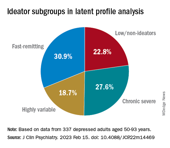
At baseline, the researchers assessed the presence or absence of suicidal behavior history and the number and lethality of attempts. They prospectively assessed suicidal ideation and attempts at least once annually thereafter over a period ranging from 3 months to 14 years (median, 3 years; IQR, 1.6-4 years).
At baseline and at follow-ups, they assessed ideation severity.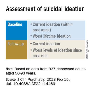
They also assessed depression severity, impulsivity, and personality measures, as well as perception of social support, social problem solving, cognitive performance, and physical comorbidities.
Personalized prevention
Of the original cohort, 92 patients died during the follow-up period, with 13 dying of suicide (or suspected suicide).
Over half (60%) of the chronic severe as well as the highly variable groups and almost half (48%) of the fast-remitting group had a history of past suicide attempt – all significantly higher than the low-nonideators (0%).
Despite comparable current ideation severity at baseline, the risk of suicide attempt/death was greater for chronic severe ideators versus fast-remitting ideators, but not greater than for highly variable ideators. On the other hand, highly variable ideators were at greater risk, compared with fast-remitting ideators.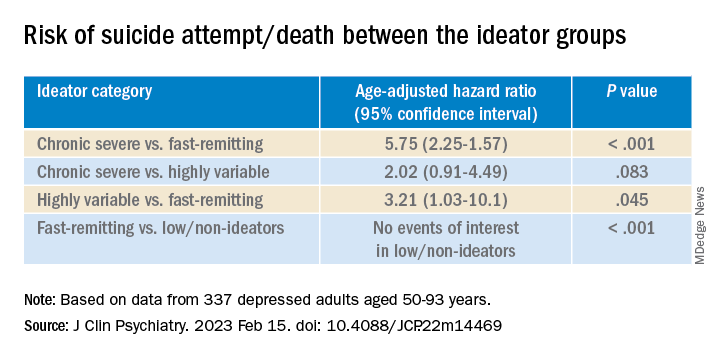
Cognitive factors “did not significantly discriminate between the ideation profiles, although ... lower global cognitive performance predicted suicidal behavior during follow-up,” the authors wrote.
This finding “aligns with prior studies indicating that late-life suicidal behavior but not ideation may be related to cognition ... and instead, ideation and cognition may act as independent risk factors for suicidal behavior,” they added.
“Patients in the fluctuating ideator group generally had moderate or high levels of worst suicidal ideation between visits, but not when asked about current ideation levels at the time of the follow-up assessment,” Dr. Galfalvy noted. “For them, the time frame of the question made a difference as to the level of ideation reported.”
The study “identified several clinical differences among these subgroups which could lead to more personalized suicide prevention efforts and further research into the heterogeneity of suicidal behavior,” she suggested.
New insight
Commenting on the study, Ari Cuperfain, MD, of the University of Toronto said the study “adds to the nuanced understanding of how changes in suicidal ideation over time can lead to suicidal actions and behavior.”
The study “sheds light on the notion of how older adults who die by suicide can demonstrate a greater degree of premeditated intent relative to younger cohorts, with chronic severe ideators portending the highest risk for suicide in this sample,” added Dr. Cuperfain, who was not involved with the current research.
“Overall, the paper highlights the importance of both screening for current levels of suicidal ideation in addition to the evolution of suicidal ideation in developing a risk assessment and in finding interventions to reduce this risk when it is most prominent,” he stated.
The research was supported by the National Institutes of Health. The authors and Dr. Cuperfain disclosed no relevant financial relationships.
A version of this article first appeared on Medscape.com.
FROM THE JOURNAL OF CLINICAL PSYCHIATRY
Causal AI quantifies CV risk, providing patient-specific goals
NEW ORLEANS – Causal artificial intelligence (AI) can translate polygenic scores (PGS) and other genetic information into risk reduction strategies for coronary artery disease (CAD) that is tailored for each individual patient, according to an analysis presented at the joint scientific sessions of the American College of Cardiology and the World Heart Federation.
Tested for LDL cholesterol (LDL-C) and systolic blood pressure (SBP), causal AI explained how much each of these risk factors must improve at the level of each individual patient “to overcome overall inherited risk,” reported Brian Ference, MD, MPhil, director of translational therapeutics, University of Cambridge (England).

Unlike the “black box” risk assessments common to machine learning, which relies on disparate forms of information of often unknown relative significance, causal AI explains cause and effect. In the case of CAD, its ability to encode the biological causes means that it can “both predict outcomes and prescribe specific actions to change those outcomes,” Dr. Ference explained.
The concept is testable against observed biology using randomized evidence, which was the objective of the study Dr. Ference presented in the late-breaker session.
Causal AI trained on nearly 2 million patients
This study employed a causal AI platform trained on roughly 1.3 million participants in Mendelian randomization studies, as well as more than 500,000 participants in randomized clinical trials. The PGS estimate of inherited risk was constructed from almost 4.1 million variants from genomewide association studies.
To test the ability of causal AI to reveal how much LDL-C or SBP had to be reduced to overcome the inherited risk of CAD based on PGS, it was applied to 445,765 participants of European ancestry in the UK Biobank. The goal was to determine how much those with greater than average risk would need to lower their LDL-C or SBP to achieve average CAD risk.
When validated against observed rates of events, causal AI accurately characterized risk before estimating what reductions in LDL-C, SBP, or both would attenuate that risk.
Providing examples, Dr. Ference explained that a PGS in the 80th percentile can be overcome by lowering LDL-C by 14 mg/dL. Alternatively, the 80th percentile risk could also be overcome by simultaneously lowering LDL-C and SBP by 7 mg/dL and 2.5 mm Hg, respectively.
Required risk factor reductions increase with age because of the increased risk of the events. For example, while a 14.8 mg/dL reduction in LDL-C would be adequate to overcome risk defined by a PGS in the 80th percentile at age 35, reductions of 18.2 mg/dL, 28.9 mg/dL, and 42.6 mg/dL would be required, respectively, at ages 45, 55, and 65 years. The values climb similarly for SBP.
Family history of CAD adds an independent variable that further contributes to the ability of causal AI to estimate risk and the degree of risk factor attenuation to overcome the risk.
Even though family history is equivalent to having PGS above the 95th percentile, it is an independent and additive variable, according to Dr. Ference. As a result, inherited risk of CAD depends on both.
Still when family history is factored into the analysis, “causal AI accurately estimated the magnitude of lower LDL-C, SBP, or both needed to overcome overall inherited risk at all levels of higher or lower PGS,” he reported.
According to Dr. Ference, the value of causal AI is that it can generate very specific goals for each patient regarding modifiable risk factors. Causal effects of risk factors encoded in time units of exposure allow the patient and the clinician to understand the biology and the basis of the disease burden.
Treatments become understandable to patients
“Encoding biology creates algorithms that are deeply explainable because they reveal why a person is at risk, how to reduce that risk, and how much each person will benefit from specific actions to reduce risk,” Dr. Ference said.
A real-world, randomized trial to confirm that the information from causal AI can reduce the risk of CAD is expected to start in 2023, but Dr. Ference thinks that causal AI for managing CAD risk, independent of this planned trial, is essentially inevitable. PGS, which he thinks will be performed routinely in all individuals within 10 years, is only likely to improve. He foresees large advantages of this form of personalized medicine.

Ami Bhatt, MD, chief innovation officer for the American College of Cardiology, Washington, agreed, seeing a direct relationship between precision health as the pathway to improvements in population health.
By explaining risk factors in terms of mechanisms and specific goals to ameliorate these risks, it “engages our patients with agency,” said Dr. Bhatt. She suggested that the information provided by causal AI has the potential to empower patients while creating a collaborative approach with clinicians to CAD prevention.
With patient-specific information provided in the context of the disease biology, “you increase the sense of transparency,” Dr. Bhatt said.
She suggested this direction of research is wholly consistent with initiatives such as those from the World Health Organization to improve precision medicine as a step toward equipping patients to manage their own health.
Dr. Ference reported financial relationships with Amgen, AstraZeneca, CiVi Pharma, Daiichi Sankyo, DalCOR, Esperion, Eli Lilly, Ionis Pharmaceuticals, KrKA, Medicines Company, Merck, Mylan, Novo Nordisk, Novartis, and Sanofi, and Viatris. Dr. Bhatt reported no potential conflicts of interest.
NEW ORLEANS – Causal artificial intelligence (AI) can translate polygenic scores (PGS) and other genetic information into risk reduction strategies for coronary artery disease (CAD) that is tailored for each individual patient, according to an analysis presented at the joint scientific sessions of the American College of Cardiology and the World Heart Federation.
Tested for LDL cholesterol (LDL-C) and systolic blood pressure (SBP), causal AI explained how much each of these risk factors must improve at the level of each individual patient “to overcome overall inherited risk,” reported Brian Ference, MD, MPhil, director of translational therapeutics, University of Cambridge (England).
Unlike the “black box” risk assessments common to machine learning, which relies on disparate forms of information of often unknown relative significance, causal AI explains cause and effect. In the case of CAD, its ability to encode the biological causes means that it can “both predict outcomes and prescribe specific actions to change those outcomes,” Dr. Ference explained.
The concept is testable against observed biology using randomized evidence, which was the objective of the study Dr. Ference presented in the late-breaker session.
Causal AI trained on nearly 2 million patients
This study employed a causal AI platform trained on roughly 1.3 million participants in Mendelian randomization studies, as well as more than 500,000 participants in randomized clinical trials. The PGS estimate of inherited risk was constructed from almost 4.1 million variants from genomewide association studies.
To test the ability of causal AI to reveal how much LDL-C or SBP had to be reduced to overcome the inherited risk of CAD based on PGS, it was applied to 445,765 participants of European ancestry in the UK Biobank. The goal was to determine how much those with greater than average risk would need to lower their LDL-C or SBP to achieve average CAD risk.
When validated against observed rates of events, causal AI accurately characterized risk before estimating what reductions in LDL-C, SBP, or both would attenuate that risk.
Providing examples, Dr. Ference explained that a PGS in the 80th percentile can be overcome by lowering LDL-C by 14 mg/dL. Alternatively, the 80th percentile risk could also be overcome by simultaneously lowering LDL-C and SBP by 7 mg/dL and 2.5 mm Hg, respectively.
Required risk factor reductions increase with age because of the increased risk of the events. For example, while a 14.8 mg/dL reduction in LDL-C would be adequate to overcome risk defined by a PGS in the 80th percentile at age 35, reductions of 18.2 mg/dL, 28.9 mg/dL, and 42.6 mg/dL would be required, respectively, at ages 45, 55, and 65 years. The values climb similarly for SBP.
Family history of CAD adds an independent variable that further contributes to the ability of causal AI to estimate risk and the degree of risk factor attenuation to overcome the risk.
Even though family history is equivalent to having PGS above the 95th percentile, it is an independent and additive variable, according to Dr. Ference. As a result, inherited risk of CAD depends on both.
Still when family history is factored into the analysis, “causal AI accurately estimated the magnitude of lower LDL-C, SBP, or both needed to overcome overall inherited risk at all levels of higher or lower PGS,” he reported.
According to Dr. Ference, the value of causal AI is that it can generate very specific goals for each patient regarding modifiable risk factors. Causal effects of risk factors encoded in time units of exposure allow the patient and the clinician to understand the biology and the basis of the disease burden.
Treatments become understandable to patients
“Encoding biology creates algorithms that are deeply explainable because they reveal why a person is at risk, how to reduce that risk, and how much each person will benefit from specific actions to reduce risk,” Dr. Ference said.
A real-world, randomized trial to confirm that the information from causal AI can reduce the risk of CAD is expected to start in 2023, but Dr. Ference thinks that causal AI for managing CAD risk, independent of this planned trial, is essentially inevitable. PGS, which he thinks will be performed routinely in all individuals within 10 years, is only likely to improve. He foresees large advantages of this form of personalized medicine.
Ami Bhatt, MD, chief innovation officer for the American College of Cardiology, Washington, agreed, seeing a direct relationship between precision health as the pathway to improvements in population health.
By explaining risk factors in terms of mechanisms and specific goals to ameliorate these risks, it “engages our patients with agency,” said Dr. Bhatt. She suggested that the information provided by causal AI has the potential to empower patients while creating a collaborative approach with clinicians to CAD prevention.
With patient-specific information provided in the context of the disease biology, “you increase the sense of transparency,” Dr. Bhatt said.
She suggested this direction of research is wholly consistent with initiatives such as those from the World Health Organization to improve precision medicine as a step toward equipping patients to manage their own health.
Dr. Ference reported financial relationships with Amgen, AstraZeneca, CiVi Pharma, Daiichi Sankyo, DalCOR, Esperion, Eli Lilly, Ionis Pharmaceuticals, KrKA, Medicines Company, Merck, Mylan, Novo Nordisk, Novartis, and Sanofi, and Viatris. Dr. Bhatt reported no potential conflicts of interest.
AT ACC 2023
Biomarkers linked to elevated T2D MACE risk in DECLARE-TIMI 58

The researchers found that N-terminal pro-B-type natriuretic peptide (NT-proBNP) and high-sensitivity cardiac troponin T (hsTnT) levels helped identify a subset of T2D patients at higher risk of major adverse cardiovascular events who would benefit most from dapagliflozin.
“We’ve shown previously that these two biomarkers are very robust risk indicators for cardiovascular death and heart failure events,” senior study author David A. Morrow, MD, of Harvard University, Boston, said in an interview. “In this study, we now show that the two biomarkers also yield important prognostic information for MACE [major adverse cardiovascular events].”
Although NT-proBNP is typically measured to diagnose heart failure, and hsTnT to diagnose acute MI, Dr. Morrow pointed out that this analysis demonstrated the potential for using the two tests to evaluate risks in T2D patients.
Study results
The secondary analysis included 14,565 patients in the DECLARE-TIMI 58 trial. The patients had T2D and multiple risk factors for atherosclerotic cardiovascular disease (about 60%) or established ASCVD (about 40%). All patients had available blood samples and the data were collected from May 2013 to September 2018. The primary outcome was MACE, a composite of MI, ischemic stroke, and cardiovascular death. The results were reported online in JAMA Cardiology.
The analysis found that higher baseline concentrations of NT-proBNP increased MACE risks by 62% (95% confidence interval, 1.49-1.76) and hsTnT elevated those risks by 59% (95% CI, 1.46-1.74).
Among placebo patients, when divided into risk quartiles, those in the highest quartile had significantly higher risk with both elevated NT-proBNP and hsTnT, compared with those with low concentrations. For example, patients with established ASCVD had a 22.9% risk vs. 9.5% with elevated NT-proBNP (P < .001) and a 24.2% vs. 7.2% risk with elevated hsTnT (P < .001). The gap was similar for patients with multiple risk factors.
Dr. Morrow noted that the main DECLARE-TIMI 58 trial showed that dapagliflozin reduced the rates of cardiovascular death or hospitalization for heart failure in patients with T2D, when compared to placebo, but didn’t reach statistical significance for MACE (N Engl J Med. 2019;380:347-57).
“We have previously shown that among patients with T2D who have high risk indicators, such as prior MI or long-standing diabetes, dapagliflozin also appeared to reduce MACE,” Dr. Morrow said. “In this study, we find that these two widely available biomarkers also identify a high-risk group who may have even more potential benefits from treatment with an SGLT2i.”
Dr. Morrow noted that the study design – a nested prospective biomarker study within a randomized, double-blind, placebo-controlled clinical trial – “is a particular strength.”
Results clarify which patients will benefit
This secondary analysis of DECLARE-TIMI 58 brings more clarity to the types of T2D patients who will get the most cardiovascular benefits from dapagliflozin, said Matthew J. Budoff, MD, professor of medicine at University of California, Los Angeles, and Endowed Chair of Preventive Cardiology at the Lundquist Institute in Torrance, Calif.

“The big picture is, we’ve known for some time from epidemiologic studies that these biomarkers, when they’re elevated, mean that the patient is at higher risk of having a cardiovascular event,” he said, “but I think what it helps us with is in knowing in whom to use dapagliflozin for prevention of ASCVD. The effect in the DECLARE-TIMI 58 trial was quite modest, but if you can subgroup it, in these high-risk people there’s a more profound effect. It helps in risk stratification because the absolute benefit is larger.”
The specific biomarkers, NT-proBNP and hsTnT, “haven’t been explored very much in clinical trials,” Dr. Budoff said, “so I do think that it’s nice that in a randomized trial it plays out the way we might expect.”
He added that “for many clinicians this is novel, because I don’t think they were aware of the biomarker data, so I think that this does add some clinical benefit in that context.” The findings also strengthen the case to get T2D patients with higher ASCVD risk onto SGLT2 inhibitors if they’re not already, he said.
Dr. Morrow disclosed relationships with AstraZeneca, Roche Diagnostics, Abbott Laboratories, Anthos Therapeutics, ARCA Biopharma, Merck, Novartis, Pfizer, Regeneron, Siemens, and InCarda outside the reported work.
Dr. Budoff has no relevant disclosures.
The researchers found that N-terminal pro-B-type natriuretic peptide (NT-proBNP) and high-sensitivity cardiac troponin T (hsTnT) levels helped identify a subset of T2D patients at higher risk of major adverse cardiovascular events who would benefit most from dapagliflozin.
“We’ve shown previously that these two biomarkers are very robust risk indicators for cardiovascular death and heart failure events,” senior study author David A. Morrow, MD, of Harvard University, Boston, said in an interview. “In this study, we now show that the two biomarkers also yield important prognostic information for MACE [major adverse cardiovascular events].”
Although NT-proBNP is typically measured to diagnose heart failure, and hsTnT to diagnose acute MI, Dr. Morrow pointed out that this analysis demonstrated the potential for using the two tests to evaluate risks in T2D patients.
Study results
The secondary analysis included 14,565 patients in the DECLARE-TIMI 58 trial. The patients had T2D and multiple risk factors for atherosclerotic cardiovascular disease (about 60%) or established ASCVD (about 40%). All patients had available blood samples and the data were collected from May 2013 to September 2018. The primary outcome was MACE, a composite of MI, ischemic stroke, and cardiovascular death. The results were reported online in JAMA Cardiology.
The analysis found that higher baseline concentrations of NT-proBNP increased MACE risks by 62% (95% confidence interval, 1.49-1.76) and hsTnT elevated those risks by 59% (95% CI, 1.46-1.74).
Among placebo patients, when divided into risk quartiles, those in the highest quartile had significantly higher risk with both elevated NT-proBNP and hsTnT, compared with those with low concentrations. For example, patients with established ASCVD had a 22.9% risk vs. 9.5% with elevated NT-proBNP (P < .001) and a 24.2% vs. 7.2% risk with elevated hsTnT (P < .001). The gap was similar for patients with multiple risk factors.
Dr. Morrow noted that the main DECLARE-TIMI 58 trial showed that dapagliflozin reduced the rates of cardiovascular death or hospitalization for heart failure in patients with T2D, when compared to placebo, but didn’t reach statistical significance for MACE (N Engl J Med. 2019;380:347-57).
“We have previously shown that among patients with T2D who have high risk indicators, such as prior MI or long-standing diabetes, dapagliflozin also appeared to reduce MACE,” Dr. Morrow said. “In this study, we find that these two widely available biomarkers also identify a high-risk group who may have even more potential benefits from treatment with an SGLT2i.”
Dr. Morrow noted that the study design – a nested prospective biomarker study within a randomized, double-blind, placebo-controlled clinical trial – “is a particular strength.”
Results clarify which patients will benefit
This secondary analysis of DECLARE-TIMI 58 brings more clarity to the types of T2D patients who will get the most cardiovascular benefits from dapagliflozin, said Matthew J. Budoff, MD, professor of medicine at University of California, Los Angeles, and Endowed Chair of Preventive Cardiology at the Lundquist Institute in Torrance, Calif.
“The big picture is, we’ve known for some time from epidemiologic studies that these biomarkers, when they’re elevated, mean that the patient is at higher risk of having a cardiovascular event,” he said, “but I think what it helps us with is in knowing in whom to use dapagliflozin for prevention of ASCVD. The effect in the DECLARE-TIMI 58 trial was quite modest, but if you can subgroup it, in these high-risk people there’s a more profound effect. It helps in risk stratification because the absolute benefit is larger.”
The specific biomarkers, NT-proBNP and hsTnT, “haven’t been explored very much in clinical trials,” Dr. Budoff said, “so I do think that it’s nice that in a randomized trial it plays out the way we might expect.”
He added that “for many clinicians this is novel, because I don’t think they were aware of the biomarker data, so I think that this does add some clinical benefit in that context.” The findings also strengthen the case to get T2D patients with higher ASCVD risk onto SGLT2 inhibitors if they’re not already, he said.
Dr. Morrow disclosed relationships with AstraZeneca, Roche Diagnostics, Abbott Laboratories, Anthos Therapeutics, ARCA Biopharma, Merck, Novartis, Pfizer, Regeneron, Siemens, and InCarda outside the reported work.
Dr. Budoff has no relevant disclosures.
FROM JAMA CARDIOLOGY
NUDGE-FLU: Electronic ‘nudges’ boost flu shot uptake in seniors
Two types of electronically delivered letter strategies – a letter highlighting potential cardiovascular benefits of influenza vaccination and a repeat reminder letter – increased flu shot uptake, compared with usual care alone, in a national study of seniors in Denmark.
And in a prespecified subanalysis focusing on older adults with cardiovascular disease, these two strategies were also effective in boosting vaccine uptake in those with or without CVD.
The findings are from the Nationwide Utilization of Danish Government Electronic Letter System for Increasing Influenza Vaccine Uptake (NUDGE-FLU) trial, which compared usual care alone with one of nine different electronic letter “behavioral nudge” strategies during the 2022-2023 flu season in people aged 65 years and older. 
Niklas Dyrby Johansen, MD, Hospital–Herlev and Gentofte and Copenhagen University, presented the main study findings in a late-breaking clinical trial session at the joint scientific sessions of the American College of Cardiology and the World Heart Federation, and the article was simultaneously published in The Lancet
The subanalysis in patients with CVD was published online March 5 in Circulation.
“Despite modest effect sizes, the results may have important implications when translated to a population level,” Dr. Dyrby Johansen concluded during his presentation. Still, the authors write, “the low-touch (no person-to-person interaction), inexpensive, and highly scalable nature of these electronic letters might have important population-level public health implications.”
They note that, among approximately 63 million Medicare beneficiaries in the United States, a 0.89–percentage point absolute increase in vaccination rate achieved through the most successful electronic letter in NUDGE-FLU, the one highlighting cardiovascular gain, would be expected to lead to 500,000 additional vaccinations and potentially prevent 7,849 illnesses, 4,395 medical visits, 714 hospitalizations, and 66 deaths each year.
Electronic letter systems similar to the one used in this trial are already in place in several European countries, including Sweden, Norway, and Ireland, the researchers note.
In countries such as the United States, where implementing a nationwide government electronic letter system might not be feasible, nudges could be done via email, text message, or other systems, but whether this would be as effective remains to be seen.
Commenting on the findings, David Cho, MD, UCLA Health and chair of the ACC Health Care Innovation Council, commended the researchers on engaging patients with more than a million separate nudges sent out during one flu season, and randomly assigning participants to 10 different types of nudges, calling it “impressive.”
“I think the concept that the nudge is to plant an idea that leads to an action is pretty much the basis of a lot of these health care interventions, which seems like a small way to have a big impact at outcome,” Dr. Cho noted. “The behavioral science aspects of the nudges are also fascinating to me personally, and I think to a lot of the cardiologists in the audience – about how you actually get people to act. I think it’s been a lifelong question for people in general, how do you get people to follow through on an action?”
“So I found the fact that secondary gain from a cardiovascular health standpoint, but also the repeated nudges were sort of simple ways that you could have people take ownership and get their flu vaccination,” he said.
“This is ACC, this is a cardiovascular conference, but the influence of vaccine is not just a primary care problem, it is also directly affecting cardiovascular disease,” Dr. Cho concluded.
‘Small but important effect’
In an accompanying editorial (Lancet. 2023 Mar 5. doi: 10.1016/S0140-6736(23)00453-1), Melissa Stockwell, MD, Columbia University, New York, writes, “The study by Johansen and colleagues highlights the small but still important effect of scalable, digital interventions across an entire at-risk population.”
A difference of 0.89% in the entire study population of over 960,000 adults age 65 years or older would be more than 8,500 additional adults protected, she notes. “That increase is important for a scalable intervention that has a low cost per letter.”
Moreover, “that the cardiovascular gain–framed messages worked best in those who had not been vaccinated in the previous season further highlights the potential impact on a more vaccine-hesitant population,” Dr. Stockwell notes.
However, with the mandatory government electronic notification system in Denmark, “notifications are sent via regular email and SMS message, and recipients log in through a portal or smartphone app to view the letter.” Similar studies in the United States that included this extra step of needing to sign in online have not been effective in older populations.
Another limitation is that the intervention may have a different effect in populations for which there is a digital divide between people with or without Internet access of sufficient data on their mobile phones.
First-of-its kind, nationwide pragmatic trial
The NUDGE-FLU protocol was previously published in the American Heart Journal. NUDGE-FLU is a first-of-its kind nationwide, pragmatic, registry-based, cluster-randomized implementation trial of electronically delivered nudges to increase influenza vaccination uptake, the researchers note.
They identified 964,870 individuals who were 65 years or older (or would turn 65 by Jan. 15, 2023) who lived in one of 691,820 households in Denmark.
This excluded individuals who lived in a nursing home or were exempt from the government’s mandatory electronic letter system that is used for official communications.
Households were randomly assigned 9:1:1:1:1:1:1:1:1:1 to receive usual care alone or to one of nine electronic letter strategies based on different behavioral science approaches to encourage influenza vaccination uptake:
- Standard electronic letter
- Standard electronic letter sent at randomization and again 14 days later (repeated letter)
- Depersonalized letter without the recipient’s name
- Gain-framing nudge (“Vaccinations help end pandemics, like COVID-19 and the flu. Protect yourself and your loved ones.”)
- Loss-framing nudge (“When too few people get vaccinated, pandemics from diseases like COVID-19 and the flu can spread and place you and your loved ones at risk.”)
- Collective-goal nudge (“78% of Danes 65 and above were vaccinated against influenza last year. Help us achieve an even higher goal this year.”)
- Active choice or implementation-intention prompt (“We encourage you to record your appointment time here.”)
- Cardiovascular gain–framing nudge (“In addition to its protection against influenza infection, influenza vaccination also seems to protect against cardiovascular disease such as heart attacks and heart failure.”)
- Expert-authority statement (“I recommend everyone over the age of 65 years to get vaccinated against influenza – Tyra Grove Krause, Executive Vice President, Statens Serum Institut.”)
The electronic letters were sent out Sept. 16, 2022, and the primary endpoint was vaccine receipt on or before Jan. 1, 2023.
All individuals received an informative vaccination encouragement letter from the Danish Health Authority (usual care) delivered via the same electronic letter system during Sept. 17 through Sept. 21, 2022.
The individuals had a mean age of 73.8 years, 51.5% were women, and 27.4% had chronic cardiovascular disease.
The analyses were done in one randomly selected individual per household.
Influenza vaccination rates were significantly higher in the cardiovascular gain–framing nudge group vs. usual care (81.00% vs. 80.12%; difference, 0.89 percentage points; P < .0001) and in the repeat-letter group vs. usual care (80.85% vs 80.12%; difference, 0.73 percentage points; P = .0006).
These two strategies also improved vaccination rates across major subgroups.
The cardiovascular gain–framed letter was particularly effective among participants who had not been vaccinated for influenza in the previous season.
The seven other letter strategies did not increase flu shot uptake.
Subanalysis in CVD
In the prespecified subanalysis of the NUDGE-FLU trial of patients aged 65 and older that focused on patients with CVD, Daniel Modin, MB, and colleagues report that 83.1% of patients with CVD vs. 79.2% of patients without CVD received influenza vaccination within the requested time (P < .0001).
The two nudging strategies – a letter highlighting potential cardiovascular benefits of influenza vaccination or a repeat letter – that were effective in boosting flu shot rates in the main analysis were also effective in all major CVD subgroups (ischemic heart disease, pulmonary heart disease, heart failure, atrial fibrillation, cerebrovascular disease, atherosclerotic CVD, embolic or thrombotic disease, and congenital heart disease).
Despite strong guideline endorsement, “influenza vaccination rates remain suboptimal in patients with high-risk cardiovascular disease,” Dr. Morin and colleagues write, possibly because of “insufficient knowledge among patients and providers of potential clinical benefits, concerns about vaccine safety, and other forms of vaccine hesitancy.”
Their findings suggest that “select digital behaviorally informed nudges delivered in advance of vaccine availability might be utilized to increase influenza vaccinate uptake in individuals with cardiovascular disease.”
NUDGE-HF was funded by Sanofi. Dr. Johansen and Dr. Modin have no disclosures. The disclosures of the other authors are listed with the articles. Dr. Stockwell has no disclosures.
A version of this article first appeared on Medscape.com.
Two types of electronically delivered letter strategies – a letter highlighting potential cardiovascular benefits of influenza vaccination and a repeat reminder letter – increased flu shot uptake, compared with usual care alone, in a national study of seniors in Denmark.
And in a prespecified subanalysis focusing on older adults with cardiovascular disease, these two strategies were also effective in boosting vaccine uptake in those with or without CVD.
The findings are from the Nationwide Utilization of Danish Government Electronic Letter System for Increasing Influenza Vaccine Uptake (NUDGE-FLU) trial, which compared usual care alone with one of nine different electronic letter “behavioral nudge” strategies during the 2022-2023 flu season in people aged 65 years and older.
Niklas Dyrby Johansen, MD, Hospital–Herlev and Gentofte and Copenhagen University, presented the main study findings in a late-breaking clinical trial session at the joint scientific sessions of the American College of Cardiology and the World Heart Federation, and the article was simultaneously published in The Lancet
The subanalysis in patients with CVD was published online March 5 in Circulation.
“Despite modest effect sizes, the results may have important implications when translated to a population level,” Dr. Dyrby Johansen concluded during his presentation. Still, the authors write, “the low-touch (no person-to-person interaction), inexpensive, and highly scalable nature of these electronic letters might have important population-level public health implications.”
They note that, among approximately 63 million Medicare beneficiaries in the United States, a 0.89–percentage point absolute increase in vaccination rate achieved through the most successful electronic letter in NUDGE-FLU, the one highlighting cardiovascular gain, would be expected to lead to 500,000 additional vaccinations and potentially prevent 7,849 illnesses, 4,395 medical visits, 714 hospitalizations, and 66 deaths each year.
Electronic letter systems similar to the one used in this trial are already in place in several European countries, including Sweden, Norway, and Ireland, the researchers note.
In countries such as the United States, where implementing a nationwide government electronic letter system might not be feasible, nudges could be done via email, text message, or other systems, but whether this would be as effective remains to be seen.
Commenting on the findings, David Cho, MD, UCLA Health and chair of the ACC Health Care Innovation Council, commended the researchers on engaging patients with more than a million separate nudges sent out during one flu season, and randomly assigning participants to 10 different types of nudges, calling it “impressive.”
“I think the concept that the nudge is to plant an idea that leads to an action is pretty much the basis of a lot of these health care interventions, which seems like a small way to have a big impact at outcome,” Dr. Cho noted. “The behavioral science aspects of the nudges are also fascinating to me personally, and I think to a lot of the cardiologists in the audience – about how you actually get people to act. I think it’s been a lifelong question for people in general, how do you get people to follow through on an action?”
“So I found the fact that secondary gain from a cardiovascular health standpoint, but also the repeated nudges were sort of simple ways that you could have people take ownership and get their flu vaccination,” he said.
“This is ACC, this is a cardiovascular conference, but the influence of vaccine is not just a primary care problem, it is also directly affecting cardiovascular disease,” Dr. Cho concluded.
‘Small but important effect’
In an accompanying editorial (Lancet. 2023 Mar 5. doi: 10.1016/S0140-6736(23)00453-1), Melissa Stockwell, MD, Columbia University, New York, writes, “The study by Johansen and colleagues highlights the small but still important effect of scalable, digital interventions across an entire at-risk population.”
A difference of 0.89% in the entire study population of over 960,000 adults age 65 years or older would be more than 8,500 additional adults protected, she notes. “That increase is important for a scalable intervention that has a low cost per letter.”
Moreover, “that the cardiovascular gain–framed messages worked best in those who had not been vaccinated in the previous season further highlights the potential impact on a more vaccine-hesitant population,” Dr. Stockwell notes.
However, with the mandatory government electronic notification system in Denmark, “notifications are sent via regular email and SMS message, and recipients log in through a portal or smartphone app to view the letter.” Similar studies in the United States that included this extra step of needing to sign in online have not been effective in older populations.
Another limitation is that the intervention may have a different effect in populations for which there is a digital divide between people with or without Internet access of sufficient data on their mobile phones.
First-of-its kind, nationwide pragmatic trial
The NUDGE-FLU protocol was previously published in the American Heart Journal. NUDGE-FLU is a first-of-its kind nationwide, pragmatic, registry-based, cluster-randomized implementation trial of electronically delivered nudges to increase influenza vaccination uptake, the researchers note.
They identified 964,870 individuals who were 65 years or older (or would turn 65 by Jan. 15, 2023) who lived in one of 691,820 households in Denmark.
This excluded individuals who lived in a nursing home or were exempt from the government’s mandatory electronic letter system that is used for official communications.
Households were randomly assigned 9:1:1:1:1:1:1:1:1:1 to receive usual care alone or to one of nine electronic letter strategies based on different behavioral science approaches to encourage influenza vaccination uptake:
- Standard electronic letter
- Standard electronic letter sent at randomization and again 14 days later (repeated letter)
- Depersonalized letter without the recipient’s name
- Gain-framing nudge (“Vaccinations help end pandemics, like COVID-19 and the flu. Protect yourself and your loved ones.”)
- Loss-framing nudge (“When too few people get vaccinated, pandemics from diseases like COVID-19 and the flu can spread and place you and your loved ones at risk.”)
- Collective-goal nudge (“78% of Danes 65 and above were vaccinated against influenza last year. Help us achieve an even higher goal this year.”)
- Active choice or implementation-intention prompt (“We encourage you to record your appointment time here.”)
- Cardiovascular gain–framing nudge (“In addition to its protection against influenza infection, influenza vaccination also seems to protect against cardiovascular disease such as heart attacks and heart failure.”)
- Expert-authority statement (“I recommend everyone over the age of 65 years to get vaccinated against influenza – Tyra Grove Krause, Executive Vice President, Statens Serum Institut.”)
The electronic letters were sent out Sept. 16, 2022, and the primary endpoint was vaccine receipt on or before Jan. 1, 2023.
All individuals received an informative vaccination encouragement letter from the Danish Health Authority (usual care) delivered via the same electronic letter system during Sept. 17 through Sept. 21, 2022.
The individuals had a mean age of 73.8 years, 51.5% were women, and 27.4% had chronic cardiovascular disease.
The analyses were done in one randomly selected individual per household.
Influenza vaccination rates were significantly higher in the cardiovascular gain–framing nudge group vs. usual care (81.00% vs. 80.12%; difference, 0.89 percentage points; P < .0001) and in the repeat-letter group vs. usual care (80.85% vs 80.12%; difference, 0.73 percentage points; P = .0006).
These two strategies also improved vaccination rates across major subgroups.
The cardiovascular gain–framed letter was particularly effective among participants who had not been vaccinated for influenza in the previous season.
The seven other letter strategies did not increase flu shot uptake.
Subanalysis in CVD
In the prespecified subanalysis of the NUDGE-FLU trial of patients aged 65 and older that focused on patients with CVD, Daniel Modin, MB, and colleagues report that 83.1% of patients with CVD vs. 79.2% of patients without CVD received influenza vaccination within the requested time (P < .0001).
The two nudging strategies – a letter highlighting potential cardiovascular benefits of influenza vaccination or a repeat letter – that were effective in boosting flu shot rates in the main analysis were also effective in all major CVD subgroups (ischemic heart disease, pulmonary heart disease, heart failure, atrial fibrillation, cerebrovascular disease, atherosclerotic CVD, embolic or thrombotic disease, and congenital heart disease).
Despite strong guideline endorsement, “influenza vaccination rates remain suboptimal in patients with high-risk cardiovascular disease,” Dr. Morin and colleagues write, possibly because of “insufficient knowledge among patients and providers of potential clinical benefits, concerns about vaccine safety, and other forms of vaccine hesitancy.”
Their findings suggest that “select digital behaviorally informed nudges delivered in advance of vaccine availability might be utilized to increase influenza vaccinate uptake in individuals with cardiovascular disease.”
NUDGE-HF was funded by Sanofi. Dr. Johansen and Dr. Modin have no disclosures. The disclosures of the other authors are listed with the articles. Dr. Stockwell has no disclosures.
A version of this article first appeared on Medscape.com.
FROM ACC 2023
EHR alerts boosted MRA prescribing to patients with HFrEF
NEW ORLEANS – EHR-embedded alerts that a patient with heart failure with reduced ejection fraction (HFrEF) is a great candidate for treatment with a mineralocorticoid receptor antagonist (MRA) more than doubled prescribing of this “pillar” class for HFrEF, compared with control practices that used usual care and no alerts.
That’s according to results of BETTER CARE-HF, a single-center, randomized trial with more than 2,000 patients and involving 180 cardiologists.
“EHR-embedded tools cans be a rapid, low-cost, and high-impact method to increase prescription of life-saving therapies across large populations,” said Amrita Mukhopadhyay, MD, at the joint scientific sessions of the American College of Cardiology and the World Heart Federation.

Her study targeted underprescribing of an MRA – spironolactone or eplerenone (Inspra) – because of its “vastly underprescribed” status in U.S. practice, where roughly two-thirds of patients with HFrEF do not receive an MRA despite clear recommendations from several medical groups that it is an essential part of treatment for most patients with HFrEF. Dr. Mukhopadhyay estimated that more comprehensive prescribing of MRAs to U.S. patients with HFrEF could prevent more than 20,000 deaths annually.
She also explained that the EHR-embedded alert was carefully devised, through interviews with cardiologists and pilot testing, to optimize the nudge so that it was less intrusive but effective for capturing attention and initiating action.
‘Clinically relevant, impressive results’
“This is a really important study, because despite overwhelming evidence for more than a decade favoring MRA use for patients with HFrEF there is an incredibly large treatment gap. MRAs can reduce all-cause death in people with HFrEF by 25%-30%, as well as reduce hospitalizations for heart failure, at a cost of less than $50 a year,” commented Gregg C. Fonarow, MD, interim chief of cardiology at the University of California, Los Angeles. The study showed “very clinically relevant, impressive results” for individualized, patient-specific alerts to prescribe an MRA and order the laboratory tests, particularly for serum potassium levels, needed to safely start the treatment, Dr. Fonarow said in an interview.
The BETTER CARE-HF study ran at more than 60 practices in the New York City region operated by the NYU Langone Health system, which sponsored the study. The trial randomized 180 cardiologists from these practices in a cluster format to one of three study arms: Sixty cardiologists received the EHR-embedded alerts for their relevant patients (755 patients) when the patient was in the physician’s office; another 60 cardiologists received a less tailored, monthly message that flagged all patients with HFrEF in a cardiologist’s practice who remained untreated candidates for MRA intervention (812 patients); and a third arm of 60 cardiologists and their HFrEF patients served as controls where the clinicians received no alert or message (644 patients).
The study included 2,211 patients with HFrEF and not on MRA treatment at baseline who were all identified as good candidates for starting treatment with the class, with no contraindications, no preexisting hyperkalemia, and no advanced-stage renal dysfunction.
The study’s primary outcome was the percentage of patients in each subgroup who received a new prescription for an MRA. This occurred in 29.6% of the patients whose physicians received an alert, in 15.6% of the patients whose physicians received a monthly message, and in 11.7% of patients in the control practices. Statistical analyses showed that the alerts led to a significant 2.53-fold increase in MRA prescribing, while the messages linked with a significant 67% increase in prescribing, compared with the control practices, reported Dr. Mukhopadhyay, a health services researcher at NYU Langone Health in New York. Simultaneously with her report, the results also appeared in the Journal of the American College of Cardiology.
The findings also showed that the alert and message had no significant impact on the prescribing of any other medication classes for HFrEF, compared with the controls. And the alert intervention had minimal adverse effects. While patients in the alert arm showed a significant, 45% relative increase in the incidence of hyperkalemia episodes, compared with control patients (because of a 4.5% absolute increase in hyperkalemia events), the rate of “significant” hyperkalemia with a value of at least 5.5 mmol/L, occurred in 5.0% of patients in the alert group and 5.1% of patients in the control arm.
Potassium testing poses another barrier
Even though the alerts substantially improved MRA prescribing, 70% of patients deemed MRA eligible in the alert subgroup still failed to receive a prescription. One additional barrier specific to MRA prescribing is the need it triggers for serial laboratory testing to monitor serum potassium levels. “Potassium testing generates additional work outside the index visit, which along with the risk for hyperkalemia exists as a barrier,” commented Lee R. Goldberg, MD, a heart failure specialist and professor at the University of Pennsylvania in Philadelphia. “This may be the next aspect to focus on to improve MRA uptake,” he said as a designated discussant for the report.

“It’s not enough to just prompt medication treatment. We also need to prompt appropriate laboratory testing,” noted Dr. Fonarow.
He also said that the approach tested by Dr. Mukhopadhyay could now be expanded to outpatient cardiologists. “The onus is on everyone involved in caring for patients with HFrEF failure to explain why maximum effort is not being made to deploy” all of the guideline-directed medical therapies for the disorder.
EHR alerts “are one way to bridge the prescribing gap, but we need multiple approaches so that all eligible patients receive guideline-directed medical therapy,” Dr. Fonarow said.
BETTER CARE-HF received no commercial funding, and Dr. Mukhopadhyay had no disclosures. Dr. Fonarow has been a consultant to AstraZeneca, Amgen, Cytokinetics, Lilly, Merck, Novartis, and Pfizer. Dr. Goldberg has received personal fees from Abbott, VisCardia, and Zoll/Respircardia.
NEW ORLEANS – EHR-embedded alerts that a patient with heart failure with reduced ejection fraction (HFrEF) is a great candidate for treatment with a mineralocorticoid receptor antagonist (MRA) more than doubled prescribing of this “pillar” class for HFrEF, compared with control practices that used usual care and no alerts.
That’s according to results of BETTER CARE-HF, a single-center, randomized trial with more than 2,000 patients and involving 180 cardiologists.
“EHR-embedded tools cans be a rapid, low-cost, and high-impact method to increase prescription of life-saving therapies across large populations,” said Amrita Mukhopadhyay, MD, at the joint scientific sessions of the American College of Cardiology and the World Heart Federation.
Her study targeted underprescribing of an MRA – spironolactone or eplerenone (Inspra) – because of its “vastly underprescribed” status in U.S. practice, where roughly two-thirds of patients with HFrEF do not receive an MRA despite clear recommendations from several medical groups that it is an essential part of treatment for most patients with HFrEF. Dr. Mukhopadhyay estimated that more comprehensive prescribing of MRAs to U.S. patients with HFrEF could prevent more than 20,000 deaths annually.
She also explained that the EHR-embedded alert was carefully devised, through interviews with cardiologists and pilot testing, to optimize the nudge so that it was less intrusive but effective for capturing attention and initiating action.
‘Clinically relevant, impressive results’
“This is a really important study, because despite overwhelming evidence for more than a decade favoring MRA use for patients with HFrEF there is an incredibly large treatment gap. MRAs can reduce all-cause death in people with HFrEF by 25%-30%, as well as reduce hospitalizations for heart failure, at a cost of less than $50 a year,” commented Gregg C. Fonarow, MD, interim chief of cardiology at the University of California, Los Angeles. The study showed “very clinically relevant, impressive results” for individualized, patient-specific alerts to prescribe an MRA and order the laboratory tests, particularly for serum potassium levels, needed to safely start the treatment, Dr. Fonarow said in an interview.
The BETTER CARE-HF study ran at more than 60 practices in the New York City region operated by the NYU Langone Health system, which sponsored the study. The trial randomized 180 cardiologists from these practices in a cluster format to one of three study arms: Sixty cardiologists received the EHR-embedded alerts for their relevant patients (755 patients) when the patient was in the physician’s office; another 60 cardiologists received a less tailored, monthly message that flagged all patients with HFrEF in a cardiologist’s practice who remained untreated candidates for MRA intervention (812 patients); and a third arm of 60 cardiologists and their HFrEF patients served as controls where the clinicians received no alert or message (644 patients).
The study included 2,211 patients with HFrEF and not on MRA treatment at baseline who were all identified as good candidates for starting treatment with the class, with no contraindications, no preexisting hyperkalemia, and no advanced-stage renal dysfunction.
The study’s primary outcome was the percentage of patients in each subgroup who received a new prescription for an MRA. This occurred in 29.6% of the patients whose physicians received an alert, in 15.6% of the patients whose physicians received a monthly message, and in 11.7% of patients in the control practices. Statistical analyses showed that the alerts led to a significant 2.53-fold increase in MRA prescribing, while the messages linked with a significant 67% increase in prescribing, compared with the control practices, reported Dr. Mukhopadhyay, a health services researcher at NYU Langone Health in New York. Simultaneously with her report, the results also appeared in the Journal of the American College of Cardiology.
The findings also showed that the alert and message had no significant impact on the prescribing of any other medication classes for HFrEF, compared with the controls. And the alert intervention had minimal adverse effects. While patients in the alert arm showed a significant, 45% relative increase in the incidence of hyperkalemia episodes, compared with control patients (because of a 4.5% absolute increase in hyperkalemia events), the rate of “significant” hyperkalemia with a value of at least 5.5 mmol/L, occurred in 5.0% of patients in the alert group and 5.1% of patients in the control arm.
Potassium testing poses another barrier
Even though the alerts substantially improved MRA prescribing, 70% of patients deemed MRA eligible in the alert subgroup still failed to receive a prescription. One additional barrier specific to MRA prescribing is the need it triggers for serial laboratory testing to monitor serum potassium levels. “Potassium testing generates additional work outside the index visit, which along with the risk for hyperkalemia exists as a barrier,” commented Lee R. Goldberg, MD, a heart failure specialist and professor at the University of Pennsylvania in Philadelphia. “This may be the next aspect to focus on to improve MRA uptake,” he said as a designated discussant for the report.
“It’s not enough to just prompt medication treatment. We also need to prompt appropriate laboratory testing,” noted Dr. Fonarow.
He also said that the approach tested by Dr. Mukhopadhyay could now be expanded to outpatient cardiologists. “The onus is on everyone involved in caring for patients with HFrEF failure to explain why maximum effort is not being made to deploy” all of the guideline-directed medical therapies for the disorder.
EHR alerts “are one way to bridge the prescribing gap, but we need multiple approaches so that all eligible patients receive guideline-directed medical therapy,” Dr. Fonarow said.
BETTER CARE-HF received no commercial funding, and Dr. Mukhopadhyay had no disclosures. Dr. Fonarow has been a consultant to AstraZeneca, Amgen, Cytokinetics, Lilly, Merck, Novartis, and Pfizer. Dr. Goldberg has received personal fees from Abbott, VisCardia, and Zoll/Respircardia.
AT ACC 2023
BIOVASC: Immediate complete revascularization beneficial in ACS
Immediate complete revascularization during the index procedure might become the new treatment paradigm in patients with an acute coronary syndrome (ACS) and multivessel disease, based on results of the BIOVASC trial.
In the trial, in patients presenting with ACS and multivessel disease, immediate complete revascularization was noninferior to staged complete revascularization for the primary composite outcome and was associated with a reduction in myocardial infarction and unplanned ischemia-driven revascularization.
The BIOVASC trial was presented on March 5 by Roberto Diletti, MD, Erasmus University Medical Center, Rotterdam, the Netherlands, at the joint scientific sessions of the American College of Cardiology and the World Heart Federation. The study was simultaneously published online in The Lancet.
“We did not detect an early safety signal against an immediate complete revascularization strategy,” the authors state in the Lancet paper, adding: “Treating physicians should not be concerned about potential risks associated with immediate treatment of nonculprit lesions.”
They note, “This strategy might be particularly effective in patients with only two-vessel disease and reasonably simple lesions, with a high likelihood of procedural success without excessive use of radiation, contrast dye, or other resources.”
The trial also showed a shorter hospital stay with an immediate complete revascularization strategy.
“Immediate complete revascularization might have potential health economic benefits because of the lower rate of myocardial infarction, including spontaneous myocardial infarction, and unplanned revascularizations, and the shorter overall hospital stay,” the researchers conclude.
Introducing his presentation, Dr. Diletti explained that multiple studies have established the clinical benefit of complete coronary revascularization as compared with exclusive reperfusion of the culprit lesion, but the optimal timing for nonculprit lesion revascularization remains unclear.
The BIOVASC trial, conducted in Belgium, Italy, the Netherlands, and Spain, involved 1,525 patients with ST-segment elevation MI (STEMI) or non-STEMI ACS and multivessel coronary artery disease with a clearly identifiable culprit lesion.
They were randomly assigned to undergo immediate complete revascularization (percutaneous coronary intervention [PCI] of the culprit lesion first, followed by other nonculprit lesions deemed to be clinically significant by the operator during the index procedure) or staged complete revascularization (PCI of only the culprit lesion during the index procedure and PCI of all nonculprit lesions deemed to be clinically significant within 6 weeks after the index procedure).
The primary outcome was the composite of all-cause mortality, MI, any unplanned ischemia-driven revascularization, or cerebrovascular events at 1 year after the index procedure.
The trial had a noninferiority design, with noninferiority of immediate to staged complete revascularization considered to be met if the upper boundary of the 95% confidence interval of the hazard ratio for the primary outcome did not exceed 1.39.
Among the trial population, 40% of patients had STEMI, 52% had non-STEMI, and 8% had unstable angina.
In the immediate complete revascularization group, 16 patients did not receive complete revascularization during the index procedure primarily because of prolonged procedure time, procedural complexity, or excessive contrast dye use.
In the staged group, 30% of patients underwent all subsequent procedures during the index hospitalization.
Results showed that the primary composite outcome at 1 year occurred in 7.6% of the immediate revascularization group and in 9.4% of the staged group, meeting the noninferiority criteria (HR, 0.78; 95% CI, 0.55-1.11; P for noninferiority = .0011).
Superiority of the immediate over the staged complete revascularization strategy was not met at 1-year follow-up (P for superiority = .17).
However, in the prespecified analysis of clinical events at 30 days after the index procedure, immediate complete revascularization was superior to staged revascularization in terms of the composite primary outcome (2.2% vs. 5.8%; HR, 0.38; P for superiority = .0007).
One-year results showed no difference in all-cause death between the two groups, but the immediate complete revascularization group appeared to have a reduction in MI (1.9% vs. 4.5%) and fewer unplanned ischemia-driven revascularizations (4.2% vs. 6.7%).
The difference in MI was mainly driven by spontaneous MIs (not procedure related) that predominantly occurred in the time window between the index procedure and the planned date for the staged intervention, and an originally nonculprit lesion was identified as the cause for these events in almost all cases.
Subgroup analysis showed similar results across the patient population, including age, sex, and STEMI vs. non-STEMI presentation.
High rate of MI in staged group
Discussant of the study at the ACC session, Dipti Itchhaporia, MD, University of California, Irvine, said this was a “very important trial.”
She expressed surprise over the “remarkably high rate” of MI in the staged procedure group and asked Dr. Diletti why that might have occurred.
He responded that the operator may have misjudged the culprit lesion or that patients with ACS may have multiple unstable plaques and “treating the culprit lesion alone does not do the job.”
He added: “We need to look at the data more thoroughly to better understand this, but in both scenarios, immediate complete revascularization would prevent these events.”
Dr. Itchhaporia also pointed out a low rate of functional imaging used in the study.
Dr. Diletti replied that this reflected current European practice, but he acknowledged that, “in my opinion this reduces our ability to detect the culprit lesion.”
Commenting at an ACC press conference, David Moliterno, MD, Gill Heart and Vascular Institute, Lexington, Ky., said the trial poses the question “Can we fix it all at once?” and the results suggest “Yes, we can.”
He said this approach had the advantage of removing any uncertainly as to which was the culprit lesion. “We just fix everything – leave no blockage behind.”
But he pointed out that for some patients this may not be appropriate, such as those with compromised renal function, in whom excessive amounts of contrast material should be avoided.
CABG still needs to be considered
In a comment accompanying the Lancet publication, Tobias Pustjens, MD, Pieter Vriesendorp, MD, and Arnoud W.J. van’t Hof, MD, Cardiovascular Research Institute Maastricht (the Netherlands), note that more than half of the patients presenting with an ACS have multivessel coronary disease.
They say the trial results suggest that “pursuing an immediate complete revascularisation strategy, especially in times of reduced hospital capacity and staff scarcity, not only benefits the individual patient in clinical outcomes but can also safely reduce the pressure on health care systems.”
But they also point out that the possibility of coronary artery bypass grafting (CABG) should not be omitted, and that CABG is still the treatment of choice in patients with diabetes or complex coronary artery disease.
They conclude: “The results of the BIOVASC study move clinical practice forward from culprit-only to an immediate, complete revascularisation strategy. … However, further fine tuning of this treatment strategy to substantiate a role for intracoronary physiology assessment, intracoronary imaging, and guidance of the heart team decision is needed.”
The BIOVASC trial was supported by an unrestricted research grant from Biotronik AG. Dr. Diletti has received institutional research grants from Biotronik, Medtronic, ACIST Medical Systems, and Boston Scientific. Dr. van’t Hof has received institutional research grants from Biotronik.
A version of this article first appeared on Medscape.com.
FROM ACS 2023
When intravascular imaging guides complex PCI, MACE risk is lowered
NEW ORLEANS – In patients undergoing percutaneous intervention (PCI) for complex coronary lesions, intravascular imaging is superior to angiography for reducing the risk of target lesion failure (TLF), according to results of a randomized trial.
Previous studies have produced the same conclusion, but the advantage was demonstrated this time in a multicenter well-powered randomized trial, principal investigator Joo Yong Hahn, MD, PhD, said at the joint scientific sessions of the American College of Cardiology and the World Heart Federation.

The earlier studies “were not definitive,” said Dr. Hahn, pointing out that even those that were randomized lacked sufficient duration of follow-up or were not inclusive of a broad array of types of complex PCI.
In this clinical outcomes–driven study, called RENOVATE-COMPLEX-PCI, 1,639 patients undergoing complex PCI in 20 South Korean treatment centers were randomized in a 2:1 ratio to PCI guided by intravascular imaging or angiography alone. There were nine types of complex PCI eligible for trial entry, including bifurcated lesions, long lesions (expected stent length ≥ 38 mm), total coronary occlusions, lesions requiring multiple stents, severely calcified lesions, and lesions in multiple vessels.
Intravascular imaging in the experimental arm could be performed with either intravascular ultrasound (IVUS) or optical coherence tomography (OCT), according to Dr. Hahn. Because one might be better than the other for specific patient and lesions characteristics, the type of intravascular imaging in the experimental group was selected at the discretion of the treating investigator, reported Dr. Hahn, of the Heart Vascular Stroke Institute, Samsung Medical Center, Sungkyunkwan University, Seoul.
The primary TLF endpoint was defined as death from cardiovascular causes, target-vessel-related MI, and target-vessel revascularization.
Risk reduction of > 35% observed
After a median of 2.1 years of follow-up, the lower TLF incidence in the group with PCI guided by intravascular imaging (7.7% vs. 12.3%) translated into a 36% reduction in risk (hazard ratio, 0.64; P = .008).
Intravascular imaging was associated with a numerical reduction of each component of TLF. In the case of death from cardiovascular causes, the confidence interval remained below the line of unity (HR 0.47; 95% CI, 0.24-0.93).
Although this was not true for target vessel–related MI (HR, 0.74, 95% CI, 0.45-1.22) or target vessel revascularization (HR, 0.66; 95% CI, 0.36-1.22), it was also true of TLF without procedural-related MI (HR, 0.59; 95% CI, 0.39-0.90) and cardiac death or target vessel–related MI (HR, 0.63; 95% CI, 0.42-0.93).
With few exceptions, all of the secondary outcomes “moved in the right direction” to favor intravascular imaging, including death from any cause (HR 0.71, 95% CI, 0.44-1.15), reported Dr. Hahn, who noted that the results were simultaneously published in the New England Journal of Medicine.
When compared, there were no major baseline differences in the 1,092 patients with PCI guided by intravascular imaging relative to the 547 guided by angiography. The median age was 65.5 years. Most (79%) were male. About half (51%) had an acute coronary syndrome and the remainder had stable ischemic heart disease. The proportions of patients with hypertension (61%), dyslipidemia (51%), and diabetes (38%) were substantial. About 18% of patients were current smokers, 24% had a previous PCI, and 7% had a previous MI.
Stent types were similar in the two groups, and they were delivered by radial access. Procedural success was achieved in about 98% of both groups. Almost all patients were discharged on a statin, aspirin, and a P2Y12 inhibitor, and the other specific postprocedural medications were comparable in the two groups.
Advantage of intravascular imaging consistent
Of the complex lesions, most (55%) had diffuse long coronary artery lesions, but other types of complex PCI, including bifurcated lesions (22%), chronic total occlusions (20%), severely calcified lesions (14%), and ostial lesions of a major coronary artery (15%) were represented. Across these lesion types, intravascular imaging was favored over angiography for TLF at least numerically. The potential exceptions were lesions requiring at least three stents (HR, 1.24; 95% CI, 0.49-3.18), but confidence intervals were wide.
The trial was unblinded, but Dr. Hahn reported that imaging analyses were performed at a core laboratory and events were adjudicated by a committee with members unaware of trial group assignments.
One unanswered question is cost. Because intravascular imaging adds cost to PCI relative to angiography, cost-effectiveness analyses are needed to provide context for the decision to use this approach in all complex PCI patients. These analyses are planned.
Based on the consistency of these trial results with previous studies, almost all of which showed the same thing, “the intravascular imaging world has spoken,” said Wayne B. Batchelor, MD, director of interventional cardiology, Inova Heart and Vascular Institute, Fairfax, Va. “The only question now is when will the interventional community is going to listen.”
Dr. Batchelor predicted that these data will change the mindset of many practitioners “to shift the debate to why not do it [intravascular imaging] from why do it.”
“Only about 15% of PCI is performed with intravascular imaging in the United States, and these [results] argue that this number needs to go up,” Dr. Batchelor said. Although he said there are technical reasons, such as diffuse lesions or small vessels, that prevent intravascular imaging from being used in every complex patient, he suggested the data are compelling.
“If you apply this to the one million patients undergoing PCI in the United States, this will translate potentially into tens of thousands of patients protected from the TVF endpoint,” Dr. Batchelor said.
Dr. Hahn reports no potential conflicts of interest, but this investigator-initiated trial received funding from Boston Scientific and Abbott Vascular. Dr. Batchelor reports financial relationships with Abbott Vascular, Boston Scientific, Idorsia, Medtronic, and V-Wave Medical.
NEW ORLEANS – In patients undergoing percutaneous intervention (PCI) for complex coronary lesions, intravascular imaging is superior to angiography for reducing the risk of target lesion failure (TLF), according to results of a randomized trial.
Previous studies have produced the same conclusion, but the advantage was demonstrated this time in a multicenter well-powered randomized trial, principal investigator Joo Yong Hahn, MD, PhD, said at the joint scientific sessions of the American College of Cardiology and the World Heart Federation.
The earlier studies “were not definitive,” said Dr. Hahn, pointing out that even those that were randomized lacked sufficient duration of follow-up or were not inclusive of a broad array of types of complex PCI.
In this clinical outcomes–driven study, called RENOVATE-COMPLEX-PCI, 1,639 patients undergoing complex PCI in 20 South Korean treatment centers were randomized in a 2:1 ratio to PCI guided by intravascular imaging or angiography alone. There were nine types of complex PCI eligible for trial entry, including bifurcated lesions, long lesions (expected stent length ≥ 38 mm), total coronary occlusions, lesions requiring multiple stents, severely calcified lesions, and lesions in multiple vessels.
Intravascular imaging in the experimental arm could be performed with either intravascular ultrasound (IVUS) or optical coherence tomography (OCT), according to Dr. Hahn. Because one might be better than the other for specific patient and lesions characteristics, the type of intravascular imaging in the experimental group was selected at the discretion of the treating investigator, reported Dr. Hahn, of the Heart Vascular Stroke Institute, Samsung Medical Center, Sungkyunkwan University, Seoul.
The primary TLF endpoint was defined as death from cardiovascular causes, target-vessel-related MI, and target-vessel revascularization.
Risk reduction of > 35% observed
After a median of 2.1 years of follow-up, the lower TLF incidence in the group with PCI guided by intravascular imaging (7.7% vs. 12.3%) translated into a 36% reduction in risk (hazard ratio, 0.64; P = .008).
Intravascular imaging was associated with a numerical reduction of each component of TLF. In the case of death from cardiovascular causes, the confidence interval remained below the line of unity (HR 0.47; 95% CI, 0.24-0.93).
Although this was not true for target vessel–related MI (HR, 0.74, 95% CI, 0.45-1.22) or target vessel revascularization (HR, 0.66; 95% CI, 0.36-1.22), it was also true of TLF without procedural-related MI (HR, 0.59; 95% CI, 0.39-0.90) and cardiac death or target vessel–related MI (HR, 0.63; 95% CI, 0.42-0.93).
With few exceptions, all of the secondary outcomes “moved in the right direction” to favor intravascular imaging, including death from any cause (HR 0.71, 95% CI, 0.44-1.15), reported Dr. Hahn, who noted that the results were simultaneously published in the New England Journal of Medicine.
When compared, there were no major baseline differences in the 1,092 patients with PCI guided by intravascular imaging relative to the 547 guided by angiography. The median age was 65.5 years. Most (79%) were male. About half (51%) had an acute coronary syndrome and the remainder had stable ischemic heart disease. The proportions of patients with hypertension (61%), dyslipidemia (51%), and diabetes (38%) were substantial. About 18% of patients were current smokers, 24% had a previous PCI, and 7% had a previous MI.
Stent types were similar in the two groups, and they were delivered by radial access. Procedural success was achieved in about 98% of both groups. Almost all patients were discharged on a statin, aspirin, and a P2Y12 inhibitor, and the other specific postprocedural medications were comparable in the two groups.
Advantage of intravascular imaging consistent
Of the complex lesions, most (55%) had diffuse long coronary artery lesions, but other types of complex PCI, including bifurcated lesions (22%), chronic total occlusions (20%), severely calcified lesions (14%), and ostial lesions of a major coronary artery (15%) were represented. Across these lesion types, intravascular imaging was favored over angiography for TLF at least numerically. The potential exceptions were lesions requiring at least three stents (HR, 1.24; 95% CI, 0.49-3.18), but confidence intervals were wide.
The trial was unblinded, but Dr. Hahn reported that imaging analyses were performed at a core laboratory and events were adjudicated by a committee with members unaware of trial group assignments.
One unanswered question is cost. Because intravascular imaging adds cost to PCI relative to angiography, cost-effectiveness analyses are needed to provide context for the decision to use this approach in all complex PCI patients. These analyses are planned.
Based on the consistency of these trial results with previous studies, almost all of which showed the same thing, “the intravascular imaging world has spoken,” said Wayne B. Batchelor, MD, director of interventional cardiology, Inova Heart and Vascular Institute, Fairfax, Va. “The only question now is when will the interventional community is going to listen.”
Dr. Batchelor predicted that these data will change the mindset of many practitioners “to shift the debate to why not do it [intravascular imaging] from why do it.”
“Only about 15% of PCI is performed with intravascular imaging in the United States, and these [results] argue that this number needs to go up,” Dr. Batchelor said. Although he said there are technical reasons, such as diffuse lesions or small vessels, that prevent intravascular imaging from being used in every complex patient, he suggested the data are compelling.
“If you apply this to the one million patients undergoing PCI in the United States, this will translate potentially into tens of thousands of patients protected from the TVF endpoint,” Dr. Batchelor said.
Dr. Hahn reports no potential conflicts of interest, but this investigator-initiated trial received funding from Boston Scientific and Abbott Vascular. Dr. Batchelor reports financial relationships with Abbott Vascular, Boston Scientific, Idorsia, Medtronic, and V-Wave Medical.
AT ACC 2023
Viability-guided PCI doubted in stable severe CAD: REVIVED-BCIS2
There is no magical amount of viable ventricular myocardium that makes percutaneous coronary intervention (PCI) an effective addition to optimal medical therapy (OMT) in stable patients with coronary disease and poor ventricular function, suggests an analysis from a major trial.
The REVIVED-BCIS2 trial recently made waves when it showed no clinical advantage from adding PCI to OMT in stable patients with severe ischemic left ventricular (LV) dysfunction. All the patients had shown viable but dysfunctional myocardium that could potentially be revascularized.
But in a secondary analysis, extent of such hibernating heart muscle was not a good predictor of clinical outcomes, which in the trial meant death from any cause or hospitalization for heart failure (HHF).
Burden of myocardial scar tissue, however, turned out to be a potent predictor of clinical risk regardless of coronary disease severity or even LV ejection fraction (LVEF).
Because myocardial viability tracks poorly with outcomes in patients like those enrolled in the trial, as the new analysis suggests, conventional viability testing isn’t an effective guide for deciding who among them should get PCI, Divaka Perera, MD, said in an interview.
Dr. Perera, of King’s College London and the trial’s principal investigator, presented the REVIVED-BCIS2 secondary results at the joint scientific sessions of the American College of Cardiology and the World Heart Federation..
Viability testing for ischemia, he noted, is often used in practice to aid revascularization decisions. As the extent of myocardial viability can vary, it’s been asked – ever since the trial’s primary publication – whether there could be “a sweet spot or Goldilocks zone of viability that would allow prediction of which patients will do better with PCI compared to medical therapy,” Dr. Perera said. “The trial conclusively shows that is not the case.”
That the extent of hibernating myocardium, which is viable but dysfunctional, didn’t predict clinical outcomes or LV functional recovery “is disruptive of current practice and challenges a view that’s been held for decades.”
The trial’s 700 patients receiving OMT had been randomly assigned to undergo PCI or not, 347 and 353 patients, respectively. About 12% of the total were women.
About 70% of patients underwent baseline and follow-up myocardial viability testing using cardiac magnetic resonance (CMR) imaging with late gadolinium enhancement for estimation of scar burden; the remainder underwent dobutamine-stress echocardiography. All imaging assessments were conducted at independent core laboratories, Dr. Perera reported.
Extent of myocardial viability was defined three ways: volume of hibernating heart muscle, total volume of viable myocardium, and scar burden – all expressed as a percentage of total LV volume.
Every 10% increment in LV volume found to be hibernating related to a hazard ratio of 0.98 (95% confidence interval, 0.93-1.04; P = .56) for all-cause mortality or HHF at a median of 3.3 years. The analysis was adjusted for age, sex, diabetes, previous HHF, chronic renal failure, extent of CAD, type of viability testing, and baseline LVEF.
The adjusted HR for the same percentage increment in total viable myocardium was marginally significantly reduced at 0.93 (95% CI, 0.87-1.00; P = .048).
The correlation with scar burden was stronger. The adjusted composite-endpoint HR per 10% increment in scar burden was significantly increased at 1.18 (95% CI, 1.04-1.33; P = .009).
Extent of myocardial viability by tertiles, regardless of viability definition, did not highlight any group with a reduced risk for death or HHF, or group with better LV functional recovery, from OMT plus PCI, compared with OMT alone.
The findings appear to suggest that scar burden, but not extent of viability as it’s usually measured, may effectively guide PCI decisions in such patients, Dr. Perera said.
“I would say that viability testing as we understand it now, based on the paradigm of hibernating myocardium, is very useful,” he said, “but that is not the only information we can get from a viability test.”
Scar burden can also be determined from the same tests but isn’t typically looked at. “We’re actually collecting this information but not using it,” Dr. Perera said. “When we do, it is really powerfully predictive” of both clinical outcomes and LV functional recovery. “Yet scar burden is not in any of the guidelines for stratifying risk.”
REVIVED-BCIS2 was funded by the National Institute for Health and Care Research Health Technology Assessment Program. Dr. Perera had no disclosures.
A version of this article first appeared on Medscape.com.
FROM ACC 2023
Atorvastatin cut anthracycline cardiac dysfunction in lymphoma
NEW ORLEANS – Atorvastatin treatment of patients with lymphoma undergoing treatment with an anthracycline significantly cut the incidence of incident cardiac dysfunction by about two-thirds during 12 months of treatment, in a multicenter, randomized trial with 300 enrolled patients.
“These data support the use of atorvastatin among patients with lymphoma being treated with anthracyclines where prevention of cardiac systolic dysfunction is important,” concluded Tomas G. Neilan, MD, at the joint scientific sessions of the American College of Cardiology and the World Heart Federation.

He highlighted that an important difference between the new study, STOP-CA, and a major prior study with a neutral effect published in 2022, was that STOP-CA “was powered for a major change” in cardiac function as the study’s primary outcome, a decline from baseline in left ventricular ejection fraction (LVEF) of at least 10% that also reduced ejection fraction to less than 55%.
“We can consider these medications [atorvastatin] for patients at higher risk for cardiac toxicity from anthracyclines, such as patients who receive a higher dose of an anthracycline, older patients, people with obesity, and women, commented Anita Deswal, MD, professor and chair of the department of cardiology at the University of Texas MD Anderson Cancer Center, Houston, who was not involved with the study.
A basis for an ‘important discussion’ with patients
“For patients receiving higher doses of anthracyclines, the STOP-CA trial says that whether to start a statin for cardiac protection is now an important discussion” for these patients to have with their treating clinicians. ”That was not the case before today,” commented Ronald M. Witteles, MD, a cardiologist and professor who specializes in cardio-oncology at Stanford (Calif.) University.
“For a patient being treated for lymphoma or for another cancer and treated with equal or higher anthracycline doses, such as patients with a sarcoma, this trial’s results at the very least warrant a discussion between physicians and patients to make the decision,” Dr. Witteles, who was not involved in the study, said in an interview. But he also cautioned that “whether an individual patient should take a statin in this scenario is still not a no-brainer. While the trial was positive, it was for an imaging rather than for a clinical endpoint.”
Experts noted that a similar study with the clinical endpoint of heart failure would require both many more randomized patients as well as much longer follow-up. STOP-CA was not powered for this endpoint. During its 12-month duration, a total of 11 patients developed heart failure, with no between group difference.
STOP-CA enrolled adults with lymphoma (Hodgkin or non-Hodgkin) and scheduled to undergo anthracycline treatment at eight U.S. centers and one in Canada, and excluded patients already on statin treatment or those for whom a statin was already indicated. Of the 300 enrolled patients, 286 had 12-month follow-up. Randomization assigned patients to receive either 40 mg daily of atorvastatin or placebo.
Their cumulative, median anthracycline dose was 300 mg/m2, which is typical for treating lymphoma, but higher than the typical dose use for patients with breast cancer. At baseline, average LVEF was 63%, and after 12 months this had declined to 59%. Forty-six of the 286 patients assessed after 12 months fulfilled the primary outcome of at least a 10–percentage point reduction from baseline in their LVEF and a decline in LVEF to less than 55%. Researchers used cardiac MR to assess LVEF at baseline, and in most patients at follow-up, but a minority of patients had their follow-up assessments by echocardiography because of logistical issues. Greater than 90% of patients were adherent to their assigned regimen.
Tripled incidence of cardiac dysfunction in placebo patients
The incidence of this outcome was 9% among the patients who received atorvastatin, and 22% among those on placebo, a significant difference. The calculated odds of the primary outcome was 2.9-fold more likely among the patients treated with placebo, compared with those who received atorvastatin, also a significant difference.
The study’s secondary outcome was patients who had at least a 5% drop from baseline in their LVEF and with a LVEF of less than 55% after 12 months. This outcome occurred in 13% of patients treated with atorvastatin and in 29% of those who received placebo, a significant difference.
The atorvastatin and placebo arms showed no significant differences in adverse events during the study, with roughly similar incidence rates for muscle pain, elevated liver enzymes, and renal failure. None of the enrolled patients developed myositis.
Atorvastatin treatment also produced an expected average 37% decline from baseline in levels of LDL cholesterol.
“This was a well-designed and important trial,” said Dr. Witteles. “Anthracyclines remain a mainstay of cancer therapies for a number of malignancies, such as lymphoma and sarcoma, and the cardiac side effects of development of cardiac dysfunction are unequivocally real.”
The importance of a clinically meaningful effect
The results especially contrast with the findings from the PREVENT study, published in 2022, which compared a daily, 40-mg atorvastatin treatment with placebo in 279 randomized patients with breast cancer and treated for 24 months. However, patients in PREVENT had a cumulative, median anthracycline dose of 240 mg/m2, and the study’s primary outcome was the average change from baseline in LVEF after 24 months of treatment, which was a reduction of 0.08 percentage points in the placebo arm, a nonsignificant difference.
In STOP-CA, the average change in LVEF from baseline was a 1–percentage point reduction in the placebo arm, compared with the atorvastatin-treated patients, a difference that was statistically significant, but “not clinically significant,” said Dr. Neilan, director of the cardio-oncology program at Massachusetts General Hospital, Boston. He cited the good fortune of the STOP-CA investigators when they received a recommendation from reviewers early on to design their study to track a clinically meaningful change in LVEF rather than just looking at the average overall change.

Dr. Deswal also noted that it is unlikely that future studies will examine the efficacy of a statin for preventing LVEF in patients across the range of cancers that are eligible for anthracycline treatment. As a result, she predicted that “we may have to extrapolate” the results from STOP-CA to patients with other cancer types.
STOP-CA received no commercial funding. Dr. Neilan has been a consultant for and received fees from Abbvie, Amgen, Bristol-Myers Squibb, CRC Oncology, Genentech, Roche, and Sanofi, and has received grant funding from AstraZeneca and Bristol Myers Squib. Dr. Deswal and Dr. Witteles had no relevant disclosures.
NEW ORLEANS – Atorvastatin treatment of patients with lymphoma undergoing treatment with an anthracycline significantly cut the incidence of incident cardiac dysfunction by about two-thirds during 12 months of treatment, in a multicenter, randomized trial with 300 enrolled patients.
“These data support the use of atorvastatin among patients with lymphoma being treated with anthracyclines where prevention of cardiac systolic dysfunction is important,” concluded Tomas G. Neilan, MD, at the joint scientific sessions of the American College of Cardiology and the World Heart Federation.
He highlighted that an important difference between the new study, STOP-CA, and a major prior study with a neutral effect published in 2022, was that STOP-CA “was powered for a major change” in cardiac function as the study’s primary outcome, a decline from baseline in left ventricular ejection fraction (LVEF) of at least 10% that also reduced ejection fraction to less than 55%.
“We can consider these medications [atorvastatin] for patients at higher risk for cardiac toxicity from anthracyclines, such as patients who receive a higher dose of an anthracycline, older patients, people with obesity, and women, commented Anita Deswal, MD, professor and chair of the department of cardiology at the University of Texas MD Anderson Cancer Center, Houston, who was not involved with the study.
A basis for an ‘important discussion’ with patients
“For patients receiving higher doses of anthracyclines, the STOP-CA trial says that whether to start a statin for cardiac protection is now an important discussion” for these patients to have with their treating clinicians. ”That was not the case before today,” commented Ronald M. Witteles, MD, a cardiologist and professor who specializes in cardio-oncology at Stanford (Calif.) University.
“For a patient being treated for lymphoma or for another cancer and treated with equal or higher anthracycline doses, such as patients with a sarcoma, this trial’s results at the very least warrant a discussion between physicians and patients to make the decision,” Dr. Witteles, who was not involved in the study, said in an interview. But he also cautioned that “whether an individual patient should take a statin in this scenario is still not a no-brainer. While the trial was positive, it was for an imaging rather than for a clinical endpoint.”
Experts noted that a similar study with the clinical endpoint of heart failure would require both many more randomized patients as well as much longer follow-up. STOP-CA was not powered for this endpoint. During its 12-month duration, a total of 11 patients developed heart failure, with no between group difference.
STOP-CA enrolled adults with lymphoma (Hodgkin or non-Hodgkin) and scheduled to undergo anthracycline treatment at eight U.S. centers and one in Canada, and excluded patients already on statin treatment or those for whom a statin was already indicated. Of the 300 enrolled patients, 286 had 12-month follow-up. Randomization assigned patients to receive either 40 mg daily of atorvastatin or placebo.
Their cumulative, median anthracycline dose was 300 mg/m2, which is typical for treating lymphoma, but higher than the typical dose use for patients with breast cancer. At baseline, average LVEF was 63%, and after 12 months this had declined to 59%. Forty-six of the 286 patients assessed after 12 months fulfilled the primary outcome of at least a 10–percentage point reduction from baseline in their LVEF and a decline in LVEF to less than 55%. Researchers used cardiac MR to assess LVEF at baseline, and in most patients at follow-up, but a minority of patients had their follow-up assessments by echocardiography because of logistical issues. Greater than 90% of patients were adherent to their assigned regimen.
Tripled incidence of cardiac dysfunction in placebo patients
The incidence of this outcome was 9% among the patients who received atorvastatin, and 22% among those on placebo, a significant difference. The calculated odds of the primary outcome was 2.9-fold more likely among the patients treated with placebo, compared with those who received atorvastatin, also a significant difference.
The study’s secondary outcome was patients who had at least a 5% drop from baseline in their LVEF and with a LVEF of less than 55% after 12 months. This outcome occurred in 13% of patients treated with atorvastatin and in 29% of those who received placebo, a significant difference.
The atorvastatin and placebo arms showed no significant differences in adverse events during the study, with roughly similar incidence rates for muscle pain, elevated liver enzymes, and renal failure. None of the enrolled patients developed myositis.
Atorvastatin treatment also produced an expected average 37% decline from baseline in levels of LDL cholesterol.
“This was a well-designed and important trial,” said Dr. Witteles. “Anthracyclines remain a mainstay of cancer therapies for a number of malignancies, such as lymphoma and sarcoma, and the cardiac side effects of development of cardiac dysfunction are unequivocally real.”
The importance of a clinically meaningful effect
The results especially contrast with the findings from the PREVENT study, published in 2022, which compared a daily, 40-mg atorvastatin treatment with placebo in 279 randomized patients with breast cancer and treated for 24 months. However, patients in PREVENT had a cumulative, median anthracycline dose of 240 mg/m2, and the study’s primary outcome was the average change from baseline in LVEF after 24 months of treatment, which was a reduction of 0.08 percentage points in the placebo arm, a nonsignificant difference.
In STOP-CA, the average change in LVEF from baseline was a 1–percentage point reduction in the placebo arm, compared with the atorvastatin-treated patients, a difference that was statistically significant, but “not clinically significant,” said Dr. Neilan, director of the cardio-oncology program at Massachusetts General Hospital, Boston. He cited the good fortune of the STOP-CA investigators when they received a recommendation from reviewers early on to design their study to track a clinically meaningful change in LVEF rather than just looking at the average overall change.
Dr. Deswal also noted that it is unlikely that future studies will examine the efficacy of a statin for preventing LVEF in patients across the range of cancers that are eligible for anthracycline treatment. As a result, she predicted that “we may have to extrapolate” the results from STOP-CA to patients with other cancer types.
STOP-CA received no commercial funding. Dr. Neilan has been a consultant for and received fees from Abbvie, Amgen, Bristol-Myers Squibb, CRC Oncology, Genentech, Roche, and Sanofi, and has received grant funding from AstraZeneca and Bristol Myers Squib. Dr. Deswal and Dr. Witteles had no relevant disclosures.
AT ACC 2023