User login
Bench-to-Bedside Translation of Targeted Therapies in Multiple Myeloma
Multiple myeloma (MM) is characterized by excess monoclonal plasma cells in the bone marrow (BM), in most cases associated with monoclonal protein in blood or urine. Nearly 50 years ago, the use of combined melphalan and prednisone was shown to extend median survival of patients with MM to 2-3 years. In an approach pioneered by Prof. Tim McElwain in the 1970s, high-dose melphalan followed by BM transplantation in the 1980s and peripheral blood stem cell rescue in the 1990s further increased median survival to 3-4 years. Since 1998, MM has represented a new paradigm in drug development due to the remarkable therapeutic efficacy of targeting tumor cells in their microenvironment1,2—an approach perhaps best exemplified by the use of the proteasome inhibitor bortezomib and immunomodulatory drugs (IMiDs) thalidomide and lenalidomide to target the MM cell in the BM microenvironment. This approach has rapidly translated from bench to bedside, producing six new Food and Drug Administration (FDA)-approved treatments in the past 7 years and a doubling of patient survival from 3-4 to 7-8 years as a direct result.3 My colleagues and I have made contributions in the areas of identifying novel targets in the tumor and microenvironment, confirming the activity of inhibitors directed at these targets, and then leading clinical trials assessing the efficacy and safety of these agents...
*For a PDF of the full article, click on the link to the left of this introduction.
Therapeutic optimization of aromatase inhibitor–associated arthralgia: etiology, onset, resolution, and symptom management in early breast cancer
Third-generation aromatase inhibitors (AIs) used in the treatment of hormone-responsive breast cancer are associated with arthralgia, which is the most common reason for treatment discontinuation. This review characterizes the observed arthralgia and describes its variable definitions in key clinical trials; its typical onset and duration; symptom management strategies; and symptom resolution. The symptomatic manifestations of AI-associated arthralgia are highly variable, with typical onset occurring 2-6 months after treatment initiation. Aromatase inhibitor-associated arthralgia is most often bilateral and symmetrical, involving hands and wrists. Other common locations include knees, hips, lower back, shoulders, and feet. To improve standardization of care as well as patient quality of life, we propose a diagnostic algorithm for the management of patients who receive AIs and who develop arthralgia or worsening symptoms from preexisting joint pain. We conclude that although arthralgia is often associated with AI therapy, prompt diagnosis and management of musculoskeletal symptoms may ensure continued AI treatment and improve quality of life...
*For a PDF of the full article, click on the link to the left of this introduction.
Racial, Cultural Diversity Still Lacking among Hospital Executives
Less than 15% of healthcare professionals believe hospitals have closed the cultural and racial diversity gap in their leadership positions over the past five years, according to the results of a new survey (PDF).
Hospitalist leaders need to recognize the value of "cultural competence," particularly in an age when patients often tie their satisfaction to such questions as, "Does my doctor look like me?" and "Can I relate to my doctor?" according to James Gauss, senior vice president at executive search firm Witt/Kieffer, which penned the report.
Racial and ethnic disparity also will be important to address under provisions in the Affordable Care Act, which tie an economic impact to an organization's ability to deal with diverse populations of patients, says Gauss.
"We are now finally getting to a point where people might get paid correctly for dealing with these diversity issues because quality outcomes are going to be rewarded," he says.
Gauss says techniques that can help hospitals better balance the ethnic and racial makeup of their leadership include:
• implementing formal mentoring programs;
• understanding the patient and physician diversity of the geographic areas they serve; and
• establishing diversity recruiting goals.
For some groups, those goals might be as simple as recognizing that an organization is not doing enough. For others, the goals can be formal quotas.
"Some organizations set targets and they're very adamant about it," Gauss says. "But for some places, that may be two or three years down the road. … It depends on your starting point."
In the Literature: Research You Need to Know
Clinical question: How does a brief period of CPR with early analysis of rhythm compare with the strategy of a longer period of CPR with delayed analysis of rhythm in patients with out-of-hospital cardiac arrest?
Background: Based on current guidelines, emergency medical service (EMS) personnel could provide two minutes of CPR before the first analysis of cardiac rhythm. However, there is a paucity of data on the outcomes of this strategy versus the short CPR and early rhythm analysis.
Study design: The EMS groups participating in the study were cluster-randomized to one strategy or the other.
Settings: The Resuscitation Outcome Consortium (ROC) is a clinical trial consortium comprising 10 U.S. and Canadian universities and their regional EMS systems. The trial was conducted at 150 of the 260 EMS agencies participating in the ROC.
Synopsis: This is a cluster-randomized trial involving adults with out-of-hospital cardiac arrest. Patients in the early-analysis group were assigned to receive 30 to 60 seconds of EMS-administered CPR, and those in the later-analysis group were assigned to receive 180 seconds of CPR, before the initial electrocardiographic analysis. The primary outcome was survival to hospital discharge with satisfactory functional status (a modified Rankin scale score of =3, on a scale of 0 to 6, with higher scores indicating greater disability).
The study included 9,933 patients, of whom 5,290 were assigned to early analysis of cardiac rhythm and 4,643 to later analysis. A total of 273 patients (5.9%) in the later-analysis group and 310 patients (5.9%) in the early-analysis group met the criteria for the primary outcome, with a cluster-adjusted difference of -0.2 percentage points (95% confidence interval, -1.1 to 0.7; P=0.59).
Analyses of the data with adjustment for confounding factors, as well as subgroup analyses, showed no survival benefit for either study group.
Bottom line: Among patients who had an out-of-hospital cardiac arrest, there is no difference in the outcomes with a brief period, as compared with a longer period, of EMS-administered CPR before the first analysis of cardiac rhythm.
Citation: Stiell IG, Nichol G, Leroux BG, et al. Early versus later rhythm analysis in patients with out-of-hospital cardiac arrest. N Engl J Med. 2011;365;787-797.
Check out more physician reviews of HM-relevant literature on our website.
Gender Pay Gaps in Hospital Medicine

Roberta Gebhard, DO, thought that her 20 years of experience as a physician in the U.S., 10 of them as a hospitalist, would mean she would get paid more than a new graduate just out of residency would.
She was wrong.
Dr. Gebhard was working at a hospital run by the U.S. Department of Veterans Affairs when she learned that the less experienced doctor—a man—was making $10,000 more a year than she was.
“After that, the job was no longer interesting to me,” says Dr. Gebhard, who left the hospital over the pay discrepancy and now works as a hospitalist at WCA Hospital in Jamestown, N.Y. “Women think that things should be fair, so they assume that they are. I’m a good negotiator, and when that happened to me, I was like, ‘Wait a minute! I didn’t just take what they offered me.’ I pushed a few times and was basically told it was a government position, there was no wiggle room, and I couldn’t get more salary.
“It happens, and women need to know that it happens,” she says.
Earnings data and research show that the gender pay gap lingers. More problematic is pinpointing why the gap won’t close. Explanations range from ignorance of the issue and trading in compensation for other job benefits to women’s lack of negotiating skills to subtle gender discrimination.
Because gender pay discrepancies persist and because theories abound as to the cause, the issue will be addressed during a “Women in Hospital Medicine” session at HM12 next month in San Diego, along with such topics as leadership challenges and work-life balance, says Patience Reich, MD, SFHM, a hospitalist and assistant professor of medicine at Wake Forest University School of Medicine in Winston-Salem, N.C.
“When we conceived the session, we were actually thinking about women in leadership, but decided to go for more general topics that affect women hospitalists, whether they are in leadership or not,” says Dr. Reich, a member of SHM’s Leadership Committee who helped coordinate the HM12 session.
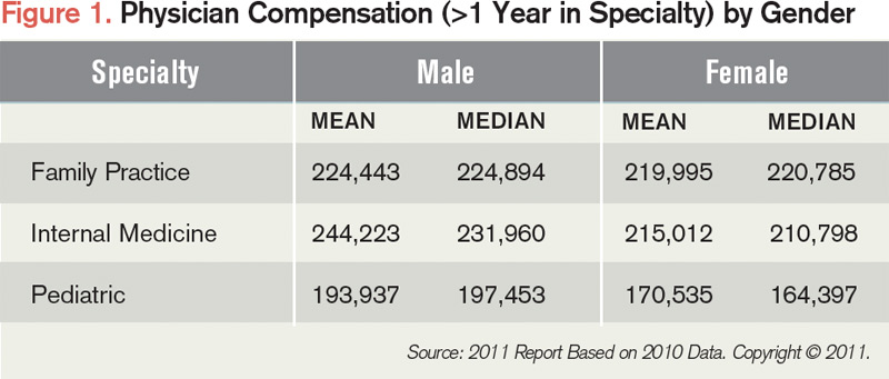
According to the 2011 SHM-MGMA compensation and productivity survey, mean annual compensation for female hospitalists in family practice, internal medicine, and pediatrics is lower than that of their male counterparts. For example, female hospitalists in family practice, internal medicine, and pediatrics have mean annual compensations of $219,995, $215,012, and $170,535, respectively, or $4,448, $29,211, and $23,402 less than male counterparts in similar positions (see Figure 1). Such factors as practice location, practice ownership, and productivity have an effect on compensation and could be the reason behind the disparity, says Liz Boten, a spokeswoman for Englewood, Colo.-based Medical Group Management Association (MGMA).
But research that is controlled for numerous observable factors has shown that the gender earnings gap continues to exist among physicians.1,2,3,4 Of particular note are two studies, including one focused on hospital medicine.
In 2004, a study authored by Timothy J. Hoff, PhD, an associate professor at State University of New York at Albany, controlled for a wide range of work and non-work variables, including clinical workload, compensation type, employer type, tenure, marital status, and tenure in hospital medicine.5 The data show that female hospitalists earned approximately $22,000 less per year than male hospitalists, despite similar work patterns. Additional study results showed that married female hospitalists with children worked just as much and carried as heavy a clinical workload as married male hospitalists who had children.
—Roberta Gebhard, DO, hospitalist, WCA Hospital, Jamestown, N.Y., American Medical Women’s Association’s Gender Equity Task Force co-chair
Last year, a study in Health Affairs generated considerable interest when it found that male physicians newly trained in New York state made on average $16,819 more than newly trained female physicians in 2008, compared with a $3,600 difference in 1999.6 The authors controlled for specialty type, hours worked, designation of hours, immigration status, age, and practice location. And by focusing on starting salaries, factors such as job tenure, institutional rank, and job productivity didn’t come into play, signifying that the experiences of married female and male hospitalists with children differed less than one might presume based upon perceptions that women with families sacrifice work commitments to take care of their spouses and children.
“It is studies like this that are going to be critically important for us to move forward,” says Janet Nagamine, RN, MD, SFHM, a hospitalist at Kaiser Permanente Medical Center in Santa Clara, Calif., and an SHM board member who is assisting with the “Women in Hospital Medicine” session at HM12. “As we talk about a pay gap, we need to be more evidence-based.”
Studies that show a gender earnings gap even among highly skilled professionals don’t surprise researchers. The U.S. Bureau of Labor Statistics collects earnings data on hundreds of occupations, including physicians, and men outearn women across the board, regardless of educational requirements, says Mary Gatta, PhD, past director of gender and workforce policy at the Center for Women and Work at Rutgers University in New Brunswick, N.J., and current senior research scholar at Wider Opportunities for Women, a Washington, D.C.-based organization that focuses on opportunity equality for women.
The Gap that Won’t Close
So why, in 2012, do gender-based pay discrepancies remain?
How much people earn typically is not public information, so women often don’t know they aren’t being paid equally and, therefore, don’t have information on which to act, Dr. Gatta says.
“My opinion on it is women don’t know about the pay gap,” says Dr. Gebhard, co-chair of the American Medical Women’s Association’s (AMWA) Gender Equity Task Force. She recalls a salary negotiation lecture she helped lead after which a woman finishing residency raised her hand to say she was joining a faculty where everyone was paid the same. “The entire room just groaned,” she says. “Clearly, women out there think everything is fair and people are paid the same. They don’t know they’re being paid less.”
In trying to explain the widening pay gap, authors of the 2011 Health Affairs study posited that the influx of women into the physician workforce is reshaping the practice and business of medicine.6
“The notion we suggest is that the increasing gender gap can be explained by new women physicians increasingly demanding non-pecuniary aspects of their jobs, and because of the greater aggregate presence of women in the physician labor market, being able to get it,” says lead author Anthony Lo Sasso, a professor and senior research scientist at the School of Public Health at the University of Illinois at Chicago. “Remember, cash wages are but one part of the compensation package in any job.”
Hoff’s study also uncovered gender differences in employment preferences, with men attracted to HM for the compensation possibilities and women for the predictable hours and lifestyle flexibility. For this reason, Hoff suggested, hospitalist employers can use different recruiting pitches for women than men and, to the extent they hire female hospitalists, save money (see “Negotiating Strategies for Better Compensation,” below).
“The Hoff paper is a goldmine,” says Linda Brodsky, MD, a pediatric otolaryngologist in Buffalo, N.Y., who co-chairs AMWA’s Gender Equity Task Force and whose organization, Expediting the Inevitable, advocates for gender equity in healthcare. “How is it that when you have shift work, women are getting paid less per shift? Because Hoff tells you at the end, employers can get away with it. Even if they know they are underpaying women, they will take the chance because it’s so hard for anybody to take legal action.”
Women also find themselves in a double bind when it comes to negotiating higher compensation, says Barbara Gault, PhD, executive director of the Institute for Women’s Policy Research in Washington, D.C. Some suggest a pay gap exists because women are not negotiating for themselves, but research shows women tend to be perceived as less likable when they are more assertive about higher wages, she says.
Erin Stucky Fisher, MD, MHM, has seen this phenomena play out in her roles as medical director for quality at Rady Children’s Hospital in San Diego and associate program director for the University of California at San Diego Pediatric Residency Program.
“I do a lot of interviewing for the hospital, and it does seem, in general, that women are less likely to promote themselves than men in the same situation,” says Dr. Fisher, an SHM board member who is also assisting with the HM12 “Women in Hospital Medicine” session. “There are reasons behind it that might have to do with women not wanting to be perceived as self-serving or arrogant.”

—Erin Stucky Fisher, MD, FAAP, MHM, medical director for quality, Rady Children’s Hospital, associate program director, University of California at San Diego Pediatric Residency Program, SHM board member
Dr. Brodsky agrees with the perception issues facing physicians.
“Women are supposed to be grateful, accommodating, and get along, which are excellent qualities,” she says. “But when you’re expected to do that and you instead negotiate with any kind of spirit, it’s perceived as troublemaking, whereas in men, it’s perceived as strength.”
Subtle forms of gender discrimination continue to exist in workplaces, Dr. Gatta says. Beliefs remain that men have a family to support, so they should be paid more, and that women are in the workforce just for extra money, even though recent data show that women’s income is key to families’ well-being, she notes.
Common patterns of gender bias will be discussed at the HM12 session, says Dr. Reich, who was a victim of gender pay disparity when she worked a locum tenens job earlier in her career.
“There was no logical explanation. The men did less work by all measures, and the other woman and I didn’t have young children at home, so explanations of women trading in money for time with family didn’t apply,” she explains. “Why did they think I should be paid less? I didn’t understand, and I never got a logical reason.”
Potential Solutions
Shortly after arriving at Wake Forest in 2004, Dr. Reich had the opportunity to build the hospitalist program. A set of thorough, transparent criteria for determining compensation were established almost immediately to help prevent pay discrepancies.
“We tried to be as objective as we could, and we involved the group in talking about it because we felt it was important,” she says.

—Linda Brodsky, MD, pediatric otolaryngologist, Buffalo, N.Y., co-chair, AMWA Gender Equity Task Force
Organizations can conduct self-audits to assess whether men and women are being compensated equitably, then make adjustments when necessary, Dr. Gault says. Policies can be adopted to promote pay transparency and allow employees to discuss compensation and suggest ways it can be improved, she adds. In workplaces where compensation discussions are discouraged, women can try to informally speak with their male friends to gather information and determine if there’s a pay gap problem.
Equal pay laws, such as the Lily Ledbetter Fair Pay Act, exist at the federal level. But Dr. Gatta says work must be done on the enforcement end to make a real difference. Similarly, Dr. Brodsky describes the Equal Employment Opportunity Commission as a toothless oversight agency with limited power to investigate complaints and assess fines.
“It’s on women to go and be the whistleblower, the policeman, and pay for legal action. It’s impossible,” she says. “When you utter the words ‘gender discrimination,’ immediately, retaliation goes into high gear.”
While employers and enforcement agencies have a significant role in closing the pay gap, women themselves must collectively advocate for equal pay, Dr. Gault says.
“Any woman physician who is in the senior ranks must be obligated to start changing the culture and making sure the fairness issue gets raised and is an important part of their agenda,” Dr. Brodsky says. “Enough women are now part of the physician population. They have to start saying, ‘We are a group to be reckoned with, and we are going to make changes.’”
Lisa Ryan is a freelance writer based in New Jersey.
References
- Wright AL, Schwindt LA, Bassford TL, et al. Gender differences in academic advancement: patterns, causes, and potential solutions in one US College of Medicine. Acad Med. 2003;78(5):500-508.
- Ash AS, Carr PL, Goldstein R, et al. Compensation and advancement of women in academic medicine: is there equity? Ann Intern Med. 2004;141(3):205-212.
- Ness RB, Ukoli F, Hunt S, et al. Salary equity among male and female internists in Pennsylvania. Ann Intern Med. 2000;133(2):104-110.
- Weeks WB, Wallace TA, Wallace AE. How do race and sex affect the earnings of primary care physicians? Health Aff (Millwood). 2009;28(2):557-566.
- Hoff TJ. Doing the same and earning less: male and female physicians in a new medical specialty. Inquiry. 2004;41:301-315.
- Lo Sasso AT, Richards MR, Chou C, Gerber SE. The $16,819 pay gap for newly trained physicians: the unexplained trend of men earning more than women. Health Aff (Millwood). 2011;30:193-201.
Roberta Gebhard, DO, thought that her 20 years of experience as a physician in the U.S., 10 of them as a hospitalist, would mean she would get paid more than a new graduate just out of residency would.
She was wrong.
Dr. Gebhard was working at a hospital run by the U.S. Department of Veterans Affairs when she learned that the less experienced doctor—a man—was making $10,000 more a year than she was.
“After that, the job was no longer interesting to me,” says Dr. Gebhard, who left the hospital over the pay discrepancy and now works as a hospitalist at WCA Hospital in Jamestown, N.Y. “Women think that things should be fair, so they assume that they are. I’m a good negotiator, and when that happened to me, I was like, ‘Wait a minute! I didn’t just take what they offered me.’ I pushed a few times and was basically told it was a government position, there was no wiggle room, and I couldn’t get more salary.
“It happens, and women need to know that it happens,” she says.
Earnings data and research show that the gender pay gap lingers. More problematic is pinpointing why the gap won’t close. Explanations range from ignorance of the issue and trading in compensation for other job benefits to women’s lack of negotiating skills to subtle gender discrimination.
Because gender pay discrepancies persist and because theories abound as to the cause, the issue will be addressed during a “Women in Hospital Medicine” session at HM12 next month in San Diego, along with such topics as leadership challenges and work-life balance, says Patience Reich, MD, SFHM, a hospitalist and assistant professor of medicine at Wake Forest University School of Medicine in Winston-Salem, N.C.
“When we conceived the session, we were actually thinking about women in leadership, but decided to go for more general topics that affect women hospitalists, whether they are in leadership or not,” says Dr. Reich, a member of SHM’s Leadership Committee who helped coordinate the HM12 session.
According to the 2011 SHM-MGMA compensation and productivity survey, mean annual compensation for female hospitalists in family practice, internal medicine, and pediatrics is lower than that of their male counterparts. For example, female hospitalists in family practice, internal medicine, and pediatrics have mean annual compensations of $219,995, $215,012, and $170,535, respectively, or $4,448, $29,211, and $23,402 less than male counterparts in similar positions (see Figure 1). Such factors as practice location, practice ownership, and productivity have an effect on compensation and could be the reason behind the disparity, says Liz Boten, a spokeswoman for Englewood, Colo.-based Medical Group Management Association (MGMA).
But research that is controlled for numerous observable factors has shown that the gender earnings gap continues to exist among physicians.1,2,3,4 Of particular note are two studies, including one focused on hospital medicine.
In 2004, a study authored by Timothy J. Hoff, PhD, an associate professor at State University of New York at Albany, controlled for a wide range of work and non-work variables, including clinical workload, compensation type, employer type, tenure, marital status, and tenure in hospital medicine.5 The data show that female hospitalists earned approximately $22,000 less per year than male hospitalists, despite similar work patterns. Additional study results showed that married female hospitalists with children worked just as much and carried as heavy a clinical workload as married male hospitalists who had children.
—Roberta Gebhard, DO, hospitalist, WCA Hospital, Jamestown, N.Y., American Medical Women’s Association’s Gender Equity Task Force co-chair
Last year, a study in Health Affairs generated considerable interest when it found that male physicians newly trained in New York state made on average $16,819 more than newly trained female physicians in 2008, compared with a $3,600 difference in 1999.6 The authors controlled for specialty type, hours worked, designation of hours, immigration status, age, and practice location. And by focusing on starting salaries, factors such as job tenure, institutional rank, and job productivity didn’t come into play, signifying that the experiences of married female and male hospitalists with children differed less than one might presume based upon perceptions that women with families sacrifice work commitments to take care of their spouses and children.
“It is studies like this that are going to be critically important for us to move forward,” says Janet Nagamine, RN, MD, SFHM, a hospitalist at Kaiser Permanente Medical Center in Santa Clara, Calif., and an SHM board member who is assisting with the “Women in Hospital Medicine” session at HM12. “As we talk about a pay gap, we need to be more evidence-based.”
Studies that show a gender earnings gap even among highly skilled professionals don’t surprise researchers. The U.S. Bureau of Labor Statistics collects earnings data on hundreds of occupations, including physicians, and men outearn women across the board, regardless of educational requirements, says Mary Gatta, PhD, past director of gender and workforce policy at the Center for Women and Work at Rutgers University in New Brunswick, N.J., and current senior research scholar at Wider Opportunities for Women, a Washington, D.C.-based organization that focuses on opportunity equality for women.
The Gap that Won’t Close
So why, in 2012, do gender-based pay discrepancies remain?
How much people earn typically is not public information, so women often don’t know they aren’t being paid equally and, therefore, don’t have information on which to act, Dr. Gatta says.
“My opinion on it is women don’t know about the pay gap,” says Dr. Gebhard, co-chair of the American Medical Women’s Association’s (AMWA) Gender Equity Task Force. She recalls a salary negotiation lecture she helped lead after which a woman finishing residency raised her hand to say she was joining a faculty where everyone was paid the same. “The entire room just groaned,” she says. “Clearly, women out there think everything is fair and people are paid the same. They don’t know they’re being paid less.”
In trying to explain the widening pay gap, authors of the 2011 Health Affairs study posited that the influx of women into the physician workforce is reshaping the practice and business of medicine.6
“The notion we suggest is that the increasing gender gap can be explained by new women physicians increasingly demanding non-pecuniary aspects of their jobs, and because of the greater aggregate presence of women in the physician labor market, being able to get it,” says lead author Anthony Lo Sasso, a professor and senior research scientist at the School of Public Health at the University of Illinois at Chicago. “Remember, cash wages are but one part of the compensation package in any job.”
Hoff’s study also uncovered gender differences in employment preferences, with men attracted to HM for the compensation possibilities and women for the predictable hours and lifestyle flexibility. For this reason, Hoff suggested, hospitalist employers can use different recruiting pitches for women than men and, to the extent they hire female hospitalists, save money (see “Negotiating Strategies for Better Compensation,” below).
“The Hoff paper is a goldmine,” says Linda Brodsky, MD, a pediatric otolaryngologist in Buffalo, N.Y., who co-chairs AMWA’s Gender Equity Task Force and whose organization, Expediting the Inevitable, advocates for gender equity in healthcare. “How is it that when you have shift work, women are getting paid less per shift? Because Hoff tells you at the end, employers can get away with it. Even if they know they are underpaying women, they will take the chance because it’s so hard for anybody to take legal action.”
Women also find themselves in a double bind when it comes to negotiating higher compensation, says Barbara Gault, PhD, executive director of the Institute for Women’s Policy Research in Washington, D.C. Some suggest a pay gap exists because women are not negotiating for themselves, but research shows women tend to be perceived as less likable when they are more assertive about higher wages, she says.
Erin Stucky Fisher, MD, MHM, has seen this phenomena play out in her roles as medical director for quality at Rady Children’s Hospital in San Diego and associate program director for the University of California at San Diego Pediatric Residency Program.
“I do a lot of interviewing for the hospital, and it does seem, in general, that women are less likely to promote themselves than men in the same situation,” says Dr. Fisher, an SHM board member who is also assisting with the HM12 “Women in Hospital Medicine” session. “There are reasons behind it that might have to do with women not wanting to be perceived as self-serving or arrogant.”
—Erin Stucky Fisher, MD, FAAP, MHM, medical director for quality, Rady Children’s Hospital, associate program director, University of California at San Diego Pediatric Residency Program, SHM board member
Dr. Brodsky agrees with the perception issues facing physicians.
“Women are supposed to be grateful, accommodating, and get along, which are excellent qualities,” she says. “But when you’re expected to do that and you instead negotiate with any kind of spirit, it’s perceived as troublemaking, whereas in men, it’s perceived as strength.”
Subtle forms of gender discrimination continue to exist in workplaces, Dr. Gatta says. Beliefs remain that men have a family to support, so they should be paid more, and that women are in the workforce just for extra money, even though recent data show that women’s income is key to families’ well-being, she notes.
Common patterns of gender bias will be discussed at the HM12 session, says Dr. Reich, who was a victim of gender pay disparity when she worked a locum tenens job earlier in her career.
“There was no logical explanation. The men did less work by all measures, and the other woman and I didn’t have young children at home, so explanations of women trading in money for time with family didn’t apply,” she explains. “Why did they think I should be paid less? I didn’t understand, and I never got a logical reason.”
Potential Solutions
Shortly after arriving at Wake Forest in 2004, Dr. Reich had the opportunity to build the hospitalist program. A set of thorough, transparent criteria for determining compensation were established almost immediately to help prevent pay discrepancies.
“We tried to be as objective as we could, and we involved the group in talking about it because we felt it was important,” she says.
—Linda Brodsky, MD, pediatric otolaryngologist, Buffalo, N.Y., co-chair, AMWA Gender Equity Task Force
Organizations can conduct self-audits to assess whether men and women are being compensated equitably, then make adjustments when necessary, Dr. Gault says. Policies can be adopted to promote pay transparency and allow employees to discuss compensation and suggest ways it can be improved, she adds. In workplaces where compensation discussions are discouraged, women can try to informally speak with their male friends to gather information and determine if there’s a pay gap problem.
Equal pay laws, such as the Lily Ledbetter Fair Pay Act, exist at the federal level. But Dr. Gatta says work must be done on the enforcement end to make a real difference. Similarly, Dr. Brodsky describes the Equal Employment Opportunity Commission as a toothless oversight agency with limited power to investigate complaints and assess fines.
“It’s on women to go and be the whistleblower, the policeman, and pay for legal action. It’s impossible,” she says. “When you utter the words ‘gender discrimination,’ immediately, retaliation goes into high gear.”
While employers and enforcement agencies have a significant role in closing the pay gap, women themselves must collectively advocate for equal pay, Dr. Gault says.
“Any woman physician who is in the senior ranks must be obligated to start changing the culture and making sure the fairness issue gets raised and is an important part of their agenda,” Dr. Brodsky says. “Enough women are now part of the physician population. They have to start saying, ‘We are a group to be reckoned with, and we are going to make changes.’”
Lisa Ryan is a freelance writer based in New Jersey.
References
- Wright AL, Schwindt LA, Bassford TL, et al. Gender differences in academic advancement: patterns, causes, and potential solutions in one US College of Medicine. Acad Med. 2003;78(5):500-508.
- Ash AS, Carr PL, Goldstein R, et al. Compensation and advancement of women in academic medicine: is there equity? Ann Intern Med. 2004;141(3):205-212.
- Ness RB, Ukoli F, Hunt S, et al. Salary equity among male and female internists in Pennsylvania. Ann Intern Med. 2000;133(2):104-110.
- Weeks WB, Wallace TA, Wallace AE. How do race and sex affect the earnings of primary care physicians? Health Aff (Millwood). 2009;28(2):557-566.
- Hoff TJ. Doing the same and earning less: male and female physicians in a new medical specialty. Inquiry. 2004;41:301-315.
- Lo Sasso AT, Richards MR, Chou C, Gerber SE. The $16,819 pay gap for newly trained physicians: the unexplained trend of men earning more than women. Health Aff (Millwood). 2011;30:193-201.
HM12 Features High-Profile Speakers, RIV Sessions, Expanded Breakouts

The world was a different place in 2008, the last time SHM’s annual meeting was in San Diego. Attendance at the yearly confab of hospitalists was almost half of what it is expected to be this year, healthcare reform was still just a bullet point for a presidential candidate’s talking points, and society leaders were drumming up interest in a new fellowship program they’d created.
Now, more than 2,000 hospitalists are expected to fill the San Diego Convention Center April 1-4. Reform isn’t just a political buzzword; it’s the subject of landmark legislation and three plenary addresses—including the requisite convention-ender from HM pioneer Robert Wachter, MD, MHM. And the now-three-tiered fellowship track is more than 1,000 physicians strong.
“Hospitalists really are on the radar of policymakers both inside and outside of our field,” says Jeff Glasheen, MD, SFHM, HM12 course director. “We really have evolved from an insular hospitalist group to the bigger picture that really touches healthcare in many different ways.”
And nowhere is that breadth on display more than the annual meting.
Among the most popular components are the April 1 pre-courses. The schedule features popular sessions on practice management, billing and coding, and ABIM Maintenance of Certification, along with a couple new offerings. The debut class generating the most interest is “How to Improve Performance in CMS’s Value-Based Purchasing Program,” led by SHM senior vice president Joseph Miller and Patrick Torcson, MD, MMM, FAACP, SFHM, chair of SHM’s Performance Measurement and Reporting Committee.
Then there is the Research, Innovations, and Clinical Vignettes (RIV) poster competition. So many entries came in for HM12 that the poster sessions have been split up to give attendees more time to interact with their authors. The Research and Innovations poster reception will be from 5 to 7 p.m. April 2. The 90-minute Vignettes session will start at noon April 3.
And, of course, the featured speakers always draw a crowd. This year offers three keynote addresses. First up will be Patrick Conway, MD, MSc, FAAP, SFHM, a pediatric hospitalist and chief medical officer of the Centers for Medicare & Medicaid Services (CMS). His talk is titled “Affordable Care Act Implementation and How Hospital Medicine Can Help Lead Health Care System Transformation.” Political commentator and analyst Norman Ornstein, PhD, MA, BA, will follow with a presentation dubbed “Making Health Policy in an Age of Dysfunctional Politics.” HM12 also continues the tradition of having Dr. Wachter give the last talk, “The Great Physician, Circa 2012: How Hospitalists Must Lead Efforts to Identify and Become This New Breed.”
Throw in a new offering of educational sessions, a new way for attendees to design a thematic schedule of classes that cross different categorical tracks, and a sunny seaside locale, and this year’s meeting would seem destined for success.
“Hopefully the weather will hold up,” says local hospitalist Pedro Ramos, MD, assistant clinical professor of medicine at the University of California at San Diego. “My fear is it’s going to be rainy that one week.”
Richard Quinn is a freelance writer in New Jersey.
Six Keys to a Successful Annual Meeting

Michael Pistoria, DO, FACP, SFHM, is as guilty as you are of not always getting the most out of the annual meeting—and he’s one of the people planning it.
Dr. Pistoria, HM12 assistant course director and hospitalist at Lehigh Valley Health Network in Allentown, Pa., always suggests that if an HM group sends more than one person to the annual meeting, those people should split up and go to as many different sessions as possible. But then, at HM11 in Dallas, an interesting course about an accountable-care unit caught the attention of three people from his office.
“I went, our senior vice president of care continuum went, and the director of our program—we were all there,” Dr. Pistoria says. “I’m thinking, ‘Well this is good, we’re all going to get a slightly different take on it and we’re all going to be listening for different things, and, when we do sit down to compare notes, this is going to be helpful.’ But there were other sessions we could be in.”
To be clear, there is no wrong way to attend an annual meeting. But like knowing which nurse shepherds a smoother discharge, there’s always a way to do it better. Tips from Dr. Pistoria and HM12 course director Jeff Glasheen, MD, SFHM, include:

—Jeff Glasheen, MD, SFHM, HM12 course director
- Prepare. Set up at least a basic schedule or pick out a few sessions to sit in on before arriving at HM12, or you risk trying to build a successful meeting on the fly. “I would treat this much like you would your first semester of college,” Dr. Glasheen says. “You wouldn’t just show up and see what course you wanted to go to. You’d spend some time prior to showing up....If you don’t have a game plan, you’re going to end up missing out on something you want to learn.”
- Go where you friends don’t. As Dr. Pistoria attests, it’s a natural tendency to want to attend sessions with colleagues and critique together. However, this is a once-a-year opportunity. Attend different sessions and compare notes.
- Attend the keynote addresses. This year’s lineup features a national election analyst for “CBS News,” the chief medical officer of the Centers for Medicare & Medicaid Services (CMS), and the incoming chair of the American Board of Internal Medicine (ABIM). Between them, there probably are a few good nuggets to glean.
- Attend the special-interest forums. SHM holds a series of small-group sessions focused on such hyperlocal topics as rural HM and health information technology (HIT), among others. Society staff and leadership take notes and let attendees drive the conversation.
- Network, network, network. Everyone had that first meeting where they didn’t know who was who, so feel free to introduce yourself to anybody. Get business cards from everybody. And hit the hotel hotspots armed with questions and a willingness to buy a potential new relationship a drink.
- “There’s a tremendous willingness to help new people, to answer questions, to have the hallway questions, the conversations that occur at 11 o’clock at night at the bar,” Dr. Pistoria says. “If you see somebody and you say, ‘Hey, you know, I was in your session earlier, can I ask you a couple of follow-up questions?’ People are going to say, ‘Hell yes, sit down. Let’s talk.’ If people remember that and aren’t shy, I think people really can maximize the potential benefit.”
- Share what you learn. Start a quality initiative or talk with your C-suite about starting a project. Email some of your new contacts to start a dialogue. Don’t go home, give one brown-bag session to your colleagues, and wait until next year’s annual meeting to think about the “big picture” again.
“It’s easy to get back home and fall right back into the patterns you were doing before,” Dr. Glasheen says. “If you get excited by something at the annual meeting, go back home and really work at putting that into place.”
Richard Quinn is a freelance writer in New Jersey.
To get involved, visit www.hospitalmedicine2012.org/schedule
Michael Pistoria, DO, FACP, SFHM, is as guilty as you are of not always getting the most out of the annual meeting—and he’s one of the people planning it.
Dr. Pistoria, HM12 assistant course director and hospitalist at Lehigh Valley Health Network in Allentown, Pa., always suggests that if an HM group sends more than one person to the annual meeting, those people should split up and go to as many different sessions as possible. But then, at HM11 in Dallas, an interesting course about an accountable-care unit caught the attention of three people from his office.
“I went, our senior vice president of care continuum went, and the director of our program—we were all there,” Dr. Pistoria says. “I’m thinking, ‘Well this is good, we’re all going to get a slightly different take on it and we’re all going to be listening for different things, and, when we do sit down to compare notes, this is going to be helpful.’ But there were other sessions we could be in.”
To be clear, there is no wrong way to attend an annual meeting. But like knowing which nurse shepherds a smoother discharge, there’s always a way to do it better. Tips from Dr. Pistoria and HM12 course director Jeff Glasheen, MD, SFHM, include:
—Jeff Glasheen, MD, SFHM, HM12 course director
- Prepare. Set up at least a basic schedule or pick out a few sessions to sit in on before arriving at HM12, or you risk trying to build a successful meeting on the fly. “I would treat this much like you would your first semester of college,” Dr. Glasheen says. “You wouldn’t just show up and see what course you wanted to go to. You’d spend some time prior to showing up....If you don’t have a game plan, you’re going to end up missing out on something you want to learn.”
- Go where you friends don’t. As Dr. Pistoria attests, it’s a natural tendency to want to attend sessions with colleagues and critique together. However, this is a once-a-year opportunity. Attend different sessions and compare notes.
- Attend the keynote addresses. This year’s lineup features a national election analyst for “CBS News,” the chief medical officer of the Centers for Medicare & Medicaid Services (CMS), and the incoming chair of the American Board of Internal Medicine (ABIM). Between them, there probably are a few good nuggets to glean.
- Attend the special-interest forums. SHM holds a series of small-group sessions focused on such hyperlocal topics as rural HM and health information technology (HIT), among others. Society staff and leadership take notes and let attendees drive the conversation.
- Network, network, network. Everyone had that first meeting where they didn’t know who was who, so feel free to introduce yourself to anybody. Get business cards from everybody. And hit the hotel hotspots armed with questions and a willingness to buy a potential new relationship a drink.
- “There’s a tremendous willingness to help new people, to answer questions, to have the hallway questions, the conversations that occur at 11 o’clock at night at the bar,” Dr. Pistoria says. “If you see somebody and you say, ‘Hey, you know, I was in your session earlier, can I ask you a couple of follow-up questions?’ People are going to say, ‘Hell yes, sit down. Let’s talk.’ If people remember that and aren’t shy, I think people really can maximize the potential benefit.”
- Share what you learn. Start a quality initiative or talk with your C-suite about starting a project. Email some of your new contacts to start a dialogue. Don’t go home, give one brown-bag session to your colleagues, and wait until next year’s annual meeting to think about the “big picture” again.
“It’s easy to get back home and fall right back into the patterns you were doing before,” Dr. Glasheen says. “If you get excited by something at the annual meeting, go back home and really work at putting that into place.”
Richard Quinn is a freelance writer in New Jersey.
To get involved, visit www.hospitalmedicine2012.org/schedule
Policy Experts Say Hospital Medicine Should be Ready to Tackle Reform Challenges
In a restaurant, it’s called being in the weeds: when the duties of one’s job become so overwhelming that you can’t keep up with the pace. Think five new admissions at the end of a 12-hour workday.
That’s where Norm Ornstein, one of the keynote speakers at HM12 in San Diego, can help. A resident scholar at the American Enterprise Institute in Washington, D.C., and a longtime observer of all things political, Ornstein is in the weeds of healthcare reform on a partisan and policy level. He pens a weekly column for the Congressional newspaper Roll Call and also provides election analysis for “CBS News.” He has authored or edited multiple books, including “Intensive Care: How Congress Shapes Health Policy” and “The Permanent Campaign and Its Future.”
So hospitalists who too often feel that, between patient care and clinical research, they don’t have time to study the political landscape should probably plan to attend Ornstein’s address, tentatively dubbed “Making Health Policy in an Age of Dysfunctional Politics.” The Hospitalist spent a few moments with Ornstein as he prepared for his HM12 address.
What do you plan to speak about and what do you want hospitalists to take away from your talk?
We’re really going to be looking toward the fall matchup in the presidential campaign. And we know that health policy is going to be not the No. 1 issue—it’s pretty clear now that the economy and jobs will be the No. 1 issue—but it will be following pretty closely behind at No. 2. And we know we’re going to get a very hot debate on whether we can or should repeal the Affordable Care Act (ACA). … But we also know that, repeal or not … health policy is up in the air. We’re going to go through some tumultuous changes. Everybody is on the line; who’s actually delivering services, or trying to, is going to have enormous challenges. It’s a perfect time to talk about these issues.
Healthcare professionals aren’t used to being part of the political maelstrom. What is their role in this process?
If anything, the last year or two should make it very clear to them they better be involved. … Whatever happens with the Affordable Care Act, we’re going to be making adjustments in public policy, and we’re going to be seeing massive adjustments in the private sector for years to come. Lots of people are going to be looking for the best ideas and looking for ways to make sure we can bend the cost curve, to use the cliché, but not sacrifice significantly services to people—and maybe even improve those services. It’s time, I think, for people in this world to understand it’s in their own self-interest to be engaged.
How nervous should hospitalists be that there really is a chance the ACA can be repealed?
I don’t think they should be worried that the whole kit and caboodle will be replaced. I think they should be concerned that we will see a combination of some elements of it perhaps repealed, but even more that the corrosive nature of our politics is such that Republicans, if they can’t repeal it, will do everything they can to bollocks up its implementation so that they can both score political points but also say, “See, we told you so.” This would be a challenge under the best of circumstances to make this work. But I think everybody has to be braced for what I think will be a series of some substantial bumps and jolts in the road along the way.
That is a lot of uncertainty for a hospitalist. How can they prepare?
I don’t think one can prepare adequately for the changes that are going to take place. What hospitalists have to do first is to make sure that they fulfill their own roles in caring for patients, but they should also be thinking about innovative approaches and ways of delivering services that are good for patents but also good for the system.
Is that enough—being at the vanguard of care delivery?
No, and I can’t give people assurances that there is any specific thing they can do that will make things work, or work better, because we’re moving into an uncertain environment—an uncertain environment in the real world where we’re going to see health as a share of GDP probably continue to go up for a while, but there will be some pushback against that; uncertainty as we try to integrate the public and private halves of our healthcare system a little bit better than we have; uncertainty as we find that the fiscal realities of America mean that you’ve got to put even more of a squeeze on Medicare, Medicaid, and even veterans’ health. Those things don’t happen in isolation.
Should doctors see this as an exciting time?
I think there is a bright side to this, which is there really is an opportunity here for innovation in ways to fulfill their oaths. There is an opportunity to find ways to provide better healthcare for people, better services to make their lives better.
Richard Quinn is a freelance writer in New Jersey.
To get involved, visit www.hospitalmedicine.org/advocacy
CME Credit at HM12
HM12 is planned and implemented in accordance with the Essential Areas and Policies of the Accreditation Council for Continuing Medical Education (ACCME) through the joint sponsorship of Blackwell Futura Media Services (BFMS) and SHM. BFMS is accredited by ACCME to provide CME for physicians.
HM12 registration is available at www.hospitalmedicine2012.org until March 5. SHM also permits walk-up registration at the San Diego Convention Center.
BFMS designates the educational activity for SHM’s annual meeting at a maximum of 19.75 Category 1 credits toward the AMA Physician’s Recognition Award. Each physician should claim only those hours of credit they actually spend in each educational activity. BFMS has designated a credit schedule for HM12’s pre-courses on April 1 as follows:
- ABIM MOC learning session, 6.5 credits;
- Advanced Interactive Critical Care, 7.75 credits;
- CMS’s Value-Based Purchasing Program, 3.75 credits;
- Medical Procedures, 7.5 credits;
- Portable Ultrasounds, 3.75 credits;
- Perioperative Medicine, 7.75 credits; and
- Practice Management, 7.5 credits.
Source: www.hospitalmedicine.org
HM12 Organizers Strive for ‘Meaningful’ Educational Offerings for Hospitalists
Jeff Glasheen, MD, SFHM, has been going to SHM’s annual meetings for a decade. He’s played the part of poster presenter, session leader, physician editor of The Hospitalist, and even just a healthcare consumer eager to hear what a keynote speaker has to say.
This year, he’s added a new title: HM12 course director. The sobriquet has given him a new appreciation for the work that goes into the four-day summit, April 1-4 at the San Diego Convention Center.
“It’s been a great experience to see how much work really goes into these meetings behind the scenes and how thoughtful people are to try to program content that all different types of hospitalists would be interested in,” says Dr. Glasheen, associate professor of medicine and director of the hospital medicine group at the University of Colorado Denver. “Trying to target enough stuff so that it’s meaningful for everybody ends up being incredibly challenging.”
The challenge is important because continuing medical education (CME) arguably is the biggest draw for the annual meeting. From pre-courses offering Category 1 credits toward the AMA Physician’s Recognition Award to rapid-fire sessions offering “Jeopardy!”-like chances to test current knowledge, a majority of past attendees say CME and pedagogy keep them coming back.
“It’s one of the top reasons people come, and it should be,” Dr. Glasheen adds. “This is the opportunity for people to come and hear new ideas. And I don’t mean that just in terms of clinical medicine, but also nonclinical medicine. This is our opportunity to pull ourselves out of the weeds … and really get a sense of what’s coming down the pike.”
Dr. Glasheen and assistant course director Michael Pistoria, DO, FACP, SFHM, say the goals of each year’s offerings are to keep things current and user-friendly.
—Jeff Glasheen, MD, SFHM, HM12 course director
Take “How to Improve Performance in CMS’s Value-Based Purchasing Program,” a debut pre-course designed as a primer for the new VBP rules going into effect Oct. 1. The session is led by SHM’s senior vice president and chief solutions officer Joseph Miller and Patrick Torcson, MD, MMM, FACP, SFHM, director of hospital medicine at St. Tammany Parish Hospital in Covington, La. The new pre-course joins the now well-established American Board of Internal Medicine’s (ABIM) Maintenance of Certification (MOC) course as one of the sessions that, although they didn’t exist a few years ago, are viewed as crucial to the specialty’s future development.
“It’s the natural evolution of what hospitalists do,” Dr. Glasheen says. “If we went back to the first year of this meeting, it was primarily a clinical meeting. But I think over time, just as my role as a hospitalist has evolved over 10 years, so has the society’s annual meeting. Yes, we have the clinical core that everybody needs. But there’s a lot of other stuff that’s happening in the world around us—including healthcare policy, payment reform—and we need to know about those things.”
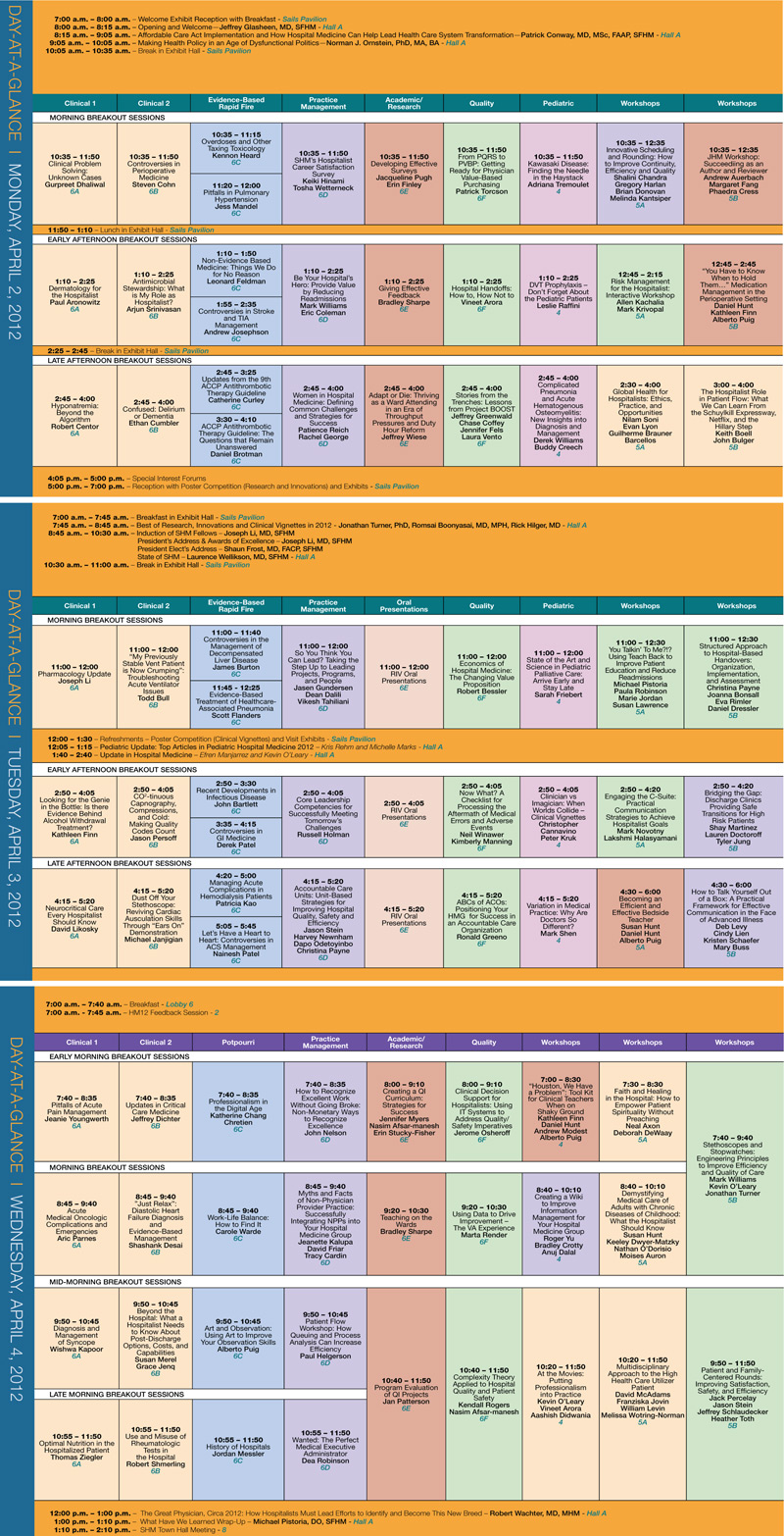
Constant change to the HM12 course menu keeps things fresh, says Dr. Pistoria, HM13’s course director, who works at Lehigh Valley Health Network in Allentown, Pa. “Quite frankly, it allows us to some degree to keep the faculty fresh as well,” he says. “If you give them a year off, they come back fully charged with new ideas. Even if it’s the same information, maybe they’ve got some new ideas of how to present it.”
Another area where the importance of the meeting’s educational offerings becomes self-evident is the Research, Innovations, and Clinical Vignettes (RIV) poster competition. The popularity of the competition continues to grow; the review committee was deluged with 200 more submissions for HM12 than for HM11. Organizers felt that was too many posters to give attendees enough quality time with their presenters, so the session has been expanded and broken up into two sessions—one for Research and Innovations (Monday, April 2, 5-7 p.m.), and one for Vignettes (Tuesday, April 3, noon-1:30 p.m.).
“It speaks to the fact that academic hospital medicine is really growing up,” Dr. Glasheen says. “Ten years ago, we were all just trying to get enough docs to take care of patients. Now we’re getting to the point where academic hospitalists really can develop a career as a scholar and a researcher.”
An educational twist to this year’s meeting is an approach called “Pathways.” The tack is an online map of interrelated courses that may appear in different categorical tracks. For example, the “pathway” highlights for attendees how they can find sessions devoted to early-career hospitalist issues, palliative care, or transitions of care. So if two people from one institution wanted to break up the sessions by a topic, they now have an easier way.
“It affords people the opportunity to plan,” Dr. Pistoria says. “And again, it keeps us fresh. As the annual meeting committee, we’re constantly looking: What can we do what can we do differently? What can we do better?”
Richard Quinn is a freelance writer in New Jersey.
Jeff Glasheen, MD, SFHM, has been going to SHM’s annual meetings for a decade. He’s played the part of poster presenter, session leader, physician editor of The Hospitalist, and even just a healthcare consumer eager to hear what a keynote speaker has to say.
This year, he’s added a new title: HM12 course director. The sobriquet has given him a new appreciation for the work that goes into the four-day summit, April 1-4 at the San Diego Convention Center.
“It’s been a great experience to see how much work really goes into these meetings behind the scenes and how thoughtful people are to try to program content that all different types of hospitalists would be interested in,” says Dr. Glasheen, associate professor of medicine and director of the hospital medicine group at the University of Colorado Denver. “Trying to target enough stuff so that it’s meaningful for everybody ends up being incredibly challenging.”
The challenge is important because continuing medical education (CME) arguably is the biggest draw for the annual meeting. From pre-courses offering Category 1 credits toward the AMA Physician’s Recognition Award to rapid-fire sessions offering “Jeopardy!”-like chances to test current knowledge, a majority of past attendees say CME and pedagogy keep them coming back.
“It’s one of the top reasons people come, and it should be,” Dr. Glasheen adds. “This is the opportunity for people to come and hear new ideas. And I don’t mean that just in terms of clinical medicine, but also nonclinical medicine. This is our opportunity to pull ourselves out of the weeds … and really get a sense of what’s coming down the pike.”
Dr. Glasheen and assistant course director Michael Pistoria, DO, FACP, SFHM, say the goals of each year’s offerings are to keep things current and user-friendly.
—Jeff Glasheen, MD, SFHM, HM12 course director
Take “How to Improve Performance in CMS’s Value-Based Purchasing Program,” a debut pre-course designed as a primer for the new VBP rules going into effect Oct. 1. The session is led by SHM’s senior vice president and chief solutions officer Joseph Miller and Patrick Torcson, MD, MMM, FACP, SFHM, director of hospital medicine at St. Tammany Parish Hospital in Covington, La. The new pre-course joins the now well-established American Board of Internal Medicine’s (ABIM) Maintenance of Certification (MOC) course as one of the sessions that, although they didn’t exist a few years ago, are viewed as crucial to the specialty’s future development.
“It’s the natural evolution of what hospitalists do,” Dr. Glasheen says. “If we went back to the first year of this meeting, it was primarily a clinical meeting. But I think over time, just as my role as a hospitalist has evolved over 10 years, so has the society’s annual meeting. Yes, we have the clinical core that everybody needs. But there’s a lot of other stuff that’s happening in the world around us—including healthcare policy, payment reform—and we need to know about those things.”
Constant change to the HM12 course menu keeps things fresh, says Dr. Pistoria, HM13’s course director, who works at Lehigh Valley Health Network in Allentown, Pa. “Quite frankly, it allows us to some degree to keep the faculty fresh as well,” he says. “If you give them a year off, they come back fully charged with new ideas. Even if it’s the same information, maybe they’ve got some new ideas of how to present it.”
Another area where the importance of the meeting’s educational offerings becomes self-evident is the Research, Innovations, and Clinical Vignettes (RIV) poster competition. The popularity of the competition continues to grow; the review committee was deluged with 200 more submissions for HM12 than for HM11. Organizers felt that was too many posters to give attendees enough quality time with their presenters, so the session has been expanded and broken up into two sessions—one for Research and Innovations (Monday, April 2, 5-7 p.m.), and one for Vignettes (Tuesday, April 3, noon-1:30 p.m.).
“It speaks to the fact that academic hospital medicine is really growing up,” Dr. Glasheen says. “Ten years ago, we were all just trying to get enough docs to take care of patients. Now we’re getting to the point where academic hospitalists really can develop a career as a scholar and a researcher.”
An educational twist to this year’s meeting is an approach called “Pathways.” The tack is an online map of interrelated courses that may appear in different categorical tracks. For example, the “pathway” highlights for attendees how they can find sessions devoted to early-career hospitalist issues, palliative care, or transitions of care. So if two people from one institution wanted to break up the sessions by a topic, they now have an easier way.
“It affords people the opportunity to plan,” Dr. Pistoria says. “And again, it keeps us fresh. As the annual meeting committee, we’re constantly looking: What can we do what can we do differently? What can we do better?”
Richard Quinn is a freelance writer in New Jersey.