User login
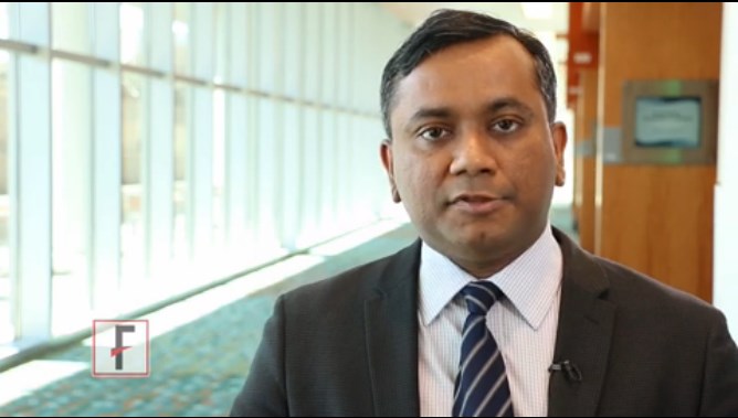
Predicting outcomes of allo-HSCT in ALL
![]()
Photo by Chad McNeeley
SAN DIEGO—A retrospective study has revealed a few factors that may predict outcomes of allogeneic hematopoietic stem cell transplant (allo-HSCT) in adults with acute lymphoblastic leukemia (ALL).
The study showed that cytogenetics at diagnosis did not impact survival rates, although having high-risk cytogenetics was associated with an increased incidence of relapse in patients who were transplanted in their first complete remission (CR1).
Patients who were not in CR1 at transplant tended to have worse survival and higher relapse rates.
And patients who received a tacrolimus/sirolimus-based regimen as graft-vs-host disease (GVHD) prophylaxis had better survival rates than their peers, but their relapse rates did not differ.
Ibrahim Aldoss, MD, of City of Hope Medical Center in Duarte, California, presented these findings at the 2015 BMT Tandem Meetings as abstract 69.*
Dr Aldoss said there is a lack of data addressing individual ALL-related prognostic factors for transplant outcomes. So he and his colleagues decided to analyze 358 adult ALL patients who received allo-HSCT at the City of Hope from January 2004 through March 2014.
The patients’ median age was 38 (range, 18 to 72), and most patients (91%) had B-cell disease. At diagnosis, 2% of patients had good-risk cytogenetics, 43% had intermediate-risk, and 46% had poor-risk. For 9% of patients, the cytogenetic risk group was unknown.
At transplant, 60% of patients were in CR1, 17% were in CR2, and 23% were in a subsequent CR or had refractory disease.
Most patients received peripheral blood stem cell transplant (86%), 7% of patients received bone marrow, and the same percentage received cord blood. Fifty-four percent of patients had a matched sibling donor, 45% had an unrelated donor, and 1% had a related donor.
Eighty-one percent of patients received myeloablative conditioning, and the same percentage received a tacrolimus/sirolimus-based regimen for GVHD prophylaxis.
The 3-year estimated overall survival (OS) rate was 54%, leukemia-free survival (LFS) was 47%, and the cumulative incidence of relapse (CIR) was 27%. The 1-year non-relapse mortality (NRM) rate was 19%.
In multivariable analyses, disease status at allo-HSCT was an independent predictor of OS, LFS, and CIR. For OS, when the researchers compared patients in CR1 to those in CR2, the hazard ratio (HR) was 1.87 (P<0.01). When patients in CR1 were compared to other patients, the HR was 2.79 (P<0.01).
For LFS, the HRs were 1.69 (P=0.02) for CR1 vs CR2 and 2.94 (P<0.01) for CR1 vs others. And for CIR, the HRs were 2.21 (P<0.01) and 3.55 (P<0.01), respectively.
The analyses also revealed that tacrolimus/sirolimus-based GVHD prophylaxis was an independent predictor of OS, LFS, and NRM. The HRs were 1.58 (P=0.03), 1.5 (P=0.03), and 1.75 (P=0.03), respectively.
“So cytogenetics at diagnosis did not impact overall survival or leukemia-free survival among adult ALL patients who underwent allogeneic stem cell transplantation,” Dr Aldoss said in closing. “However, high-risk cytogenetics was associated with an increased cumulative incidence of relapse in patients transplanted in CR1.”
“Non-CR1 status at the time of transplant adversely affected overall survival, leukemia-free survival, and cumulative incidence of relapse. And a tacrolimus/sirolimus-based GVHD prophylaxis regimen was associated with improved overall survival, leukemia-free survival, and non-relapse mortality but did not influence the cumulative incidence of relapse.” ![]()
*Information in the abstract differs from that presented at the meeting.
Silk-based bone marrow system produces functional platelets

proplatelets (green) that will
become mature platelets
Image from Tufts University
Researchers say they’ve developed a 3-dimensional system that reproduces the structure and physiology of human bone marrow.
Using this silk-based bone marrow niche tissue system, the team was able to manufacture functional human platelets.
The system might also prove useful for studying platelet-related diseases and predicting the efficacy of new drugs, according to the researchers, who said the new system could be a more precise and cheaper alternative to animal models.
“There are many diseases where platelet production or function is impaired,” said Alessandra Balduini, MD, of Tufts University in Medford, Massachusetts.
“New insight into the formation of platelets would have a major impact on patients and healthcare. In this tissue system, we can culture patient-derived megakaryocytes—the bone marrow cells that make platelets—and also endothelial cells, which are found in bone marrow and promote platelet production, to design patient-specific drug administration regimens.”
Dr Balduini and her colleagues described the system in Blood.
The system combined microtubes spun of silk, collagen, and fibronectin surrounded by a porous silk sponge. Megakaryocytes—some derived from patients—were seeded into the engineered microvasculature.
The researchers were able to increase platelet production in the bioreactor by embedding the silk with active endothelial cells and endothelial-related molecular proteins that support platelet formation.
The special properties of silk protein were essential to successfully mimicking the bone marrow microenvironment, said study author David Kaplan, PhD, of Tufts University.
“Silk protein possesses a unique molecular structure that enables it to be modeled in a wide variety of forms and stiffnesses, characteristics that have been shown to affect platelet formation and release,” Dr Kaplan said.
“Furthermore, silk is biocompatible and has the ability to stabilize bioactive agents at normal temperatures. Therefore, we can ‘functionalize’ it by adding such agents.”
In addition, the silk is nonactivating to platelets, which allowed the researchers to collect functional platelets from the bioreactor. Tests showed the platelets were able to aggregate and clot.
Although the number of platelets produced per megakaryocyte was lower than normally made in the body, the researchers said the system represents a significant advance over previous models. The scalable nature of the bioreactor system provides engineering options to increase the yield of platelets in ongoing studies.
In addition to providing a platform for studying the processes that regulate platelet production and related diseases, the researchers hope the platelets produced can be used as a source of growth factors for wound healing in regenerative medicine.
“The need for platelet production systems to treat patients with related diseases is significant,” Dr Kaplan said. “This patient-specific system could provide new insight and options for clinical treatments.”
“Further, the platelets can be generated on demand, avoiding the complications of storage problems, and in greater quantities and with better quality and control in terms of morphology and function.” ![]()
FDA approves drug for newly diagnosed MM

The US Food and Drug Administration (FDA) has expanded the existing indication for lenalidomide (Revlimid)—in combination with dexamethasone—to include patients with newly diagnosed multiple myeloma (MM).
The FDA previously approved lenalidomide in combination with dexamethasone to treat MM patients who had received at least 1 prior therapy.
Lenalidomide is also FDA-approved to treat mantle cell lymphoma patients who have failed 2 prior therapies, including bortezomib.
And the drug is approved to treat patients with transfusion-dependent anemia due to low- or intermediate-1-risk myelodysplastic syndromes associated with 5q deletion, with or without additional cytogenetic abnormalities.
“The approval of Revlimid as an option for use in all patients with multiple myeloma represents a new paradigm in the management of this disease,” said Kenneth Anderson, MD, of Dana-Farber/Brigham and Women’s Cancer Center in Boston, Massachusetts.
“We now have clinical evidence demonstrating that starting and keeping newly diagnosed multiple myeloma patients on Revlimid significantly improves progression-free survival.”
The FDA’s latest approval of lenalidomide was based on safety and efficacy results from phase 3 studies, particularly the FIRST trial.
The FIRST trial
In this phase 3 trial, researchers enrolled 1623 patients who were newly diagnosed with MM and not eligible for stem cell transplant.
Patients were randomized to receive lenalidomide and dexamethasone (Rd) in 28-day cycles until disease progression (n=535), 18 cycles of lenalidomide and dexamethasone (Rd18) for 72 weeks (n=541), or melphalan, prednisone, and thalidomide (MPT) for 72 weeks (n=547).
Response rates were significantly better with continuous Rd (75%) and Rd18 (73%) than with MPT (62%, P<0.001 for both comparisons). Complete response rates were 15%, 14%, and 9%, respectively.
The median progression-free survival was 25.5 months with continuous Rd, 20.7 months with Rd18, and 21.2 months with MPT.
This resulted in a 28% reduction in the risk of progression or death for patients treated with continuous Rd compared with those treated with MPT (hazard ratio[HR]=0.72, P<0.001) and a 30% reduction compared with Rd18 (HR=0.70, P<0.001).
The pre-planned interim analysis of overall survival showed a 22% reduction in the risk of death for continuous Rd vs MPT (HR=0.78, P=0.02), but the difference did not cross the pre-specified superiority boundary (P<0.0096).
Adverse events reported in 20% or more of patients in the continuous Rd, Rd18, or MPT arms included diarrhea (45.5%, 38.5%, 16.5%), anemia (43.8%, 35.7%, 42.3%), neutropenia (35.0%, 33.0%, 60.6%), fatigue (32.5%, 32.8%, 28.5%), back pain (32.0%, 26.9%, 21.4%), insomnia (27.6%, 23.5%, 9.8%), asthenia (28.2%, 22.8%, 22.9%), rash (26.1%, 28.0%, 19.4%), decreased appetite (23.1%, 21.3%, 13.3%), cough (22.7%, 17.4%, 12.6%), pyrexia (21.4%, 18.9%, 14.0%), muscle spasms (20.5%, 18.9%, 11.3%) and abdominal pain (20.5%, 14.4%, 11.1%).
The most frequently reported grade 3/4 events in the continuous Rd arm (until disease progression) were neutropenia (27.8%), anemia (18.2%), thrombocytopenia (8.3%), pneumonia (11.3%), asthenia (7.7%), fatigue (7.3%), back pain (7%), hypokalemia (6.6%), rash (7.3%), cataract (5.8%), dyspnea (5.6%), deep vein thrombosis (5.6%), and hyperglycemia (5.3%).
The incidence of invasive second primary malignancies was 3% in the continuous Rd arm, 6% in the Rd18 arm, and 5% in the MPT arm. The overall incidence of solid tumors was identical in the continuous Rd and MPT arms (3%) and 5% in the Rd18 arm. ![]()
Researchers map the human epigenome

to the Epigenome Roadmap
Image by Nik Spencer/Nature
Scientists have created new maps of the human epigenome that may help unravel the complex links between DNA and disease.
Researchers supported by the Common Fund’s Epigenomics Program have mapped the epigenomes of more than 100 types of cells and tissues, providing new insight into which parts of the genome are used to make a particular type of cell.
The group published an article describing the epigenome maps in the journal Nature.
Twenty-three additional papers in Nature Publishing Group journals show how these maps can be used to study human biology.
The papers are available on Nature’s Epigenome Roadmap site.
For the Roadmap Epigenomics Project, scientists compared epigenomic signatures and established their differences across a variety of cell types. The resulting information may help us understand how changes to the genome and epigenome can lead to cancers and other conditions.
“These 111 reference epigenome maps are essentially a vocabulary book that helps us decipher each DNA segment in distinct cell and tissue types,” said Bing Ren, PhD, of the University of California, San Diego.
For cancer research, the new data will hasten a merging of genomic and epigenomic perspectives that was already underway, according to Joseph F. Costello, PhD, of the University of California, San Francisco.
“You’ve had cancer researchers studying the genome—the role of mutations, deletions, and so on—and others studying epigenomes,” Dr Costello said. “They’ve almost been working on parallel tracks, and they didn’t talk to each other all that much.”
“Over the past 5 or 6 years, there’s been a reframing of the discussion, because the most recurrent mutations in cancer affect epigenomic regulators. So the way mutations in the genome play out is through epigenomic mechanisms, and major pharmaceutical companies now view epigenomes as an important target.” ![]()
It’s time to take a stand against vaccine refusers
The challenges in primary care are many, and one of increasing importance is what to say to vaccine refusers. After much debate and thoughtful discussion, my medical partner, Dr. Janet Casey at Legacy Pediatrics, decided that the practice would refuse to care for the refusers.
Over the years, I have accepted such patients into my practice and worked with them to gain their confidence and debunk the many myths about the safety of vaccination that are so visible on the Internet. The approach worked well, and by the time the children were 1 year of age, I cannot remember but a handful of parents who did not come around to realize that it was best to vaccinate. However, with the recent measles outbreak at Disneyland in California, pertussis at epidemic proportions in pockets of the United States and elsewhere in the world, and the antivaccine voices gaining more and more attention, I agree, it is time to take a stand.
When a family brings their unvaccinated or undervaccinated child into the waiting room of a physician’s practice, that family is potentially exposing others in that waiting room to serious infectious diseases – that is not fair. In the waiting room may well be a patient who is on chemotherapy or immunotherapy or otherwise immunocompromised, and he relies on the “herd immunity” achieved by vaccinations of those who can safely be vaccinated for individual protection and public health. Those patients who have weakened immune systems did not choose to have their medical condition, whereas the vaccine refusers are choosing not to vaccinate their child (or typically themselves as well). And the reasons they are choosing not to vaccinate are based on misrepresentation of medical facts, fabrications of safety concerns, long ago disproven speculations by well-meaning and not so well-meaning physicians and scientists, pseudoscience published in pseudoscientific journals, and/or general distrust of the federal government that mandates vaccinations for the good of the public health.
My personal experience with vaccine scares dates back to a time when whole-cell pertussis vaccine was the only pertussis vaccine available. I was a medical student, resident, and then an infectious diseases fellow during the escalating debate about the significant side effects of vaccines. I joined in the chorus of voices questioning the need for clear data on the problem, and then the pursuit of a safer acellular pertussis vaccine. The physician community and the public were ready for change, and the National Institutes of Health took the lead in organizing multiple studies and clinical trials leading to eventual replacement of the whole cell pertussis vaccine with the current acellular vaccines.
Much more recently, at the request of National Institutes of Health, I led studies of the safety of thimerosal preservative in multidose vaccine vials that appeared in the Lancet (2002;360:1737-41); Pediatrics (2008;121:e208-14) and the Journal of Pediatrics (2009;155:495-9). Using the data from those three studies, the World Health Organization (WHO), the United Nations, the Institute of Medicine, and other organizations were able to see that the metabolism and elimination from the body of ethylmercury in thimerosal was dramatically faster, compared with methylmercury in fish. Therefore, the presumption of possible accumulation of mercury in the body of infants receiving vaccines from multidose vials when such vaccines were closely spaced was disproven by scientific data.
In plain language, there was never a known risk from thimerosal, but a premature, hurried decision was made to mandate removal of thimerosal from vaccines given to children in the United States and western Europe; thereby the myth lives on that thimerosal is not safe. Yet thimerosal is safe, and the WHO continues to advocate use of thimerosal in multidose vaccine vials. Nevertheless, I have been criticized personally on the Internet for this work. The accusation is that I, the rest of the scientists who participated in the study, and the NIH oversight were biased because our academic institutions had previously received funding from vaccine companies to perform clinical and translational research. I received many hate e-mails and even a death threat.
To close this column with a sense of humor, I suggest you Google the responses by U.S. presidential hopefuls on their stand with regard to vaccine refusers. The comments, then the reversal and “corrections” to their comments is amusing. The presidential hopefuls quickly recognized that the right to choose may not be the best policy for the public health of American citizens. Refusing to vaccinate a child potentially harms the child and may harm others!
Dr. Pichichero, a specialist in pediatric infectious diseases, is director of the Research Institute, Rochester (N.Y.) General Hospital. He is also a pediatrician at Legacy Pediatrics in Rochester. Dr. Pichichero said he had no relevant financial disclosures. E-mail him at [email protected].
From the Washington Office
Reform of graduate medical education has been raised as a potential agenda item for the 114th Congress.
In an open letter released Dec. 6, 2014, eight members of the House Energy and Commerce Committee’s Health Subcommittee requested input on the structure, financing, and governance of graduate medical education. In order to frame and organize the responses, the members posed seven specific questions. Over a period of several weeks, through a process incorporating input from more than 250 Fellows in leadership positions, the ACS Health Policy and Advocacy Group, faculty of the University of North Carolina’s Cecil G. Sheps Center for Health Services Research, Chapel Hill, and ACS staff of both the Chicago and Washington offices, a letter on behalf of ACS was drafted and submitted on Jan. 15, 2015. The submitted response not only provided answers to the seven questions but also included a set of principles that ACS advocates should guide reform efforts. Those principles are as follows:
1. Education and training are essential mechanisms in the process by which new medical discovery and excellence in new therapy are achieved. In order to foster and preserve the innovation for which our country’s medical system is noted, graduate medical education (GME) should continue to be supported as a public good.
2. Surgical GME has unique needs linked to the skills training required for an additional set of technical competencies. Accordingly, in order to acquire and achieve mastery of those skills, it is imperative that those unique training needs be recognized.
3. Reforms should focus on creating a system that produces the optimal workforce of physicians to meet our country’s medical needs. The population of the United States deserves consistent service across the board.
4. Given that the practice of medicine is dynamic and therefore what we need today is not necessarily what we will need in 10 years, the system should be nimble enough to adjust rapidly to the changing medical landscape. Methodologies to project workforce needs will need to be developed and continually refined as data becomes available. This methodology should be used to distribute funding in a way that meets workforce needs, not vested political or financial interests.
5. There must be accountability and transparency built into the system, not only to certify that funds are being spent appropriately to support the training of physicians, but also to ensure quality and the readiness of the physicians emerging from training. A combined governance system with articulated goals and measured outcomes is needed.
6. Programs that produce high-quality graduates in an efficient manner and consistent with workforce needs should be rewarded through financial incentives or higher levels of support. Similarly, funds should be set aside to support innovation in GME, which will incentivize higher quality training.
It is our intent that these principles be used to inform decisions on any proposal affecting how surgeons and other physicians are trained and how such training is financed. The Energy and Commerce Committee is currently reviewing the responses received from ACS and other stakeholders and is expected to use the information received to compose draft reform legislation in the late spring or early summer.
As of yet, neither of the other two committees with jurisdiction over GME reform, (House Ways and Means or Senate Finance), have expressed interest in the topic in the new 114th Congress just underway. However, it is entirely possible such could change if hearings held by the Energy and Commerce Committee generate such interest or if there are broader conversations about Medicare financing in general.
In addition to plans for ACS Division of Advocacy and Health Policy (DAHP) staff to visit and personally present the ACS perspective on GME reform to each member office of the Energy and Commerce Committee, the DAHP is also in the early stages of planning a summit in the Washington office to further explore the topic. The goal would be the development and proposal of innovative solutions designed to improve the way our physician workforce is trained based on our principles listed above.
The topic of GME has long been one of the top priorities for the College and accordingly, this office. Fellows can be assured that the staff of the DAHP recognize such and are diligently directing their efforts into assuring that the surgeon’s perspective is well represented as discussions and deliberations on GME reform get underway on Capitol Hill.
Until next month ...
Dr. Bailey is a pediatric surgeon and Medical Director, Advocacy, for the Division of Advocacy and Health Policy, in the ACS offices in Washington, D.C.

William J. Baker, MD, FACS (1915-1993): A Rural Surgeon
We are currently embroiled in a health care crisis, characterized by the need for malpractice reform, pressures of the 80-hour work week regulations on resident education, and a decreasing interest in pursuing a career in surgery. As we face these difficult problems, we should reflect upon one of the aspects that makes surgery a great profession – namely, devotion to our patients and our craft. As we look forward to the future, I would like to share my personal reminiscences of a rural surgeon from the 20th century who affected my personal development and growth as a general and trauma surgeon.

My father, William J. Baker, M.D., was the first surgeon I ever knew. His effects on me (both as a parent and as a mentor) have been profound and long lasting. Born in Cambridge, Mass., Bill Baker attended Cambridge Latin, Harvard College (BS, 1936), Tufts University (MS in psychology, 1937), and Harvard Medical School (MD, 1941). He then worked as an intern at the Massachusetts General Hospital, where he met Jean “Pinky” Houghton who was working as a scrub nurse for Dr. Robert Linton. Pinky and Bill were married in Hawaii just before he joined the Navy in 1942. He was assigned to a Marine infantry assault division in the Philippines, where he was honored with a Purple Heart and the Silver Star.
>After the war, Dr. Baker returned to Boston where he trained under Dr. Richard Warren at the West Roxbury VA. In 1950 he and Pinky moved to the small town of Laconia in central New Hampshire; he was the first board-certified general surgeon in the State of New Hampshire outside of Dartmouth (which is, after all, almost in Vermont). He performed the first thoracic operation at the Laconia Hospital and brought a high standard of surgical care to the Lakes Region. At the end of his career, he served as Chief of Surgery at the Brockton VA from 1981 to 1985, allowing him to go back to his roots as a Visiting Attending at the West Roxbury VA.
Although Bill Baker did not pursue a career in academic surgery, he made major contributions to surgical care in New Hampshire. He was a charter member of the Northeast Medical Association (NEMA), founded in 1957, which was devoted to improving the care of injured skiers. I remember attending the second meeting at Stowe, Vt., in 1958 (at the age of 10). We both enjoyed our participation in the National Ski Patrol Association, and we were both very proud when I was able to join him as a member of the NEMA in 1984. As a strong advocate for prevention in the area of trauma, Bill Baker spearheaded efforts that led to legislation for the mandatory use of seatbelts and motorcycle helmets in New Hampshire. In the field of breast cancer, he developed an informal but well-organized group of breast cancer survivors (whom he lovingly called his “bosom buddies”). These ladies connected to women who had recently undergone mastectomy for breast cancer. This initiative preceded the Reach to Recovery program that was later sponsored by the American Cancer Society.
As a parent, Bill Baker taught me many things. As a rural surgeon, he evinced a dedication to excellent patient care, a legacy of life-long learning, and a strong commitment to community service and the prevention of injury. He served as President of the New Hampshire Chapter of the American College of Surgeons and was an active member of the New England Surgical Society. As a father, he was a great role model, who taught me the satisfaction that a career in rural general surgery could provide. As the quintessential rural surgeon, Bill Baker made multiple contributions to his community and his adopted state of New Hampshire. His death at the age of 78 was mourned by his family, friends, and the many patients whose lives he had affected as a surgeon, combined with his special mixture of a personal touch and compassion.
Dr. William Baker practiced general surgery from 1950 to 1985, in what some have called “the golden age of medicine.” What insights can be learned from his story for today’s rural surgeon? Rural surgeons today still work in hospitals with fewer resources and lateral support systems than are typically available in larger, urban hospitals. Although these conditions create problems, they mean that the rural surgeon can enjoy closer relationships with patients, nursing staff, colleagues in other specialties, and administrators. And rural surgeons can become influential community leaders and strong advocates for change and improvements in systems of care. The impact of activism is all the greater in rural communities because of the unique role of rural surgeons in the community.
Taking an active role in regional societies is key for rural surgeons. Participation in these societies helps individual surgeons develop networks of like-minded colleagues. Meetings help to “recharge batteries,” both intellectually and emotionally. Knowing that other surgeons are facing similar problems enhances solidarity, leads to creative solutions to issues, and develops bonds of friendship and support. Participation in the state chapter of the ACS can provide resources and leadership opportunities, particularly involvement in the Committee on Rural Surgery. Individuals such as Dr. Phil Caropreso and Dr. Tyler Hughes have been powerful spokespersons for rural surgery with the ACS leadership and their activism has produced results. The Listserve and the ACS Communities that they have developed provide rural surgeons with unprecedented networking and communication channels, the potential of which is only beginning to be understood.
Finally, consider partnering with a regional surgical program so that you can participate in the Transition to Practice program. This could be an opportunity to mentor and welcome energetic young surgeons to rural practice. Although this program is in its early stages, some trainees have decided to remain in those communities and partner with their senior mentor.
As a number of rural surgeons age, they will need to be replaced by dedicated young surgeons. Having interviewed resident applicants for 30 years, I have observed that today’s applicants have a strong commitment to service. Rural surgery can be a challenging career, but the rewards are substantial. One can elevate the standard of care in smaller communities, and the joy of caring for patients and improving their lives with surgical procedures is unparalleled. Living in a small community allows the rural surgeon to maintain a good standard of living and a positive work-life balance, allowing quality time with one’s family. As the son of a rural surgeon, I can personally attest to these advantages.
Dr. Christopher Baker has practiced as a general and trauma surgeon in a number of academic medical centers. He is currently chair of surgery at Carilion Clinic, and professor of surgery at the newly formed Virginia Tech Carilion School in Roanoke, Va. He is proud of the fact that the majority of Carilion’s surgery residents go directly into practice, often in rural settings.
We are currently embroiled in a health care crisis, characterized by the need for malpractice reform, pressures of the 80-hour work week regulations on resident education, and a decreasing interest in pursuing a career in surgery. As we face these difficult problems, we should reflect upon one of the aspects that makes surgery a great profession – namely, devotion to our patients and our craft. As we look forward to the future, I would like to share my personal reminiscences of a rural surgeon from the 20th century who affected my personal development and growth as a general and trauma surgeon.
My father, William J. Baker, M.D., was the first surgeon I ever knew. His effects on me (both as a parent and as a mentor) have been profound and long lasting. Born in Cambridge, Mass., Bill Baker attended Cambridge Latin, Harvard College (BS, 1936), Tufts University (MS in psychology, 1937), and Harvard Medical School (MD, 1941). He then worked as an intern at the Massachusetts General Hospital, where he met Jean “Pinky” Houghton who was working as a scrub nurse for Dr. Robert Linton. Pinky and Bill were married in Hawaii just before he joined the Navy in 1942. He was assigned to a Marine infantry assault division in the Philippines, where he was honored with a Purple Heart and the Silver Star.
>After the war, Dr. Baker returned to Boston where he trained under Dr. Richard Warren at the West Roxbury VA. In 1950 he and Pinky moved to the small town of Laconia in central New Hampshire; he was the first board-certified general surgeon in the State of New Hampshire outside of Dartmouth (which is, after all, almost in Vermont). He performed the first thoracic operation at the Laconia Hospital and brought a high standard of surgical care to the Lakes Region. At the end of his career, he served as Chief of Surgery at the Brockton VA from 1981 to 1985, allowing him to go back to his roots as a Visiting Attending at the West Roxbury VA.
Although Bill Baker did not pursue a career in academic surgery, he made major contributions to surgical care in New Hampshire. He was a charter member of the Northeast Medical Association (NEMA), founded in 1957, which was devoted to improving the care of injured skiers. I remember attending the second meeting at Stowe, Vt., in 1958 (at the age of 10). We both enjoyed our participation in the National Ski Patrol Association, and we were both very proud when I was able to join him as a member of the NEMA in 1984. As a strong advocate for prevention in the area of trauma, Bill Baker spearheaded efforts that led to legislation for the mandatory use of seatbelts and motorcycle helmets in New Hampshire. In the field of breast cancer, he developed an informal but well-organized group of breast cancer survivors (whom he lovingly called his “bosom buddies”). These ladies connected to women who had recently undergone mastectomy for breast cancer. This initiative preceded the Reach to Recovery program that was later sponsored by the American Cancer Society.
As a parent, Bill Baker taught me many things. As a rural surgeon, he evinced a dedication to excellent patient care, a legacy of life-long learning, and a strong commitment to community service and the prevention of injury. He served as President of the New Hampshire Chapter of the American College of Surgeons and was an active member of the New England Surgical Society. As a father, he was a great role model, who taught me the satisfaction that a career in rural general surgery could provide. As the quintessential rural surgeon, Bill Baker made multiple contributions to his community and his adopted state of New Hampshire. His death at the age of 78 was mourned by his family, friends, and the many patients whose lives he had affected as a surgeon, combined with his special mixture of a personal touch and compassion.
Dr. William Baker practiced general surgery from 1950 to 1985, in what some have called “the golden age of medicine.” What insights can be learned from his story for today’s rural surgeon? Rural surgeons today still work in hospitals with fewer resources and lateral support systems than are typically available in larger, urban hospitals. Although these conditions create problems, they mean that the rural surgeon can enjoy closer relationships with patients, nursing staff, colleagues in other specialties, and administrators. And rural surgeons can become influential community leaders and strong advocates for change and improvements in systems of care. The impact of activism is all the greater in rural communities because of the unique role of rural surgeons in the community.
Taking an active role in regional societies is key for rural surgeons. Participation in these societies helps individual surgeons develop networks of like-minded colleagues. Meetings help to “recharge batteries,” both intellectually and emotionally. Knowing that other surgeons are facing similar problems enhances solidarity, leads to creative solutions to issues, and develops bonds of friendship and support. Participation in the state chapter of the ACS can provide resources and leadership opportunities, particularly involvement in the Committee on Rural Surgery. Individuals such as Dr. Phil Caropreso and Dr. Tyler Hughes have been powerful spokespersons for rural surgery with the ACS leadership and their activism has produced results. The Listserve and the ACS Communities that they have developed provide rural surgeons with unprecedented networking and communication channels, the potential of which is only beginning to be understood.
Finally, consider partnering with a regional surgical program so that you can participate in the Transition to Practice program. This could be an opportunity to mentor and welcome energetic young surgeons to rural practice. Although this program is in its early stages, some trainees have decided to remain in those communities and partner with their senior mentor.
As a number of rural surgeons age, they will need to be replaced by dedicated young surgeons. Having interviewed resident applicants for 30 years, I have observed that today’s applicants have a strong commitment to service. Rural surgery can be a challenging career, but the rewards are substantial. One can elevate the standard of care in smaller communities, and the joy of caring for patients and improving their lives with surgical procedures is unparalleled. Living in a small community allows the rural surgeon to maintain a good standard of living and a positive work-life balance, allowing quality time with one’s family. As the son of a rural surgeon, I can personally attest to these advantages.
Dr. Christopher Baker has practiced as a general and trauma surgeon in a number of academic medical centers. He is currently chair of surgery at Carilion Clinic, and professor of surgery at the newly formed Virginia Tech Carilion School in Roanoke, Va. He is proud of the fact that the majority of Carilion’s surgery residents go directly into practice, often in rural settings.

The Right Choice? The importance of sometimes saying “no”
When I was a resident, one of the surgery faculty who often performed big, high-risk operations liked to say, “If the patient can tolerate a haircut, he can tolerate an operation.” By this, he meant that there were not patients who were too sick for surgery if the operation was indicated. However, over the last 2 decades, I have seen a handful of patients for whom the risks of the operation far outweigh the potential benefits and for whom I have said I am not offering surgery as an option.
Recently, I had a chance to discuss troubling ethics cases with group of thoughtful surgical residents. They raised concerns over the common scenario of being consulted in the middle of the night on the critically ill patient in the intensive care unit for whom the risks of surgery are extremely high. These residents asked the question of whether it is ever acceptable for surgeons to simply refuse to take such patients to the operating room if the alternative to surgery is virtually certain death. The overriding concern among the residents was whether saying “no” to a request for operative intervention in a critically ill patient can ever be justified since the surgeon is essentially “playing God” by not offering the possibility of intervention.
There is no question that there can be very sick patients who have a poor prognosis and the decision is appropriately made to recommend surgery even though the risks are very high. I also believe that there are patients for whom the risks of surgery are so high, and the prospects for a good outcome are so low, that surgery should not be recommended. However, it is important to distinguish two different scenarios. In one scenario, the surgical consultant decides that surgery is an option, but then tries to convince the surrogate decision makers (usually the patient’s family) to decline surgery because of the very high risks. In the second scenario, the surgeon decides that the risks to the patient are so high that it would be wrong to even take the patient to the operating room.
In both scenarios, the patient does not get an operation and in the vast majority of such cases, the patient will die in a short period of time. The question remains whether it is better to give families a choice or not. I believe that posing the question in this manner is misleading and presents a false dichotomy.
Although the distinctions can be subtle, it is critical for the surgeon to decide whether each patient has a high enough chance for survival that the operation is medically justifiable. If the answer is “yes,” then the next question will be one for the surrogate decision makers to decide whether to consent to the surgery or not. Based on the importance of respecting the autonomous choices of patients or their surrogates, it is important that surgeons respect the choice not to have an operation even if one is being recommended. If the answer to the question of whether the operation is medically justifiable is “no,” to offer surgery to family and then try to convince them to decline it by overstating the risks is misleading. Although such a strategy would give the family a sense of control over the situation, it would also give the false impression that surgery is truly an option. To act this way would allow the surgeon the ability to avoid “playing God” since the family is “making the decision”. However, I believe that taking that decision away from families when there is not really a reasonable choice for surgery is a better way to eliminate their potential guilt. Not only is it ethically acceptable to decline to offer an operation to an extremely high-risk patient, I would argue that such behavior is actually the ethical responsibility of the surgeon. We should take on the burden of saying “no” when surgery should NOT be performed. Forcing such a decision on families in the name of respecting autonomy is to shirk our responsibility and something that we must avoid doing whenever possible.
Dr. Angelos is an ACS Fellow; the Linda Kohler Anderson Professor of Surgery and Surgical Ethics; chief, endocrine surgery; and associate director of the MacLean Center for Clinical Medical Ethics at the University of Chicago.

Rising to the therapeutic challenge of head and neck cancer
As a significant cause of cancer-related mortality, head and neck cancer presents an important therapeutic challenge that has proven relatively resistant to attempts to improve patient outcomes over the past several decades. In recent years, molecular profiling of head and neck cancers has provided greater insight into their significant genetic heterogeneity, creating potential opportunities for novel therapies. Here, we discuss the most promising advances.
Limited progress in HNSCC treatment
Cancers of the nasal cavity, sinuses, mouth, lips, salivary glands, throat, and larynx, collectively called head and neck cancers, are the sixth leading cause of cancer-related death worldwide. The majority of head and neck cancer arises in the epithelial cells that line the mucosal surfaces of the head and neck and is known as squamous cell carcinoma (HNSCC). If caught in the early stages, HNSCC has a high cure rate with single-modality treatment with either surgery or radiation therapy (RT).1 However, a substantial proportion of patients present with advanced disease that requires multimodality therapy and has significantly poorer outcomes. Locally advanced HNSCC is typically treated with various combinations of surgery, RT, and chemotherapy and survival rates for all patients at 5 years are 40%- 60%, compared with 70%-90% for patients with early-stage disease.1,3 Up to half of locally advanced tumors relapse within the first 2 years after treatment. For patients with recurrent/metastatic disease, various chemotherapeutic regimens are available but median survival is typically less than a year.3-5
Click on the PDF icon at the top of this introduction to read the full article.
VIDEO: Many stroke patients may miss out on clot-retrieval options
NASHVILLE, TENN. – A 1 million–patient analysis of the Get With the Guidelines database found striking improvements in outcomes after ischemic stroke.
Despite the proven benefits of new-generation clot-retrieval devices, however, only about 2% of such stroke patients undergo endovascular therapy.
In a video interview at the International Stroke Conference, Dr. Bijoy Menon of the department of clinical neurosciences at the University of Calgary (Alta.), who presented the findings at the conference, discussed this apparent clinical paradox.
The video associated with this article is no longer available on this site. Please view all of our videos on the MDedge YouTube channel
Dr. Menon’s study echoes findings of the landmark Endovascular Treatment for Small Core and Proximal Occlusion Ischemic Stroke (ESCAPE) trial, which found dramatically improved stroke outcomes after endovascular therapy.
But the benefits of such treatment should never outweigh the need for thoroughly screening patients and carefully considering their recovery prospects, Dr. Michael Hill, professor of neurology at the University of Calgary and primary investigator on the ESCAPE study, cautioned in another video interview at the meeting.
The video associated with this article is no longer available on this site. Please view all of our videos on the MDedge YouTube channel
On Twitter @alz_gal
AT THE INTERNATIONAL STROKE CONFERENCE