User login
Antibody can turn AML cells against each other

Image by Joshua Stokes
Scientists say they have discovered an antibody that can transform leukemic cells into immune cells that target their former “siblings.”
This agonist antibody, which is highly specific for the thrombopoietin receptor, can turn acute myeloid leukemia (AML) cells into dendritic cells.
With extended exposure to the antibody, these dendritic cells can then transform into natural killer (NK)-like cells that target AML cells.
The scientists described these discoveries in PNAS.
“It’s a totally new approach to cancer, and we’re working to test it in human patients as soon as possible,” said study author Richard A. Lerner, MD, of The Scripps Research Institute in La Jolla, California.
Unexpected transformation
Dr Lerner’s lab has pioneered techniques to generate and screen large libraries of antibodies to find therapeutic antibodies that bind to a desired target or activate a desired receptor on cells.
During the course of this work, the team discovered a phenomenon they call “receptor pleiotropism,” in which agonist antibodies against known receptors cause cell fates that are extremely different from those induced by the natural agonist to the receptor.
Why this happens is unclear, but the discovery led the scientists to wonder if they could use the method to convert leukemia cells into non-cancerous cells.
To find out, they tested 20 of their recently discovered receptor-activating antibodies against AML cells from patients. This revealed the transformative properties of the agonist antibody that is highly specific for the thrombopoietin receptor.
When this antibody was applied to healthy, immature bone marrow cells, it caused them to mature into megakaryocytes. However, when the antibody was applied to AML cells, they were converted into dendritic cells.
With longer exposures to the antibody and certain other in vitro conditions, the induced dendritic cells morphed into cells that closely resemble NK cells.
“That antibody could have turned those acute myeloid leukemia cells into a lot of other cell types, but, somehow, we were lucky enough to get NK cells,” Dr Lerner said.
The team examined these induced NK cells with electron microscopy and observed that many of the cells had extended tendrils through the outer membranes of neighboring AML cells. In in vitro tests, a modest number of these NK cells wiped out about 15% of the surrounding AML cell population in 24 hours.
The scientists also noted that the induced NK cells’ cancer-killing effects appeared to be purely “fratricidal.” Unrelated breast cancer cells did not die off in large numbers in the presence of the NK cells.
Fratricidin therapy
Why the induced NK cells appear to target only closely related cells isn’t yet clear. However, the finding suggests there may be antibodies—and even small-molecule compounds—that would turn other cancerous cell types into fratricidal NK cells by activating other receptors expressed on those cells.
Such fratricidal therapies, which Dr Lerner calls “fratricidins,” would have several potential advantages. First, especially if they are antibodies, they could be clinically useful with little or no further modification.
Second, their high specificity for their target receptors, and the resulting NK cells’ specificity for related cancer cells, should reduce the likelihood of adverse effects.
Finally, the peculiar dynamics of fratricidin therapy, in which every cancerous cell is potentially convertible to a cancer-killing NK cell, suggests that—if the strategy works—it might not just reduce the targeted cancer-cell population in a patient but eliminate it altogether.
“We’re in discussions with pharmaceutical companies to take this straight into humans after the appropriate preclinical toxicity studies,” Dr Lerner said.
This research was supported by the JPB Foundation and Zebra Biologics. ![]()
Malpractice premiums flat in 2015, but changes could be ahead
Physicians paid about the same in liability insurance premiums in 2015 as in 2014, and analysts don’t see costs changing anytime soon. A nationwide survey of insurers by the Medical Liability Monitor shows that 71% of insurance premiums did not change this year, while 17% of rates rose and 12% fell.
Internists experienced an average premium increase of 0.6% in 2015, while general surgeons saw a 0.2% average rate decrease, and ob.gyns experienced an average 0.5% rate increase.
The static premium market is being largely driven by the low number of lawsuits filed by patients and family members in recent years, said survey coauthor Paul Greve Jr., executive vice president/senior consultant for the Willis Health Care Practice, a global risk management consultant firm.
“It’s amazing to see the continuing stability in claim frequency,” Mr. Greve said in an interview. “The claims counts are just not rising. Its great for the industry, and it’s great for physicians, but it is puzzling because you wonder what has caused what amounts to a sea change in the attitudes of the general public toward malpractice litigation such that the claim counts were drop off.”
Premiums continue to vary geographically. Southern Florida internists for example, will pay $47,707 for malpractice insurance this year, while their counterparts in Minnesota will pay $3,375. For ob.gyns., premiums range from $214,999 in southern New York to $16,240 in central California. General surgeons in Southern Florida will pay $190,829 this year, while Wisconsin surgeons will pay $10,868.
Various factors influence premium amounts, including the overall legal climate and the rate of insurer competition in each state, said Susan J. Forray, principal and consulting actuary with the Milwaukee office of Milliman, a global provider of actuarial services.
“The dollar amounts themselves are a function of the litigation environment [and] the cost level of medicine or living within the state,” Ms. Forray said in an interview. “In terms of rate changes, we are seeing certain environments where there is more competition. Obviously, those more competitive markets are more likely to have rate decreases or perhaps, stable rates, where perhaps markets with less competition are more likely to see increased rates.”
On a regional basis, Southern physicians experienced the largest rate increases, while doctors in the Northeast, West, and Midwest continued to see decreases. The Midwest’s 0.8% rate decrease was the largest decline, while Western states experienced a 0.2% average rate decrease. On average, the South showed a rate increase of 0.9% and the Northeast experienced a 0.1% average decrease. Doctors in Georgia, North Carolina, and Texas saw rate increases in excess of 5%, while Iowa physicians experienced an 11% rate decrease. Only three western states experienced rate increases: New Mexico at 2.5%, Oregon at 2%, and Idaho at 1%. Premium changes for Northeastern doctors fluctuated from Rhode Island’s 7% increase to Pennsylvania’s 8% decrease. Additionally, for the first time in 8 years, the premium market experienced an average overall increase of 0.3% in 2015, compared with an average overall decrease of 1.5% last year.
The jury is still out on how the Affordable Care Act and other health reforms will impact the malpractice premium market, according to Mr. Greve. He said that he believes the majority of upcoming health reforms will improve patient safety, thus reducing liability for doctors. However, as more physicians become part of larger networks to deliver new models of care, their contractual liability spreads, he said. However, as value-based care becomes the law of the land, new claims could arise.
“We’re just beginning to see the tip of the iceberg here,” Mr. Greve said. “In the past, it was overutilization, [the claim] that you did something in order to put money in your pocket. With putting providers at financial risk with capitated or bundled payments or global payments, then the argument is going to be, ‘You didn’t deliver enough care,’ or ‘You [used that device] because it was less expensive.’ ”
The MLM survey, published yearly in October, gathered July 1 premium data from the major malpractice insurers and examines rates for mature, claims-made policies with $1 million/$3 million limits for internists, general surgeons, and ob.gyns.
On Twitter @legal_med

APA-IPS: Disaster psychiatry – Nepal, Ebola, and beyond
NEW YORK – After the earthquake in Nepal earlier this year, Disaster Psychiatry Outreach sent in volunteers who found preexisting issues that made their mental health response challenging at best, Dr. Ram Suresh Mahato reported at the American Psychiatric Association’s Institute on Psychiatric Services.
Nepal was recovering from armed conflict that lasted from 1996 to 2006 and resulted in what some have called a “postconflict identity crisis” (Int J Educ Dev. 2014;34:42-50). The caste system in the country was abolished in 1963, but social inequality continued to persist. In addition, more than 60 languages are spoken in Nepal, and at least 25% of the population lives below the poverty line, said Dr. Mahato, a Disaster Psychiatry Outreach (DPO) volunteer who was part of the needs-assessment team dispatched to the country in May.
Other complicating factors included high rates of domestic violence. Nepali women are at greater risk of depression, anxiety, and posttraumatic stress than are males (Lancet. 2008 May;371[9625]:1664) and (J Affect Disord. 2007 Sep;102[1-3]:219-25), and a culture of silence prevails, Dr. Mahato said. The literature describes informal social networks in Nepal in which community members share their distress and symptoms, “as well as traditional (shamanistic) healing practices for those suffering mental health complaints in relation to political violence” (Soc Sci Med. 2010 Jan;70[1]:35-44).
Dr. Mahato spoke at a workshop, sponsored by DPO, aimed at urging psychiatrists to be prepared in providing mental health services to disaster survivors across the globe and here at home. “The room was full last year,” said Dr. Sander Koyfman, DPO’s president, referring to the intense interest in Ebola at the height of the outbreak in 2014. “This year, it’s more of a challenge, as interest wanes from disaster to disaster,” but their organization would like to “sustain the desire in mental health providers and disaster responders to learn how to help most effectively,” Dr. Koyfman said in an interview.
The presentation focused on the mental health aspects of the Ebola response and the more recent DPO work following the earthquake in Nepal that killed 10,000 people. In a striking similarity, about 10,900 people died in the wake of the Ebola epidemic in West Africa and its rolling impact across many regions. (In May, the World Health Organization declared Liberia free of Ebola but said on Oct. 14 that a preliminary study published in the New England Journal of Medicine shows that the virus can persist in the semen of some survivors for at least 9 months.)
Vulnerable suffer most
Over the last 10 years, more than 1.4 million people have been injured and about 23 million have been left homeless across the globe because of man-made and natural disasters, according to a 2015 United Nations report. “Overall, more than 1.5 billion people have been affected by disasters in various ways, with women, children, and people in vulnerable situations disproportionately affected,” the report says.
DPO, a New York–based nonprofit, launched in 1998, has sent volunteers to an average of one disaster per year, said Dr. Koyfman, also medical director for EmblemHealth Insurance, New York.
“We at DPO learn to caution folks and say, ‘Look, it’s important and it’s critical to do everything you can, but do appreciate one thing: The key is what happens 3 to 6 months from today,’ ” he said. “The mental health component will happen then. This is very different from a typical disaster mentality.”
Before the earthquake in Nepal, manpower and resources were limited: The country has about 80 psychiatrists, or about 1 for every million people, said Dr. Mahato, chief psychiatry resident, PGY-4, at Mount Sinai/Elmhurst Hospital Center, New York. After the earthquake and more than 300 aftershocks, about 2.8 million people were in need of humanitarian assistance. The DPO team partnered with the Psychiatrists’ Association of Nepal by visiting affected districts and participating in health camps. “The challenges we saw involved developing communication and training materials in a culturally appropriate framework,” Dr. Mahato said.
Portable intervention used
One intervention used by DPO teams in Nepal was Psychological First Aid (PFA), Dr. Javier Garcia said.
PFA has grown in popularity and acceptance, especially when it became increasingly clear after the attacks of Sept. 11, 2001, that psychological debriefing was not as universally useful or safe modality as it was once thought to be, said Dr. Garcia of Richmond University Medical Center, New York.
In contrast, PFA is an intervention based on principles of resilience that focuses on safety, calming, connectedness, self-community, efficacy, and hope. “PFA assumes that people can have maladaptive reactions,” Dr. Garcia said. “ But is designed to reduce the initial distress and foster short- and long-term adaptive functioning.” He said all first responders, including fire, police and crisis response teams, health care professionals, and paraprofessionals can be trained to use PFA. In fact, another model of PFA was created for school staff in the 1990s in response to school shootings.
The first goal after a disaster is to ensure physical safety. After that, teams try to protect those traumatized from additional trauma. Emotionally overwhelmed and disoriented survivors must be stabilized, and medications generally are not recommended during this part of the process. Medications might be helpful in cases involving addiction or sleep, but such cases are exceptions, Dr. Garcia said. In general, the same strict clinical criteria for use of psychiatric medications are applicable in postdisaster environments and are specific to the episode and the individual. PFA attempts to be culturally informed and delivered in a flexible manner, Dr. Garcia said. “It’s evidence informed but not evidence based. So, we need more research.”
PFA, along with effective risk communications, frequently are the mainstay of an effective mental health response. Where PFA informs the “what” of the mental health conversation, risk communications, as Dr. Grant H. Brenner pointed out at the meeting, is the key “how” of getting the right message out the right way. Dr. Brenner, DPO board member, is a faculty member of Mount Sinai Hospital, director of the William Alanson White Institute Trauma Service, and an editor of Creating Spiritual and Psychological Resilience: Integrating Care in Disaster Relief Work (New York: Routledge, 2009).
After the Ebola work on the ground, volunteers often found complicated terrain in the United States. As the example of the single New York City Ebola patient showed, medical and psychological preparedness and the ability of the authorities to effectively communicate safety information to the public were tested. DPO worked with a nonprofit group called More Than Me to offer mental health support services to returning volunteers and to the few people who were under quarantine orders in New York.
Each disaster is different, but a few common themes are apparent. “There’s huge value in presence and human touch,” Dr. Koyfman said.
DPO offers training sessions for new volunteers. Psychiatrists interested in volunteering can send a message to [email protected] or call 646-867-3514. For more on risk communication, check out the information on emergency preparedness and response provided by the Centers for Disease Control and Prevention. Other useful resources are the American Psychiatric Association’s Committee on Psychiatric Dimensions of Disaster and Resiliency in the Face of Disaster and Terrorism: 10 Things to Do to Survive (Personhood Press, 2005).
On Twitter @ginalhenderson
EXPERT ANALYSIS AT THE INSTITUTE ON PSYCHIATRIC SERVICES
Worsening of lesions on MRI predicts knee OA
People without x-ray evidence of osteoarthritis in their knees but with MRI-identified lesions that worsen over time are significantly likelier to develop knee OA, compared with people whose lesions remain stable, according to results from a prospective cohort study.
For their research, published Oct. 14 in Annals of the Rheumatic Diseases, Dr. Leena Sharma of Northwestern University in Chicago and her colleagues recruited more than 1,000 patients at elevated risk of knee OA but with no radiographic evidence yet of disease (Kellgren/Lawrence measures of 0 [KL0] in both knees) to test their hypothesis that lesions seen worsening over time on MRI were predictive of knee OA within 4 years and of persistent symptoms between 4 and 7 years. Patients in the cohort (56% women, mean age 59.6 years) were assessed for cartilage damage, meniscal tears, meniscal extrusions, and bone marrow lesions at 12 and 48 months. Study inclusion required that patients remain at KL0 in both knees at 12 months to continue, and 849 patients had complete data at 12 and 48 months.

Patients with lesions that had worsened on MRI between 12 and 48 months had significantly higher risk of incident radiographic KL1 and KL2 by 48 months, compared with patients whose lesions had not worsened. For example, 6.3% of patients with tibiofemoral cartilage damage that was stable at 48 months developed mild (KL1) disease at 48 months, compared with 9.5% of patients whose damage had worsened in that interval (odds ratio, 2.69; 95% confidence interval, 1.50-4.84). Half of patients with worsening meniscal extrusion developed mild knee OA by the endpoint, compared with 13.6% of patients with a stable lesion (OR, 5.73; 95% CI, 2.94-11.16). Higher risk of KL1 or KL2 at 48 months was significant for all the lesion types studied except bone marrow lesions. Worsening of these lesions between 12 and 48 months was also significantly associated with having persistent symptoms between 4 and 7 years. Having more lesion types that worsened was significantly associated with worse outcomes.
The findings, Dr. Sharma and her colleagues wrote, support the idea of stable and progressive disease phases with early indicators of each and that worsening lesions represent early osteoarthritis. “Given the absence of disease-modifying therapy for OA, widespread clinical application of MRI is difficult to justify,” the investigators wrote in their analysis (Ann Rheum Dis. 2015 Oct 14. doi: 10.1136/annrheumdis-2015-208129).
Nevertheless, they concluded, “prevention or delay of worsening of early-stage lesions should be considered as a target for emerging pharmacological and nonpharmacological treatments in an effort to prevent or delay full-blown disease. Candidate interventions should be studied at this stage, when they are more likely to be effective.” Investigators acknowledged that one limitation of the study was that its findings may not apply in populations not already at higher risk for knee OA.
The study was funded by the Osteoarthritis Initiative, a public-private partnership of the National Institutes of Health and Merck, Novartis, GlaxoSmithKline, and Pfizer, with industry funding administered by NIH.
People without x-ray evidence of osteoarthritis in their knees but with MRI-identified lesions that worsen over time are significantly likelier to develop knee OA, compared with people whose lesions remain stable, according to results from a prospective cohort study.
For their research, published Oct. 14 in Annals of the Rheumatic Diseases, Dr. Leena Sharma of Northwestern University in Chicago and her colleagues recruited more than 1,000 patients at elevated risk of knee OA but with no radiographic evidence yet of disease (Kellgren/Lawrence measures of 0 [KL0] in both knees) to test their hypothesis that lesions seen worsening over time on MRI were predictive of knee OA within 4 years and of persistent symptoms between 4 and 7 years. Patients in the cohort (56% women, mean age 59.6 years) were assessed for cartilage damage, meniscal tears, meniscal extrusions, and bone marrow lesions at 12 and 48 months. Study inclusion required that patients remain at KL0 in both knees at 12 months to continue, and 849 patients had complete data at 12 and 48 months.
Patients with lesions that had worsened on MRI between 12 and 48 months had significantly higher risk of incident radiographic KL1 and KL2 by 48 months, compared with patients whose lesions had not worsened. For example, 6.3% of patients with tibiofemoral cartilage damage that was stable at 48 months developed mild (KL1) disease at 48 months, compared with 9.5% of patients whose damage had worsened in that interval (odds ratio, 2.69; 95% confidence interval, 1.50-4.84). Half of patients with worsening meniscal extrusion developed mild knee OA by the endpoint, compared with 13.6% of patients with a stable lesion (OR, 5.73; 95% CI, 2.94-11.16). Higher risk of KL1 or KL2 at 48 months was significant for all the lesion types studied except bone marrow lesions. Worsening of these lesions between 12 and 48 months was also significantly associated with having persistent symptoms between 4 and 7 years. Having more lesion types that worsened was significantly associated with worse outcomes.
The findings, Dr. Sharma and her colleagues wrote, support the idea of stable and progressive disease phases with early indicators of each and that worsening lesions represent early osteoarthritis. “Given the absence of disease-modifying therapy for OA, widespread clinical application of MRI is difficult to justify,” the investigators wrote in their analysis (Ann Rheum Dis. 2015 Oct 14. doi: 10.1136/annrheumdis-2015-208129).
Nevertheless, they concluded, “prevention or delay of worsening of early-stage lesions should be considered as a target for emerging pharmacological and nonpharmacological treatments in an effort to prevent or delay full-blown disease. Candidate interventions should be studied at this stage, when they are more likely to be effective.” Investigators acknowledged that one limitation of the study was that its findings may not apply in populations not already at higher risk for knee OA.
The study was funded by the Osteoarthritis Initiative, a public-private partnership of the National Institutes of Health and Merck, Novartis, GlaxoSmithKline, and Pfizer, with industry funding administered by NIH.
FROM ANNALS OF THE RHEUMATIC DISEASES
Key clinical point: People with knee cartilage damage, meniscal tear, meniscal extrusion, and bone marrow lesions on MRI were likelier to have developed knee osteoarthritis at 48 months, compared with people with stable lesions; more lesion types at baseline were associated with worse outcomes.
Major finding: Higher-risk mild or moderate radiographic knee OA at 48 months was significant for most types of lesions that had worsened after 12 months.
Data source: A prospective cohort study of 849 people at high risk of knee osteoarthritis evaluated on radiography (for evidence of knee OA) and MRI (for lesions) at baseline, 12 months, and 48 months and followed up for symptoms through 84 months.
Disclosures: The study was funded by the Osteoarthritis Initiative, a public-private partnership of the National Institutes of Health and Merck, Novartis, GlaxoSmithKline, and Pfizer, with industry funding administered by NIH. The study authors disclosed no conflicts of interest.

Resident Involvement in Policy-Making
As dermatology residents, we hear rumblings of the political aspects of medicine on the news, at conferences, or from our attending physicians. Most of us conveniently ignore them until after we graduate; however, once we start practicing, we may be shocked to discover just how much politics affect the practice of medicine. In this article, the role of the American Academy of Dermatology Association (AADA) in policy-making is discussed as well as some ways residents can be involved in the process and emphasize just how vital our participation is, even this early on in our careers.
Role of the AADA in Policy-Making
The AADA advocates on behalf of its members and dermatology patients with the US Congress, state legislatures, and regulatory agencies on issues of concern to the specialty and offers members several opportunities to become involved in advocacy at the state, national, and grassroots levels.1 The AAD provides several sources of information on the Web, including the Dermatology Advocacy Network (http://www.aad-dan.com/), which includes information on how to contact your member of Congress through the AADA; updates on activities and topics discussed at the AADA Legislative Conference (https://www.aad.org/meetings/legislative -conference) and information on how to participate in future conferences; resources on getting involved in advocacy at the grassroots level (https://www.aad.org /members/practice-and-advocacy/get-involved /grassroots-advocacy); and information on how to become involved with SkinPAC (https://www.skinpac.org/), the AADA’s political action committee (PAC). A PAC is organized for the purpose of advancing a particular legislative agenda, whatever the issue may be. SkinPAC ensures that the concerns of dermatologists and their patients are heard on Capitol Hill.
Opportunities for Resident Involvement
There are several ways residents can be involved in policy-making, including (1) attending the annual AADA Legislative Conference, which offers participants a unique opportunity to attend advocacy training sessions taught by health policy experts, discuss dermatology issues with colleagues, and become trusted and influential voices that members of Congress can rely on,1 (2) sending a letter to your state or federal officials through the AADA Dermatology Advocacy Network, (3) becoming a state advocacy leader, especially through your state’s PAC, and (4) reading the AAD’s bi-weekly Dermatology Advocate e-newsletter (https://www.aad.org /members/publications/dermatology-advocate), which includes information on congressional actions, federal agency and administration activities, state-level legislative and regulatory news, actions by private payers, and the AADA’s active engagement in these arenas. Also featured in the Dermatology Advocate e-newsletter is news and information about how members can get involved in advocacy efforts by the AADA and SkinPAC.
AADA Legislative Conference
The 2015 AADA Legislative Conference took place in September in Washington, DC. This conference offers a unique opportunity for residents to learn how federal legislation will impact the future of dermatology. The AADA awards several scholarships to residents who commit to one year of involvement in AADA grassroots advocacy.2 The AADA covers all expenses to attend the conference for each scholarship recipient, and residents are not required to have any political knowledge or experience in order to attend the conference or receive a scholarship. Advocacy training, which is offered by a panel of health policy experts, covers all aspects of the legislative process as well as information about the legislators themselves.
In addition to the opportunities to get involved on a national level through the AADA, most states also have their own PACs with which physicians can work on grassroots-level issues, such as advocating for state laws prohibiting minors under 18 years of age from using indoor tanning beds, or larger issues including the Medicare sustainable growth rate or global period codes. Additionally, some subspecialties also have their own advocacy groups, including the American Society for Dermatologic Surgery Association’s State-based Advocacy Network for Dermatology Surgery (http://asdsa.asds.net/ResidentStateAdvocacy.aspx).
The Importance and Influence of Involvement in Policy-Making
It has been shown that many residents are in fact interested in joining PACs that are relevant to their specialty after learning about the roles these committees play in policy-making but that time constraints and obligations of residency often interfere with their participation.3 As residents, involvement by our attending physicians plays a huge role. A recent study showed more resident involvement when faculty members set an example of civic involvement and PAC support.3
I was inspired to write this column by my residency program’s impressive involvement with SkinPAC last year as well as my own personal experience being an active advocate. During medical school, I helped testify for the Texas State Senate Committee on Health and Human Services in Austin about the risks of indoor tanning and witnessed the eventual ban on indoor tanning use by minors in the state of Texas, which came as a direct result of our advocacy and push for change. I used the University of Texas Medical Branch Dermatology Interest blog (of which I was the Editor during medical school) to help educate others on this issue, lay down the facts to be discussed in an organized and powerful way, and provide contact information for state legislators (http://digutmb.blogspot.com/2013/05 /act-now-tell-governor-perry-to-support.html).
Final Thoughts
As dermatology residents, a substantial amount of what we do in the routine care of our patients is influenced by policy decisions made by legislators at the state and federal levels, who often do not understand the impact their decisions have on our ability to effectively practice medicine. Learning about these issues now and becoming involved in efforts to affect change is something every resident can do, so get active!
Acknowledgement—I would like to thank the faculty members of the Department of Dermatology and Cutaneous Surgery at the University of South Florida, Tampa, for their active involvement in SkinPAC and the AADA Legislative Conference.
1. Get involved: AADA advocacy. American Academy of Dermatology Web site. https://www.aad.org /members/practice-and-advocacy//get-involved. Accessed September 21, 2015.
2. Resident scholarship to legislative conference. American Academy of Dermatology Web site. https://www.aad.org /education/awards-grants-and-scholarships/resident -scholarship-to-legislative-conference. Accessed October 19, 2015.
3. Shah RP, Froelich, JM, Weinstein SL, et al. Factors influencing resident participation in the AAOS Political Action Committee. Orthopedics. 2013;36:826-830.
As dermatology residents, we hear rumblings of the political aspects of medicine on the news, at conferences, or from our attending physicians. Most of us conveniently ignore them until after we graduate; however, once we start practicing, we may be shocked to discover just how much politics affect the practice of medicine. In this article, the role of the American Academy of Dermatology Association (AADA) in policy-making is discussed as well as some ways residents can be involved in the process and emphasize just how vital our participation is, even this early on in our careers.
Role of the AADA in Policy-Making
The AADA advocates on behalf of its members and dermatology patients with the US Congress, state legislatures, and regulatory agencies on issues of concern to the specialty and offers members several opportunities to become involved in advocacy at the state, national, and grassroots levels.1 The AAD provides several sources of information on the Web, including the Dermatology Advocacy Network (http://www.aad-dan.com/), which includes information on how to contact your member of Congress through the AADA; updates on activities and topics discussed at the AADA Legislative Conference (https://www.aad.org/meetings/legislative -conference) and information on how to participate in future conferences; resources on getting involved in advocacy at the grassroots level (https://www.aad.org /members/practice-and-advocacy/get-involved /grassroots-advocacy); and information on how to become involved with SkinPAC (https://www.skinpac.org/), the AADA’s political action committee (PAC). A PAC is organized for the purpose of advancing a particular legislative agenda, whatever the issue may be. SkinPAC ensures that the concerns of dermatologists and their patients are heard on Capitol Hill.
Opportunities for Resident Involvement
There are several ways residents can be involved in policy-making, including (1) attending the annual AADA Legislative Conference, which offers participants a unique opportunity to attend advocacy training sessions taught by health policy experts, discuss dermatology issues with colleagues, and become trusted and influential voices that members of Congress can rely on,1 (2) sending a letter to your state or federal officials through the AADA Dermatology Advocacy Network, (3) becoming a state advocacy leader, especially through your state’s PAC, and (4) reading the AAD’s bi-weekly Dermatology Advocate e-newsletter (https://www.aad.org /members/publications/dermatology-advocate), which includes information on congressional actions, federal agency and administration activities, state-level legislative and regulatory news, actions by private payers, and the AADA’s active engagement in these arenas. Also featured in the Dermatology Advocate e-newsletter is news and information about how members can get involved in advocacy efforts by the AADA and SkinPAC.
AADA Legislative Conference
The 2015 AADA Legislative Conference took place in September in Washington, DC. This conference offers a unique opportunity for residents to learn how federal legislation will impact the future of dermatology. The AADA awards several scholarships to residents who commit to one year of involvement in AADA grassroots advocacy.2 The AADA covers all expenses to attend the conference for each scholarship recipient, and residents are not required to have any political knowledge or experience in order to attend the conference or receive a scholarship. Advocacy training, which is offered by a panel of health policy experts, covers all aspects of the legislative process as well as information about the legislators themselves.
In addition to the opportunities to get involved on a national level through the AADA, most states also have their own PACs with which physicians can work on grassroots-level issues, such as advocating for state laws prohibiting minors under 18 years of age from using indoor tanning beds, or larger issues including the Medicare sustainable growth rate or global period codes. Additionally, some subspecialties also have their own advocacy groups, including the American Society for Dermatologic Surgery Association’s State-based Advocacy Network for Dermatology Surgery (http://asdsa.asds.net/ResidentStateAdvocacy.aspx).
The Importance and Influence of Involvement in Policy-Making
It has been shown that many residents are in fact interested in joining PACs that are relevant to their specialty after learning about the roles these committees play in policy-making but that time constraints and obligations of residency often interfere with their participation.3 As residents, involvement by our attending physicians plays a huge role. A recent study showed more resident involvement when faculty members set an example of civic involvement and PAC support.3
I was inspired to write this column by my residency program’s impressive involvement with SkinPAC last year as well as my own personal experience being an active advocate. During medical school, I helped testify for the Texas State Senate Committee on Health and Human Services in Austin about the risks of indoor tanning and witnessed the eventual ban on indoor tanning use by minors in the state of Texas, which came as a direct result of our advocacy and push for change. I used the University of Texas Medical Branch Dermatology Interest blog (of which I was the Editor during medical school) to help educate others on this issue, lay down the facts to be discussed in an organized and powerful way, and provide contact information for state legislators (http://digutmb.blogspot.com/2013/05 /act-now-tell-governor-perry-to-support.html).
Final Thoughts
As dermatology residents, a substantial amount of what we do in the routine care of our patients is influenced by policy decisions made by legislators at the state and federal levels, who often do not understand the impact their decisions have on our ability to effectively practice medicine. Learning about these issues now and becoming involved in efforts to affect change is something every resident can do, so get active!
Acknowledgement—I would like to thank the faculty members of the Department of Dermatology and Cutaneous Surgery at the University of South Florida, Tampa, for their active involvement in SkinPAC and the AADA Legislative Conference.
1. Get involved: AADA advocacy. American Academy of Dermatology Web site. https://www.aad.org /members/practice-and-advocacy//get-involved. Accessed September 21, 2015.
2. Resident scholarship to legislative conference. American Academy of Dermatology Web site. https://www.aad.org /education/awards-grants-and-scholarships/resident -scholarship-to-legislative-conference. Accessed October 19, 2015.
3. Shah RP, Froelich, JM, Weinstein SL, et al. Factors influencing resident participation in the AAOS Political Action Committee. Orthopedics. 2013;36:826-830.

Pharmacist Intervention Can Help Reduce Readmissions
A new study has found that a pharmacist-led intervention featuring three outreach phone calls in the 30-day postdischarge period can help reduce patients' readmissions and ED visits.
Recently published in the Journal of Hospital Medicine, the report found that 39% of patients who received only one postdischarge call at the end of the 30-day time frame were either readmitted to the hospital or visited the ED within 30 days of discharge. By comparison, 24.8% of patients who received three phone calls, at days 3, 14, and 30 postdischarge, had a readmission or ED visit.
"The unique thing about this is we added three postdischarge phone calls," says senior author Michael Postelnick, RPh, BCPS, senior infectious diseases pharmacist at Northwestern Memorial Hospital in Chicago. "Most studies look at one or, at most, two. But we thought of the midpoint of the 30-day period postdischarge as a very hazardous time, a time that would benefit from pharmacist contact to make sure that patients were continuing on their plan and not having any problems."
The research showed that the number of outreach calls in the 30-day postdischarge period did not significantly impact the number of adverse drug events or medication errors reported nor did it affect patients’ knowledge about their medications as measured by Hospital Consumer Assessment of Healthcare Providers and Systems scores.
Postelnick says he thinks a larger study may bear evidence that pharmacist-led interventions can impact those areas, as well. Either way, the research suggests that multiple "touch points" are needed to reinforce postdischarge instructions given at a "very chaotic time," he adds.
"All the [patient] education one does upon discharge, there's likely to be little retention of that," Postelnick adds. "As patients settle more into their usual routine, they become more receptive to learning about what they need to do to ensure that they can maintain their health. Even at 72 hours, they're starting to settle, but by the time you hit the 14-day period, you can have a good discussion with them."
Visit our website for more information on pharmacists and care transitions.
Thrombectomy within Eight Hours of Stroke Onset Reduces Poststroke Disability
Clinical question: Does thrombectomy, in conjunction with medical therapy, improve functional independence in patients with an acute proximal anterior stroke?
Background: Revascularization of proximal anterior strokes with alteplase alone occurs less than 50% of the time. First-generation thrombectomy devices (i.e., Merci and Penumbra) have not shown improvement in revascularization or functional outcomes; however, the development of thrombectomy stent retriever devices has led to more promising results, with several recent studies demonstrating functional improvement using endovascular retrieval in addition to medical therapy in proximal anterior circulation strokes.
Study design: Prospective, multicenter, randomized, sequential, open-label, phase 3 study with blinded evaluation.
Setting: Four hospitals in Spain.
Synopsis: Approximately 200 patients who were diagnosed within eight hours of onset of a large vessel anterior stroke were randomly assigned to medical therapy (alteplase) plus endovascular treatment versus medical therapy alone. In order to reduce selection bias, the study was conducted within a population-based registry of acute stroke patients from the same area. The major exclusion criterion was evidence of a large infarct on imaging. The primary outcome was severity of disability at 90 days based on the modified Rankin scale.
Study results showed a significant improvement in functional status in the thrombectomy group, with 66% of patients demonstrating revascularization. The rate of death and intracranial hemorrhage was similar between both groups. The trial stopped recruitment after the first interim analysis given lack of equipoise, with emerging literature supporting endovascular therapy.
Bottom line: Thrombectomy performed in proximal, large vessel anterior circulation strokes within eight hours of onset of symptoms improves functional status at 90 days.
Citation: Jovin TG, Chamorro A, Cobo E, et al. Thrombectomy within 8 hours after symptom onset in ischemic stroke. New Engl J Med. 2015;372(24):2296–2306.
Visit our website for more hospitalist reviews of HM-focused research.
Nonmelanoma skin cancer initially misdiagnosed in 36% of small cohort
In a small sample study, 10 (36%) of 28 children and young adults diagnosed with nonmelanoma skin cancer were given a misdiagnosis initially, suggesting that young patients with NMSC risk factors may require heightened monitoring from health care providers, according to a study published in Journal of the American Academy of Dermatology (2015 doi: 10.1016/j.jaad.2015.08.007).
In their efforts to identify potential risk factors and gaps in care associated with NMSC in pediatric populations, lead author Hasan Khosravi of Harvard Medical School, Boston, and his associates examined records from 28 patients and 182 occurrences of NMSC, collected from Boston Children’s Hospital between 1993 and 2014.
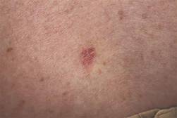
Thirteen (46%) of the 28 pediatric NMSC patients had a history of prolonged immunosuppression, radiation therapy, chemotherapy, voriconazole use, or a combination of these. Among these 28 patients, 19 were diagnosed with basal cell carcinoma (BCC), 7 were diagnosed with squamous cell carcinoma (SCC), and 2 were diagnosed with both BCC and SCC.
The authors noted significant delays in the initial diagnosis in both types of cancer; the mean number of days from time of lesion onset to diagnosis was 667 for SCC and 1,176 for BCC. When misdiagnosed, carcinomas were incorrectly identified as viral wart and graft-versus-host disease for SCC, and psoriasis, acrochordon, wart, nevus, and atypical nevus for BCC.
Even so, interventions were effective in the pediatric population, the authors noted.
“Although most of our patients developed subsequent NMSC after their initial diagnosis, the majority of cases were treated successfully with surgical excision, without recurrence or spread of disease. This suggests that interventions in children and young adults that involve prevention of subsequent disease may be most impactful,” they wrote.
The researchers had no conflicts to declare.
In a small sample study, 10 (36%) of 28 children and young adults diagnosed with nonmelanoma skin cancer were given a misdiagnosis initially, suggesting that young patients with NMSC risk factors may require heightened monitoring from health care providers, according to a study published in Journal of the American Academy of Dermatology (2015 doi: 10.1016/j.jaad.2015.08.007).
In their efforts to identify potential risk factors and gaps in care associated with NMSC in pediatric populations, lead author Hasan Khosravi of Harvard Medical School, Boston, and his associates examined records from 28 patients and 182 occurrences of NMSC, collected from Boston Children’s Hospital between 1993 and 2014.
Thirteen (46%) of the 28 pediatric NMSC patients had a history of prolonged immunosuppression, radiation therapy, chemotherapy, voriconazole use, or a combination of these. Among these 28 patients, 19 were diagnosed with basal cell carcinoma (BCC), 7 were diagnosed with squamous cell carcinoma (SCC), and 2 were diagnosed with both BCC and SCC.
The authors noted significant delays in the initial diagnosis in both types of cancer; the mean number of days from time of lesion onset to diagnosis was 667 for SCC and 1,176 for BCC. When misdiagnosed, carcinomas were incorrectly identified as viral wart and graft-versus-host disease for SCC, and psoriasis, acrochordon, wart, nevus, and atypical nevus for BCC.
Even so, interventions were effective in the pediatric population, the authors noted.
“Although most of our patients developed subsequent NMSC after their initial diagnosis, the majority of cases were treated successfully with surgical excision, without recurrence or spread of disease. This suggests that interventions in children and young adults that involve prevention of subsequent disease may be most impactful,” they wrote.
The researchers had no conflicts to declare.
FROM JOURNAL OF THE AMERICAN ACADEMY OF DERMATOLOGY
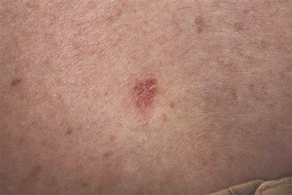
Bipolar patients, relatives slow to gauge facial emotions
Both bipolar disorder (BD) patients and their first-degree relatives were slower than were healthy controls in an emotion recognition task, suggesting facial recognition may be an endophenotype in bipolar disorder, according to a study published in Psychiatry Research.
Dr. Esther Vierck of the University of Otago in New Zealand and her associates compared 36 BD patients and 40 healthy control participants in a computerized facial emotion recognition task – 24 of the BD patient group’s first-degree relatives also were measured.
The researchers noted that bipolar patients were less accurate in recognizing emotional expressions than were controls, but did not find any evidence for emotion specificity within the BD or BD relative groups.
Read the article here: doi:10.1016/j.psychres.2015.08.033.
FROM PSYCHIATRY RESEARCH
David Henry's JCSO podcast, October 2015
Dr David Henry’s October podcast for The Journal of Community and Supportive Oncology, begins with a discussion of the recent approval of panobinostat for the treatment of relapsed and refractory multiple myeloma and of dinutuximab combination therapy as a first-line option for high-risk neuroblastoma in children. He also highlights two Review articles, one on cancer-related pain management, and another on current practice with endocrine therapy in metastatic breast cancer, both of which provide the reader with detailed, up-to-date overviews of current literature on the topics and clinical practice. Two Original Reports examine drugs costs and outcomes as they pertain to the practicing oncologist. The first article looks at the value of anticancer drugs in metastatic castrate-resistant prostate cancer; the second examines the implications of hospitalizations of 5 or more days on outcomes among patients with head and neck cancer who have received radiotherapy. Dr Henry rounds off his podcast with Case Reports on zoledronic acid-induced hypocalcemia in hyercalcemia of malignancy and neuroendocrine carcinaoma of the larynx with metastasis to the eyelid.
Click on the download icon at the top of this introduction to listen to the podcast.