User login
ADHD, Part 2: How important is a teacher’s assessment?
Dr. David Pickar and Dr. Lillian M. Beard address the importance of flagging ADHD in the classroom, addressing family resistance to treatment, and situations that might call for the involvement of other mental health colleagues.
“I find a lot of resistance [to the ADHD diagnosis], particularly among African American families,” says Dr. Beard, physician director, Children’s Pediatricians and Associates, Silver Spring, Md.
“An untreated ADHD kid is difficult for the teacher, but it is toughest on the kid,” says Dr. Pickar, adjunct professor of psychiatry at Johns Hopkins University, Baltimore. “Families may not hear it all, but a doctor pays a lot of attention to what the teacher is saying about that child in the classroom.”
The video associated with this article is no longer available on this site. Please view all of our videos on the MDedge YouTube channel

ADHD, Part 1: Is prevalence overstated in children?
Primary care physicians are on the front lines for diagnosing and prescribing to treat attention-deficit/hyperactivity disorder in children. In this video, Dr. David Pickar and Dr. Lillian M. Beard discuss what primary care physicians should know to feel confident when diagnosing and treating ADHD.
“[The first time] you see a patient with ADHD respond to stimulants, you see it is lifesaving … [the result] takes your breath away,” Dr. Pickar says.
The video associated with this article is no longer available on this site. Please view all of our videos on the MDedge YouTube channel

Collaborative Care, Part 2: What is 'enough' team care training?
Medical students are better prepared for practice when they learn to integrate mental health and team-based care in the academic setting.
“All schools of health professions’ education are moving in that direction,” says Dr. Lawrence “Bopper” Deyton, senior associate dean for public health at George Washington University, Washington. “Nobody has it perfect yet, but I think we’re seeing changes in that direction.”
In this installment of Mental Health Consult, Dr. Deyton and Dr. April Barbour, director of general internal medicine and the primary care residency program at George Washington University, Washington, discuss approaches to teaching integrated mental health care.
The video associated with this article is no longer available on this site. Please view all of our videos on the MDedge YouTube channel

Understanding the Deeds’ family tragedy
Last week, the Washington Post reported that Virginia state Sen. Creigh Deeds has filed a $6 million wrongful death lawsuit against the state of Virginia and the mental health agency that employed an evaluator who saw his son during a psychiatric crisis. In November 2013, Sen. Deeds’ son, Gus, had been brought to an emergency department for evaluation. He was judged to be both psychotic and dangerous, but a bed could not be located for him within the state-mandated 6-hour time frame, and Gus Deeds was released. Soon after, he repeatedly stabbed his father, then took his own life with a firearm.
My knowledge of the case is limited to what has been reported in the media, but as a psychiatrist practicing in Maryland where there are no time limits on locating beds, the story is unthinkable. Why would a very ill and very violent – and perhaps very treatable – young man be released from an emergency department because the gong went off in someone’s ill-conceived game of human “beat the clock?” While I have no inside knowledge of the Deeds’ family tragedy, I do want to talk about the Virginia laws that may have enabled a predictable calamity.

Most psychiatrists are familiar with the practice of psychiatric boarding, or holding a patient in the emergency department for a prolonged period of time because a bed cannot be located on a psychiatry inpatient unit. It’s not unusual for EDs to take many hours, or even several days, to transfer psychiatry patients to treatment units. In some places, it can take even longer. In Vermont, where the state hospital was demolished by a tropical storm, there were instances when patients remained in the ED for up to 6 weeks. On Native American reservations without hospital facilities, very ill psychiatric patients may be housed in jails, again for weeks, while an appropriate hospital bed is located off the reservation. Boarding is, by any measure, inhumane, and in 2014, the Washington state Supreme Court ruled it unlawful, though it did not say exactly where the patients were to be held pending bed availability.
With the civil rights of patients in mind, there is something to be said for mandating that beds be located for patients in a timely manner, and for the patient who is distressed, 6 hours may seem to be quite a long enough time. A drop-dead time, however, leaves no room for unforeseen issues, or even for the possibility that medication could be administered in the ED that might avert the need for an involuntary admission. A time limit of this type forces an immediate decision before a thoughtful assessment can be done and other interventions can be explored. In 2014, as one response to the Deeds’ family tragedy, the law was changed to lengthen the period to 8 hours.
In Virginia, the logistics of admitting a patient are more complex than in most states. Any citizen who is concerned about another person’s mental state and safety can apply to a magistrate for an emergency custody order. The patient is then transported by the sheriff, and the clock starts ticking on the 8-hour time frame. In the ED, the patient is seen by the emergency physician, but regardless of how obviously ill and dangerous the patient is, the physician has no authority to involuntarily admit a psychiatric patient. An outside agency – a local community service board (CSB) – is contacted, and an emergency services staff person is summoned to evaluate the patient, perform preadmission screening, and locate a bed.
Virginia is divided into 41 catchment areas, each served by a community service board. Each CSB is required to have crisis staff who assess patients for civil commitment. Virginia is a state with both urban and rural areas consisting of nearly 43,000 square miles. While the CSB prescreener drives in, performs the evaluation, and searches for an available bed, the clock continues to tick.
The qualifications to be a screener are not precise. The website in the county where Gus Deeds lived advertises: “QUALIFICATIONS: Master’s Degree in human services related field, mental health and/or substance abuse experience; or equivalent combination of education and experience including BSN RN’s with Psychiatric Nursing experience in outpatient or inpatient settings. Extensive knowledge of the DSM, mental health and substance abuse assessments, treatment, crisis assessment and intervention strategies for clients of all ages; must be certified or eligible to be certified as a prescreener by the CSB and Virginia DBHDS” (Department of Behavioral Health & Developmental Services). The prescreener has the authority to override the ED physician and release a patient the physician believes should be admitted.
It’s not uncommon to hear people compare the treatment of an acute myocardial infarction to the treatment of an agitated psychosis, especially when the topic of parity is raised. Could one imagine a scenario where an ED physician could not admit a patient with chest pain without first waiting hours for an outside consultant to come – a consultant who was not even a physician? Presumably, this is not an issue meant to defy parity but to prevent financial conflicts of interest in situations where patient civil rights are a sensitive concern.
Given all the obstacles, what’s surprising is that the vast majority of the time, if it’s determined that a patient needs involuntary admission, a temporary detention order (TDO) is issued and a bed is located. A study done in 2013 looked at the outcome of TDOs issued in a 3-month period. Of an estimated 5,000 TDOs, beds were located for 98.5% of the patients. Still, 72 patients who had been found to be in need of admission were released because beds could not be found before the emergency custody order expired. The practice of releasing patients in crisis has been assigned its own jargon: “streeting.” It’s not quite that simple, however: Not everyone was released as the gong struck. In 273 instances, patients were admitted after the TDO expired, with an average time to admission of just over 16 hours.
A major issue is that Virginia, like nearly all states, has a shortage of psychiatric beds. In Virginia, the problems may be unnecessarily complex with the added requirement to pull in an outside agency and not leave admission decisions to ED clinicians, coupled with a short time limit for transport, treatment, and admission of a complex, sometimes dangerous patient population. According to the Post article, the Deeds suit is particularly troublesome, because beds were available: The CSB prescreener did not call every available hospital, and he attempted to contact one nearby hospital by fax. The fax number he had was wrong, so the request for the bed was never received by the facility. But the facility did, in fact, have a bed. What hasn’t been legislated is how fast a screener needs to drive to the hospital, or how fast he or she is expected to dial for beds, or what happens if he gets a flat tire or if the responses don’t come fast enough.
Tragedy often leads to change, and laws in Virginia changed after the Virginia Tech shootings, and again after the Deeds’ family tragedy. Legislative gaps remain, however, and bed shortages, time limits, and the requirement for an outside agency assessment continue to leave room for the possibility of more tragedy. Clearly, there is more to be done.
With thanks to Dr. Anita Everett for her consultation.
Dr. Miller is coauthor of the forthcoming book, “Committed: The Battle Over Involuntary Psychiatric Care.”
Last week, the Washington Post reported that Virginia state Sen. Creigh Deeds has filed a $6 million wrongful death lawsuit against the state of Virginia and the mental health agency that employed an evaluator who saw his son during a psychiatric crisis. In November 2013, Sen. Deeds’ son, Gus, had been brought to an emergency department for evaluation. He was judged to be both psychotic and dangerous, but a bed could not be located for him within the state-mandated 6-hour time frame, and Gus Deeds was released. Soon after, he repeatedly stabbed his father, then took his own life with a firearm.
My knowledge of the case is limited to what has been reported in the media, but as a psychiatrist practicing in Maryland where there are no time limits on locating beds, the story is unthinkable. Why would a very ill and very violent – and perhaps very treatable – young man be released from an emergency department because the gong went off in someone’s ill-conceived game of human “beat the clock?” While I have no inside knowledge of the Deeds’ family tragedy, I do want to talk about the Virginia laws that may have enabled a predictable calamity.
Most psychiatrists are familiar with the practice of psychiatric boarding, or holding a patient in the emergency department for a prolonged period of time because a bed cannot be located on a psychiatry inpatient unit. It’s not unusual for EDs to take many hours, or even several days, to transfer psychiatry patients to treatment units. In some places, it can take even longer. In Vermont, where the state hospital was demolished by a tropical storm, there were instances when patients remained in the ED for up to 6 weeks. On Native American reservations without hospital facilities, very ill psychiatric patients may be housed in jails, again for weeks, while an appropriate hospital bed is located off the reservation. Boarding is, by any measure, inhumane, and in 2014, the Washington state Supreme Court ruled it unlawful, though it did not say exactly where the patients were to be held pending bed availability.
With the civil rights of patients in mind, there is something to be said for mandating that beds be located for patients in a timely manner, and for the patient who is distressed, 6 hours may seem to be quite a long enough time. A drop-dead time, however, leaves no room for unforeseen issues, or even for the possibility that medication could be administered in the ED that might avert the need for an involuntary admission. A time limit of this type forces an immediate decision before a thoughtful assessment can be done and other interventions can be explored. In 2014, as one response to the Deeds’ family tragedy, the law was changed to lengthen the period to 8 hours.
In Virginia, the logistics of admitting a patient are more complex than in most states. Any citizen who is concerned about another person’s mental state and safety can apply to a magistrate for an emergency custody order. The patient is then transported by the sheriff, and the clock starts ticking on the 8-hour time frame. In the ED, the patient is seen by the emergency physician, but regardless of how obviously ill and dangerous the patient is, the physician has no authority to involuntarily admit a psychiatric patient. An outside agency – a local community service board (CSB) – is contacted, and an emergency services staff person is summoned to evaluate the patient, perform preadmission screening, and locate a bed.
Virginia is divided into 41 catchment areas, each served by a community service board. Each CSB is required to have crisis staff who assess patients for civil commitment. Virginia is a state with both urban and rural areas consisting of nearly 43,000 square miles. While the CSB prescreener drives in, performs the evaluation, and searches for an available bed, the clock continues to tick.
The qualifications to be a screener are not precise. The website in the county where Gus Deeds lived advertises: “QUALIFICATIONS: Master’s Degree in human services related field, mental health and/or substance abuse experience; or equivalent combination of education and experience including BSN RN’s with Psychiatric Nursing experience in outpatient or inpatient settings. Extensive knowledge of the DSM, mental health and substance abuse assessments, treatment, crisis assessment and intervention strategies for clients of all ages; must be certified or eligible to be certified as a prescreener by the CSB and Virginia DBHDS” (Department of Behavioral Health & Developmental Services). The prescreener has the authority to override the ED physician and release a patient the physician believes should be admitted.
It’s not uncommon to hear people compare the treatment of an acute myocardial infarction to the treatment of an agitated psychosis, especially when the topic of parity is raised. Could one imagine a scenario where an ED physician could not admit a patient with chest pain without first waiting hours for an outside consultant to come – a consultant who was not even a physician? Presumably, this is not an issue meant to defy parity but to prevent financial conflicts of interest in situations where patient civil rights are a sensitive concern.
Given all the obstacles, what’s surprising is that the vast majority of the time, if it’s determined that a patient needs involuntary admission, a temporary detention order (TDO) is issued and a bed is located. A study done in 2013 looked at the outcome of TDOs issued in a 3-month period. Of an estimated 5,000 TDOs, beds were located for 98.5% of the patients. Still, 72 patients who had been found to be in need of admission were released because beds could not be found before the emergency custody order expired. The practice of releasing patients in crisis has been assigned its own jargon: “streeting.” It’s not quite that simple, however: Not everyone was released as the gong struck. In 273 instances, patients were admitted after the TDO expired, with an average time to admission of just over 16 hours.
A major issue is that Virginia, like nearly all states, has a shortage of psychiatric beds. In Virginia, the problems may be unnecessarily complex with the added requirement to pull in an outside agency and not leave admission decisions to ED clinicians, coupled with a short time limit for transport, treatment, and admission of a complex, sometimes dangerous patient population. According to the Post article, the Deeds suit is particularly troublesome, because beds were available: The CSB prescreener did not call every available hospital, and he attempted to contact one nearby hospital by fax. The fax number he had was wrong, so the request for the bed was never received by the facility. But the facility did, in fact, have a bed. What hasn’t been legislated is how fast a screener needs to drive to the hospital, or how fast he or she is expected to dial for beds, or what happens if he gets a flat tire or if the responses don’t come fast enough.
Tragedy often leads to change, and laws in Virginia changed after the Virginia Tech shootings, and again after the Deeds’ family tragedy. Legislative gaps remain, however, and bed shortages, time limits, and the requirement for an outside agency assessment continue to leave room for the possibility of more tragedy. Clearly, there is more to be done.
With thanks to Dr. Anita Everett for her consultation.
Dr. Miller is coauthor of the forthcoming book, “Committed: The Battle Over Involuntary Psychiatric Care.”

Collaborative Care, Part 1: The move toward collaborative care
Neil Kirschner, Ph.D., senior associate for health policy and regulatory affairs for the American College of Physicians, explains why the solo practitioner model of health care needs to give way to collaborative care. “Care is turning into a team-based approach [that] allows you to have staff to provide effective integration of care, but to have a team, you need funding,” he says. “One-third of our health care spending is on ‘wasted care’ … [and that money] could be leveraged toward providing effective care.”
The video associated with this article is no longer available on this site. Please view all of our videos on the MDedge YouTube channel

Poorer survival of African Americans with kidney cancer may be due to genomic differences
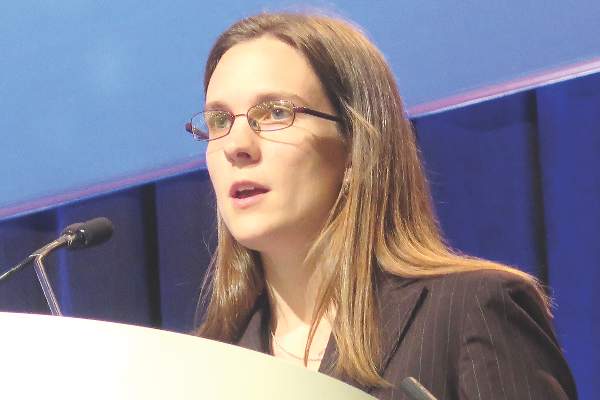
SAN FRANCISCO – African American patients with clear-cell renal cell carcinoma may have poorer survival in part because of genomic factors that render tumors more aggressive and less sensitive to anti-angiogenic therapy, suggests a study reported at the Genitourinary Cancers Symposium.

Genomic analysis in 438 patients with metastatic disease found that African Americans were about half as likely as Caucasians to have mutations of the von Hippel–Lindau (VHL) tumor suppressor gene, reported lead investigator Dr. Tracy Lynn Rose, a hematology-oncology fellow at the University of North Carolina at Chapel Hill and Lineberger Comprehensive Cancer Center. Mutational inactivation of this gene leads to increased signaling in the vascular endothelial growth factor (VEGF) pathway.
African Americans also were more likely to have the clear cell B molecular subtype and had less up-regulation of pathways associated with hypoxia-inducible factor (HIF), which collectively suggest a less angiogenic profile and activation of non-VEGF pathways.
A companion analysis of 35,152 patients treated during a 14-year period found that African American patients with metastatic renal cell carcinoma still had a higher risk of death than white peers in the contemporary era, after introduction of multiple agents that target the VEGF pathway and similar increases in receipt of systemic therapy.
“Our findings indicate clear differences in the biology of clear-cell renal cell carcinoma between African Americans and Caucasians. These differences could suggest a larger proportion of tumors from African Americans have a HIF- and VEGF-independent propensity for aggressiveness, and they also suggest perhaps increased resistance of African Americans to VEGF-targeted therapy,” Dr. Rose said at the symposium, sponsored by the American Society of Clinical Oncology, ASTRO, and the Society of Urologic Oncology.
“Overall, our data lend support to a role for tumor biology in the survival disparity observed between African American and Caucasian patients,” she said.

The study may be a step toward precision medicine, whereby race is used to guide treatment decisions, according to invited discussant Dr. Guru Sonpavde, director of the Genitourinary Malignancy Program at the University of Alabama at Birmingham. “That’s an exciting possibility,” he said.
“It is plausible that African American patients are less VEGF driven, less responsive to VEGF inhibitors, but I think the results right now are not ready for use in the clinic,” he further commented. “We need validation in a larger number of African American patients. But even more importantly, we need to focus more on molecular measures of VEGF-driven tumors, to select patients for VEGF inhibitors since we don’t have any biomarkers of this sort in the clinic today.”
“Finally, somatic differences may be driven by differences in the germline, and we may want to focus on integrating the germline and somatic alterations into a molecular panel which might be even better at predicting benefit from specific agents,” he concluded.
Previous studies have found a small but consistent elevation of the risk of death for African American patients with renal cell carcinoma, Dr. Rose noted when introducing the study. Initial hypotheses were that this disparity might be due to differences in comorbidities and use of nephrectomy.
She and her colleagues performed genomic analyses in a discovery cohort of 438 African American and Caucasian patients with metastatic clear-cell renal cell carcinoma from The Cancer Genome Atlas database.
Results indicated that African Americans in this discovery cohort were less likely to have a VHL mutation (17% vs. 50%, P = .036), a finding that was confirmed in a validation cohort of 135 similar patients (40% vs. 81%, P = .008).
African American patients were less likely to have several VEGF and HIF signatures relative to Caucasian counterparts. On the other hand, they were more likely to have the clear cell B molecular subtype (79% vs. 45%, P less than .01), which has been associated with decreased activation of angiogenic pathways and poorer prognosis.
The investigators next analyzed data from the National Cancer Database, assessing survival among 35,152 patients who received a diagnosis of metastatic clear-cell renal cell carcinoma between 1998 and 2011.
The proportion receiving systemic therapy over time was similar for African American and Caucasian patients, with both seeing a rise in 2006, corresponding to the introduction of VEGF-targeted therapies. The poorer median survival for African American versus Caucasian patients seen during 1998-2004 (6.0 vs. 7.6 months; adjusted hazard ratio, 1.07; P less than .01) was still evident in 2006-2011 (6.5 vs. 9.2 months; adjusted hazard ratio, 1.08; P less than .01).
SAN FRANCISCO – African American patients with clear-cell renal cell carcinoma may have poorer survival in part because of genomic factors that render tumors more aggressive and less sensitive to anti-angiogenic therapy, suggests a study reported at the Genitourinary Cancers Symposium.
Genomic analysis in 438 patients with metastatic disease found that African Americans were about half as likely as Caucasians to have mutations of the von Hippel–Lindau (VHL) tumor suppressor gene, reported lead investigator Dr. Tracy Lynn Rose, a hematology-oncology fellow at the University of North Carolina at Chapel Hill and Lineberger Comprehensive Cancer Center. Mutational inactivation of this gene leads to increased signaling in the vascular endothelial growth factor (VEGF) pathway.
African Americans also were more likely to have the clear cell B molecular subtype and had less up-regulation of pathways associated with hypoxia-inducible factor (HIF), which collectively suggest a less angiogenic profile and activation of non-VEGF pathways.
A companion analysis of 35,152 patients treated during a 14-year period found that African American patients with metastatic renal cell carcinoma still had a higher risk of death than white peers in the contemporary era, after introduction of multiple agents that target the VEGF pathway and similar increases in receipt of systemic therapy.
“Our findings indicate clear differences in the biology of clear-cell renal cell carcinoma between African Americans and Caucasians. These differences could suggest a larger proportion of tumors from African Americans have a HIF- and VEGF-independent propensity for aggressiveness, and they also suggest perhaps increased resistance of African Americans to VEGF-targeted therapy,” Dr. Rose said at the symposium, sponsored by the American Society of Clinical Oncology, ASTRO, and the Society of Urologic Oncology.
“Overall, our data lend support to a role for tumor biology in the survival disparity observed between African American and Caucasian patients,” she said.
The study may be a step toward precision medicine, whereby race is used to guide treatment decisions, according to invited discussant Dr. Guru Sonpavde, director of the Genitourinary Malignancy Program at the University of Alabama at Birmingham. “That’s an exciting possibility,” he said.
“It is plausible that African American patients are less VEGF driven, less responsive to VEGF inhibitors, but I think the results right now are not ready for use in the clinic,” he further commented. “We need validation in a larger number of African American patients. But even more importantly, we need to focus more on molecular measures of VEGF-driven tumors, to select patients for VEGF inhibitors since we don’t have any biomarkers of this sort in the clinic today.”
“Finally, somatic differences may be driven by differences in the germline, and we may want to focus on integrating the germline and somatic alterations into a molecular panel which might be even better at predicting benefit from specific agents,” he concluded.
Previous studies have found a small but consistent elevation of the risk of death for African American patients with renal cell carcinoma, Dr. Rose noted when introducing the study. Initial hypotheses were that this disparity might be due to differences in comorbidities and use of nephrectomy.
She and her colleagues performed genomic analyses in a discovery cohort of 438 African American and Caucasian patients with metastatic clear-cell renal cell carcinoma from The Cancer Genome Atlas database.
Results indicated that African Americans in this discovery cohort were less likely to have a VHL mutation (17% vs. 50%, P = .036), a finding that was confirmed in a validation cohort of 135 similar patients (40% vs. 81%, P = .008).
African American patients were less likely to have several VEGF and HIF signatures relative to Caucasian counterparts. On the other hand, they were more likely to have the clear cell B molecular subtype (79% vs. 45%, P less than .01), which has been associated with decreased activation of angiogenic pathways and poorer prognosis.
The investigators next analyzed data from the National Cancer Database, assessing survival among 35,152 patients who received a diagnosis of metastatic clear-cell renal cell carcinoma between 1998 and 2011.
The proportion receiving systemic therapy over time was similar for African American and Caucasian patients, with both seeing a rise in 2006, corresponding to the introduction of VEGF-targeted therapies. The poorer median survival for African American versus Caucasian patients seen during 1998-2004 (6.0 vs. 7.6 months; adjusted hazard ratio, 1.07; P less than .01) was still evident in 2006-2011 (6.5 vs. 9.2 months; adjusted hazard ratio, 1.08; P less than .01).
AT THE GENITOURINARY CANCERS SYMPOSIUM
Key clinical point: The poorer survival of African American patients vs. Caucasian patients with clear-cell renal cell carcinoma may be due in part to genomic factors.
Major finding: Compared with Caucasian patients, African American patients were less likely to have mutations of the VHL gene (17% vs. 50%) and more likely to have the clear cell B molecular subtype (79% vs. 45%).
Data source: Analyses of a genomic cohort of 438 patients with metastatic clear-cell renal cell carcinoma from The Cancer Genome Atlas database and a survival cohort of 35,152 from the National Cancer Database.
Disclosures: Dr. Rose disclosed that she had no relevant conflicts of interest. Dr. Sonpavde disclosed that he has a consulting or advisory role with Bayer, Genentech, Merck, Novartis, Pfizer, and Sanofi, and that his institution receives research funding from Bayer, Boehringer Ingelheim, and Onyx.
NSAIDs for UTI
Urinary tract infections (UTIs) unfortunately present an abundant opportunity for us to reflexively and mindlessly contribute to filling up the globe with multidrug resistant bacteria. Many of us have patients with recurrent UTIs, who, despite our best efforts at trying to reduce recurrence through non–medication approaches, frequently call for treatment. We stand by helplessly as we watch the resistance of these organisms increase.
Is there another way?

Investigators in Germany evaluated the benefit and harms of prescribing ibuprofen in place of an antibiotic in women with symptoms of a UTI and no risk factors (BMC Med. 2010 May 26;8:30). Women aged 18-65 years were randomized to a single dose of the broad-spectrum antibiotic fosfomycin 3 g (n = 246) or ibuprofen three 400-mg doses (n = 248) for 3 days. Patients were excluded if they had any signs of upper urinary tract infection, pregnancy, urinary catheterization, gastrointestinal ulcers, or chronic conditions. Antibiotics were prescribed for persistent, worsening, or recurrent symptoms.
Women in the ibuprofen group received significantly fewer antibiotic prescriptions (283 in the fosfomycin group and 94 in the ibuprofen group; incidence rate reduction of 66.5%; 95% confidence interval, 58.8%-74.4%; P less than .001). The ibuprofen group had more symptoms that lasted a day longer. On day 4, 56% of women in the fosfomycin vs. 39% of participants in the ibuprofen group were symptom free, which increased to 82% and 70% by day 7. The ibuprofen group were more likely to develop pyelonephritis (five cases in the ibuprofen group and one in the fosfomycin group; P = .12), but all women were treated and recovered fully. Four events led to “hospital referrals,” only one of which was related to trial drug (gastrointestinal hemorrhage).
In summary, two-thirds of women in the ibuprofen group recovered without antibiotic treatment and one-third received antibiotics for persistent or worsening symptoms. The authors concluded that ibuprofen was inferior to fosfomycin for initial symptomatic treatment. The nonstatistically higher rate of upper urinary tract infection with ibuprofen may make some clinicians nervous.
However, perhaps this is worth exploring for select patients with mild symptoms. The investigators mention data suggesting women may be aware of disadvantages of antibiotics and may be open to the idea of delaying or avoiding treatment, which opens the door to informed discussions. I am planning to discuss it with my patient who has frequent UTIs to see if we can delay intravenous antibiotics for UTI. Intravenous antibiotics for UTI make me uncomfortable for a variety of reasons.
Dr. Ebbert is professor of medicine, a general internist at the Mayo Clinic in Rochester, Minn., and a diplomate of the American Board of Addiction Medicine. The opinions expressed are those of the author and do not necessarily represent the views and opinions of the Mayo Clinic. The opinions expressed in this article should not be used to diagnose or treat any medical condition nor should they be used as a substitute for medical advice from a qualified, board-certified practicing clinician. Dr. Ebbert has no relevant financial disclosures about this article.
Urinary tract infections (UTIs) unfortunately present an abundant opportunity for us to reflexively and mindlessly contribute to filling up the globe with multidrug resistant bacteria. Many of us have patients with recurrent UTIs, who, despite our best efforts at trying to reduce recurrence through non–medication approaches, frequently call for treatment. We stand by helplessly as we watch the resistance of these organisms increase.
Is there another way?
Investigators in Germany evaluated the benefit and harms of prescribing ibuprofen in place of an antibiotic in women with symptoms of a UTI and no risk factors (BMC Med. 2010 May 26;8:30). Women aged 18-65 years were randomized to a single dose of the broad-spectrum antibiotic fosfomycin 3 g (n = 246) or ibuprofen three 400-mg doses (n = 248) for 3 days. Patients were excluded if they had any signs of upper urinary tract infection, pregnancy, urinary catheterization, gastrointestinal ulcers, or chronic conditions. Antibiotics were prescribed for persistent, worsening, or recurrent symptoms.
Women in the ibuprofen group received significantly fewer antibiotic prescriptions (283 in the fosfomycin group and 94 in the ibuprofen group; incidence rate reduction of 66.5%; 95% confidence interval, 58.8%-74.4%; P less than .001). The ibuprofen group had more symptoms that lasted a day longer. On day 4, 56% of women in the fosfomycin vs. 39% of participants in the ibuprofen group were symptom free, which increased to 82% and 70% by day 7. The ibuprofen group were more likely to develop pyelonephritis (five cases in the ibuprofen group and one in the fosfomycin group; P = .12), but all women were treated and recovered fully. Four events led to “hospital referrals,” only one of which was related to trial drug (gastrointestinal hemorrhage).
In summary, two-thirds of women in the ibuprofen group recovered without antibiotic treatment and one-third received antibiotics for persistent or worsening symptoms. The authors concluded that ibuprofen was inferior to fosfomycin for initial symptomatic treatment. The nonstatistically higher rate of upper urinary tract infection with ibuprofen may make some clinicians nervous.
However, perhaps this is worth exploring for select patients with mild symptoms. The investigators mention data suggesting women may be aware of disadvantages of antibiotics and may be open to the idea of delaying or avoiding treatment, which opens the door to informed discussions. I am planning to discuss it with my patient who has frequent UTIs to see if we can delay intravenous antibiotics for UTI. Intravenous antibiotics for UTI make me uncomfortable for a variety of reasons.
Dr. Ebbert is professor of medicine, a general internist at the Mayo Clinic in Rochester, Minn., and a diplomate of the American Board of Addiction Medicine. The opinions expressed are those of the author and do not necessarily represent the views and opinions of the Mayo Clinic. The opinions expressed in this article should not be used to diagnose or treat any medical condition nor should they be used as a substitute for medical advice from a qualified, board-certified practicing clinician. Dr. Ebbert has no relevant financial disclosures about this article.

Bert B. Vargas, MD

AHA: Poor real-world adherence to NOACs
ORLANDO – Adherence to the novel oral anticoagulants (NOACs) is surprisingly poor in clinical practice, Xiaoxi Yao, Ph.D., reported at the American Heart Association scientific sessions.
Her retrospective study of nearly 65,000 patients with atrial fibrillation who initiated therapy with apixaban, dabigatran, rivaroxaban, or warfarin showed that during a median 1.1 years of follow-up fewer than half of all patients were treatment adherent, with adherence defined as possession of sufficient medication to cover at least 80% of days.

Adherence rates, while uniformly suboptimal, nevertheless varied considerably: lowest at 38.5% for dabigatran, followed by 40.2% for warfarin, 50.5% for rivaroxaban, and 61.9% for apixaban.
This poor adherence to NOACs in real-world clinical practice is surprising in light of the drugs’ greater convenience, with fewer drug interactions than warfarin and no need for laboratory monitoring, observed Dr. Yao of the Mayo Clinic in Rochester, Minn.
It’s possible, although speculative, that the NOACs’ greater convenience paradoxically contributes to the low adherence rates, since unlike warfarin, NOACs don’t require regular interactions with the health care system for INR monitoring. And then there is the hefty cost of the novel agents, she added.
The study population consisted of 3,900 patients with atrial fibrillation who initiated oral anticoagulation with apixaban (Eliquis), 10,235 who started on dabigatran (Pradaxa), 12,366 on rivaroxaban (Xarelto), and 38,190 on warfarin. The analysis utilized claims data from a large U.S. commercial insurance database.
Adherence rates were better among patients with greater stroke risk as reflected by their CHA2DS2-VASc scores. For example, at the high end of the adherence spectrum, the adherence rate for apixaban was 50% in patients with a CHA2DS2-VASc score of 0-1, rising to 62% with a score of 2-3 and 64% with a score of 4 or more. The corresponding adherence rates for dabigatran were 25% in patients with a CHA2DS2-VASc of 0-1, 40% among those with a score of 2-3, and 42% in patients with a score of 4 or higher.
Dr. Yao and coinvestigators were interested in whether lower adherence to oral anticoagulation was associated with worse outcomes. This proved to be the case with regard to stroke rate for patients with a CHA2DS2-VASc score of 2 or more, where a clear dose-response relationship was evident between the event rate and cumulative time off oral anticoagulation during follow-up.
Among patients with a CHA2DS2-VASc of 2 or 3, the stroke rate was nearly twice as high among those off oral anticoagulation for a total of 3-6 months and three times greater if off therapy for more than 6 months than in those with a total time off of less than 1 week. The stroke rate was even higher in patients with a CHA2DS2-VASc of 4 or more who had suboptimal adherence.
An unexpected finding, she continued, was that among patients with a CHA2DS2-VASc score of 2 or more there was no significant relationship between cumulative time off oral anticoagulation and the risk of major bleeding unless they were off treatment for a total of 6 months or more; only then was the major bleeding risk lower than in patients whose total time off therapy was less than a week. Also, one would expect that when patients are off oral anticoagulation they should be at significantly lower risk of intracranial hemorrhage than when on-therapy, but this proved not to be the case.
For patients at substantial stroke risk as indicated by a CHA2DS2-VASc score of at least 2, this finding about off-treatment bleeding risk actually constitutes a good argument for sticking to their medication, in Dr. Yao’s view.
“Physicians and patients often fear bleeding, especially intracranial hemorrhage, but we found that for patients at higher risk for stroke there is little difference in intracranial hemorrhage risk whether you’re on or off of oral anticoagulation. So higher-risk patients should definitely adhere to their medication because of the stroke prevention benefit. However, in low-risk patients with a CHA2DS2-VASc of 0-1, the benefits of oral anticoagulation may not always outweigh the harm,” she said.
Dr. Yao reported having no financial conflicts of interest regarding her study.
ORLANDO – Adherence to the novel oral anticoagulants (NOACs) is surprisingly poor in clinical practice, Xiaoxi Yao, Ph.D., reported at the American Heart Association scientific sessions.
Her retrospective study of nearly 65,000 patients with atrial fibrillation who initiated therapy with apixaban, dabigatran, rivaroxaban, or warfarin showed that during a median 1.1 years of follow-up fewer than half of all patients were treatment adherent, with adherence defined as possession of sufficient medication to cover at least 80% of days.
Adherence rates, while uniformly suboptimal, nevertheless varied considerably: lowest at 38.5% for dabigatran, followed by 40.2% for warfarin, 50.5% for rivaroxaban, and 61.9% for apixaban.
This poor adherence to NOACs in real-world clinical practice is surprising in light of the drugs’ greater convenience, with fewer drug interactions than warfarin and no need for laboratory monitoring, observed Dr. Yao of the Mayo Clinic in Rochester, Minn.
It’s possible, although speculative, that the NOACs’ greater convenience paradoxically contributes to the low adherence rates, since unlike warfarin, NOACs don’t require regular interactions with the health care system for INR monitoring. And then there is the hefty cost of the novel agents, she added.
The study population consisted of 3,900 patients with atrial fibrillation who initiated oral anticoagulation with apixaban (Eliquis), 10,235 who started on dabigatran (Pradaxa), 12,366 on rivaroxaban (Xarelto), and 38,190 on warfarin. The analysis utilized claims data from a large U.S. commercial insurance database.
Adherence rates were better among patients with greater stroke risk as reflected by their CHA2DS2-VASc scores. For example, at the high end of the adherence spectrum, the adherence rate for apixaban was 50% in patients with a CHA2DS2-VASc score of 0-1, rising to 62% with a score of 2-3 and 64% with a score of 4 or more. The corresponding adherence rates for dabigatran were 25% in patients with a CHA2DS2-VASc of 0-1, 40% among those with a score of 2-3, and 42% in patients with a score of 4 or higher.
Dr. Yao and coinvestigators were interested in whether lower adherence to oral anticoagulation was associated with worse outcomes. This proved to be the case with regard to stroke rate for patients with a CHA2DS2-VASc score of 2 or more, where a clear dose-response relationship was evident between the event rate and cumulative time off oral anticoagulation during follow-up.
Among patients with a CHA2DS2-VASc of 2 or 3, the stroke rate was nearly twice as high among those off oral anticoagulation for a total of 3-6 months and three times greater if off therapy for more than 6 months than in those with a total time off of less than 1 week. The stroke rate was even higher in patients with a CHA2DS2-VASc of 4 or more who had suboptimal adherence.
An unexpected finding, she continued, was that among patients with a CHA2DS2-VASc score of 2 or more there was no significant relationship between cumulative time off oral anticoagulation and the risk of major bleeding unless they were off treatment for a total of 6 months or more; only then was the major bleeding risk lower than in patients whose total time off therapy was less than a week. Also, one would expect that when patients are off oral anticoagulation they should be at significantly lower risk of intracranial hemorrhage than when on-therapy, but this proved not to be the case.
For patients at substantial stroke risk as indicated by a CHA2DS2-VASc score of at least 2, this finding about off-treatment bleeding risk actually constitutes a good argument for sticking to their medication, in Dr. Yao’s view.
“Physicians and patients often fear bleeding, especially intracranial hemorrhage, but we found that for patients at higher risk for stroke there is little difference in intracranial hemorrhage risk whether you’re on or off of oral anticoagulation. So higher-risk patients should definitely adhere to their medication because of the stroke prevention benefit. However, in low-risk patients with a CHA2DS2-VASc of 0-1, the benefits of oral anticoagulation may not always outweigh the harm,” she said.
Dr. Yao reported having no financial conflicts of interest regarding her study.
AT THE AHA SCIENTIFIC SESSIONS
Key clinical point: Adherence to the novel oral anticoagulants by patients with atrial fibrillation is surprisingly poor outside the clinical trial setting.
Major finding: More than half of patients with atrial fibrillation who started on a novel oral anticoagulant were medication adherent less than 80% of the time.
Data source: This was a retrospective study of nearly 65,000 patients with atrial fibrillation who initiated oral anticoagulant therapy and were then followed for a median of 1.1 years.
Disclosures: The presenter reported having no financial conflicts of interest regarding the study.,

Beta-Blockers May Increase Risk of Perioperative MACEs in Patients with Uncomplicated Hypertension
Clinical question: Does taking a perioperative beta-blocker increase the risk of major adverse cardiovascular events (MACEs) and all-cause mortality in low-risk patients with essential hypertension (HTN)?

Background: Guidelines for the use of perioperative beta-blockers are being reevaluated due to concerns about validity of prior studies that supported the use of perioperative beta-blockers. This study sought to evaluate effectiveness and safety of beta-blockers in patients with uncomplicated HTN.
Study design: Observational cohort study.
Setting: Denmark.
Synopsis: This study included 55,320 hypertensive patients using at least two antihypertensive drugs who underwent non-cardiac surgery. Of these, 14,644 patients were treated with a beta-blocker. Patients with secondary cardiovascular conditions, renal disease, or liver disease were excluded; 30-day MACEs and all-cause mortality were analyzed.
In patients treated with a beta-blocker, the incidence of 30-day MACEs was 1.32% compared with 0.84% in the non-beta-blockers group; 30-day mortality in those treated with beta-blocker was 1.9% compared with 1.3% in the non-beta-blocker group. Risk of beta-blocker-associated MACEs was higher in patients 70 and older. Causality cannot be concluded based on observational data.
Bottom line: In patients with uncomplicated HTN, treatment with a beta-blocker may be associated with increased 30-day risk of perioperative MACEs after non-cardiac surgery.
Citation: Jorgensen ME, Hlatky MA, Kober L, et al. Beta-blocker-associated risks in patients with uncomplicated hypertension undergoing noncardiac surgery. JAMA Intern Med. 2015;175(12):1923-1931.
Clinical question: Does taking a perioperative beta-blocker increase the risk of major adverse cardiovascular events (MACEs) and all-cause mortality in low-risk patients with essential hypertension (HTN)?
Background: Guidelines for the use of perioperative beta-blockers are being reevaluated due to concerns about validity of prior studies that supported the use of perioperative beta-blockers. This study sought to evaluate effectiveness and safety of beta-blockers in patients with uncomplicated HTN.
Study design: Observational cohort study.
Setting: Denmark.
Synopsis: This study included 55,320 hypertensive patients using at least two antihypertensive drugs who underwent non-cardiac surgery. Of these, 14,644 patients were treated with a beta-blocker. Patients with secondary cardiovascular conditions, renal disease, or liver disease were excluded; 30-day MACEs and all-cause mortality were analyzed.
In patients treated with a beta-blocker, the incidence of 30-day MACEs was 1.32% compared with 0.84% in the non-beta-blockers group; 30-day mortality in those treated with beta-blocker was 1.9% compared with 1.3% in the non-beta-blocker group. Risk of beta-blocker-associated MACEs was higher in patients 70 and older. Causality cannot be concluded based on observational data.
Bottom line: In patients with uncomplicated HTN, treatment with a beta-blocker may be associated with increased 30-day risk of perioperative MACEs after non-cardiac surgery.
Citation: Jorgensen ME, Hlatky MA, Kober L, et al. Beta-blocker-associated risks in patients with uncomplicated hypertension undergoing noncardiac surgery. JAMA Intern Med. 2015;175(12):1923-1931.
