User login
VIDEO: EVLP may extend lung preservation, quality for transplants
BALTIMORE – The use of ex vivo lung perfusion (EVLP) may allow for the safe transplantation of lungs preserved for more than 12 hours, according to a study presented at the annual meeting of the American Association for Thoracic Surgery.
A research team at the University of Toronto evaluated the outcomes of transplant patients who received a lung with a preservation time of over 12 hours between January 2006 and April 2015 and compared them to the general lung transplant population. Median hospital and ICU length of stay were similar between the two groups, and Kaplan-Meier survival curves between the two groups did not show any difference. Preservation time, donor PO2, and use of EVLP were not significant variables affecting survival.
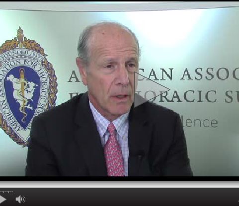
Dr. Bartley P. Griffith, chief of cardiac surgery at the University of Maryland, Baltimore, and a discussant on the paper at the meeting, said that the findings of the study open up the possibility of a more “planned” approach to transplantation.
“Anything that not only extends preservation time, but perhaps even improves quality of preservation, would be a godsend,” Dr. Griffith said in a video interview. He cautioned that the “devil is in the details,” and that the data had to be examined closely. Nevertheless, Dr. Griffith said transplant surgeons should be grateful for the important work done by the University of Toronto team.
Dr. Griffith reported no relevant financial disclosures.
The video associated with this article is no longer available on this site. Please view all of our videos on the MDedge YouTube channel
On Twitter @richpizzi
BALTIMORE – The use of ex vivo lung perfusion (EVLP) may allow for the safe transplantation of lungs preserved for more than 12 hours, according to a study presented at the annual meeting of the American Association for Thoracic Surgery.
A research team at the University of Toronto evaluated the outcomes of transplant patients who received a lung with a preservation time of over 12 hours between January 2006 and April 2015 and compared them to the general lung transplant population. Median hospital and ICU length of stay were similar between the two groups, and Kaplan-Meier survival curves between the two groups did not show any difference. Preservation time, donor PO2, and use of EVLP were not significant variables affecting survival.
Dr. Bartley P. Griffith, chief of cardiac surgery at the University of Maryland, Baltimore, and a discussant on the paper at the meeting, said that the findings of the study open up the possibility of a more “planned” approach to transplantation.
“Anything that not only extends preservation time, but perhaps even improves quality of preservation, would be a godsend,” Dr. Griffith said in a video interview. He cautioned that the “devil is in the details,” and that the data had to be examined closely. Nevertheless, Dr. Griffith said transplant surgeons should be grateful for the important work done by the University of Toronto team.
Dr. Griffith reported no relevant financial disclosures.
The video associated with this article is no longer available on this site. Please view all of our videos on the MDedge YouTube channel
On Twitter @richpizzi
AT THE AATS ANNUAL MEETING
VIDEO: CBT interventions abound for psychosis
ATLANTA – Now that cognitive-behavioral therapy (CBT) for psychosis is an established intervention, what are the novel ways it is being used, either alone or combined with cognitive remediation therapy, to help outpatients with mild to moderate symptoms?
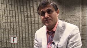
In this video interview, Dr. Farooq Naeem, an associate professor of psychiatry at Queen’s University in Kingston, Ont., discusses how a variety of CBT therapies are proving cost effective with good patient outcomes. Dr. Naeem covers what is known as brief CBT, as well as online self-help CBT, and CBT combination therapies.
He pointed to success with brief CBT for psychosis that is delivered in 6 to 10 sessions, compared with the standard, which is 12 to 20 sessions.
Dr. Naeem also offered perspective on where CBT for psychosis is being delivered across the globe. “In England, possibly, you have the best coverage,” Dr. Naeem said at the annual meeting of the American Psychiatric Association. Australia and New Zealand have had some success with CBT, he said, “but Canada and [the] U.S. are far behind.”
Dr. Naeem did not have any relevant disclosures.
The video associated with this article is no longer available on this site. Please view all of our videos on the MDedge YouTube channel
On Twitter @whitneymcknight
ATLANTA – Now that cognitive-behavioral therapy (CBT) for psychosis is an established intervention, what are the novel ways it is being used, either alone or combined with cognitive remediation therapy, to help outpatients with mild to moderate symptoms?
In this video interview, Dr. Farooq Naeem, an associate professor of psychiatry at Queen’s University in Kingston, Ont., discusses how a variety of CBT therapies are proving cost effective with good patient outcomes. Dr. Naeem covers what is known as brief CBT, as well as online self-help CBT, and CBT combination therapies.
He pointed to success with brief CBT for psychosis that is delivered in 6 to 10 sessions, compared with the standard, which is 12 to 20 sessions.
Dr. Naeem also offered perspective on where CBT for psychosis is being delivered across the globe. “In England, possibly, you have the best coverage,” Dr. Naeem said at the annual meeting of the American Psychiatric Association. Australia and New Zealand have had some success with CBT, he said, “but Canada and [the] U.S. are far behind.”
Dr. Naeem did not have any relevant disclosures.
The video associated with this article is no longer available on this site. Please view all of our videos on the MDedge YouTube channel
On Twitter @whitneymcknight
EXPERT ANALYSIS FROM the APA ANNUAL MEETING
CVS MinuteClinics: A Cure for Long Wait Times at Veterans Affairs?
Struggling with long wait times, the Veterans Affairs Health Care System is trying something new: a partnership with the CVS Pharmacy chain to offer urgent care services to more than 65,000 veterans.
The experiment begins today at the VA’s operations in Palo Alto, California.
Veterans can visit 14 “MinuteClinics” operated by CVS in the San Francisco Bay area and Sacramento, where staff will treat them for conditions such as respiratory infections, order lab tests and prescribe medications, which can be filled at CVS pharmacies.
The care will be free for veterans, and the VA will reimburse CVS for the treatment and medications. Whether the partnership will spread to other VA locales isn’t yet clear.
The collaboration comes amid renewed scrutiny of the nation’s troubled VA health system, which has tried without much success to improve long wait times for veterans needing health care.
Despite a $10 billion “Veterans Choice” program allowing veterans to receive care outside the closed VA system, vets nationwide wait for an appointment even longer than they did before the program started in 2014, according to a federal audit.
The MinuteClinic partnership is not part of the Veterans Choice program.
“The concern has always been, how do we make sure veterans get the care they need in a timely way and in a way that works for the veteran?” said Dr. Stephen Ezeji-Okoye, the Palo Alto VA’s deputy chief of staff. The deal indicates that the VA is willing to try outside partnerships to meet veterans’ needs, he said. “We want to have not just timely access but geographic access to care.”
Sarah Russell, the Palo Alto VA’s chief medical informatics officer, came up with the idea, said Ezeji-Okoye.
The VA will integrate MinuteClinics’ patient records with its own electronic health records to provide consistency of care, Ezeji-Okoye said.
The Palo Alto VA fares better than some other facilities nationwide in providing timely care to veterans, according to VA data, and Ezeji-Okoye said most patients with urgent care needs are seen quickly.
But the system was so busy in the past year that about 11 percent of appointments at its network of hospitals and clinics — which stretch south from Sonora to Monterey — could not be scheduled within 30 or fewer days, which is considered an acceptable timeframe, VA data show. That includes appointments that would require urgent care.
More than 5,000 appointments system-wide were scheduled more than 30 days out, but each hospital and clinic’s performance varied widely. At a Fremont clinic, less than 2 percent of appointment requests could not be scheduled within 30 days. At the VA’s rural Modesto clinic, by contrast, more than 17 percent of requests were not be scheduled within 30 days.
Once the MinuteClinic operation is well underway, Ezeji-Okoye anticipates that between 10 and 15 veterans — from among the estimated 150 who call the Palo Alto VA’s advice nurse hotline daily — will be treated at the retail clinics on any given day.
About 95,000 veterans are eligible to use the Palo Alto system, one of the VA’s largest in the Western United States. About 65,000 use it every year.
The $330,000 pilot project will be evaluated after one year. CVS’ MinuteClinic president, Dr. Andrew Sussman, hopes it can be rolled out nationally if it succeeds. CVS is by far the biggest player in retail pharmacy clinics, operating 1,135 of them in 35 states.
“We’d love to have that opportunity to expand after we go through this phase,” Sussman said. “We’re well suited to help because of our large footprint and ability to see people on a quick basis.”
It is unclear, however, what the VA’s nationwide plans are. The Veterans Health Administration office did not respond to Kaiser Health News’ request for comment.
Blake Schindler, a retired Army major who lives in Santa Clara near one of the participating MinuteClinics, was intrigued, but cautious about the MinuteClinics. He counts himself lucky because unlike some other veterans, he has access to the U.S. military’s TRICARE health insurance program for active and some retired service members.
“It could make a big difference, but how much access are the veterans going to have? That was the big problem with the Veterans Choice program; it didn’t end up the way it was supposed to,” said Schindler, 58.
“I’m always hopeful when I hear about these things; I keep an open mind until I have experience with it,” he added.
Interested veterans served by the VA Palo Alto can call its advice nurse line at 800-455-0057.
This story was produced by Kaiser Health News, which publishes California Healthline, a service of the California Health Care Foundation.
This story also ran on NPR.
Repeat SICU admissions should trigger palliative care consult
ICU readmission was most predictive of the need for palliative care among patients in the surgical intensive care unit, based on a study of six potential trigger criteria associated with in-hospital death or discharge to hospice.
To facilitate proactive case findings of patients who would benefit from a palliative care consult, a team of surgical ICU and palliative care clinicians at the Icahn School of Medicine at Mount Sinai, N.Y., developed and tested a system of palliative care triggers. The study was published online in the Journal of Critical Care (http://dx.doi.org/10.1016/j.jcrc.2016.04.010).
Based on a literature review, the researchers created a six-item list of potential triggers for palliative care: length of stay over 10 days, ICU readmission, intensivist referral, status post cardiac arrest, metastatic cancer, and a match of two or more on a set of secondary criteria.

Data were collected for the period from Sept. 4, 2013, through May 30, 2014, at the surgical ICU of a 1,170-bed tertiary medical center. Patients who received a palliative care consultation were compared with those who did not, and the trigger list was tested for accuracy in predicting patient outcomes. The primary outcomes were hospital death, hospice discharge, and a combined endpoint of these two outcomes. Patients who died in the hospital or were released to hospice care were assumed to be those most in need of a palliative care consult.
Bivariate analysis was done to calculate the unadjusted odds ratios of individual triggers to each of these outcomes. Then, the team used logistic regression analysis to calculate the adjusted odds ratios of triggers to outcomes.
Of the 512 patients admitted to the SICU in the study period, those not discharged by the end of the study were excluded, leaving 492 patients in the study.
Bivariate analysis found that all of the triggers were significantly associated with in-hospital death. With the multivariate analysis and adjusted odds ratios, SICU readmission, status post cardiac arrest, metastatic cancer, and secondary triggers were significantly associated with hospital death.
For the combined outcome of hospital death or release to hospice care, the relationships were stronger. In particular, repeat SICU readmissions and metastatic cancer triggers were strongly associated with the combined outcome (odds ratio, 19.41, CI 5.81-54.86 and OR, 16.40, CI 4.69-57.36, respectively). The secondary triggers did not show the same strength of association, although they were associated significantly with the combined outcome (OR, 4.41, CI 2.05-9.53).
The most prominent finding is the strength of repeat SICU admissions with the hospital death or release to hospice. The strong relationship between repeat SICU admission and outcomes led the researchers to conclude “that one might consider adapting this clinical criterion as a standalone criterion. This would require all patients who are readmitted to the SICU to be seen by palliative care to assess their overall goals of care and understanding of their serious illness. This approach may be particularly useful for smaller palliative care teams that do not have the resources to screen daily with a series of triggers.”
The American Federation of Aging Research and the National Institute on Aging funded the study.
ICU readmission was most predictive of the need for palliative care among patients in the surgical intensive care unit, based on a study of six potential trigger criteria associated with in-hospital death or discharge to hospice.
To facilitate proactive case findings of patients who would benefit from a palliative care consult, a team of surgical ICU and palliative care clinicians at the Icahn School of Medicine at Mount Sinai, N.Y., developed and tested a system of palliative care triggers. The study was published online in the Journal of Critical Care (http://dx.doi.org/10.1016/j.jcrc.2016.04.010).
Based on a literature review, the researchers created a six-item list of potential triggers for palliative care: length of stay over 10 days, ICU readmission, intensivist referral, status post cardiac arrest, metastatic cancer, and a match of two or more on a set of secondary criteria.
Data were collected for the period from Sept. 4, 2013, through May 30, 2014, at the surgical ICU of a 1,170-bed tertiary medical center. Patients who received a palliative care consultation were compared with those who did not, and the trigger list was tested for accuracy in predicting patient outcomes. The primary outcomes were hospital death, hospice discharge, and a combined endpoint of these two outcomes. Patients who died in the hospital or were released to hospice care were assumed to be those most in need of a palliative care consult.
Bivariate analysis was done to calculate the unadjusted odds ratios of individual triggers to each of these outcomes. Then, the team used logistic regression analysis to calculate the adjusted odds ratios of triggers to outcomes.
Of the 512 patients admitted to the SICU in the study period, those not discharged by the end of the study were excluded, leaving 492 patients in the study.
Bivariate analysis found that all of the triggers were significantly associated with in-hospital death. With the multivariate analysis and adjusted odds ratios, SICU readmission, status post cardiac arrest, metastatic cancer, and secondary triggers were significantly associated with hospital death.
For the combined outcome of hospital death or release to hospice care, the relationships were stronger. In particular, repeat SICU readmissions and metastatic cancer triggers were strongly associated with the combined outcome (odds ratio, 19.41, CI 5.81-54.86 and OR, 16.40, CI 4.69-57.36, respectively). The secondary triggers did not show the same strength of association, although they were associated significantly with the combined outcome (OR, 4.41, CI 2.05-9.53).
The most prominent finding is the strength of repeat SICU admissions with the hospital death or release to hospice. The strong relationship between repeat SICU admission and outcomes led the researchers to conclude “that one might consider adapting this clinical criterion as a standalone criterion. This would require all patients who are readmitted to the SICU to be seen by palliative care to assess their overall goals of care and understanding of their serious illness. This approach may be particularly useful for smaller palliative care teams that do not have the resources to screen daily with a series of triggers.”
The American Federation of Aging Research and the National Institute on Aging funded the study.
FROM THE JOURNAL OF CRITICAL CARE
Key clinical point: A list of tested triggers can predict the need of surgical ICU patients for a palliative care consultation.
Major finding: Readmission to the surgical ICU was strongly associated with the study endpoint of hospital death or release to hospice (odds ratio 19.41, CI 5.81-54.86).
Data source: A case review of all 492 patients admitted to the surgical intensive care facility at a 1,170-bed, tertiary care medical center.
Disclosures: The American Federation of Aging Research and the National Institute on Aging funded the study.

SSRIs don’t boost risk in patients with bleeding peptic ulcers
SAN DIEGO – Taking selective serotonin reuptake inhibitors (SSRIs) was not associated with an increased risk of endoscopy-refractory bleeding, rebleeding, or in-hospital mortality among 14,343 consecutive patients admitted with bleeding peptic ulcer in Denmark.
The study suggests that patients with bleeding peptic ulcers can “continue SSRI treatment if the treatment is well indicated,” and that these patients can otherwise receive treatment similar to that of other patients with peptic ulcer bleeding, Dr. Stig B. Laursen reported at the annual Digestive Disease Week.
Observational research has linked use of SSRI antidepressants to an approximately 1.5-fold increase in upper GI bleeding in peptic ulcer bleeding cases, said Dr. Laursen of Odense (Denmark) University Hospital. Studies, mostly in vitro ones, indicate SSRIs decrease the function of thrombocytes and fibrin formation via lowered levels of serotonin. Therefore, it has been suggested that temporarily discontinuing SSRIs may benefit patients with bleeding peptic ulcers.
However, sudden cessation of SSRIs can be associated with withdrawal symptoms, which have been reported in up to one-third of such patients.
Dr. Laursen and his associates prospectively analyzed data for 14,343 consecutive patients admitted with bleeding peptic ulcers in Denmark from 2006 to 2014. They investigated associations between SSRI use and several outcomes, adjusted for confounding factors including age, sex, comorbidity, anemia, medication use, circulatory failure at hospital admission, location of ulcer, stigmata of bleeding, and weekend admission.
After adjustment for confounding risk factors, SSRIs were not associated with increased risk of endoscopy-refractory bleeding (odds ratio [OR] 95% confidence interval [CI]: 1.01 [0.78-1.30]), rebleeding (OR [95% CI]: 0.94 [0.81-1.10]), or in-hospital mortality (hazard ratio [95% CI]: 0.84 [0.68-1.05]).
“Patients taking SSRIs have the same outcomes following peptic ulcer bleeding as non-SSRI users. This suggests that there is no clinically significant impairment of hemostatic function,” Dr. Laursen said. “Therefore, it seems safe to continue SSRI treatment in patients developing peptic ulcer bleeding and thereby avoid development of withdrawal symptoms.”
Dr. Laursen noted that the study has several limitations. It’s not a randomized controlled trial, he said, and there’s no information about the types of SSRIs that the patients were taking. Also, there’s a lack of information about possible discontinuation of SSRIs during hospitalization. However, he said, “that doesn’t seem to be a major confounder.”
During the question-and-answer session following his presentation, Dr. Laursen said the increased risk of poor outcome is quite low, but “there may be a small risk that we haven’t found yet. But if it’s small, maybe it doesn’t have an impact in day-to-day practice.”
The research is hospital-funded. Dr. Laursen had no financial disclosures to report.
AT DDW 2016
Key clinical point: Patients with bleeding peptic ulcers who are on SSRIs may be able to avoid cessation and withdrawal symptoms because outcomes aren’t affected despite worsened bleeding.
Major finding: After adjustment for confounding factors, SSRIs didn’t significantly worsen endoscopy-refractory bleeding (OR 1.01), rebleeding (OR 0.94) or in-hospital mortality (HR 0.84).
Data source: Analysis of 14,343 consecutive patients with bleeding peptic ulcers in Denmark from 2006 to 2014.
Disclosures: The research is hospital funded. Dr. Laursen had no financial disclosures to report.

Resection of recurrence shows survival benefit for adrenocortical carcinoma
BALTIMORE – Patients who have recurrent adrenocortical carcinoma appear to have one option to increase their survival: surgery that includes complete tumor resection – but it may a viable path only if the recurrence occurred a year or more after the initial resection and diagnosis, according to an retrospective study of patients at five French university hospitals. “Adrenocortical carcinoma (ACC) is a rare, malignant tumor that has a poor prognosis, and recurrence of the tumor is considerable with 75% recurrence at 5 years,” Dr. Claire Blanchard of the Digestive and Endocrine Surgery Clinic at the Central University Hospital, Nantes, France, reported at the annual meeting of the American Association of Endocrine Surgeons. “Complete resection of recurrence is the only curative treatment.”
The researchers conducted a retrospective study of patients with at least one recurrence, diagnosed between 1980 and 2014, after initial resection of ACC, comparing outcomes in 29 patients who underwent surgery with 30 who had non-operative treatment, mainly chemotherapy and radiation.

Patients who had an operation for recurrence more often had local recurrence, 75% vs. 10% in the nonoperative group, and more frequently had a unique site of recurrence, 97% vs. 45%, than the nonoperative patients, Dr. Blanchard said.
These other demographic and tumor characteristics were similar between the operative and nonoperative groups, respectively: age, 49 years and 53 years; gender, 63% and 79% female; Weiss score, 6 and 7; Ki-67 protein index, 23% and 24%; tumor size, 99.2 mm and 115.5 mm; ENSAT stage, 65% and 45% stages I and II; and R0 resection status of the primary tumor at initial surgery, 83% and 71%.
The univariate analysis showed that appearance of the first recurrence more than 12 months after the initial diagnosis increased a patient’s chance of survival after treatment for recurrence, Dr. Blanchard said.
“Recurrences occurred at median delay of 12 months after the initial surgery,” Dr. Blanchard said. “In the 59 patients, 24 had local recurrences and 35 had distant metastases.”
Overall median survival after the first recurrence was 91 months for patients who had surgery vs. 15 months for those who did not. Overall median survival after initial resection of the primary tumor was 133 months, with a range of 14 to 252 months, in operated patients vs. 32 months, ranging from 21 to 43 months, in those who had no surgery, Dr. Blanchard said.
Of the 29 surgery patients in the surgery group, 22 had local-regional resections, 6 of whom had adjunctive radiation of the tumor bed.
“The type of resection in recurrent ACC depends on the location of the recurrence,” senior coauthor Dr. Eric Mirallié said. “In the case of local recurrence, we resected the adrenalectomy bed and all the adjacent invaded organs.”
In this series, 6 patients had resection of the tumor bed and 16 had adjacent organ resections; 8 patients (28%) had two or more operations for recurrences. These operations involved eight splenectomies, seven resections for abdominal nodules, six nephrectomies, three distal pancreatectomies, three segmental colectomies, and two minor hepatectomies. All operations were by laparotomy.
“In cases of distant recurrence, complete metastasectomy was performed,” Dr. Mirallié said. The series reported two right hepatectomies, one liver tumorectomy, one lung tumorectomy, and one brain tumorectomy.
“Nonoperative management is reserved for nonresectable patients with recurrent adrenocortical carcinoma,” Dr. Mirallié said. “Oral chemotherapy like mitotane was always given when possible. In cases of nonresectable local recurrence, radiotherapy can be used.”
During the discussion, Dr. Bradford K. Mitchell of Michigan State University, East Lansing, said that the benefit of improved survival in surgical patients in the study may have been a function of selection bias as patients who were not operated on may have had more advanced disease.
Dr. Blanchard and her coauthors had no financial relationships to disclose.
BALTIMORE – Patients who have recurrent adrenocortical carcinoma appear to have one option to increase their survival: surgery that includes complete tumor resection – but it may a viable path only if the recurrence occurred a year or more after the initial resection and diagnosis, according to an retrospective study of patients at five French university hospitals. “Adrenocortical carcinoma (ACC) is a rare, malignant tumor that has a poor prognosis, and recurrence of the tumor is considerable with 75% recurrence at 5 years,” Dr. Claire Blanchard of the Digestive and Endocrine Surgery Clinic at the Central University Hospital, Nantes, France, reported at the annual meeting of the American Association of Endocrine Surgeons. “Complete resection of recurrence is the only curative treatment.”
The researchers conducted a retrospective study of patients with at least one recurrence, diagnosed between 1980 and 2014, after initial resection of ACC, comparing outcomes in 29 patients who underwent surgery with 30 who had non-operative treatment, mainly chemotherapy and radiation.
Patients who had an operation for recurrence more often had local recurrence, 75% vs. 10% in the nonoperative group, and more frequently had a unique site of recurrence, 97% vs. 45%, than the nonoperative patients, Dr. Blanchard said.
These other demographic and tumor characteristics were similar between the operative and nonoperative groups, respectively: age, 49 years and 53 years; gender, 63% and 79% female; Weiss score, 6 and 7; Ki-67 protein index, 23% and 24%; tumor size, 99.2 mm and 115.5 mm; ENSAT stage, 65% and 45% stages I and II; and R0 resection status of the primary tumor at initial surgery, 83% and 71%.
The univariate analysis showed that appearance of the first recurrence more than 12 months after the initial diagnosis increased a patient’s chance of survival after treatment for recurrence, Dr. Blanchard said.
“Recurrences occurred at median delay of 12 months after the initial surgery,” Dr. Blanchard said. “In the 59 patients, 24 had local recurrences and 35 had distant metastases.”
Overall median survival after the first recurrence was 91 months for patients who had surgery vs. 15 months for those who did not. Overall median survival after initial resection of the primary tumor was 133 months, with a range of 14 to 252 months, in operated patients vs. 32 months, ranging from 21 to 43 months, in those who had no surgery, Dr. Blanchard said.
Of the 29 surgery patients in the surgery group, 22 had local-regional resections, 6 of whom had adjunctive radiation of the tumor bed.
“The type of resection in recurrent ACC depends on the location of the recurrence,” senior coauthor Dr. Eric Mirallié said. “In the case of local recurrence, we resected the adrenalectomy bed and all the adjacent invaded organs.”
In this series, 6 patients had resection of the tumor bed and 16 had adjacent organ resections; 8 patients (28%) had two or more operations for recurrences. These operations involved eight splenectomies, seven resections for abdominal nodules, six nephrectomies, three distal pancreatectomies, three segmental colectomies, and two minor hepatectomies. All operations were by laparotomy.
“In cases of distant recurrence, complete metastasectomy was performed,” Dr. Mirallié said. The series reported two right hepatectomies, one liver tumorectomy, one lung tumorectomy, and one brain tumorectomy.
“Nonoperative management is reserved for nonresectable patients with recurrent adrenocortical carcinoma,” Dr. Mirallié said. “Oral chemotherapy like mitotane was always given when possible. In cases of nonresectable local recurrence, radiotherapy can be used.”
During the discussion, Dr. Bradford K. Mitchell of Michigan State University, East Lansing, said that the benefit of improved survival in surgical patients in the study may have been a function of selection bias as patients who were not operated on may have had more advanced disease.
Dr. Blanchard and her coauthors had no financial relationships to disclose.
AT AAES 2016
Key clinical point: Surgery carries a significant survival benefit in patients with recurrent adrenocortical carcinoma (ACC).
Major finding: Overall median survival after the first recurrence was 91 months in patients who had surgery vs. 32 months in those who did not.
Data source: Retrospective case-control study of 59 patients at five French teaching hospitals who had treatment for recurrent ACC from 1980-2014.
Disclosures: Dr. Blanchard and her coauthors reported having no financial disclosures.
David Henry's JCSO podcast, May 2016
For the May podcast for The Journal of Community and Supportive Oncology, Dr David Henry highlights original reports on the prognostic significance of HPV status in postoperative squamous cell carcinoma of the head and neck, the contributions of caregivers in American Indian cancer patients, and the omission of dexamethasone from antiemetics treatment in breast cancer patients with hepatitis B infection or diabetes. He also describes how providers at a comprehensive cancer center devised a framework for implementing survivorship care planning, and, in keeping with the ‘how we do it’ theme, he discusses an article by 2 practicing on oncologists on managing and treating multiple myeloma. The line-up is rounded off with articles on the effects of exercise interventions during different treatments in patients with breast cancer, the approval of trabectedin for some forms of advanced soft tissue sarcoma, and a Case Report on a rare case of hypoglycemia induced by a classical gastro-intestinal stromal tumor.
Listen to the podcast below.
Stella Fitzgibbons, MD, FHM, Relishes the Variety, Interactions of Hospitalist Practice
Stella Fitzgibbons, MD, FHM, was an engineer for several years after college. But there wasn’t enough working with people for her taste. So she moved into internal medicine. But then there was, how to put this, something lacking in office work.

“I realized how bored I was with office practice and how much more interesting were the problems at the hospital than outpatient ones,” Dr. Fitzgibbons says.
So she went to work in hospitals. She hasn’t left.
Dr. Fitzgibbons is a hospitalist and ED practitioner with Mint Physician Staffing, primarily in the Apollo Hospital System in The Woodlands, Texas. And the best part of the job for Dr. Fitzgibbons, one of eight new members of Team Hospitalist, the volunteer editorial advisory board of The Hospitalist newsmagazine, is easy to pick.
“Seeing sick people get better,” she says.
Question: Switching careers from engineering to medicine is a big step. What motivated that?
Answer: I wanted to see my efforts helping people I could actually see, and I thought—and still do—that medicine uses my talents better and is far more interesting.
Q: You say office practice became a bit boring. How so? What appealed about the inpatient setting?
A: An internist in the office only sees a tiny fraction of the interesting problems that our field covers. Rheumatoid arthritis is diagnosed by a rheumatologist, who then makes all the decisions. Abdominal pain gets sent to the hospital, where all the diagnostic tests are done.
Fortunately, my multispecialty group arranged for about a quarter of its internists [the youngest quarter in most cases] to manage hospital patients; I figured out pretty quickly that it was only there that I got to see pulmonary hypertension, congestive heart failure, and acute abdomens. Even night call was better at the hospital since office doctors only answered phone calls and never had a chance to do any real evaluation and treatment no matter how sick the caller was.
And a problem at the office was something that made me run behind that odious and impractical appointment schedule; at the hospital I was seeing real illnesses, not people who wanted a prescription med for their sore throat so they wouldn’t be bothered with it on their vacation.
Q: What is your biggest professional challenge?
A: EHRs.
Q: What is your biggest professional reward?
A: When a patient says, “Thanks for taking care of me, doctor.”
Q: What does teaching mean to you, and how has it been gratifying in your career?
A: Teaching means paying it forward, in gratitude to those who taught me, with the reward of seeing light bulbs go off behind the eyes of students and younger doctors who are eager to learn.
Q: When you aren’t working, what is important to you?
A: Family and music and church.
Q: Faith is obviously important to you. How does that help your work as a care provider?
A: I don’t think anybody goes around being religious all the time. But it sometimes makes all the difference knowing that a higher power is looking out for me and the patients.
Q: You’ve described mentoring as fun for you. What exactly do you mean by fun?
A: Mentoring is what we do. Patients, nurses—anybody we work with—need explanations and clarifications. About the third day of med school, docs in training realize that anybody who can help us understand and retain the huge stream of information directed at us is performing a necessary service. Throughout the training period, residents teach students, fellows teach residents, and attending faculty teach everybody. Doctors in training are bright people who want to learn both the facts and how to deal with patients’ side of things, and feeding their desires is very enjoyable.
Q: You’d like to see more physicians than MBAs in decision-making positions. Why? What real changes do you think that would effectuate?
A: Physicians and nurses were administrators for decades before insurance company penny-pinching and government regulations led hospitals to hire “bean counters” to replace them. It is a tremendous change for the worse, to have people making decisions for patients whose primary consideration is the bottom line.
Q: What’s next professionally?
A: Small-volume ERs, where I don’t have to do discharge planning while being harassed by insurance company reps.
Q: Where do you see yourself in 10 years?
A: Retired.
Q: If you weren’t a doctor, what would you be doing right now?
A: Law enforcement.
Q: Devices like iPhones and tablets can take away from patient face time. But they can also be valuable. How do you balance that? How do you encourage younger docs to do so, particularly when they’re much more used to having smartphones glued to their hands?
A: I use my iPhone when I’m with patients … but only when they can see the reason I need it to help them, such as looking up the side effects of a medication. Electronic health records can work on an iPad, but I hesitate to use them unless the patient knows just what I am doing, such as looking up results of a lab test that concerns them. Taking a computer on wheels into a patient’s room means that I spend part of the visit looking at a screen instead of at the patient, and I prefer to avoid it if at all possible.
Q: What’s the best book you’ve read recently? Why?
A: The House of Silk by Anthony Horowitz. Great continuation of the Holmes stories, with a seamless link to [Sir Arthur] Conan Doyle’s style.
Q: How many Apple products (phones, iPods, tablets, iTunes, etc.) do you interface with in a given week?
A: Three.
Q: What’s your favorite social network? Do you use it all for work or professional development?
A: Facebook. Heck no, it’s just fun.
Q: What’s next in your Netflix queue?
A: Last two episodes of Game of Thrones season 5.
Richard Quinn is a freelance writer in New Jersey.
Stella Fitzgibbons, MD, FHM, was an engineer for several years after college. But there wasn’t enough working with people for her taste. So she moved into internal medicine. But then there was, how to put this, something lacking in office work.
“I realized how bored I was with office practice and how much more interesting were the problems at the hospital than outpatient ones,” Dr. Fitzgibbons says.
So she went to work in hospitals. She hasn’t left.
Dr. Fitzgibbons is a hospitalist and ED practitioner with Mint Physician Staffing, primarily in the Apollo Hospital System in The Woodlands, Texas. And the best part of the job for Dr. Fitzgibbons, one of eight new members of Team Hospitalist, the volunteer editorial advisory board of The Hospitalist newsmagazine, is easy to pick.
“Seeing sick people get better,” she says.
Question: Switching careers from engineering to medicine is a big step. What motivated that?
Answer: I wanted to see my efforts helping people I could actually see, and I thought—and still do—that medicine uses my talents better and is far more interesting.
Q: You say office practice became a bit boring. How so? What appealed about the inpatient setting?
A: An internist in the office only sees a tiny fraction of the interesting problems that our field covers. Rheumatoid arthritis is diagnosed by a rheumatologist, who then makes all the decisions. Abdominal pain gets sent to the hospital, where all the diagnostic tests are done.
Fortunately, my multispecialty group arranged for about a quarter of its internists [the youngest quarter in most cases] to manage hospital patients; I figured out pretty quickly that it was only there that I got to see pulmonary hypertension, congestive heart failure, and acute abdomens. Even night call was better at the hospital since office doctors only answered phone calls and never had a chance to do any real evaluation and treatment no matter how sick the caller was.
And a problem at the office was something that made me run behind that odious and impractical appointment schedule; at the hospital I was seeing real illnesses, not people who wanted a prescription med for their sore throat so they wouldn’t be bothered with it on their vacation.
Q: What is your biggest professional challenge?
A: EHRs.
Q: What is your biggest professional reward?
A: When a patient says, “Thanks for taking care of me, doctor.”
Q: What does teaching mean to you, and how has it been gratifying in your career?
A: Teaching means paying it forward, in gratitude to those who taught me, with the reward of seeing light bulbs go off behind the eyes of students and younger doctors who are eager to learn.
Q: When you aren’t working, what is important to you?
A: Family and music and church.
Q: Faith is obviously important to you. How does that help your work as a care provider?
A: I don’t think anybody goes around being religious all the time. But it sometimes makes all the difference knowing that a higher power is looking out for me and the patients.
Q: You’ve described mentoring as fun for you. What exactly do you mean by fun?
A: Mentoring is what we do. Patients, nurses—anybody we work with—need explanations and clarifications. About the third day of med school, docs in training realize that anybody who can help us understand and retain the huge stream of information directed at us is performing a necessary service. Throughout the training period, residents teach students, fellows teach residents, and attending faculty teach everybody. Doctors in training are bright people who want to learn both the facts and how to deal with patients’ side of things, and feeding their desires is very enjoyable.
Q: You’d like to see more physicians than MBAs in decision-making positions. Why? What real changes do you think that would effectuate?
A: Physicians and nurses were administrators for decades before insurance company penny-pinching and government regulations led hospitals to hire “bean counters” to replace them. It is a tremendous change for the worse, to have people making decisions for patients whose primary consideration is the bottom line.
Q: What’s next professionally?
A: Small-volume ERs, where I don’t have to do discharge planning while being harassed by insurance company reps.
Q: Where do you see yourself in 10 years?
A: Retired.
Q: If you weren’t a doctor, what would you be doing right now?
A: Law enforcement.
Q: Devices like iPhones and tablets can take away from patient face time. But they can also be valuable. How do you balance that? How do you encourage younger docs to do so, particularly when they’re much more used to having smartphones glued to their hands?
A: I use my iPhone when I’m with patients … but only when they can see the reason I need it to help them, such as looking up the side effects of a medication. Electronic health records can work on an iPad, but I hesitate to use them unless the patient knows just what I am doing, such as looking up results of a lab test that concerns them. Taking a computer on wheels into a patient’s room means that I spend part of the visit looking at a screen instead of at the patient, and I prefer to avoid it if at all possible.
Q: What’s the best book you’ve read recently? Why?
A: The House of Silk by Anthony Horowitz. Great continuation of the Holmes stories, with a seamless link to [Sir Arthur] Conan Doyle’s style.
Q: How many Apple products (phones, iPods, tablets, iTunes, etc.) do you interface with in a given week?
A: Three.
Q: What’s your favorite social network? Do you use it all for work or professional development?
A: Facebook. Heck no, it’s just fun.
Q: What’s next in your Netflix queue?
A: Last two episodes of Game of Thrones season 5.
Richard Quinn is a freelance writer in New Jersey.
IMF denounces report on newer MM drugs

Photo by Bill Branson
A report assessing the value of newer multiple myeloma (MM) treatments “dangerously oversimplifies” a complex issue and could limit patients’ access to treatment, according to the International Myeloma Foundation (IMF).
The report, which was drafted by the Institute for Clinical and Economic Review (ICER), is scheduled to be discussed at the inaugural meeting of the Midwest Comparative Effectiveness Public Advisory Council (Midwest CEPAC) in St. Louis, Missouri, on May 26.
The main conclusion of ICER’s report was that newer second- and third-line treatment regimens for MM appear to confer clinical benefits, but the estimated cost-effectiveness of these regimens exceeds commonly cited thresholds.
For example, ICER said that, based on the available data, there was “moderate certainty” that carfilzomib (CFZ), ixazomib (IX), or elotuzumab (ELO) given in combination with lenalidomide and dexamethasone (LEN+DEX) can provide an incremental or better net health benefit for second-line, third-line, or subsequent therapy in adults with relapsed/refractory MM, relative to LEN+DEX alone.
However, the estimated cost-effectiveness, compared to LEN+DEX, was $200,000 per quality-adjusted life year (QALY) gained for CFZ+LEN+DEX, $428,000 for ELO+LEN+DEX, and $434,000 for IX+LEN+DEX. All of these exceed commonly cited thresholds of $50,000 to $150,000 per QALY.
ICER said achieving levels of value more closely aligned with patient benefit would require substantial discounts from the list price in many cases. In other cases, there is no realistic price for the newest agents that would achieve these thresholds.
IMF’s response
IMF said ICER’s report has a few flaws—namely, the absence of many newer MM drugs and combinations, the use of inaccurate data, and an underestimation of QALYs.
Furthermore, IMF said it is concerned that, if ICER's recommendations were to be adopted by the Centers for Medicare & Medicaid Services, patients might be required to “fail first” before other, possibly more effective drugs would be an option.
“We believe that the IMF’s research body, the International Myeloma Working Group (IMWG), will produce superior patient-centered and research-supported guidelines to effectively impact drug costs at our annual summit in June,” said IMF Chairman Brian Durie.
IMWG plans to focus on healthcare cost containment in a special session at the 2016 IMWG Summit, which is scheduled to take place June 7-9 in Copenhagen, Denmark.
IMF said the guidelines resulting from this session should be available in about 2 months. And they will spell out primary and secondary recommendations that allow for individualized therapy choices based on unique features of the disease, patient and/or physician preference, and local and/or regional access issues. ![]()
Team simplifies synthesis of anticancer agent
Photo courtesy of Jeff Fitlow
and Rice University
Researchers say they have streamlined synthesis of delta12-prostaglandin J3, a molecule that has been shown to kill leukemia cells.
Total synthesis of the molecule now requires only 6 steps from commercially available starting materials.
The researchers say this work sets the stage for large-scale synthesis of the molecule—a lipid found in nearly all animal tissues—and related compounds that can be produced as potential anticancer agents.
K.C. Nicolaou, PhD, of Rice University in Houston, Texas, and his colleagues described the work in Chemistry - A European Journal.
The prostaglandin the researchers synthesized had been isolated in 2011 as a secondary metabolite formed from eicosapentaenoic acid, which is found primarily in fish oil.
The team reported the first total synthesis of the molecule in 2014. That allowed them to confirm its structure.
Now, the researchers have established techniques to develop related disease-fighting compounds and ramp up bulk production if necessary.
Several such prostaglandin derivatives under consideration as preclinical drug candidates were detailed in a second paper published in the Journal of the American Chemical Society.
That publication described the synthesis of dozens of prostaglandin derivatives that were tested against a range of cancer cells by the National Cancer Institute.
One such derivative, macrolactone 11, is currently under evaluation as a preclinical drug candidate. Related compounds macrolactone 33 and 44 showed evidence of even higher potency against leukemia, lung cancer, colon cancer, melanoma, renal, and prostate cancer.
“The macrolactones are very good—better than the natural product—and now we’re following this lead to optimize the potency while minimizing toxicity,” Dr Nicolaou said. “It’s a balancing act.”
In addition, he and his colleagues are developing other drug candidates based on prostaglandin.
“In the process, we’ve developed a lot of nice chemistry, and we know a lot more about the biology of this compound,” Dr Nicolaou said. “We’ve advanced organic synthesis in general and also enriched the knowledge about how these kinds of molecules behave. We hope the papers provide some ideas and leads and inspiration for others to follow.” ![]()