User login
Your patient has chronic leukemia: Now what?
The advent of targeted therapies has dramatically changed the management of chronic leukemia. Chemotherapy—highly toxic, nonspecific drugs that can be dangerous to patients and providers and result in only modest success—is gradually being replaced by biologic targeting of malignancy. Scientists are rapidly identifying extracellular and intracellular targets on tumor cells and are developing and testing promising new therapies aimed at these targets. Survival of cancer patients has become so common that clinicians outside the specialties of hematology and oncology are now caring for them.
This article describes new biologic therapies for chronic myelogenous leukemia (CML) and chronic lymphocytic leukemia (CLL), along with the diagnosis of these diseases and management of survivors in the primary care setting.
CHRONIC MYELOGENOUS LEUKEMIA
A seemingly healthy person needs laboratory blood work, perhaps for an insurance physical examination or for a preoperative workup. Or a patient comes to the emergency department with a sore throat and routine blood tests are ordered. Their laboratory values:
- White blood cell count 250 × 109/L (reference range 3–11)
- Neutrophils 70% (40%–70%)
- Blasts 1% (0)
- Metacytes and myelocytes 5% (0)
- Bands 5% (0)
- Lymphocytes 10% (22%–40%)
- Monocytes 5% (0–7%)
- Basophils 3% (0–1%)
- Eosinophils 1% (0–4%)
- Hemoglobin 12.1 g/dL (11.5–15.5 in women, 13.0–17.0 in men)
- Platelet count 525 × 109/L (150–400).
Leukocytosis and a ‘left shift’
Although this scenario often raises concern for acute leukemia, a careful look shows evidence of a chronic myeloproliferative disorder instead. Specifically, this patient’s laboratory values show a “left shift”—an increase in immature neutrophils, ie, blasts, myelocytes, and bands.
This picture is characteristic of CML, an uncommon leukemia with about 4,500 new cases annually in the United States. Patients can present at any age, but the disease occurs more often in older people, with a median age of 66.1
The presentation is usually subtle: about half of cases are detected by routine laboratory testing, which typically reveals a left-shifted leukocytosis with basophilia and a few blasts. Mild anemia is common. The platelet count is elevated in 30% to 50% of patients at diagnosis. Bone marrow aspirate shows significant myeloid hyperplasia without dysplasia, and sometimes shows mild fibrosis.
Philadelphia chromosome is diagnostic
A definitive diagnosis is made by demonstration of an abnormally short chromosome 22. Described in 1960 by Peter Nowell of the University of Pennsylvania and David Hugerford of the Institute for Cancer Research,2 this abnormality, called the Philadelphia chromosome, was the first specific genetic abnormality associated with a human cancer. Later, researchers used banding techniques to find that the Philadelphia chromosome results from a reciprocal translocation of genetic material between the BCR gene on chromosome 22 and the ABL1 gene on chromosome 9, t(9:22).3,4 The resulting chimeric gene, called BCR-ABL, codes for an oncogenic protein, a tyrosine kinase with constitutive activity.
The Philadelphia chromosome is present in 95% of patients with CML and can be found in all myeloid cell lineages, including erythrocytes, granulocytes, monocytes, and megakaryocytes as well as some cells of lymphocytic lineage, indicating that malignant transformation to CML takes place at the stem cell level.
The mutation causes several problems: the abnormal tyrosine kinase increases cell proliferation, inhibits apoptosis, and alters adhesion molecules in the stroma of the bone marrow, allowing immature cells to leak into the bloodstream. Most important, the mutation increases genomic instability so that additional mutations are likelier to occur over time, making it inevitable that, without treatment, the disease will progress to a fatal blast crisis within an average of 5 years of diagnosis.
CML has three clinical phases
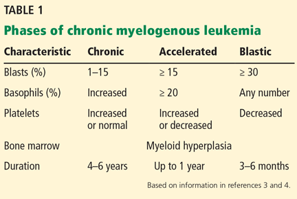
Untreated, CML progresses through three distinct phases: chronic, accelerated, and blast crisis, defined by abnormalities in the blood smear and bone marrow (Table 1).5,6 Most patients (85%) are diagnosed during the chronic phase. The accelerated and blastic phases resemble acute leukemia.
Chronic phase management
Therapies over the years have included arsenic (Fowler solution), splenic radiotherapy, busulfan, hydroxyurea, cytarabine, and interferon. All had some palliative success, but usually did not suppress leukemic progression.7
In contrast, patients undergoing allogeneic bone marrow transplant had a 5-year survival rate of 60% to 80% during the chronic phase of CML, 40% to 60% during the accelerated phase, and 10% to 20% during a blast crisis.8 Long-term survival confirmed the ability of transplant to cure CML, and bone marrow transplant with matched donors was the standard of care for younger patients until the end of the 20th century.
Tyrosine kinase inhibition
A new paradigm in treatment began with the development of imatinib, a tyrosine kinase inhibitor that directly interferes with the product of the chimeric BCR-ABL gene.9
Patients treated with imatinib during the chronic phase of CML have survival rates similar to those of people without the disease, and they usually do not progress to the accelerated and blast phases. As a result of this success, the number of transplants for CML has fallen precipitously.
Other tyrosine kinase inhibitors (dasatinib, nilotinib) that have since been developed have shown even better results in achieving remission and preventing progression. Improved survival is more difficult to demonstrate because the control groups in studies receive imatinib and have 10-year survival rates of about 90%.10–12
With the tyrosine kinase inhibitors, CML can be regarded as functionally cured.13 Patients take these drugs for life and usually experience a relapse if they stop. Patients with CML are now more likely to die of a comorbidity than of CML.
Choose therapy by tolerability
Which tyrosine kinase inhibitor to use depends more on the side-effect profile of the drug than on its efficacy. Nilotinib should be avoided in patients with vascular disease, and dasatinib avoided in patients with pulmonary disease. Each drug may be associated with some degree of nausea, diarrhea, cramps, rash, and edema.10–12
CML is not an immunosuppressive disease, nor are the drugs used to treat it. Patients with CML have an intact immune system. Therefore, precautions taken for patients with acute leukemia or lymphoid malignancy are not required for patients with CML.
Managing survivors
Since imatinib was introduced in 2000, the US Food and Drug Administration (FDA) has approved approximately 20 tyrosine kinase inhibitors for various cancers. These drugs are improving survival rates so well that patients with cancer are increasingly being seen by their primary care doctors for their medical problems.
Some problems have emerged that are consequences of this successful therapy. Angiogenesis inhibitors such as bevacizumab affect vascular endothelial growth factors, which injure endothelial cells. These effects may result in high blood pressure and arterial occlusive disease. Algorithms have been proposed for managing cardiovascular complications for patients taking tyrosine kinase inhibitors.14 Further, cardiovascular risk factors such as hyperlipidemia, diabetes, and obesity must be aggressively managed in patients taking tyrosine kinase inhibitors.
Vascular effects, rashes, and drug interactions may best be managed by primary care physicians, cardiologists, and nephrologists, who deal with such problems regularly.
CHRONIC LYMPHOCYTIC LEUKEMIA
A patient undergoes routine laboratory blood work in the emergency department or clinic, with these results:
- White blood cell count 250 × 109/L
- Neutrophils 1%
- Lymphocytes 99%
- Hemoglobin 12.1 g/dL
- Platelet count 160 × 109/L.
Like patients with CML, those with CLL usually present with no symptoms. The complete blood cell count reveals numerous white blood cells and lymphocytosis. Patients may have painless lymphadenopathy, anemia, and thrombocytopenia, but they do not typically have fever, sweats, or weight loss.
The disease is characterized by clonal proliferation and accumulation of mature-appearing neoplastic B lymphocytes in the blood, bone marrow, lymph nodes, and spleen. The peripheral blood smear shows “smudge cells,” indicating fragile lymphocytes.
The median age at diagnosis is about 70, with fewer than 15% of newly diagnosed patients under age 50.
CLL is the most common leukemia in the Western world, accounting for about 30% of cases of leukemia in adults. It is rare in Asians, probably because of genetic differences.
Monoclonal B-cell lymphocytosis precedes CLL
Monoclonal B-cell lymphocytosis is related to CLL and always precedes it. It is a common condition, detectable in up to 5% of older adults. The differential count shows a less severe lymphocytosis than in CLL.
Because monoclonal B-cell lymphocytosis does not always convert to leukemia, it is important for insurance coverage purposes not to diagnose it as a leukemia. Treatment-free survival of patients diagnosed with monoclonal B-cell lymphocytosis is 87% at 5 years.15,16
Diagnosing CLL
Lymphocytosis can indicate other low-grade lymphoproliferative diseases and malignancies, so further evaluation is critical. To diagnose CLL, the B-cell count by flow cytometry (not the absolute lymphocyte count from the complete blood cell count) must be at least 5 × 10⁹/L. Below that threshold, monoclonal B-cell lymphocytosis is diagnosed unless lymphadenopathy is present, indicating small lymphocytic lymphoma. Unlike in benign lymphoproliferations, CLL lymphocytes coexpress the B-cell marker CD19 and the T-cell marker CD5.17 Bone marrow examination is rarely needed for the diagnosis of CLL.
Two types of CLL can be defined, depending on whether the B cells carry V genes that are mutated or unmutated. B cells expressing ZAP-70 and CD38 tend to carry the unmutated gene, which is associated with a worse prognosis.18 Regardless of which type a patient has, treatments and the indications for treatment are the same.
Increasing immune dysfunction
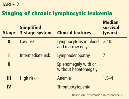
CLL is staged according to effects on lymph tissue and hematopoiesis. The Rai system for clinical staging of CLL has been used since 1975 with little alteration (Table 2).19
CLL is often an indolent lymphoproliferative malignancy and does not always progress to a fatal end stage. Therefore, treatment may be deferred, with a watch-and-wait approach until symptoms develop or the disease progresses. Approximately half of patients never require treatment.20 Progression involves increasing bone marrow impairment with greater susceptibility to infection (due to intrinsic features of CLL and its therapy) and hypogammaglobulinemia in advanced disease.21,22 Systemic infection is the cause of death for most patients.
Because CLL is a disease of the immune system, the development of autoantibodies is a cardinal feature. Autoimmune complications are almost exclusively limited to blood and can include hemolytic anemia, pure red cell aplasia, immune-mediated thrombocytopenia, and granulocytopenia. Other autoimmune diseases, such as rheumatoid arthritis, thyroiditis, and Addison disease, are uncommon.23,24
Other complications may occur in patients who have been treated with chemotherapy, and these are usually fatal. The Richter transformation (to an aggressive lymphoma) occurs in about 15%. Other less common complications include prolymphocytoid transformation and secondary malignancies, particularly carcinomas of the lung and gastrointestinal tract and acute (myeloid) leukemia.25
Survival rates in CLL have improved substantially over the past decades,26–28 with significant gains following the introduction of antibiotics and, to a lesser extent, transfusions. Median survival is generally between 6 and 9 years, but many patients live for years without requiring therapy.
Chemotherapy: The mainstay of treatment
When to begin therapy remains one of the most challenging issues of patient management. Unlike in CML, there is no advantage to starting at diagnosis when most patients are asymptomatic.29
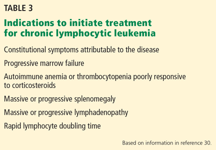
In 1996, the National Cancer Institute issued guidelines for starting treatment, which were updated in 2008 with very little change (Table 3).30 In general, the onset of symptoms and evidence of impaired marrow function, including an abnormal hemoglobin level and platelet count, are indications. The white blood cell count continuously increases during the disease course but is not usually an important factor for initiating treatment.
The therapeutic goal for most patients who require treatment has historically been palliation of symptoms. Therapy must be individualized to a patient’s age and clinical status, with a heavier reliance on chemotherapeutic agents for patients who can tolerate it and on immunotherapy for others. General strategies are as follows:
- “Go-Go” patients—young, fit, with few comorbidities, good renal function—are the minority. Recommendation: combination chemotherapy with fludarabine, cyclophosphamide, and rituximab (FCR).
- “Slo-Go” patients are reasonably fit and can tolerate chemotherapy but not FCR. Recommendation: combination therapy with either bendamustine and rituximab or chlorambucil and rituximab (for less fit patients). Recent evidence indicates ibrutinib may be useful for such patients.31
- “No-Go” patients are frail with short life expectancy. Recommendation: rituximab or observation (see below)
All CLL treatments are potentially toxic. Chemotherapy damages DNA and often causes blood cell counts to fall. Immunosuppression worsens with almost any treatment, involving a substantial risk of secondary malignancy. Although survival improves with therapy, relapse is universal.
Targeting CLL pathways
The new paradigm for cancer therapy is to identify a cellular pathway that drives oncogenesis or proliferation and interfere with it. The B-cell receptor pathway is enormously complex with numerous complex factors, making it difficult to discern the critical mutation that drives the proliferation of lymphocytes.
Bruton tyrosine kinase (Btk) is one factor that is critical for CLL proliferation. Patients with congenitally mutated or dysfunctional Btk have lymphopenia and agammaglobulinemia, making it a promising target for patients with B-cell disorders. Other experimental therapies are based on other such identified factors.
In 2014, the FDA approved two drugs for CLL—ibrutinib, a Btk inhibitor, and idelalisib, an inhibitor of phosphoinositide 3-kinase—after they were shown in clinical trials to dramatically improve outcomes in patients with relapsed CLL.32,33 Trials with these drugs are ongoing. These drugs also inhibit tyrosine kinase and so have vascular side effects in addition to their own idiosyncratic effects.
Ibrutinib has anticoagulant effects and should be stopped before surgery. It also can cause or exacerbate atrial fibrillation, making management of CLL difficult. It is associated with hypogammaglobulinemia, often requiring ongoing immunoglobulin replacement.
Idelalisib tends to cause systemic autoimmune phenomena such as pneumonitis and colitis.
Using T cells as therapy
It has long been observed that patients who undergo bone marrow transplant for leukemia have lower relapse rates if the transplant is allogeneic rather than from a twin. Further, if T cells are removed from the donor graft, graft-vs-host disease may be prevented but the risk of relapses increases. Finally, the presence of graft-vs-host disease tends to reduce the risk of relapse.34 Therefore, T cells clearly are key ingredients for success in the setting of bone marrow transplant. In fact, merely providing T cells for a relapse after allogeneic transplant can induce remission. However, because donor T cells are not targeted, acute and chronic graft-vs-host disease often can ensue.
‘Designer’ monoclonal antibodies
The B lymphocyte has multiple potential targets for new therapies for CLL as well as other cancers involving B cells. CD20 was identified on the surface of B cells in 1988 and is the target protein of the monoclonal antibody drug rituximab. Monoclonal antibodies can be modified to target other surface antigens, to link radioisotopes to deliver radiation therapy, and to deliver drugs that would otherwise be too toxic to be given systemically.35 Monoclonal antibodies can also be modified to enhance function.
Antibodies alone, however, must often rely on the host T cells for cytotoxicity and they are often compromised by either the underlying disease or treatment. Adapting the targeting function of antibodies to enhance or genetically alter T cells to recognize cancer-specific antigens is now being explored for leukemias.36
In 2014, the FDA approved blinatumomab for the treatment of relapsed or refractory acute lymphoblastic leukemia. This biopharmaceutical agent recruits T cells with one antibody-like moiety and targets the CD19 receptor of B cells with another. Given as a single intravenous treatment without chemotherapy, it has an almost 50% response rate, and those who respond tend to stay in remission. Other similar drugs are being developed, and using them earlier in treatment and for other B-cell leukemias is being explored.
New B-cell targeted therapy with CAR-Ts
Newer treatments are being developed based on chimeric antigen receptor T (CAR-T) cells. These engineered T cells express an anti-CD19 moiety that targets B cells, but also activate upon binding to them.37 CAR-T technology is being refined and shows great promise for cancer treatment.
Multiple clinical trials are currently under way in which the investigators collect autologous T cells by leukopheresis from a patient with a relapsed or refractory B-cell malignancy, transduce the T cells with retroviral vectors into anti-CD19 CAR-T cells, and then reinfuse them into the patient following modest chemotherapy.38
Study results from a small number of patients with relapsing or refractory CLL showed that some patients achieved long-term, progression-free survival.39 The most success with this therapy, however, has been in acute lymphoblastic leukemia.40 Possibly, this treatment could be applied to other lymphoid malignancies that also express CD19.
More advances
CAR-T cell therapy has drawbacks. The cells attack only the target antigen, which currently limits their use mostly to hematologic malignancies. In addition, autologous T cells are not robust. Also, the use of allogeneic T cells is restricted by their major histocompatibility complex, and the cells will be rejected by the recipient if not matched.
An attempt to overcome some of these drawbacks is to develop T cells redirected for universal cytokine killing. CAR-T cells are modified with a gene that causes them to excrete interleukin 12, which attracts macrophages and natural killer cells to the environment to better fight the tumor.41
Other modifications include editing out certain genes including the major histocompatibility complex, which avoids the problem of rejection. Another modification is to insert a “suicide gene” that allows the engineered T cells to be killed with an antidote if they do not work as planned.
Such gene-editing techniques hold great promise for curing cancers without chemotherapy in the not so distant future.
- National Cancer Institute Surveillance, Epidemiology, and End Results Program. SEER Stat Fact Sheets: Chronic Myeloid Leukemia. http://seer.cancer.gov/statfacts/html/cmyl.html. Accessed July 1, 2016.
- Nowell PC, Hungerford DA. A minute chromosome in human chronic granulocytic leukemia. Science 1960; 132:1497.
- Melo JV. The diversity of BCR-ABL fusion proteins and their relationship to leukemia phenotype. Blood 1996; 88:2375–2384.
- Pasternak G, Hochhaus A, Schultheis B, Hehlmann R. Chronic myelogenous leukemia: molecular and cellular aspects. J Cancer Res Clin Oncol 1998; 124:643–660.
- Faderl S, Kantarjian HM, Talpaz M. Chronic myelogenous leukemia: update on biology and treatment. Oncology (Williston Park) 1999; 13:169–184.
- Sawyers CL. Chronic myeloid leukemia. N Engl J Med 1999; 340:1330–1340.
- Hehlmann R, Heimpel H, Hasford J, et al. Randomized comparison of interferon-alpha with busulfan and hydroxyurea in chronic myelogenous leukemia. The German CML Study Group. Blood 1994; 84:4064–4077.
- Radich JP, Olavarria E, Apperley JF. Allogeneic hematopoietic stem cell transplantation for chronic myeloid leukemia. Hematol Oncol Clin North Am 2004; 18:685–702.
- Druker BJ. Translation of the Philadelphia chromosome into therapy for CML. Blood 2008; 112:4808–4817.
- O’Brien SG, Guilhot F, Larson RA, et al; IRIS Investigators. Imatinib compared with interferon and low-dose cytarabine for newly diagnosed chronic-phase chronic myeloid leukemia. N Engl J Med 2003; 348:994-1004.
- Kantarjian H, Shah NP, Hochhaus A, et al. Dasatinib versus imatinib in newly diagnosed chronic-phase chronic myeloid leukemia. N Engl J Med 2010; 362:2260–2270.
- Saglio G, Kim DW, Issaragrisil S, et al; ENESTnd Investigators. Nilotinib versus imatinib for newly diagnosed chronic myeloid leukemia. N Engl J Med 2010; 362:2251–2259.
- Pfirrmann M, Baccarani M, Saussele S, et al. Prognosis of long-term survival considering disease-specific death in patients with chronic myeloid leukemia. Leukemia 2016; 30:48-56.
- Li W, Croce K, Steensma DP, McDermott DF, Ben-Yehuda O, Moslehi J. Vascular and metabolic implications of novel targeted cancer therapies: focus on kinase inhibitors. J Am Coll Cardiol 2015; 66:1160–1178.
- Rawstron AC, Bennett F, Hillmen P. The biological and clinical relationship between CD5+23+ monoclonal B-cell lymphocytosis and chronic lymphocytic leukaemia. Br J Haematol 2007; 139:724–729.
- Rawstron AC, Bennett FL, O’Connor SJ, et al. Monoclonal B-cell lymphocytosis and chronic lymphocytic leukemia. N Engl J Med 2008; 359:575–583.
- Hallek M, Cheson BD, Catovsky D, et al; International Workshop on Chronic Lymphocytic Leukemia. Guidelines for the diagnosis and treatment of chronic lymphocytic leukemia: a report from the International Workshop on Chronic Lymphocytic Leukemia updating the National Cancer Institute-Working Group 1996 guidelines. Blood 2008; 111:5446–5456.
- Chiorazzi N, Rai KR, Ferrarini M. Chronic lymphocytic leukemia. N Engl J Med 2005; 352:804–815.
- Rai KR, Sawitsky A, Cronkite EP, Chanana AD, Levy RN, Pasternack BS. Clinical staging of chronic lymphocytic leukemia. Blood 1975; 46:219–234.
- Dierlamm J, Michaux L, Criel A, Wlodarska I, Van den Berghe H, Hossfeld DK. Genetic abnormalities in chronic lymphocytic leukemia and their clinical and prognostic implications. Cancer Genet Cytogenet 1997; 94:27–35.
- Rozman C, Montserrat E. Chronic lymphocytic leukemia. N Engl J Med 1995; 333:1052–1057. Erratum in: N Engl J Med 1995; 333:1515.
- Jemal A, Thomas A, Murray T, Thun M. Cancer statistics, 2002. CA Cancer J Clin 2002; 52:23-47. Errata in: CA Cancer J Clin 2002; 52:119. CA Cancer J Clin 2002; 52:181–182.
- Caligaris-Cappio F, Hamblin TJ. B-cell chronic lymphocytic leukemia: a bird of a different feather. J Clin Oncol 1999; 17:399–408.
- Keating MJ. Chronic lymphocytic leukemia. Semin Oncol 1999; 26(suppl 14):107–114.
- Kalil N, Cheson BD. Management of chronic lymphocytic leukaemia. Drugs Aging 2000; 16:9–27.
- Minot GR, Buckman TE, Isaacs R. Chronic myelogenous leukemia: age incidence, duration, and benefit derived from irradiation. JAMA 1924; 82:1489–1494.
- Reinhard EH, Neely CL, Samples DM. Radioactive phosphorus in the treatment of chronic leukemias: long-term results over a period of 15 years. Cancer 1959; 50:942–958.
- Diehl LF, Karnell LH, Menck HR. The American College of Surgeons Commission on Cancer and the American Cancer Society. The National Cancer Data Base report on age, gender, treatment, and outcomes of patients with chronic lymphocytic leukemia. Cancer 1999; 86:2684–2692.
- Chemotherapeutic options in chronic lymphocytic leukemia: a meta-analysis of the randomized trials. CLL Trialists’ Collaborative Group. J Natl Cancer Inst 1999; 91:861–868.
- Cheson BD, Bennett JM, Grever M, et al. National Cancer Institute-sponsored working group guidelines for chronic lymphocytic leukemia: revised guidelines for diagnosis and treatment. Blood 1996; 87:4990–4997.
- Burger JA, Tedeschi A, Barr PM, et al; RESONATE-2 Investigators. Ibrutinib as initial therapy for patients with chronic lymphocytic leukemia. N Engl J Med 2015; 373:2425–2437.
- Byrd JC, Brown JR, O’Brien S, et al; RESONATE Investigators. Ibrutinib versus ofatumumab in previously treated chronic lymphoid leukemia. N Engl J Med 2014; 371:213–223.
- Furman RR, Sharman JP, Coutre SE, et al. Idelalisib and rituximab in relapsed chronic lymphocytic leukemia. N Engl J Med 2014; 370:997–1007.
- Horowitz MM, Gale RP, Sondel PM, et al. Graft-versus-leukemia reactions after bone marrow transplantation. Blood 1990; 75:555–562.
- Weiner GJ. Building better monoclonal antibody-based therapeutics. Nat Rev Cancer 2015; 15:361–370.
- Kershaw MH, Westwood JA, Darcy PK. Gene-engineered T cells for cancer therapy. Nat Rev Cancer 2013; 13:525–541.
- Urba WJ, Longo DL. Redirecting T cells. N Engl J Med 2011; 365:754–757.
- Klebanoff CA, Yamamoto TN, Restifo NP. Immunotherapy: treatment of aggressive lymphomas with anti-CD19 CAR T cells. Nat Rev Clin Oncol 2014; 11:685-686.
- Porter DL, Hwang WT, Frey NV, et al. Chimeric antigen receptor T cells persist and induce sustained remissions in relapsed refractory chronic lymphocytic leukemia. Sci Transl Med 2015; 7:303ra139.
- Lee DW, Kochenderfer JN, Stetler-Stevenson M, et al. T cells expressing CD19 chimeric antigen receptors for acute lymphoblastic leukaemia in children and young adults: a phase 1 dose-escalation trial. Lancet 2015; 385:517–528.
- Chmielewski M, Hombach AA, Abken H. Of CARs and TRUCKs: chimeric antigen receptor (CAR) T cells engineered with an inducible cytokine to modulate the tumor stroma. Immunol Rev 2014; 257:83–90.
The advent of targeted therapies has dramatically changed the management of chronic leukemia. Chemotherapy—highly toxic, nonspecific drugs that can be dangerous to patients and providers and result in only modest success—is gradually being replaced by biologic targeting of malignancy. Scientists are rapidly identifying extracellular and intracellular targets on tumor cells and are developing and testing promising new therapies aimed at these targets. Survival of cancer patients has become so common that clinicians outside the specialties of hematology and oncology are now caring for them.
This article describes new biologic therapies for chronic myelogenous leukemia (CML) and chronic lymphocytic leukemia (CLL), along with the diagnosis of these diseases and management of survivors in the primary care setting.
CHRONIC MYELOGENOUS LEUKEMIA
A seemingly healthy person needs laboratory blood work, perhaps for an insurance physical examination or for a preoperative workup. Or a patient comes to the emergency department with a sore throat and routine blood tests are ordered. Their laboratory values:
- White blood cell count 250 × 109/L (reference range 3–11)
- Neutrophils 70% (40%–70%)
- Blasts 1% (0)
- Metacytes and myelocytes 5% (0)
- Bands 5% (0)
- Lymphocytes 10% (22%–40%)
- Monocytes 5% (0–7%)
- Basophils 3% (0–1%)
- Eosinophils 1% (0–4%)
- Hemoglobin 12.1 g/dL (11.5–15.5 in women, 13.0–17.0 in men)
- Platelet count 525 × 109/L (150–400).
Leukocytosis and a ‘left shift’
Although this scenario often raises concern for acute leukemia, a careful look shows evidence of a chronic myeloproliferative disorder instead. Specifically, this patient’s laboratory values show a “left shift”—an increase in immature neutrophils, ie, blasts, myelocytes, and bands.
This picture is characteristic of CML, an uncommon leukemia with about 4,500 new cases annually in the United States. Patients can present at any age, but the disease occurs more often in older people, with a median age of 66.1
The presentation is usually subtle: about half of cases are detected by routine laboratory testing, which typically reveals a left-shifted leukocytosis with basophilia and a few blasts. Mild anemia is common. The platelet count is elevated in 30% to 50% of patients at diagnosis. Bone marrow aspirate shows significant myeloid hyperplasia without dysplasia, and sometimes shows mild fibrosis.
Philadelphia chromosome is diagnostic
A definitive diagnosis is made by demonstration of an abnormally short chromosome 22. Described in 1960 by Peter Nowell of the University of Pennsylvania and David Hugerford of the Institute for Cancer Research,2 this abnormality, called the Philadelphia chromosome, was the first specific genetic abnormality associated with a human cancer. Later, researchers used banding techniques to find that the Philadelphia chromosome results from a reciprocal translocation of genetic material between the BCR gene on chromosome 22 and the ABL1 gene on chromosome 9, t(9:22).3,4 The resulting chimeric gene, called BCR-ABL, codes for an oncogenic protein, a tyrosine kinase with constitutive activity.
The Philadelphia chromosome is present in 95% of patients with CML and can be found in all myeloid cell lineages, including erythrocytes, granulocytes, monocytes, and megakaryocytes as well as some cells of lymphocytic lineage, indicating that malignant transformation to CML takes place at the stem cell level.
The mutation causes several problems: the abnormal tyrosine kinase increases cell proliferation, inhibits apoptosis, and alters adhesion molecules in the stroma of the bone marrow, allowing immature cells to leak into the bloodstream. Most important, the mutation increases genomic instability so that additional mutations are likelier to occur over time, making it inevitable that, without treatment, the disease will progress to a fatal blast crisis within an average of 5 years of diagnosis.
CML has three clinical phases
Untreated, CML progresses through three distinct phases: chronic, accelerated, and blast crisis, defined by abnormalities in the blood smear and bone marrow (Table 1).5,6 Most patients (85%) are diagnosed during the chronic phase. The accelerated and blastic phases resemble acute leukemia.
Chronic phase management
Therapies over the years have included arsenic (Fowler solution), splenic radiotherapy, busulfan, hydroxyurea, cytarabine, and interferon. All had some palliative success, but usually did not suppress leukemic progression.7
In contrast, patients undergoing allogeneic bone marrow transplant had a 5-year survival rate of 60% to 80% during the chronic phase of CML, 40% to 60% during the accelerated phase, and 10% to 20% during a blast crisis.8 Long-term survival confirmed the ability of transplant to cure CML, and bone marrow transplant with matched donors was the standard of care for younger patients until the end of the 20th century.
Tyrosine kinase inhibition
A new paradigm in treatment began with the development of imatinib, a tyrosine kinase inhibitor that directly interferes with the product of the chimeric BCR-ABL gene.9
Patients treated with imatinib during the chronic phase of CML have survival rates similar to those of people without the disease, and they usually do not progress to the accelerated and blast phases. As a result of this success, the number of transplants for CML has fallen precipitously.
Other tyrosine kinase inhibitors (dasatinib, nilotinib) that have since been developed have shown even better results in achieving remission and preventing progression. Improved survival is more difficult to demonstrate because the control groups in studies receive imatinib and have 10-year survival rates of about 90%.10–12
With the tyrosine kinase inhibitors, CML can be regarded as functionally cured.13 Patients take these drugs for life and usually experience a relapse if they stop. Patients with CML are now more likely to die of a comorbidity than of CML.
Choose therapy by tolerability
Which tyrosine kinase inhibitor to use depends more on the side-effect profile of the drug than on its efficacy. Nilotinib should be avoided in patients with vascular disease, and dasatinib avoided in patients with pulmonary disease. Each drug may be associated with some degree of nausea, diarrhea, cramps, rash, and edema.10–12
CML is not an immunosuppressive disease, nor are the drugs used to treat it. Patients with CML have an intact immune system. Therefore, precautions taken for patients with acute leukemia or lymphoid malignancy are not required for patients with CML.
Managing survivors
Since imatinib was introduced in 2000, the US Food and Drug Administration (FDA) has approved approximately 20 tyrosine kinase inhibitors for various cancers. These drugs are improving survival rates so well that patients with cancer are increasingly being seen by their primary care doctors for their medical problems.
Some problems have emerged that are consequences of this successful therapy. Angiogenesis inhibitors such as bevacizumab affect vascular endothelial growth factors, which injure endothelial cells. These effects may result in high blood pressure and arterial occlusive disease. Algorithms have been proposed for managing cardiovascular complications for patients taking tyrosine kinase inhibitors.14 Further, cardiovascular risk factors such as hyperlipidemia, diabetes, and obesity must be aggressively managed in patients taking tyrosine kinase inhibitors.
Vascular effects, rashes, and drug interactions may best be managed by primary care physicians, cardiologists, and nephrologists, who deal with such problems regularly.
CHRONIC LYMPHOCYTIC LEUKEMIA
A patient undergoes routine laboratory blood work in the emergency department or clinic, with these results:
- White blood cell count 250 × 109/L
- Neutrophils 1%
- Lymphocytes 99%
- Hemoglobin 12.1 g/dL
- Platelet count 160 × 109/L.
Like patients with CML, those with CLL usually present with no symptoms. The complete blood cell count reveals numerous white blood cells and lymphocytosis. Patients may have painless lymphadenopathy, anemia, and thrombocytopenia, but they do not typically have fever, sweats, or weight loss.
The disease is characterized by clonal proliferation and accumulation of mature-appearing neoplastic B lymphocytes in the blood, bone marrow, lymph nodes, and spleen. The peripheral blood smear shows “smudge cells,” indicating fragile lymphocytes.
The median age at diagnosis is about 70, with fewer than 15% of newly diagnosed patients under age 50.
CLL is the most common leukemia in the Western world, accounting for about 30% of cases of leukemia in adults. It is rare in Asians, probably because of genetic differences.
Monoclonal B-cell lymphocytosis precedes CLL
Monoclonal B-cell lymphocytosis is related to CLL and always precedes it. It is a common condition, detectable in up to 5% of older adults. The differential count shows a less severe lymphocytosis than in CLL.
Because monoclonal B-cell lymphocytosis does not always convert to leukemia, it is important for insurance coverage purposes not to diagnose it as a leukemia. Treatment-free survival of patients diagnosed with monoclonal B-cell lymphocytosis is 87% at 5 years.15,16
Diagnosing CLL
Lymphocytosis can indicate other low-grade lymphoproliferative diseases and malignancies, so further evaluation is critical. To diagnose CLL, the B-cell count by flow cytometry (not the absolute lymphocyte count from the complete blood cell count) must be at least 5 × 10⁹/L. Below that threshold, monoclonal B-cell lymphocytosis is diagnosed unless lymphadenopathy is present, indicating small lymphocytic lymphoma. Unlike in benign lymphoproliferations, CLL lymphocytes coexpress the B-cell marker CD19 and the T-cell marker CD5.17 Bone marrow examination is rarely needed for the diagnosis of CLL.
Two types of CLL can be defined, depending on whether the B cells carry V genes that are mutated or unmutated. B cells expressing ZAP-70 and CD38 tend to carry the unmutated gene, which is associated with a worse prognosis.18 Regardless of which type a patient has, treatments and the indications for treatment are the same.
Increasing immune dysfunction
CLL is staged according to effects on lymph tissue and hematopoiesis. The Rai system for clinical staging of CLL has been used since 1975 with little alteration (Table 2).19
CLL is often an indolent lymphoproliferative malignancy and does not always progress to a fatal end stage. Therefore, treatment may be deferred, with a watch-and-wait approach until symptoms develop or the disease progresses. Approximately half of patients never require treatment.20 Progression involves increasing bone marrow impairment with greater susceptibility to infection (due to intrinsic features of CLL and its therapy) and hypogammaglobulinemia in advanced disease.21,22 Systemic infection is the cause of death for most patients.
Because CLL is a disease of the immune system, the development of autoantibodies is a cardinal feature. Autoimmune complications are almost exclusively limited to blood and can include hemolytic anemia, pure red cell aplasia, immune-mediated thrombocytopenia, and granulocytopenia. Other autoimmune diseases, such as rheumatoid arthritis, thyroiditis, and Addison disease, are uncommon.23,24
Other complications may occur in patients who have been treated with chemotherapy, and these are usually fatal. The Richter transformation (to an aggressive lymphoma) occurs in about 15%. Other less common complications include prolymphocytoid transformation and secondary malignancies, particularly carcinomas of the lung and gastrointestinal tract and acute (myeloid) leukemia.25
Survival rates in CLL have improved substantially over the past decades,26–28 with significant gains following the introduction of antibiotics and, to a lesser extent, transfusions. Median survival is generally between 6 and 9 years, but many patients live for years without requiring therapy.
Chemotherapy: The mainstay of treatment
When to begin therapy remains one of the most challenging issues of patient management. Unlike in CML, there is no advantage to starting at diagnosis when most patients are asymptomatic.29
In 1996, the National Cancer Institute issued guidelines for starting treatment, which were updated in 2008 with very little change (Table 3).30 In general, the onset of symptoms and evidence of impaired marrow function, including an abnormal hemoglobin level and platelet count, are indications. The white blood cell count continuously increases during the disease course but is not usually an important factor for initiating treatment.
The therapeutic goal for most patients who require treatment has historically been palliation of symptoms. Therapy must be individualized to a patient’s age and clinical status, with a heavier reliance on chemotherapeutic agents for patients who can tolerate it and on immunotherapy for others. General strategies are as follows:
- “Go-Go” patients—young, fit, with few comorbidities, good renal function—are the minority. Recommendation: combination chemotherapy with fludarabine, cyclophosphamide, and rituximab (FCR).
- “Slo-Go” patients are reasonably fit and can tolerate chemotherapy but not FCR. Recommendation: combination therapy with either bendamustine and rituximab or chlorambucil and rituximab (for less fit patients). Recent evidence indicates ibrutinib may be useful for such patients.31
- “No-Go” patients are frail with short life expectancy. Recommendation: rituximab or observation (see below)
All CLL treatments are potentially toxic. Chemotherapy damages DNA and often causes blood cell counts to fall. Immunosuppression worsens with almost any treatment, involving a substantial risk of secondary malignancy. Although survival improves with therapy, relapse is universal.
Targeting CLL pathways
The new paradigm for cancer therapy is to identify a cellular pathway that drives oncogenesis or proliferation and interfere with it. The B-cell receptor pathway is enormously complex with numerous complex factors, making it difficult to discern the critical mutation that drives the proliferation of lymphocytes.
Bruton tyrosine kinase (Btk) is one factor that is critical for CLL proliferation. Patients with congenitally mutated or dysfunctional Btk have lymphopenia and agammaglobulinemia, making it a promising target for patients with B-cell disorders. Other experimental therapies are based on other such identified factors.
In 2014, the FDA approved two drugs for CLL—ibrutinib, a Btk inhibitor, and idelalisib, an inhibitor of phosphoinositide 3-kinase—after they were shown in clinical trials to dramatically improve outcomes in patients with relapsed CLL.32,33 Trials with these drugs are ongoing. These drugs also inhibit tyrosine kinase and so have vascular side effects in addition to their own idiosyncratic effects.
Ibrutinib has anticoagulant effects and should be stopped before surgery. It also can cause or exacerbate atrial fibrillation, making management of CLL difficult. It is associated with hypogammaglobulinemia, often requiring ongoing immunoglobulin replacement.
Idelalisib tends to cause systemic autoimmune phenomena such as pneumonitis and colitis.
Using T cells as therapy
It has long been observed that patients who undergo bone marrow transplant for leukemia have lower relapse rates if the transplant is allogeneic rather than from a twin. Further, if T cells are removed from the donor graft, graft-vs-host disease may be prevented but the risk of relapses increases. Finally, the presence of graft-vs-host disease tends to reduce the risk of relapse.34 Therefore, T cells clearly are key ingredients for success in the setting of bone marrow transplant. In fact, merely providing T cells for a relapse after allogeneic transplant can induce remission. However, because donor T cells are not targeted, acute and chronic graft-vs-host disease often can ensue.
‘Designer’ monoclonal antibodies
The B lymphocyte has multiple potential targets for new therapies for CLL as well as other cancers involving B cells. CD20 was identified on the surface of B cells in 1988 and is the target protein of the monoclonal antibody drug rituximab. Monoclonal antibodies can be modified to target other surface antigens, to link radioisotopes to deliver radiation therapy, and to deliver drugs that would otherwise be too toxic to be given systemically.35 Monoclonal antibodies can also be modified to enhance function.
Antibodies alone, however, must often rely on the host T cells for cytotoxicity and they are often compromised by either the underlying disease or treatment. Adapting the targeting function of antibodies to enhance or genetically alter T cells to recognize cancer-specific antigens is now being explored for leukemias.36
In 2014, the FDA approved blinatumomab for the treatment of relapsed or refractory acute lymphoblastic leukemia. This biopharmaceutical agent recruits T cells with one antibody-like moiety and targets the CD19 receptor of B cells with another. Given as a single intravenous treatment without chemotherapy, it has an almost 50% response rate, and those who respond tend to stay in remission. Other similar drugs are being developed, and using them earlier in treatment and for other B-cell leukemias is being explored.
New B-cell targeted therapy with CAR-Ts
Newer treatments are being developed based on chimeric antigen receptor T (CAR-T) cells. These engineered T cells express an anti-CD19 moiety that targets B cells, but also activate upon binding to them.37 CAR-T technology is being refined and shows great promise for cancer treatment.
Multiple clinical trials are currently under way in which the investigators collect autologous T cells by leukopheresis from a patient with a relapsed or refractory B-cell malignancy, transduce the T cells with retroviral vectors into anti-CD19 CAR-T cells, and then reinfuse them into the patient following modest chemotherapy.38
Study results from a small number of patients with relapsing or refractory CLL showed that some patients achieved long-term, progression-free survival.39 The most success with this therapy, however, has been in acute lymphoblastic leukemia.40 Possibly, this treatment could be applied to other lymphoid malignancies that also express CD19.
More advances
CAR-T cell therapy has drawbacks. The cells attack only the target antigen, which currently limits their use mostly to hematologic malignancies. In addition, autologous T cells are not robust. Also, the use of allogeneic T cells is restricted by their major histocompatibility complex, and the cells will be rejected by the recipient if not matched.
An attempt to overcome some of these drawbacks is to develop T cells redirected for universal cytokine killing. CAR-T cells are modified with a gene that causes them to excrete interleukin 12, which attracts macrophages and natural killer cells to the environment to better fight the tumor.41
Other modifications include editing out certain genes including the major histocompatibility complex, which avoids the problem of rejection. Another modification is to insert a “suicide gene” that allows the engineered T cells to be killed with an antidote if they do not work as planned.
Such gene-editing techniques hold great promise for curing cancers without chemotherapy in the not so distant future.
The advent of targeted therapies has dramatically changed the management of chronic leukemia. Chemotherapy—highly toxic, nonspecific drugs that can be dangerous to patients and providers and result in only modest success—is gradually being replaced by biologic targeting of malignancy. Scientists are rapidly identifying extracellular and intracellular targets on tumor cells and are developing and testing promising new therapies aimed at these targets. Survival of cancer patients has become so common that clinicians outside the specialties of hematology and oncology are now caring for them.
This article describes new biologic therapies for chronic myelogenous leukemia (CML) and chronic lymphocytic leukemia (CLL), along with the diagnosis of these diseases and management of survivors in the primary care setting.
CHRONIC MYELOGENOUS LEUKEMIA
A seemingly healthy person needs laboratory blood work, perhaps for an insurance physical examination or for a preoperative workup. Or a patient comes to the emergency department with a sore throat and routine blood tests are ordered. Their laboratory values:
- White blood cell count 250 × 109/L (reference range 3–11)
- Neutrophils 70% (40%–70%)
- Blasts 1% (0)
- Metacytes and myelocytes 5% (0)
- Bands 5% (0)
- Lymphocytes 10% (22%–40%)
- Monocytes 5% (0–7%)
- Basophils 3% (0–1%)
- Eosinophils 1% (0–4%)
- Hemoglobin 12.1 g/dL (11.5–15.5 in women, 13.0–17.0 in men)
- Platelet count 525 × 109/L (150–400).
Leukocytosis and a ‘left shift’
Although this scenario often raises concern for acute leukemia, a careful look shows evidence of a chronic myeloproliferative disorder instead. Specifically, this patient’s laboratory values show a “left shift”—an increase in immature neutrophils, ie, blasts, myelocytes, and bands.
This picture is characteristic of CML, an uncommon leukemia with about 4,500 new cases annually in the United States. Patients can present at any age, but the disease occurs more often in older people, with a median age of 66.1
The presentation is usually subtle: about half of cases are detected by routine laboratory testing, which typically reveals a left-shifted leukocytosis with basophilia and a few blasts. Mild anemia is common. The platelet count is elevated in 30% to 50% of patients at diagnosis. Bone marrow aspirate shows significant myeloid hyperplasia without dysplasia, and sometimes shows mild fibrosis.
Philadelphia chromosome is diagnostic
A definitive diagnosis is made by demonstration of an abnormally short chromosome 22. Described in 1960 by Peter Nowell of the University of Pennsylvania and David Hugerford of the Institute for Cancer Research,2 this abnormality, called the Philadelphia chromosome, was the first specific genetic abnormality associated with a human cancer. Later, researchers used banding techniques to find that the Philadelphia chromosome results from a reciprocal translocation of genetic material between the BCR gene on chromosome 22 and the ABL1 gene on chromosome 9, t(9:22).3,4 The resulting chimeric gene, called BCR-ABL, codes for an oncogenic protein, a tyrosine kinase with constitutive activity.
The Philadelphia chromosome is present in 95% of patients with CML and can be found in all myeloid cell lineages, including erythrocytes, granulocytes, monocytes, and megakaryocytes as well as some cells of lymphocytic lineage, indicating that malignant transformation to CML takes place at the stem cell level.
The mutation causes several problems: the abnormal tyrosine kinase increases cell proliferation, inhibits apoptosis, and alters adhesion molecules in the stroma of the bone marrow, allowing immature cells to leak into the bloodstream. Most important, the mutation increases genomic instability so that additional mutations are likelier to occur over time, making it inevitable that, without treatment, the disease will progress to a fatal blast crisis within an average of 5 years of diagnosis.
CML has three clinical phases
Untreated, CML progresses through three distinct phases: chronic, accelerated, and blast crisis, defined by abnormalities in the blood smear and bone marrow (Table 1).5,6 Most patients (85%) are diagnosed during the chronic phase. The accelerated and blastic phases resemble acute leukemia.
Chronic phase management
Therapies over the years have included arsenic (Fowler solution), splenic radiotherapy, busulfan, hydroxyurea, cytarabine, and interferon. All had some palliative success, but usually did not suppress leukemic progression.7
In contrast, patients undergoing allogeneic bone marrow transplant had a 5-year survival rate of 60% to 80% during the chronic phase of CML, 40% to 60% during the accelerated phase, and 10% to 20% during a blast crisis.8 Long-term survival confirmed the ability of transplant to cure CML, and bone marrow transplant with matched donors was the standard of care for younger patients until the end of the 20th century.
Tyrosine kinase inhibition
A new paradigm in treatment began with the development of imatinib, a tyrosine kinase inhibitor that directly interferes with the product of the chimeric BCR-ABL gene.9
Patients treated with imatinib during the chronic phase of CML have survival rates similar to those of people without the disease, and they usually do not progress to the accelerated and blast phases. As a result of this success, the number of transplants for CML has fallen precipitously.
Other tyrosine kinase inhibitors (dasatinib, nilotinib) that have since been developed have shown even better results in achieving remission and preventing progression. Improved survival is more difficult to demonstrate because the control groups in studies receive imatinib and have 10-year survival rates of about 90%.10–12
With the tyrosine kinase inhibitors, CML can be regarded as functionally cured.13 Patients take these drugs for life and usually experience a relapse if they stop. Patients with CML are now more likely to die of a comorbidity than of CML.
Choose therapy by tolerability
Which tyrosine kinase inhibitor to use depends more on the side-effect profile of the drug than on its efficacy. Nilotinib should be avoided in patients with vascular disease, and dasatinib avoided in patients with pulmonary disease. Each drug may be associated with some degree of nausea, diarrhea, cramps, rash, and edema.10–12
CML is not an immunosuppressive disease, nor are the drugs used to treat it. Patients with CML have an intact immune system. Therefore, precautions taken for patients with acute leukemia or lymphoid malignancy are not required for patients with CML.
Managing survivors
Since imatinib was introduced in 2000, the US Food and Drug Administration (FDA) has approved approximately 20 tyrosine kinase inhibitors for various cancers. These drugs are improving survival rates so well that patients with cancer are increasingly being seen by their primary care doctors for their medical problems.
Some problems have emerged that are consequences of this successful therapy. Angiogenesis inhibitors such as bevacizumab affect vascular endothelial growth factors, which injure endothelial cells. These effects may result in high blood pressure and arterial occlusive disease. Algorithms have been proposed for managing cardiovascular complications for patients taking tyrosine kinase inhibitors.14 Further, cardiovascular risk factors such as hyperlipidemia, diabetes, and obesity must be aggressively managed in patients taking tyrosine kinase inhibitors.
Vascular effects, rashes, and drug interactions may best be managed by primary care physicians, cardiologists, and nephrologists, who deal with such problems regularly.
CHRONIC LYMPHOCYTIC LEUKEMIA
A patient undergoes routine laboratory blood work in the emergency department or clinic, with these results:
- White blood cell count 250 × 109/L
- Neutrophils 1%
- Lymphocytes 99%
- Hemoglobin 12.1 g/dL
- Platelet count 160 × 109/L.
Like patients with CML, those with CLL usually present with no symptoms. The complete blood cell count reveals numerous white blood cells and lymphocytosis. Patients may have painless lymphadenopathy, anemia, and thrombocytopenia, but they do not typically have fever, sweats, or weight loss.
The disease is characterized by clonal proliferation and accumulation of mature-appearing neoplastic B lymphocytes in the blood, bone marrow, lymph nodes, and spleen. The peripheral blood smear shows “smudge cells,” indicating fragile lymphocytes.
The median age at diagnosis is about 70, with fewer than 15% of newly diagnosed patients under age 50.
CLL is the most common leukemia in the Western world, accounting for about 30% of cases of leukemia in adults. It is rare in Asians, probably because of genetic differences.
Monoclonal B-cell lymphocytosis precedes CLL
Monoclonal B-cell lymphocytosis is related to CLL and always precedes it. It is a common condition, detectable in up to 5% of older adults. The differential count shows a less severe lymphocytosis than in CLL.
Because monoclonal B-cell lymphocytosis does not always convert to leukemia, it is important for insurance coverage purposes not to diagnose it as a leukemia. Treatment-free survival of patients diagnosed with monoclonal B-cell lymphocytosis is 87% at 5 years.15,16
Diagnosing CLL
Lymphocytosis can indicate other low-grade lymphoproliferative diseases and malignancies, so further evaluation is critical. To diagnose CLL, the B-cell count by flow cytometry (not the absolute lymphocyte count from the complete blood cell count) must be at least 5 × 10⁹/L. Below that threshold, monoclonal B-cell lymphocytosis is diagnosed unless lymphadenopathy is present, indicating small lymphocytic lymphoma. Unlike in benign lymphoproliferations, CLL lymphocytes coexpress the B-cell marker CD19 and the T-cell marker CD5.17 Bone marrow examination is rarely needed for the diagnosis of CLL.
Two types of CLL can be defined, depending on whether the B cells carry V genes that are mutated or unmutated. B cells expressing ZAP-70 and CD38 tend to carry the unmutated gene, which is associated with a worse prognosis.18 Regardless of which type a patient has, treatments and the indications for treatment are the same.
Increasing immune dysfunction
CLL is staged according to effects on lymph tissue and hematopoiesis. The Rai system for clinical staging of CLL has been used since 1975 with little alteration (Table 2).19
CLL is often an indolent lymphoproliferative malignancy and does not always progress to a fatal end stage. Therefore, treatment may be deferred, with a watch-and-wait approach until symptoms develop or the disease progresses. Approximately half of patients never require treatment.20 Progression involves increasing bone marrow impairment with greater susceptibility to infection (due to intrinsic features of CLL and its therapy) and hypogammaglobulinemia in advanced disease.21,22 Systemic infection is the cause of death for most patients.
Because CLL is a disease of the immune system, the development of autoantibodies is a cardinal feature. Autoimmune complications are almost exclusively limited to blood and can include hemolytic anemia, pure red cell aplasia, immune-mediated thrombocytopenia, and granulocytopenia. Other autoimmune diseases, such as rheumatoid arthritis, thyroiditis, and Addison disease, are uncommon.23,24
Other complications may occur in patients who have been treated with chemotherapy, and these are usually fatal. The Richter transformation (to an aggressive lymphoma) occurs in about 15%. Other less common complications include prolymphocytoid transformation and secondary malignancies, particularly carcinomas of the lung and gastrointestinal tract and acute (myeloid) leukemia.25
Survival rates in CLL have improved substantially over the past decades,26–28 with significant gains following the introduction of antibiotics and, to a lesser extent, transfusions. Median survival is generally between 6 and 9 years, but many patients live for years without requiring therapy.
Chemotherapy: The mainstay of treatment
When to begin therapy remains one of the most challenging issues of patient management. Unlike in CML, there is no advantage to starting at diagnosis when most patients are asymptomatic.29
In 1996, the National Cancer Institute issued guidelines for starting treatment, which were updated in 2008 with very little change (Table 3).30 In general, the onset of symptoms and evidence of impaired marrow function, including an abnormal hemoglobin level and platelet count, are indications. The white blood cell count continuously increases during the disease course but is not usually an important factor for initiating treatment.
The therapeutic goal for most patients who require treatment has historically been palliation of symptoms. Therapy must be individualized to a patient’s age and clinical status, with a heavier reliance on chemotherapeutic agents for patients who can tolerate it and on immunotherapy for others. General strategies are as follows:
- “Go-Go” patients—young, fit, with few comorbidities, good renal function—are the minority. Recommendation: combination chemotherapy with fludarabine, cyclophosphamide, and rituximab (FCR).
- “Slo-Go” patients are reasonably fit and can tolerate chemotherapy but not FCR. Recommendation: combination therapy with either bendamustine and rituximab or chlorambucil and rituximab (for less fit patients). Recent evidence indicates ibrutinib may be useful for such patients.31
- “No-Go” patients are frail with short life expectancy. Recommendation: rituximab or observation (see below)
All CLL treatments are potentially toxic. Chemotherapy damages DNA and often causes blood cell counts to fall. Immunosuppression worsens with almost any treatment, involving a substantial risk of secondary malignancy. Although survival improves with therapy, relapse is universal.
Targeting CLL pathways
The new paradigm for cancer therapy is to identify a cellular pathway that drives oncogenesis or proliferation and interfere with it. The B-cell receptor pathway is enormously complex with numerous complex factors, making it difficult to discern the critical mutation that drives the proliferation of lymphocytes.
Bruton tyrosine kinase (Btk) is one factor that is critical for CLL proliferation. Patients with congenitally mutated or dysfunctional Btk have lymphopenia and agammaglobulinemia, making it a promising target for patients with B-cell disorders. Other experimental therapies are based on other such identified factors.
In 2014, the FDA approved two drugs for CLL—ibrutinib, a Btk inhibitor, and idelalisib, an inhibitor of phosphoinositide 3-kinase—after they were shown in clinical trials to dramatically improve outcomes in patients with relapsed CLL.32,33 Trials with these drugs are ongoing. These drugs also inhibit tyrosine kinase and so have vascular side effects in addition to their own idiosyncratic effects.
Ibrutinib has anticoagulant effects and should be stopped before surgery. It also can cause or exacerbate atrial fibrillation, making management of CLL difficult. It is associated with hypogammaglobulinemia, often requiring ongoing immunoglobulin replacement.
Idelalisib tends to cause systemic autoimmune phenomena such as pneumonitis and colitis.
Using T cells as therapy
It has long been observed that patients who undergo bone marrow transplant for leukemia have lower relapse rates if the transplant is allogeneic rather than from a twin. Further, if T cells are removed from the donor graft, graft-vs-host disease may be prevented but the risk of relapses increases. Finally, the presence of graft-vs-host disease tends to reduce the risk of relapse.34 Therefore, T cells clearly are key ingredients for success in the setting of bone marrow transplant. In fact, merely providing T cells for a relapse after allogeneic transplant can induce remission. However, because donor T cells are not targeted, acute and chronic graft-vs-host disease often can ensue.
‘Designer’ monoclonal antibodies
The B lymphocyte has multiple potential targets for new therapies for CLL as well as other cancers involving B cells. CD20 was identified on the surface of B cells in 1988 and is the target protein of the monoclonal antibody drug rituximab. Monoclonal antibodies can be modified to target other surface antigens, to link radioisotopes to deliver radiation therapy, and to deliver drugs that would otherwise be too toxic to be given systemically.35 Monoclonal antibodies can also be modified to enhance function.
Antibodies alone, however, must often rely on the host T cells for cytotoxicity and they are often compromised by either the underlying disease or treatment. Adapting the targeting function of antibodies to enhance or genetically alter T cells to recognize cancer-specific antigens is now being explored for leukemias.36
In 2014, the FDA approved blinatumomab for the treatment of relapsed or refractory acute lymphoblastic leukemia. This biopharmaceutical agent recruits T cells with one antibody-like moiety and targets the CD19 receptor of B cells with another. Given as a single intravenous treatment without chemotherapy, it has an almost 50% response rate, and those who respond tend to stay in remission. Other similar drugs are being developed, and using them earlier in treatment and for other B-cell leukemias is being explored.
New B-cell targeted therapy with CAR-Ts
Newer treatments are being developed based on chimeric antigen receptor T (CAR-T) cells. These engineered T cells express an anti-CD19 moiety that targets B cells, but also activate upon binding to them.37 CAR-T technology is being refined and shows great promise for cancer treatment.
Multiple clinical trials are currently under way in which the investigators collect autologous T cells by leukopheresis from a patient with a relapsed or refractory B-cell malignancy, transduce the T cells with retroviral vectors into anti-CD19 CAR-T cells, and then reinfuse them into the patient following modest chemotherapy.38
Study results from a small number of patients with relapsing or refractory CLL showed that some patients achieved long-term, progression-free survival.39 The most success with this therapy, however, has been in acute lymphoblastic leukemia.40 Possibly, this treatment could be applied to other lymphoid malignancies that also express CD19.
More advances
CAR-T cell therapy has drawbacks. The cells attack only the target antigen, which currently limits their use mostly to hematologic malignancies. In addition, autologous T cells are not robust. Also, the use of allogeneic T cells is restricted by their major histocompatibility complex, and the cells will be rejected by the recipient if not matched.
An attempt to overcome some of these drawbacks is to develop T cells redirected for universal cytokine killing. CAR-T cells are modified with a gene that causes them to excrete interleukin 12, which attracts macrophages and natural killer cells to the environment to better fight the tumor.41
Other modifications include editing out certain genes including the major histocompatibility complex, which avoids the problem of rejection. Another modification is to insert a “suicide gene” that allows the engineered T cells to be killed with an antidote if they do not work as planned.
Such gene-editing techniques hold great promise for curing cancers without chemotherapy in the not so distant future.
- National Cancer Institute Surveillance, Epidemiology, and End Results Program. SEER Stat Fact Sheets: Chronic Myeloid Leukemia. http://seer.cancer.gov/statfacts/html/cmyl.html. Accessed July 1, 2016.
- Nowell PC, Hungerford DA. A minute chromosome in human chronic granulocytic leukemia. Science 1960; 132:1497.
- Melo JV. The diversity of BCR-ABL fusion proteins and their relationship to leukemia phenotype. Blood 1996; 88:2375–2384.
- Pasternak G, Hochhaus A, Schultheis B, Hehlmann R. Chronic myelogenous leukemia: molecular and cellular aspects. J Cancer Res Clin Oncol 1998; 124:643–660.
- Faderl S, Kantarjian HM, Talpaz M. Chronic myelogenous leukemia: update on biology and treatment. Oncology (Williston Park) 1999; 13:169–184.
- Sawyers CL. Chronic myeloid leukemia. N Engl J Med 1999; 340:1330–1340.
- Hehlmann R, Heimpel H, Hasford J, et al. Randomized comparison of interferon-alpha with busulfan and hydroxyurea in chronic myelogenous leukemia. The German CML Study Group. Blood 1994; 84:4064–4077.
- Radich JP, Olavarria E, Apperley JF. Allogeneic hematopoietic stem cell transplantation for chronic myeloid leukemia. Hematol Oncol Clin North Am 2004; 18:685–702.
- Druker BJ. Translation of the Philadelphia chromosome into therapy for CML. Blood 2008; 112:4808–4817.
- O’Brien SG, Guilhot F, Larson RA, et al; IRIS Investigators. Imatinib compared with interferon and low-dose cytarabine for newly diagnosed chronic-phase chronic myeloid leukemia. N Engl J Med 2003; 348:994-1004.
- Kantarjian H, Shah NP, Hochhaus A, et al. Dasatinib versus imatinib in newly diagnosed chronic-phase chronic myeloid leukemia. N Engl J Med 2010; 362:2260–2270.
- Saglio G, Kim DW, Issaragrisil S, et al; ENESTnd Investigators. Nilotinib versus imatinib for newly diagnosed chronic myeloid leukemia. N Engl J Med 2010; 362:2251–2259.
- Pfirrmann M, Baccarani M, Saussele S, et al. Prognosis of long-term survival considering disease-specific death in patients with chronic myeloid leukemia. Leukemia 2016; 30:48-56.
- Li W, Croce K, Steensma DP, McDermott DF, Ben-Yehuda O, Moslehi J. Vascular and metabolic implications of novel targeted cancer therapies: focus on kinase inhibitors. J Am Coll Cardiol 2015; 66:1160–1178.
- Rawstron AC, Bennett F, Hillmen P. The biological and clinical relationship between CD5+23+ monoclonal B-cell lymphocytosis and chronic lymphocytic leukaemia. Br J Haematol 2007; 139:724–729.
- Rawstron AC, Bennett FL, O’Connor SJ, et al. Monoclonal B-cell lymphocytosis and chronic lymphocytic leukemia. N Engl J Med 2008; 359:575–583.
- Hallek M, Cheson BD, Catovsky D, et al; International Workshop on Chronic Lymphocytic Leukemia. Guidelines for the diagnosis and treatment of chronic lymphocytic leukemia: a report from the International Workshop on Chronic Lymphocytic Leukemia updating the National Cancer Institute-Working Group 1996 guidelines. Blood 2008; 111:5446–5456.
- Chiorazzi N, Rai KR, Ferrarini M. Chronic lymphocytic leukemia. N Engl J Med 2005; 352:804–815.
- Rai KR, Sawitsky A, Cronkite EP, Chanana AD, Levy RN, Pasternack BS. Clinical staging of chronic lymphocytic leukemia. Blood 1975; 46:219–234.
- Dierlamm J, Michaux L, Criel A, Wlodarska I, Van den Berghe H, Hossfeld DK. Genetic abnormalities in chronic lymphocytic leukemia and their clinical and prognostic implications. Cancer Genet Cytogenet 1997; 94:27–35.
- Rozman C, Montserrat E. Chronic lymphocytic leukemia. N Engl J Med 1995; 333:1052–1057. Erratum in: N Engl J Med 1995; 333:1515.
- Jemal A, Thomas A, Murray T, Thun M. Cancer statistics, 2002. CA Cancer J Clin 2002; 52:23-47. Errata in: CA Cancer J Clin 2002; 52:119. CA Cancer J Clin 2002; 52:181–182.
- Caligaris-Cappio F, Hamblin TJ. B-cell chronic lymphocytic leukemia: a bird of a different feather. J Clin Oncol 1999; 17:399–408.
- Keating MJ. Chronic lymphocytic leukemia. Semin Oncol 1999; 26(suppl 14):107–114.
- Kalil N, Cheson BD. Management of chronic lymphocytic leukaemia. Drugs Aging 2000; 16:9–27.
- Minot GR, Buckman TE, Isaacs R. Chronic myelogenous leukemia: age incidence, duration, and benefit derived from irradiation. JAMA 1924; 82:1489–1494.
- Reinhard EH, Neely CL, Samples DM. Radioactive phosphorus in the treatment of chronic leukemias: long-term results over a period of 15 years. Cancer 1959; 50:942–958.
- Diehl LF, Karnell LH, Menck HR. The American College of Surgeons Commission on Cancer and the American Cancer Society. The National Cancer Data Base report on age, gender, treatment, and outcomes of patients with chronic lymphocytic leukemia. Cancer 1999; 86:2684–2692.
- Chemotherapeutic options in chronic lymphocytic leukemia: a meta-analysis of the randomized trials. CLL Trialists’ Collaborative Group. J Natl Cancer Inst 1999; 91:861–868.
- Cheson BD, Bennett JM, Grever M, et al. National Cancer Institute-sponsored working group guidelines for chronic lymphocytic leukemia: revised guidelines for diagnosis and treatment. Blood 1996; 87:4990–4997.
- Burger JA, Tedeschi A, Barr PM, et al; RESONATE-2 Investigators. Ibrutinib as initial therapy for patients with chronic lymphocytic leukemia. N Engl J Med 2015; 373:2425–2437.
- Byrd JC, Brown JR, O’Brien S, et al; RESONATE Investigators. Ibrutinib versus ofatumumab in previously treated chronic lymphoid leukemia. N Engl J Med 2014; 371:213–223.
- Furman RR, Sharman JP, Coutre SE, et al. Idelalisib and rituximab in relapsed chronic lymphocytic leukemia. N Engl J Med 2014; 370:997–1007.
- Horowitz MM, Gale RP, Sondel PM, et al. Graft-versus-leukemia reactions after bone marrow transplantation. Blood 1990; 75:555–562.
- Weiner GJ. Building better monoclonal antibody-based therapeutics. Nat Rev Cancer 2015; 15:361–370.
- Kershaw MH, Westwood JA, Darcy PK. Gene-engineered T cells for cancer therapy. Nat Rev Cancer 2013; 13:525–541.
- Urba WJ, Longo DL. Redirecting T cells. N Engl J Med 2011; 365:754–757.
- Klebanoff CA, Yamamoto TN, Restifo NP. Immunotherapy: treatment of aggressive lymphomas with anti-CD19 CAR T cells. Nat Rev Clin Oncol 2014; 11:685-686.
- Porter DL, Hwang WT, Frey NV, et al. Chimeric antigen receptor T cells persist and induce sustained remissions in relapsed refractory chronic lymphocytic leukemia. Sci Transl Med 2015; 7:303ra139.
- Lee DW, Kochenderfer JN, Stetler-Stevenson M, et al. T cells expressing CD19 chimeric antigen receptors for acute lymphoblastic leukaemia in children and young adults: a phase 1 dose-escalation trial. Lancet 2015; 385:517–528.
- Chmielewski M, Hombach AA, Abken H. Of CARs and TRUCKs: chimeric antigen receptor (CAR) T cells engineered with an inducible cytokine to modulate the tumor stroma. Immunol Rev 2014; 257:83–90.
- National Cancer Institute Surveillance, Epidemiology, and End Results Program. SEER Stat Fact Sheets: Chronic Myeloid Leukemia. http://seer.cancer.gov/statfacts/html/cmyl.html. Accessed July 1, 2016.
- Nowell PC, Hungerford DA. A minute chromosome in human chronic granulocytic leukemia. Science 1960; 132:1497.
- Melo JV. The diversity of BCR-ABL fusion proteins and their relationship to leukemia phenotype. Blood 1996; 88:2375–2384.
- Pasternak G, Hochhaus A, Schultheis B, Hehlmann R. Chronic myelogenous leukemia: molecular and cellular aspects. J Cancer Res Clin Oncol 1998; 124:643–660.
- Faderl S, Kantarjian HM, Talpaz M. Chronic myelogenous leukemia: update on biology and treatment. Oncology (Williston Park) 1999; 13:169–184.
- Sawyers CL. Chronic myeloid leukemia. N Engl J Med 1999; 340:1330–1340.
- Hehlmann R, Heimpel H, Hasford J, et al. Randomized comparison of interferon-alpha with busulfan and hydroxyurea in chronic myelogenous leukemia. The German CML Study Group. Blood 1994; 84:4064–4077.
- Radich JP, Olavarria E, Apperley JF. Allogeneic hematopoietic stem cell transplantation for chronic myeloid leukemia. Hematol Oncol Clin North Am 2004; 18:685–702.
- Druker BJ. Translation of the Philadelphia chromosome into therapy for CML. Blood 2008; 112:4808–4817.
- O’Brien SG, Guilhot F, Larson RA, et al; IRIS Investigators. Imatinib compared with interferon and low-dose cytarabine for newly diagnosed chronic-phase chronic myeloid leukemia. N Engl J Med 2003; 348:994-1004.
- Kantarjian H, Shah NP, Hochhaus A, et al. Dasatinib versus imatinib in newly diagnosed chronic-phase chronic myeloid leukemia. N Engl J Med 2010; 362:2260–2270.
- Saglio G, Kim DW, Issaragrisil S, et al; ENESTnd Investigators. Nilotinib versus imatinib for newly diagnosed chronic myeloid leukemia. N Engl J Med 2010; 362:2251–2259.
- Pfirrmann M, Baccarani M, Saussele S, et al. Prognosis of long-term survival considering disease-specific death in patients with chronic myeloid leukemia. Leukemia 2016; 30:48-56.
- Li W, Croce K, Steensma DP, McDermott DF, Ben-Yehuda O, Moslehi J. Vascular and metabolic implications of novel targeted cancer therapies: focus on kinase inhibitors. J Am Coll Cardiol 2015; 66:1160–1178.
- Rawstron AC, Bennett F, Hillmen P. The biological and clinical relationship between CD5+23+ monoclonal B-cell lymphocytosis and chronic lymphocytic leukaemia. Br J Haematol 2007; 139:724–729.
- Rawstron AC, Bennett FL, O’Connor SJ, et al. Monoclonal B-cell lymphocytosis and chronic lymphocytic leukemia. N Engl J Med 2008; 359:575–583.
- Hallek M, Cheson BD, Catovsky D, et al; International Workshop on Chronic Lymphocytic Leukemia. Guidelines for the diagnosis and treatment of chronic lymphocytic leukemia: a report from the International Workshop on Chronic Lymphocytic Leukemia updating the National Cancer Institute-Working Group 1996 guidelines. Blood 2008; 111:5446–5456.
- Chiorazzi N, Rai KR, Ferrarini M. Chronic lymphocytic leukemia. N Engl J Med 2005; 352:804–815.
- Rai KR, Sawitsky A, Cronkite EP, Chanana AD, Levy RN, Pasternack BS. Clinical staging of chronic lymphocytic leukemia. Blood 1975; 46:219–234.
- Dierlamm J, Michaux L, Criel A, Wlodarska I, Van den Berghe H, Hossfeld DK. Genetic abnormalities in chronic lymphocytic leukemia and their clinical and prognostic implications. Cancer Genet Cytogenet 1997; 94:27–35.
- Rozman C, Montserrat E. Chronic lymphocytic leukemia. N Engl J Med 1995; 333:1052–1057. Erratum in: N Engl J Med 1995; 333:1515.
- Jemal A, Thomas A, Murray T, Thun M. Cancer statistics, 2002. CA Cancer J Clin 2002; 52:23-47. Errata in: CA Cancer J Clin 2002; 52:119. CA Cancer J Clin 2002; 52:181–182.
- Caligaris-Cappio F, Hamblin TJ. B-cell chronic lymphocytic leukemia: a bird of a different feather. J Clin Oncol 1999; 17:399–408.
- Keating MJ. Chronic lymphocytic leukemia. Semin Oncol 1999; 26(suppl 14):107–114.
- Kalil N, Cheson BD. Management of chronic lymphocytic leukaemia. Drugs Aging 2000; 16:9–27.
- Minot GR, Buckman TE, Isaacs R. Chronic myelogenous leukemia: age incidence, duration, and benefit derived from irradiation. JAMA 1924; 82:1489–1494.
- Reinhard EH, Neely CL, Samples DM. Radioactive phosphorus in the treatment of chronic leukemias: long-term results over a period of 15 years. Cancer 1959; 50:942–958.
- Diehl LF, Karnell LH, Menck HR. The American College of Surgeons Commission on Cancer and the American Cancer Society. The National Cancer Data Base report on age, gender, treatment, and outcomes of patients with chronic lymphocytic leukemia. Cancer 1999; 86:2684–2692.
- Chemotherapeutic options in chronic lymphocytic leukemia: a meta-analysis of the randomized trials. CLL Trialists’ Collaborative Group. J Natl Cancer Inst 1999; 91:861–868.
- Cheson BD, Bennett JM, Grever M, et al. National Cancer Institute-sponsored working group guidelines for chronic lymphocytic leukemia: revised guidelines for diagnosis and treatment. Blood 1996; 87:4990–4997.
- Burger JA, Tedeschi A, Barr PM, et al; RESONATE-2 Investigators. Ibrutinib as initial therapy for patients with chronic lymphocytic leukemia. N Engl J Med 2015; 373:2425–2437.
- Byrd JC, Brown JR, O’Brien S, et al; RESONATE Investigators. Ibrutinib versus ofatumumab in previously treated chronic lymphoid leukemia. N Engl J Med 2014; 371:213–223.
- Furman RR, Sharman JP, Coutre SE, et al. Idelalisib and rituximab in relapsed chronic lymphocytic leukemia. N Engl J Med 2014; 370:997–1007.
- Horowitz MM, Gale RP, Sondel PM, et al. Graft-versus-leukemia reactions after bone marrow transplantation. Blood 1990; 75:555–562.
- Weiner GJ. Building better monoclonal antibody-based therapeutics. Nat Rev Cancer 2015; 15:361–370.
- Kershaw MH, Westwood JA, Darcy PK. Gene-engineered T cells for cancer therapy. Nat Rev Cancer 2013; 13:525–541.
- Urba WJ, Longo DL. Redirecting T cells. N Engl J Med 2011; 365:754–757.
- Klebanoff CA, Yamamoto TN, Restifo NP. Immunotherapy: treatment of aggressive lymphomas with anti-CD19 CAR T cells. Nat Rev Clin Oncol 2014; 11:685-686.
- Porter DL, Hwang WT, Frey NV, et al. Chimeric antigen receptor T cells persist and induce sustained remissions in relapsed refractory chronic lymphocytic leukemia. Sci Transl Med 2015; 7:303ra139.
- Lee DW, Kochenderfer JN, Stetler-Stevenson M, et al. T cells expressing CD19 chimeric antigen receptors for acute lymphoblastic leukaemia in children and young adults: a phase 1 dose-escalation trial. Lancet 2015; 385:517–528.
- Chmielewski M, Hombach AA, Abken H. Of CARs and TRUCKs: chimeric antigen receptor (CAR) T cells engineered with an inducible cytokine to modulate the tumor stroma. Immunol Rev 2014; 257:83–90.
KEY POINTS
- Chronic myelogenous leukemia (CML) can now be functionally cured with tyrosine kinase inhibitors, which interfere with the product of the oncogene causing the disease.
- Patients diagnosed with CML should begin therapy immediately even if they have no symptoms.
- Tyrosine kinase inhibitors have side effects that increase cardiovascular risk.
- Chronic lymphocytic leukemia (CLL) is an immunologic disease involving clonal proliferation of B cells. Chemotherapy for CLL should begin only when symptoms or indicators of impaired marrow function reach a certain threshold.
- New treatments for CLL increase the risk of atrial fibrillation and autoimmunity.
- Experimental B-cell–targeted therapies have demonstrated encouraging results even when chemotherapy fails in CLL and other B-cell cancers.

Support Our Veterans and PA History!
If you are a veteran PA or a PA on active duty in the uniformed services, you may want to take advantage of the PA History Society’s plans to upgrade the Veteran Memorial Garden at the Eugene A. Stead, Jr, Center for Physician Assistants in Durham, North Carolina, into a “place of remembrance.”

The Society is selling 9 x 9-in engraved brick pavers for $100 each. For those interested in purchasing more than one paver, they are offering a sliding scale: 1 for $100, 2 for $175, 3 for $250, 4 for $325, and 5 for $400. The engraved paver will include the appropriate uniformed service logo and 3 lines for name, branch, and years of service. The pavers will be embedded in the wheelchair accessible walkway and in the patio area surrounding a life-size bronze combat medic statue— the centerpiece of the garden.

This is a chance to honor yourself and other PA colleagues who have served or are currently serving their country. Construction and landscaping is to begin in October 2016, with a dedication ceremony scheduled for April 2017.
Order now via the Society’s website at http://pahx.org/ to make sure that your paver is displayed prominently in the garden.
Reginald Carter, PhD, PA
Historian Emeritus
PA History Society
If you are a veteran PA or a PA on active duty in the uniformed services, you may want to take advantage of the PA History Society’s plans to upgrade the Veteran Memorial Garden at the Eugene A. Stead, Jr, Center for Physician Assistants in Durham, North Carolina, into a “place of remembrance.”
The Society is selling 9 x 9-in engraved brick pavers for $100 each. For those interested in purchasing more than one paver, they are offering a sliding scale: 1 for $100, 2 for $175, 3 for $250, 4 for $325, and 5 for $400. The engraved paver will include the appropriate uniformed service logo and 3 lines for name, branch, and years of service. The pavers will be embedded in the wheelchair accessible walkway and in the patio area surrounding a life-size bronze combat medic statue— the centerpiece of the garden.
This is a chance to honor yourself and other PA colleagues who have served or are currently serving their country. Construction and landscaping is to begin in October 2016, with a dedication ceremony scheduled for April 2017.
Order now via the Society’s website at http://pahx.org/ to make sure that your paver is displayed prominently in the garden.
Reginald Carter, PhD, PA
Historian Emeritus
PA History Society
If you are a veteran PA or a PA on active duty in the uniformed services, you may want to take advantage of the PA History Society’s plans to upgrade the Veteran Memorial Garden at the Eugene A. Stead, Jr, Center for Physician Assistants in Durham, North Carolina, into a “place of remembrance.”
The Society is selling 9 x 9-in engraved brick pavers for $100 each. For those interested in purchasing more than one paver, they are offering a sliding scale: 1 for $100, 2 for $175, 3 for $250, 4 for $325, and 5 for $400. The engraved paver will include the appropriate uniformed service logo and 3 lines for name, branch, and years of service. The pavers will be embedded in the wheelchair accessible walkway and in the patio area surrounding a life-size bronze combat medic statue— the centerpiece of the garden.
This is a chance to honor yourself and other PA colleagues who have served or are currently serving their country. Construction and landscaping is to begin in October 2016, with a dedication ceremony scheduled for April 2017.
Order now via the Society’s website at http://pahx.org/ to make sure that your paver is displayed prominently in the garden.
Reginald Carter, PhD, PA
Historian Emeritus
PA History Society

Who benefits most from immediate HIV therapy?
DURBAN, SOUTH AFRICA – Immediate initiation of antiretroviral therapy in asymptomatic treatment-naive HIV-infected adults with a CD4+ cell count greater than 500/mL brings considerably more bang for the buck in selected patient subgroups, according to a secondary analysis from the landmark START trial.
Four subgroups in START stood out as having larger absolute risk reductions and lower numbers-needed-to-treat with a strategy of immediate treatment: patients above age 50, those with a baseline Framingham Risk Score in excess of 10%, individuals whose plasma HIV RNA level exceeds 50,000 copies/mL, and patients with a CD4:CD8 ratio below 0.5, Dr. Jean-Michel Molina reported at the 21st International AIDS Conference.
“These patients might be prioritized for immediate access to ART,” observed Dr. Molina, professor of infectious diseases at the University of Paris-Diderot and head of the infectious diseases department at Saint-Louis Hospital, also in Paris.
The START (Strategic Timing of AntiRetroviral Treatment) study was a major clinical trial conducted in 35 countries. Investigators randomized 4,685 treatment-naive HIV-infected men and women with CD4+ cell counts in the normal range to immediate antiretroviral therapy or to deferral of treatment until their CD4+ cell count dropped to 350 cells/mL. After 3 years of prospective follow-up, the immediate-treatment strategy was associated with a 47% reduction in risk for the primary endpoint, a composite of AIDS, major cardiovascular or other non-AIDS events, and death. The number needed to treat immediately for 1 year in order to prevent one major event was 128 (N Engl J Med. 2015;373:795-807).
The START findings prompted a revision in World Health Organization guidelines, which now recommend universal antiretroviral treatment (ART) in patients with HIV infection regardless of their CD4+ cell count.
But some patients are reluctant to go on lifetime ART, particularly since they still feel normal while in the initial phases of HIV infection. In such cases, these new subgroup data may tip the balance in decision-making. Moreover, the new START findings should help physicians and policy makers in prioritizing access to immediate ART in settings where it isn’t universally available, according to Dr. Molina.
In the prespecified subgroup analysis, patients aged 50 and up at enrollment had a 2.9% incidence of the primary composite endpoint at 3 years if randomized to immediate ART and an 11.7% rate if they were assigned to deferred ART. The number of 50-plus-year-olds needed to treat (NNT) immediately for 1 year in order to prevent one additional case of AIDS, a major non-AIDS event, or death was just 45, compared to NNTs of 151 and 206 in patients aged 30-49 and younger than 30, respectively. Patients aged 50 and older accounted for nearly 12% of the overall study population.
For the roughly 28% of START participants whose baseline CD4:CD8 ratio was less than 0.5, the NNT for immediate rather than deferred therapy was 60, substantially more favorable than the NNTs of 214 in patients with a baseline ratio of 0.5-0.8 and 248 in patients with a CD4:CD8 ratio greater than 0.8. The incidence of the primary endpoint at 3 years of follow-up in patients with a CD4:CD8 ratio of less than 0.5 was 0.5% in the immediate ART group and 6.3% with deferred therapy.
Similarly, patients with a baseline 10-year Framingham Risk Score (FRS) of 10% or higher had an NNT of 69, compared with NNTs of 111 in subjects with an FRS of 1%-9.9% and 276 in those with an FRS of less than 1%. Patients with an FRS of 10% or more had a 2.4% incidence of the primary endpoint at 3 years if assigned to immediate ART and a 10.1% rate with deferred therapy. Patients with an FRS of 10% or more comprised only 9.6% of the study population, Dr. Molina continued.
Patients with a heavy baseline viral load as evidenced by a plasma HIV RNA level of at least 50,000 copies/mL accounted for roughly 22% of the total study sample. Their 3-year rate of the primary outcome was 2.1% with immediate ART and 6.9% with deferred treatment. The NNT was 67, compared to an NNT of 122 in patients with 3,000-49,999 copies/mL and 992 in the one-quarter of START participants with a baseline plasma HIV RNA level of less than 3,000 copies/mL.
Variables that weren’t related to the magnitude of absolute risk reduction and NNT in the START subgroup analysis were race, gender, geographic region, baseline CD4 cell count, and whether an individual resided in a high- or lower-income country.
Several audience members rose to assert that the CD4:CD8 ratio and viral load might very well be redundant predictors measuring the same thing, since they are typically tightly correlated. Dr. Molina replied that the START investigators are planning to conduct a multivariate analysis of the data in the near future, which should provide a definitive answer.
Another audience member expressed surprise at what struck him as a low cardiovascular event rate in the START study, given that HIV infection is known to be associated with accelerated atherosclerosis. Dr. Molina said the explanation for the low number of cardiovascular events lies in the fact that cardiovascular risk is so heavily age-dependent, and START participants were relatively young, with a median age of 36 years.
The START trial was carried out by the International Network for Strategic Initiatives in Global HIV Trials (INSIGHT) with funding provided mainly by the National Institutes of Health. Dr. Molina reported having no financial conflicts of interest.
DURBAN, SOUTH AFRICA – Immediate initiation of antiretroviral therapy in asymptomatic treatment-naive HIV-infected adults with a CD4+ cell count greater than 500/mL brings considerably more bang for the buck in selected patient subgroups, according to a secondary analysis from the landmark START trial.
Four subgroups in START stood out as having larger absolute risk reductions and lower numbers-needed-to-treat with a strategy of immediate treatment: patients above age 50, those with a baseline Framingham Risk Score in excess of 10%, individuals whose plasma HIV RNA level exceeds 50,000 copies/mL, and patients with a CD4:CD8 ratio below 0.5, Dr. Jean-Michel Molina reported at the 21st International AIDS Conference.
“These patients might be prioritized for immediate access to ART,” observed Dr. Molina, professor of infectious diseases at the University of Paris-Diderot and head of the infectious diseases department at Saint-Louis Hospital, also in Paris.
The START (Strategic Timing of AntiRetroviral Treatment) study was a major clinical trial conducted in 35 countries. Investigators randomized 4,685 treatment-naive HIV-infected men and women with CD4+ cell counts in the normal range to immediate antiretroviral therapy or to deferral of treatment until their CD4+ cell count dropped to 350 cells/mL. After 3 years of prospective follow-up, the immediate-treatment strategy was associated with a 47% reduction in risk for the primary endpoint, a composite of AIDS, major cardiovascular or other non-AIDS events, and death. The number needed to treat immediately for 1 year in order to prevent one major event was 128 (N Engl J Med. 2015;373:795-807).
The START findings prompted a revision in World Health Organization guidelines, which now recommend universal antiretroviral treatment (ART) in patients with HIV infection regardless of their CD4+ cell count.
But some patients are reluctant to go on lifetime ART, particularly since they still feel normal while in the initial phases of HIV infection. In such cases, these new subgroup data may tip the balance in decision-making. Moreover, the new START findings should help physicians and policy makers in prioritizing access to immediate ART in settings where it isn’t universally available, according to Dr. Molina.
In the prespecified subgroup analysis, patients aged 50 and up at enrollment had a 2.9% incidence of the primary composite endpoint at 3 years if randomized to immediate ART and an 11.7% rate if they were assigned to deferred ART. The number of 50-plus-year-olds needed to treat (NNT) immediately for 1 year in order to prevent one additional case of AIDS, a major non-AIDS event, or death was just 45, compared to NNTs of 151 and 206 in patients aged 30-49 and younger than 30, respectively. Patients aged 50 and older accounted for nearly 12% of the overall study population.
For the roughly 28% of START participants whose baseline CD4:CD8 ratio was less than 0.5, the NNT for immediate rather than deferred therapy was 60, substantially more favorable than the NNTs of 214 in patients with a baseline ratio of 0.5-0.8 and 248 in patients with a CD4:CD8 ratio greater than 0.8. The incidence of the primary endpoint at 3 years of follow-up in patients with a CD4:CD8 ratio of less than 0.5 was 0.5% in the immediate ART group and 6.3% with deferred therapy.
Similarly, patients with a baseline 10-year Framingham Risk Score (FRS) of 10% or higher had an NNT of 69, compared with NNTs of 111 in subjects with an FRS of 1%-9.9% and 276 in those with an FRS of less than 1%. Patients with an FRS of 10% or more had a 2.4% incidence of the primary endpoint at 3 years if assigned to immediate ART and a 10.1% rate with deferred therapy. Patients with an FRS of 10% or more comprised only 9.6% of the study population, Dr. Molina continued.
Patients with a heavy baseline viral load as evidenced by a plasma HIV RNA level of at least 50,000 copies/mL accounted for roughly 22% of the total study sample. Their 3-year rate of the primary outcome was 2.1% with immediate ART and 6.9% with deferred treatment. The NNT was 67, compared to an NNT of 122 in patients with 3,000-49,999 copies/mL and 992 in the one-quarter of START participants with a baseline plasma HIV RNA level of less than 3,000 copies/mL.
Variables that weren’t related to the magnitude of absolute risk reduction and NNT in the START subgroup analysis were race, gender, geographic region, baseline CD4 cell count, and whether an individual resided in a high- or lower-income country.
Several audience members rose to assert that the CD4:CD8 ratio and viral load might very well be redundant predictors measuring the same thing, since they are typically tightly correlated. Dr. Molina replied that the START investigators are planning to conduct a multivariate analysis of the data in the near future, which should provide a definitive answer.
Another audience member expressed surprise at what struck him as a low cardiovascular event rate in the START study, given that HIV infection is known to be associated with accelerated atherosclerosis. Dr. Molina said the explanation for the low number of cardiovascular events lies in the fact that cardiovascular risk is so heavily age-dependent, and START participants were relatively young, with a median age of 36 years.
The START trial was carried out by the International Network for Strategic Initiatives in Global HIV Trials (INSIGHT) with funding provided mainly by the National Institutes of Health. Dr. Molina reported having no financial conflicts of interest.
DURBAN, SOUTH AFRICA – Immediate initiation of antiretroviral therapy in asymptomatic treatment-naive HIV-infected adults with a CD4+ cell count greater than 500/mL brings considerably more bang for the buck in selected patient subgroups, according to a secondary analysis from the landmark START trial.
Four subgroups in START stood out as having larger absolute risk reductions and lower numbers-needed-to-treat with a strategy of immediate treatment: patients above age 50, those with a baseline Framingham Risk Score in excess of 10%, individuals whose plasma HIV RNA level exceeds 50,000 copies/mL, and patients with a CD4:CD8 ratio below 0.5, Dr. Jean-Michel Molina reported at the 21st International AIDS Conference.
“These patients might be prioritized for immediate access to ART,” observed Dr. Molina, professor of infectious diseases at the University of Paris-Diderot and head of the infectious diseases department at Saint-Louis Hospital, also in Paris.
The START (Strategic Timing of AntiRetroviral Treatment) study was a major clinical trial conducted in 35 countries. Investigators randomized 4,685 treatment-naive HIV-infected men and women with CD4+ cell counts in the normal range to immediate antiretroviral therapy or to deferral of treatment until their CD4+ cell count dropped to 350 cells/mL. After 3 years of prospective follow-up, the immediate-treatment strategy was associated with a 47% reduction in risk for the primary endpoint, a composite of AIDS, major cardiovascular or other non-AIDS events, and death. The number needed to treat immediately for 1 year in order to prevent one major event was 128 (N Engl J Med. 2015;373:795-807).
The START findings prompted a revision in World Health Organization guidelines, which now recommend universal antiretroviral treatment (ART) in patients with HIV infection regardless of their CD4+ cell count.
But some patients are reluctant to go on lifetime ART, particularly since they still feel normal while in the initial phases of HIV infection. In such cases, these new subgroup data may tip the balance in decision-making. Moreover, the new START findings should help physicians and policy makers in prioritizing access to immediate ART in settings where it isn’t universally available, according to Dr. Molina.
In the prespecified subgroup analysis, patients aged 50 and up at enrollment had a 2.9% incidence of the primary composite endpoint at 3 years if randomized to immediate ART and an 11.7% rate if they were assigned to deferred ART. The number of 50-plus-year-olds needed to treat (NNT) immediately for 1 year in order to prevent one additional case of AIDS, a major non-AIDS event, or death was just 45, compared to NNTs of 151 and 206 in patients aged 30-49 and younger than 30, respectively. Patients aged 50 and older accounted for nearly 12% of the overall study population.
For the roughly 28% of START participants whose baseline CD4:CD8 ratio was less than 0.5, the NNT for immediate rather than deferred therapy was 60, substantially more favorable than the NNTs of 214 in patients with a baseline ratio of 0.5-0.8 and 248 in patients with a CD4:CD8 ratio greater than 0.8. The incidence of the primary endpoint at 3 years of follow-up in patients with a CD4:CD8 ratio of less than 0.5 was 0.5% in the immediate ART group and 6.3% with deferred therapy.
Similarly, patients with a baseline 10-year Framingham Risk Score (FRS) of 10% or higher had an NNT of 69, compared with NNTs of 111 in subjects with an FRS of 1%-9.9% and 276 in those with an FRS of less than 1%. Patients with an FRS of 10% or more had a 2.4% incidence of the primary endpoint at 3 years if assigned to immediate ART and a 10.1% rate with deferred therapy. Patients with an FRS of 10% or more comprised only 9.6% of the study population, Dr. Molina continued.
Patients with a heavy baseline viral load as evidenced by a plasma HIV RNA level of at least 50,000 copies/mL accounted for roughly 22% of the total study sample. Their 3-year rate of the primary outcome was 2.1% with immediate ART and 6.9% with deferred treatment. The NNT was 67, compared to an NNT of 122 in patients with 3,000-49,999 copies/mL and 992 in the one-quarter of START participants with a baseline plasma HIV RNA level of less than 3,000 copies/mL.
Variables that weren’t related to the magnitude of absolute risk reduction and NNT in the START subgroup analysis were race, gender, geographic region, baseline CD4 cell count, and whether an individual resided in a high- or lower-income country.
Several audience members rose to assert that the CD4:CD8 ratio and viral load might very well be redundant predictors measuring the same thing, since they are typically tightly correlated. Dr. Molina replied that the START investigators are planning to conduct a multivariate analysis of the data in the near future, which should provide a definitive answer.
Another audience member expressed surprise at what struck him as a low cardiovascular event rate in the START study, given that HIV infection is known to be associated with accelerated atherosclerosis. Dr. Molina said the explanation for the low number of cardiovascular events lies in the fact that cardiovascular risk is so heavily age-dependent, and START participants were relatively young, with a median age of 36 years.
The START trial was carried out by the International Network for Strategic Initiatives in Global HIV Trials (INSIGHT) with funding provided mainly by the National Institutes of Health. Dr. Molina reported having no financial conflicts of interest.
AT AIDS 2016
Key clinical point: Four specific subgroups of asymptomatic HIV-infected adults who obtain the most clinical benefit from immediate rather than deferred antiretroviral therapy have been identified.
Major finding: The number of asymptomatic HIV-infected adults needed to treat immediately with antiretroviral therapy for 1 year instead of deferring treatment in order to avoid one case of AIDS or serious non-AIDS illness is 45 in patients aged 50 and older, compared with NNTs of 151 in 30- to 49-year-olds and 206 in patients younger than age 30.
Data source: This was a prespecified subgroup analysis of the landmark START trial, in which 4,685 treatment-naive asymptomatic HIV-infected adults with more than 500 CD4+ cells/mL were randomized to immediate or deferred antiretroviral therapy.
Disclosures: The START trial was funded chiefly by the National Institutes of Health. The presenter reported having no financial conflicts of interest.
Prophylaxis key to preventing medication overuse migraine
LAKE BUENA VISTA, FLA. – The etiology of intractable daily headaches is broad and includes life-threatening diagnoses. But a large proportion of those headaches results from overuse of therapies for migraine, making them in some cases an iatrogenic and avoidable complication, according to a headache specialist.
After ruling out serious systemic diseases, one of the first questions to ask patients with chronic daily headache is whether they have a history of migraine and, if this history is positive, how often they have been taking medications to abort symptoms, reported Wendy L. Wright, MS, a headache specialist and family nurse practitioner in private practice in Amherst, N.H.

“Use of any medicine for the treatment of migraine more than 2 or 3 times per week can result in medication overuse headache,” according to Ms. Wright, who maintained that medication overuse headache is “almost always transformed migraine.”
Medication overuse headaches do not stem from prescription drugs only, Ms. Wright said at the meeting, which was held by the American Pain Society and Global Academy for Medical Education. Global Academy and this organization are owned by the same company. She cited data indicating that acetaminophen is implicated in almost half of overuse headaches, but most patients are taking this drug or others in combinations. One study found that at the time that overuse headache developed, the average number of daily doses of headache drugs, including different types of drugs, was 5.2, Ms. Wright said.
Controlling medication overuse headaches is challenging and often requires several steps, she said. Overuse of butalbital, for example, requires tapering.
“You do not want to cold turkey individuals who have been taking high doses of butalbiltal because they can actually have a seizure,” Ms. Wright cautioned.
A more prudent strategy outlined by Ms. Wright involves a slow taper of the medication that the patient has been overusing while simultaneously uptitrating prophylactic therapies, such as beta blockers, divalproex, or topiramate. For butalbital, specifically, Ms. Wright recommended reducing the dose by about 10% per week with complete withdrawal in 2 to 3 months. For treatment of migraine, abortive medications should be used that have a different mechanism of action from the one implicated in the overuse complication.
“Here is one of my strategies: 0.5 mg to 1.0 mg per day of prednisone along with a [proton pump inhibitor],” Ms. Wright reported. “I taper the prednisone over 21 days, but at the same time I am pulling away their abortive medications.”
As migraine transforms from medication overuse into chronic daily headache, the presentation often shifts from its rapid attack-like onset into a less severe presentation, often losing the aura for those who had aura previously, Ms. Wright said. For migraine patients who develop chronic daily headache, other etiologies, such as meningitis or a tumor, must be considered. However, suspicion of an overuse syndrome should intensify for patients who report taking drugs like triptans 10 or more days per month or analgesics such as acetaminophen or nonsteroidal anti-inflammatory medications 15 days or more per days per month.
In some cases, patients take it upon themselves to increase the frequency of drugs they use to control migraine. This is particularly common for nonprescription agents, such as acetaminophen, that patients consider to be benign. However, many patients come to her specialty clinic from another provider who increased the frequency of abortive medications without understanding or considering the overuse phenomenon. Patients should be educated about the risks of medication overuse, but clinicians can avoid overuse by increasing their focus on prophylaxis.
Prophylaxis is particularly useful in patients with known triggers or a consistent pattern of migraine, such as migraine related to the menstrual cycle, Ms. Wright said. She referred to joint guidelines from the American Headache Society and the American Academy of Neurology (AHS/AAN) that have outlined available prophylactic therapies grouped by level of supporting evidence (Headache. 2012 Jun;52[6]:930-45).
Medication overuse headache is such a well-recognized phenomenon that it has been given its own ICD-10 code for reimbursement, but Ms. Wright said. In addition to prophylactic therapies recommended by AHS/AAN, she recommended pursuing adjunctive nonpharmacologic strategies for migraine prevention. Acupuncture is one such option. In addition, patients must be educated about the risks.
Ms. Wright has financial relationships with Merck, Pfizer, and Takeda.
LAKE BUENA VISTA, FLA. – The etiology of intractable daily headaches is broad and includes life-threatening diagnoses. But a large proportion of those headaches results from overuse of therapies for migraine, making them in some cases an iatrogenic and avoidable complication, according to a headache specialist.
After ruling out serious systemic diseases, one of the first questions to ask patients with chronic daily headache is whether they have a history of migraine and, if this history is positive, how often they have been taking medications to abort symptoms, reported Wendy L. Wright, MS, a headache specialist and family nurse practitioner in private practice in Amherst, N.H.
“Use of any medicine for the treatment of migraine more than 2 or 3 times per week can result in medication overuse headache,” according to Ms. Wright, who maintained that medication overuse headache is “almost always transformed migraine.”
Medication overuse headaches do not stem from prescription drugs only, Ms. Wright said at the meeting, which was held by the American Pain Society and Global Academy for Medical Education. Global Academy and this organization are owned by the same company. She cited data indicating that acetaminophen is implicated in almost half of overuse headaches, but most patients are taking this drug or others in combinations. One study found that at the time that overuse headache developed, the average number of daily doses of headache drugs, including different types of drugs, was 5.2, Ms. Wright said.
Controlling medication overuse headaches is challenging and often requires several steps, she said. Overuse of butalbital, for example, requires tapering.
“You do not want to cold turkey individuals who have been taking high doses of butalbiltal because they can actually have a seizure,” Ms. Wright cautioned.
A more prudent strategy outlined by Ms. Wright involves a slow taper of the medication that the patient has been overusing while simultaneously uptitrating prophylactic therapies, such as beta blockers, divalproex, or topiramate. For butalbital, specifically, Ms. Wright recommended reducing the dose by about 10% per week with complete withdrawal in 2 to 3 months. For treatment of migraine, abortive medications should be used that have a different mechanism of action from the one implicated in the overuse complication.
“Here is one of my strategies: 0.5 mg to 1.0 mg per day of prednisone along with a [proton pump inhibitor],” Ms. Wright reported. “I taper the prednisone over 21 days, but at the same time I am pulling away their abortive medications.”
As migraine transforms from medication overuse into chronic daily headache, the presentation often shifts from its rapid attack-like onset into a less severe presentation, often losing the aura for those who had aura previously, Ms. Wright said. For migraine patients who develop chronic daily headache, other etiologies, such as meningitis or a tumor, must be considered. However, suspicion of an overuse syndrome should intensify for patients who report taking drugs like triptans 10 or more days per month or analgesics such as acetaminophen or nonsteroidal anti-inflammatory medications 15 days or more per days per month.
In some cases, patients take it upon themselves to increase the frequency of drugs they use to control migraine. This is particularly common for nonprescription agents, such as acetaminophen, that patients consider to be benign. However, many patients come to her specialty clinic from another provider who increased the frequency of abortive medications without understanding or considering the overuse phenomenon. Patients should be educated about the risks of medication overuse, but clinicians can avoid overuse by increasing their focus on prophylaxis.
Prophylaxis is particularly useful in patients with known triggers or a consistent pattern of migraine, such as migraine related to the menstrual cycle, Ms. Wright said. She referred to joint guidelines from the American Headache Society and the American Academy of Neurology (AHS/AAN) that have outlined available prophylactic therapies grouped by level of supporting evidence (Headache. 2012 Jun;52[6]:930-45).
Medication overuse headache is such a well-recognized phenomenon that it has been given its own ICD-10 code for reimbursement, but Ms. Wright said. In addition to prophylactic therapies recommended by AHS/AAN, she recommended pursuing adjunctive nonpharmacologic strategies for migraine prevention. Acupuncture is one such option. In addition, patients must be educated about the risks.
Ms. Wright has financial relationships with Merck, Pfizer, and Takeda.
LAKE BUENA VISTA, FLA. – The etiology of intractable daily headaches is broad and includes life-threatening diagnoses. But a large proportion of those headaches results from overuse of therapies for migraine, making them in some cases an iatrogenic and avoidable complication, according to a headache specialist.
After ruling out serious systemic diseases, one of the first questions to ask patients with chronic daily headache is whether they have a history of migraine and, if this history is positive, how often they have been taking medications to abort symptoms, reported Wendy L. Wright, MS, a headache specialist and family nurse practitioner in private practice in Amherst, N.H.
“Use of any medicine for the treatment of migraine more than 2 or 3 times per week can result in medication overuse headache,” according to Ms. Wright, who maintained that medication overuse headache is “almost always transformed migraine.”
Medication overuse headaches do not stem from prescription drugs only, Ms. Wright said at the meeting, which was held by the American Pain Society and Global Academy for Medical Education. Global Academy and this organization are owned by the same company. She cited data indicating that acetaminophen is implicated in almost half of overuse headaches, but most patients are taking this drug or others in combinations. One study found that at the time that overuse headache developed, the average number of daily doses of headache drugs, including different types of drugs, was 5.2, Ms. Wright said.
Controlling medication overuse headaches is challenging and often requires several steps, she said. Overuse of butalbital, for example, requires tapering.
“You do not want to cold turkey individuals who have been taking high doses of butalbiltal because they can actually have a seizure,” Ms. Wright cautioned.
A more prudent strategy outlined by Ms. Wright involves a slow taper of the medication that the patient has been overusing while simultaneously uptitrating prophylactic therapies, such as beta blockers, divalproex, or topiramate. For butalbital, specifically, Ms. Wright recommended reducing the dose by about 10% per week with complete withdrawal in 2 to 3 months. For treatment of migraine, abortive medications should be used that have a different mechanism of action from the one implicated in the overuse complication.
“Here is one of my strategies: 0.5 mg to 1.0 mg per day of prednisone along with a [proton pump inhibitor],” Ms. Wright reported. “I taper the prednisone over 21 days, but at the same time I am pulling away their abortive medications.”
As migraine transforms from medication overuse into chronic daily headache, the presentation often shifts from its rapid attack-like onset into a less severe presentation, often losing the aura for those who had aura previously, Ms. Wright said. For migraine patients who develop chronic daily headache, other etiologies, such as meningitis or a tumor, must be considered. However, suspicion of an overuse syndrome should intensify for patients who report taking drugs like triptans 10 or more days per month or analgesics such as acetaminophen or nonsteroidal anti-inflammatory medications 15 days or more per days per month.
In some cases, patients take it upon themselves to increase the frequency of drugs they use to control migraine. This is particularly common for nonprescription agents, such as acetaminophen, that patients consider to be benign. However, many patients come to her specialty clinic from another provider who increased the frequency of abortive medications without understanding or considering the overuse phenomenon. Patients should be educated about the risks of medication overuse, but clinicians can avoid overuse by increasing their focus on prophylaxis.
Prophylaxis is particularly useful in patients with known triggers or a consistent pattern of migraine, such as migraine related to the menstrual cycle, Ms. Wright said. She referred to joint guidelines from the American Headache Society and the American Academy of Neurology (AHS/AAN) that have outlined available prophylactic therapies grouped by level of supporting evidence (Headache. 2012 Jun;52[6]:930-45).
Medication overuse headache is such a well-recognized phenomenon that it has been given its own ICD-10 code for reimbursement, but Ms. Wright said. In addition to prophylactic therapies recommended by AHS/AAN, she recommended pursuing adjunctive nonpharmacologic strategies for migraine prevention. Acupuncture is one such option. In addition, patients must be educated about the risks.
Ms. Wright has financial relationships with Merck, Pfizer, and Takeda.
EXPERT ANALYSIS FROM PAIN CARE FOR PRIMARY CARE

New SHM Members – August 2016
J. Nicholson, Alabama
N. Tangutur, MD, Alabama
E. Ali, MD, Arizona
B. Cabrera, MD, Arizona
J. Castrolondono, MD, Arizona
T. Djurisic, MD, Arizona
R. Ernst, MD, Arizona
A. M. Mendez, Arizona
B. Mozaffari, DO, Arizona
A. Sharma, MD, Arizona
R. Soni, MD, Arizona
G. Neaville, MD, Arkansas
D. Sidhu, PA-C, British Columbia
S. Sidhu, EMBA, British Columbia
G. Bean, MD, MPH, MBA, FAAP, California
K. Bechler, MD, California
K. Chauhan, MPH, MD, California
N. Dave, MD, California
G. Dhanoa, California
A. Fisher, California
Y. Youssef, MD, California
S. De La Garza, MD, Colorado
V. Gundu, MD, Colorado
B. McCoy, DO, Colorado
J. Costanzo-Brown, FNP, Delaware
G. Siu, MD, Delaware
Y. Tal, MD, Delaware
J. Browning, NP-BC, DCNP, Florida
A. Chamseddin, MD, Florida
J. Florindez, MD, Florida
I. Gadalla, PA-C, Florida
G. Guess, Florida
M. E. Huckestein, ANP, Florida
D. Keerty, Florida
M. Mayo, DO, Florida
H. Nasser, MD, Florida
S. Rothstein, MSc, Florida
L. Succari, MD, Florida
S. Zimmer, MD, Florida
C. Ezigbo, Georgia
A. Mann, MD, Georgia
D. Wilmoth, Georgia
A. M. Sanchez Varela, MD, Guam
S. Cline, PhD, MBA, RN, Idaho
K. Abe, PA-C, Illinois
S. Chaudhry-Khan, MD, Illinois
K. Gallagher, Illinois
S. Kuhns, RN, Illinois
S. Pulimi, MD, Illinois
A. Urbonas, MD, Illinois
J. Chounramany, Iowa
B. Daniel, MD, Iowa
S. Joy, ARNP, Iowa
E. Kuperman, MD, Iowa
E. Shinozaki, MD, Iowa
S. Velur, MBBS, Iowa
L. Amos, MD, Kansas
L. Olson, MD, Kansas
M. Schultz, ANP, Kansas
M. Sharma, MD, Kansas
C. Castellanos, Kentucky
B. Mauldin, Louisiana
A. Thompson Soileau, MD, Louisiana
K. Hartman, MD, Maine
K. Carr, MD, Maryland
M. J. Dales, Maryland
K. Jansen, MPAS, PA-C, Maryland
A. Jubon, PA-C, Maryland
O. Schwartz, MD, Maryland
J. Withey, MD, Maryland
J. Louloudes, PA-C, Massachusetts
A. Susheelo, Massachusetts
T. Vu, Massachusetts
K. Bhatti, PA-C, Manitoba
R. Akram, Michigan
S. Federico, APRN-BC, Michigan
A. Mohammed, MBBS, Michigan
Q. Salamah, MD, Michigan
M. Schmuker, DO, Michigan
J. Dressen, Minnesota
G. Larson, MHA, Minnesota
T. Starkey, MD, Minnesota
V. Adike, MBBS, Mississippi
A. Collins, MD, Mississippi
J. Foreman, AGNP, Mississippi
J. Grady, MD, Mississippi
K. Heintzelman, DO, Mississippi
M. Moon, MD, Mississippi
A. Pamarthy, MD, Mississippi
J. Shores, Mississippi
J. Halsey, MD, MA, Missouri
M. Hendrix, MD, Missouri
U. Inampudi, MD, Missouri
C. Paris, APRN, FNP, Missouri
D. Payton, MD, Missouri
N. Crump, MD, Nebraska
T. Langenhan, MD, Nebraska
S. Garrett, MD, Nevada
C. Szot, MD, New Hampshire
D. Abbasi, MD, MBBS, New Jersey
S. M. Abel, ACNP, New Jersey
K. Alban, New Jersey
R. Amarini, New Jersey
J. Bauer, New Jersey
M. Branca, New Jersey
A. Hamarich, DO, New Jersey
S. Jaleel, MD, New Jersey
J. Knight, New Jersey
C. Lucchese, New Jersey
J. Peterson, New Jersey
M. Sohaib, MD, New Jersey
A. Azhar, MD, New York
B. Changlai, MD, New York
D. Gerling, New York
Y. Jin, New York
E. Palermo, ACNP, New York
V. Phillips, FNP, New York
P. Shi, DO, New York
B. Wertheimer, MD, New York
M. Yarowsky, MD, New York
C. Yates, MD, New York
J. Adams, MD, North Carolina
S. Akkaladevi, North Carolina
M. Arapian, MD, North Carolina
J. Cunningham, MD, North Carolina
K. Desronvil, ACNP, North Carolina
M. Dittmer, PA-C, North Carolina
Z. Edinger, ANP, North Carolina
T. Elswick, PA-C, North Carolina
D. Goble, MD, North Carolina
O. Jeelani, MD, MBBS, North Carolina
S. Lateef, North Carolina
G. Shalhoub, MD, North Carolina
J. Townsend, North Carolina
T. Turbett, North Carolina
K. Broderick-Forsgren, MD, Ohio
D. Foote, ACNP, Ohio
R. Muriithi, MBchB, Ohio
K. Patel, MD, Ohio
P. Veeramreddy, MBBS, Ohio
Y. Chen, MD, Oklahoma
M. Langmacher, BS, MD, Oklahoma
R. Mourh, MD, Oklahoma
M. Lukban, MD, Oregon
B. Ongole, MD, Oregon
J. Brunner, BS, MBA, Pennsylvania
M. Butala, Pennsylvania
B. Y. Chen, Pennsylvania
Z. Chen, MD, Pennsylvania
R. House, CRNP, Pennsylvania
M. Mar Fan, MD, Pennsylvania
E. McCamant, Pennsylvania
O. Okonkwo, MD, FACP, Pennsylvania
A. Savini, PA-C, Pennsylvania
A. Singh, MD, Pennsylvania
B. Smith, Pennsylvania
A. Tarique, MD, Pennsylvania
A. Whitsel, CRNP, Pennsylvania
P. Woods, MD, Pennsylvania
R. Ball, MHA, South Carolina
D. Burns, South Carolina
A. Kachalia, MD, South Carolina
L. Teague, South Carolina
D. Kindelspire, South Dakota
J. Bynum, Tennessee
A. Davidson, APRN-BC, Tennessee
P. Goleb, Tennessee
P. McCain, FNP, Tennessee
S. Patel, MD, Tennessee
A. Proffitt, ACNP, ANP, APRN, MSN, Tennessee
J. Tompkins, MD, Tennessee
F. Cardona, MD, Texas
N. Civunigunta, Texas
S. Khan, Texas
M. Mann, Texas
J. Muntz, MD, Texas
L. Swift, Texas
M. Abbott, FACHE, MBA, PharmD, Virginia
M. Alfaris, MD, Virginia
H. Aros, MD, Virginia
S. Naidu, MD, Virginia
M. Shaub, Virginia
O. Adeyeri, West Virginia
F. Farahmand, MD, West Virginia
J. Guinto, ARNP, West Virginia
S. Shiveley, MD, West Virginia
J. Singh, MBBch, West Virginia
G. Johnson, DO, Wisconsin
V. McFadden, MD, PhD, Wisconsin
S. Alam, Dhaka, Bangladesh
A. Fathala, MD, Saudi Arabia
J. Nicholson, Alabama
N. Tangutur, MD, Alabama
E. Ali, MD, Arizona
B. Cabrera, MD, Arizona
J. Castrolondono, MD, Arizona
T. Djurisic, MD, Arizona
R. Ernst, MD, Arizona
A. M. Mendez, Arizona
B. Mozaffari, DO, Arizona
A. Sharma, MD, Arizona
R. Soni, MD, Arizona
G. Neaville, MD, Arkansas
D. Sidhu, PA-C, British Columbia
S. Sidhu, EMBA, British Columbia
G. Bean, MD, MPH, MBA, FAAP, California
K. Bechler, MD, California
K. Chauhan, MPH, MD, California
N. Dave, MD, California
G. Dhanoa, California
A. Fisher, California
Y. Youssef, MD, California
S. De La Garza, MD, Colorado
V. Gundu, MD, Colorado
B. McCoy, DO, Colorado
J. Costanzo-Brown, FNP, Delaware
G. Siu, MD, Delaware
Y. Tal, MD, Delaware
J. Browning, NP-BC, DCNP, Florida
A. Chamseddin, MD, Florida
J. Florindez, MD, Florida
I. Gadalla, PA-C, Florida
G. Guess, Florida
M. E. Huckestein, ANP, Florida
D. Keerty, Florida
M. Mayo, DO, Florida
H. Nasser, MD, Florida
S. Rothstein, MSc, Florida
L. Succari, MD, Florida
S. Zimmer, MD, Florida
C. Ezigbo, Georgia
A. Mann, MD, Georgia
D. Wilmoth, Georgia
A. M. Sanchez Varela, MD, Guam
S. Cline, PhD, MBA, RN, Idaho
K. Abe, PA-C, Illinois
S. Chaudhry-Khan, MD, Illinois
K. Gallagher, Illinois
S. Kuhns, RN, Illinois
S. Pulimi, MD, Illinois
A. Urbonas, MD, Illinois
J. Chounramany, Iowa
B. Daniel, MD, Iowa
S. Joy, ARNP, Iowa
E. Kuperman, MD, Iowa
E. Shinozaki, MD, Iowa
S. Velur, MBBS, Iowa
L. Amos, MD, Kansas
L. Olson, MD, Kansas
M. Schultz, ANP, Kansas
M. Sharma, MD, Kansas
C. Castellanos, Kentucky
B. Mauldin, Louisiana
A. Thompson Soileau, MD, Louisiana
K. Hartman, MD, Maine
K. Carr, MD, Maryland
M. J. Dales, Maryland
K. Jansen, MPAS, PA-C, Maryland
A. Jubon, PA-C, Maryland
O. Schwartz, MD, Maryland
J. Withey, MD, Maryland
J. Louloudes, PA-C, Massachusetts
A. Susheelo, Massachusetts
T. Vu, Massachusetts
K. Bhatti, PA-C, Manitoba
R. Akram, Michigan
S. Federico, APRN-BC, Michigan
A. Mohammed, MBBS, Michigan
Q. Salamah, MD, Michigan
M. Schmuker, DO, Michigan
J. Dressen, Minnesota
G. Larson, MHA, Minnesota
T. Starkey, MD, Minnesota
V. Adike, MBBS, Mississippi
A. Collins, MD, Mississippi
J. Foreman, AGNP, Mississippi
J. Grady, MD, Mississippi
K. Heintzelman, DO, Mississippi
M. Moon, MD, Mississippi
A. Pamarthy, MD, Mississippi
J. Shores, Mississippi
J. Halsey, MD, MA, Missouri
M. Hendrix, MD, Missouri
U. Inampudi, MD, Missouri
C. Paris, APRN, FNP, Missouri
D. Payton, MD, Missouri
N. Crump, MD, Nebraska
T. Langenhan, MD, Nebraska
S. Garrett, MD, Nevada
C. Szot, MD, New Hampshire
D. Abbasi, MD, MBBS, New Jersey
S. M. Abel, ACNP, New Jersey
K. Alban, New Jersey
R. Amarini, New Jersey
J. Bauer, New Jersey
M. Branca, New Jersey
A. Hamarich, DO, New Jersey
S. Jaleel, MD, New Jersey
J. Knight, New Jersey
C. Lucchese, New Jersey
J. Peterson, New Jersey
M. Sohaib, MD, New Jersey
A. Azhar, MD, New York
B. Changlai, MD, New York
D. Gerling, New York
Y. Jin, New York
E. Palermo, ACNP, New York
V. Phillips, FNP, New York
P. Shi, DO, New York
B. Wertheimer, MD, New York
M. Yarowsky, MD, New York
C. Yates, MD, New York
J. Adams, MD, North Carolina
S. Akkaladevi, North Carolina
M. Arapian, MD, North Carolina
J. Cunningham, MD, North Carolina
K. Desronvil, ACNP, North Carolina
M. Dittmer, PA-C, North Carolina
Z. Edinger, ANP, North Carolina
T. Elswick, PA-C, North Carolina
D. Goble, MD, North Carolina
O. Jeelani, MD, MBBS, North Carolina
S. Lateef, North Carolina
G. Shalhoub, MD, North Carolina
J. Townsend, North Carolina
T. Turbett, North Carolina
K. Broderick-Forsgren, MD, Ohio
D. Foote, ACNP, Ohio
R. Muriithi, MBchB, Ohio
K. Patel, MD, Ohio
P. Veeramreddy, MBBS, Ohio
Y. Chen, MD, Oklahoma
M. Langmacher, BS, MD, Oklahoma
R. Mourh, MD, Oklahoma
M. Lukban, MD, Oregon
B. Ongole, MD, Oregon
J. Brunner, BS, MBA, Pennsylvania
M. Butala, Pennsylvania
B. Y. Chen, Pennsylvania
Z. Chen, MD, Pennsylvania
R. House, CRNP, Pennsylvania
M. Mar Fan, MD, Pennsylvania
E. McCamant, Pennsylvania
O. Okonkwo, MD, FACP, Pennsylvania
A. Savini, PA-C, Pennsylvania
A. Singh, MD, Pennsylvania
B. Smith, Pennsylvania
A. Tarique, MD, Pennsylvania
A. Whitsel, CRNP, Pennsylvania
P. Woods, MD, Pennsylvania
R. Ball, MHA, South Carolina
D. Burns, South Carolina
A. Kachalia, MD, South Carolina
L. Teague, South Carolina
D. Kindelspire, South Dakota
J. Bynum, Tennessee
A. Davidson, APRN-BC, Tennessee
P. Goleb, Tennessee
P. McCain, FNP, Tennessee
S. Patel, MD, Tennessee
A. Proffitt, ACNP, ANP, APRN, MSN, Tennessee
J. Tompkins, MD, Tennessee
F. Cardona, MD, Texas
N. Civunigunta, Texas
S. Khan, Texas
M. Mann, Texas
J. Muntz, MD, Texas
L. Swift, Texas
M. Abbott, FACHE, MBA, PharmD, Virginia
M. Alfaris, MD, Virginia
H. Aros, MD, Virginia
S. Naidu, MD, Virginia
M. Shaub, Virginia
O. Adeyeri, West Virginia
F. Farahmand, MD, West Virginia
J. Guinto, ARNP, West Virginia
S. Shiveley, MD, West Virginia
J. Singh, MBBch, West Virginia
G. Johnson, DO, Wisconsin
V. McFadden, MD, PhD, Wisconsin
S. Alam, Dhaka, Bangladesh
A. Fathala, MD, Saudi Arabia
J. Nicholson, Alabama
N. Tangutur, MD, Alabama
E. Ali, MD, Arizona
B. Cabrera, MD, Arizona
J. Castrolondono, MD, Arizona
T. Djurisic, MD, Arizona
R. Ernst, MD, Arizona
A. M. Mendez, Arizona
B. Mozaffari, DO, Arizona
A. Sharma, MD, Arizona
R. Soni, MD, Arizona
G. Neaville, MD, Arkansas
D. Sidhu, PA-C, British Columbia
S. Sidhu, EMBA, British Columbia
G. Bean, MD, MPH, MBA, FAAP, California
K. Bechler, MD, California
K. Chauhan, MPH, MD, California
N. Dave, MD, California
G. Dhanoa, California
A. Fisher, California
Y. Youssef, MD, California
S. De La Garza, MD, Colorado
V. Gundu, MD, Colorado
B. McCoy, DO, Colorado
J. Costanzo-Brown, FNP, Delaware
G. Siu, MD, Delaware
Y. Tal, MD, Delaware
J. Browning, NP-BC, DCNP, Florida
A. Chamseddin, MD, Florida
J. Florindez, MD, Florida
I. Gadalla, PA-C, Florida
G. Guess, Florida
M. E. Huckestein, ANP, Florida
D. Keerty, Florida
M. Mayo, DO, Florida
H. Nasser, MD, Florida
S. Rothstein, MSc, Florida
L. Succari, MD, Florida
S. Zimmer, MD, Florida
C. Ezigbo, Georgia
A. Mann, MD, Georgia
D. Wilmoth, Georgia
A. M. Sanchez Varela, MD, Guam
S. Cline, PhD, MBA, RN, Idaho
K. Abe, PA-C, Illinois
S. Chaudhry-Khan, MD, Illinois
K. Gallagher, Illinois
S. Kuhns, RN, Illinois
S. Pulimi, MD, Illinois
A. Urbonas, MD, Illinois
J. Chounramany, Iowa
B. Daniel, MD, Iowa
S. Joy, ARNP, Iowa
E. Kuperman, MD, Iowa
E. Shinozaki, MD, Iowa
S. Velur, MBBS, Iowa
L. Amos, MD, Kansas
L. Olson, MD, Kansas
M. Schultz, ANP, Kansas
M. Sharma, MD, Kansas
C. Castellanos, Kentucky
B. Mauldin, Louisiana
A. Thompson Soileau, MD, Louisiana
K. Hartman, MD, Maine
K. Carr, MD, Maryland
M. J. Dales, Maryland
K. Jansen, MPAS, PA-C, Maryland
A. Jubon, PA-C, Maryland
O. Schwartz, MD, Maryland
J. Withey, MD, Maryland
J. Louloudes, PA-C, Massachusetts
A. Susheelo, Massachusetts
T. Vu, Massachusetts
K. Bhatti, PA-C, Manitoba
R. Akram, Michigan
S. Federico, APRN-BC, Michigan
A. Mohammed, MBBS, Michigan
Q. Salamah, MD, Michigan
M. Schmuker, DO, Michigan
J. Dressen, Minnesota
G. Larson, MHA, Minnesota
T. Starkey, MD, Minnesota
V. Adike, MBBS, Mississippi
A. Collins, MD, Mississippi
J. Foreman, AGNP, Mississippi
J. Grady, MD, Mississippi
K. Heintzelman, DO, Mississippi
M. Moon, MD, Mississippi
A. Pamarthy, MD, Mississippi
J. Shores, Mississippi
J. Halsey, MD, MA, Missouri
M. Hendrix, MD, Missouri
U. Inampudi, MD, Missouri
C. Paris, APRN, FNP, Missouri
D. Payton, MD, Missouri
N. Crump, MD, Nebraska
T. Langenhan, MD, Nebraska
S. Garrett, MD, Nevada
C. Szot, MD, New Hampshire
D. Abbasi, MD, MBBS, New Jersey
S. M. Abel, ACNP, New Jersey
K. Alban, New Jersey
R. Amarini, New Jersey
J. Bauer, New Jersey
M. Branca, New Jersey
A. Hamarich, DO, New Jersey
S. Jaleel, MD, New Jersey
J. Knight, New Jersey
C. Lucchese, New Jersey
J. Peterson, New Jersey
M. Sohaib, MD, New Jersey
A. Azhar, MD, New York
B. Changlai, MD, New York
D. Gerling, New York
Y. Jin, New York
E. Palermo, ACNP, New York
V. Phillips, FNP, New York
P. Shi, DO, New York
B. Wertheimer, MD, New York
M. Yarowsky, MD, New York
C. Yates, MD, New York
J. Adams, MD, North Carolina
S. Akkaladevi, North Carolina
M. Arapian, MD, North Carolina
J. Cunningham, MD, North Carolina
K. Desronvil, ACNP, North Carolina
M. Dittmer, PA-C, North Carolina
Z. Edinger, ANP, North Carolina
T. Elswick, PA-C, North Carolina
D. Goble, MD, North Carolina
O. Jeelani, MD, MBBS, North Carolina
S. Lateef, North Carolina
G. Shalhoub, MD, North Carolina
J. Townsend, North Carolina
T. Turbett, North Carolina
K. Broderick-Forsgren, MD, Ohio
D. Foote, ACNP, Ohio
R. Muriithi, MBchB, Ohio
K. Patel, MD, Ohio
P. Veeramreddy, MBBS, Ohio
Y. Chen, MD, Oklahoma
M. Langmacher, BS, MD, Oklahoma
R. Mourh, MD, Oklahoma
M. Lukban, MD, Oregon
B. Ongole, MD, Oregon
J. Brunner, BS, MBA, Pennsylvania
M. Butala, Pennsylvania
B. Y. Chen, Pennsylvania
Z. Chen, MD, Pennsylvania
R. House, CRNP, Pennsylvania
M. Mar Fan, MD, Pennsylvania
E. McCamant, Pennsylvania
O. Okonkwo, MD, FACP, Pennsylvania
A. Savini, PA-C, Pennsylvania
A. Singh, MD, Pennsylvania
B. Smith, Pennsylvania
A. Tarique, MD, Pennsylvania
A. Whitsel, CRNP, Pennsylvania
P. Woods, MD, Pennsylvania
R. Ball, MHA, South Carolina
D. Burns, South Carolina
A. Kachalia, MD, South Carolina
L. Teague, South Carolina
D. Kindelspire, South Dakota
J. Bynum, Tennessee
A. Davidson, APRN-BC, Tennessee
P. Goleb, Tennessee
P. McCain, FNP, Tennessee
S. Patel, MD, Tennessee
A. Proffitt, ACNP, ANP, APRN, MSN, Tennessee
J. Tompkins, MD, Tennessee
F. Cardona, MD, Texas
N. Civunigunta, Texas
S. Khan, Texas
M. Mann, Texas
J. Muntz, MD, Texas
L. Swift, Texas
M. Abbott, FACHE, MBA, PharmD, Virginia
M. Alfaris, MD, Virginia
H. Aros, MD, Virginia
S. Naidu, MD, Virginia
M. Shaub, Virginia
O. Adeyeri, West Virginia
F. Farahmand, MD, West Virginia
J. Guinto, ARNP, West Virginia
S. Shiveley, MD, West Virginia
J. Singh, MBBch, West Virginia
G. Johnson, DO, Wisconsin
V. McFadden, MD, PhD, Wisconsin
S. Alam, Dhaka, Bangladesh
A. Fathala, MD, Saudi Arabia
Register for Academic Hospitalist Academy
Don’t miss the eighth annual Academic Hospitalist Academy (AHA), Sept. 12–15, at the scenic Lakeway Resort and Spa in Austin, Texas. You will experience an energizing, interactive learning environment featuring didactics, small-group exercises, and skill-building breakout sessions. Each full day of learning is facilitated by leading clinician-educators, hospitalist-researchers, and clinical administrators in a 1-to-10 faculty-to-student ratio.
AHA’s principal goals are to:
- Develop junior academic hospitalists as the premier teachers and educational leaders at their institutions
- Help academic hospitalists develop scholarly work and increase scholarly output
- Enhance awareness of the value of quality improvement and patient safety work
- Support academic promotion of all attendees
Register now at www.academichospitalist.org.
Don’t miss the eighth annual Academic Hospitalist Academy (AHA), Sept. 12–15, at the scenic Lakeway Resort and Spa in Austin, Texas. You will experience an energizing, interactive learning environment featuring didactics, small-group exercises, and skill-building breakout sessions. Each full day of learning is facilitated by leading clinician-educators, hospitalist-researchers, and clinical administrators in a 1-to-10 faculty-to-student ratio.
AHA’s principal goals are to:
- Develop junior academic hospitalists as the premier teachers and educational leaders at their institutions
- Help academic hospitalists develop scholarly work and increase scholarly output
- Enhance awareness of the value of quality improvement and patient safety work
- Support academic promotion of all attendees
Register now at www.academichospitalist.org.
Don’t miss the eighth annual Academic Hospitalist Academy (AHA), Sept. 12–15, at the scenic Lakeway Resort and Spa in Austin, Texas. You will experience an energizing, interactive learning environment featuring didactics, small-group exercises, and skill-building breakout sessions. Each full day of learning is facilitated by leading clinician-educators, hospitalist-researchers, and clinical administrators in a 1-to-10 faculty-to-student ratio.
AHA’s principal goals are to:
- Develop junior academic hospitalists as the premier teachers and educational leaders at their institutions
- Help academic hospitalists develop scholarly work and increase scholarly output
- Enhance awareness of the value of quality improvement and patient safety work
- Support academic promotion of all attendees
Register now at www.academichospitalist.org.
Targeted conjugate therapy kills ALL cells

Photo courtesy of the
University of California Davis
Researchers say they have developed a targeted conjugate therapy that harnesses a monoclonal antibody to deliver antisense DNA to acute lymphoblastic leukemia (ALL) cells.
Once delivered, the therapeutic DNA reduces levels of MXD3, a protein that helps cancer cells survive.
This conjugate therapy proved cytotoxic in ALL cell lines and showed promise in animal models, destroying ALL cells while limiting other damage.
“We’ve shown, for the first time, that anti-CD22 antibody-antisense conjugates are a potential therapeutic agent for ALL,” said Noriko Satake, MD, of the University of California Davis in Sacramento.
“This could be a new type of treatment that kills leukemia cells with few side effects.”
Dr Satake and her colleagues described the treatment in Molecular Medicine.
To create the therapy, the researchers attached antisense DNA that inhibits the MXD3 protein to an antibody that binds to CD22, a protein receptor expressed almost exclusively on ALL cells and normal B cells.
Once the antibody binds to CD22, the conjugate is drawn inside the cell, allowing the antisense molecule to prevent MXD3 production. Without this anti-apoptotic protein, cells are more prone to death.
The conjugate therapy was effective against ALL cell lines and primary ALL cells in a xenograft mouse model. Animals that received the therapy survived significantly longer than those in the control group.
While the conjugate therapy does target healthy B cells along with ALL cells, it is expected to leave hematopoietic stem cells and other tissues unharmed.
“Our novel conjugate is designed so that it does not harm hair, eyes, heart, kidneys, or other types of cells,” Dr Satake said.
She and her colleagues noted that, although this study shows the conjugate therapy can knock down MXD3, it is not clear exactly how this is accomplished. So the researchers plan to investigate the mechanism.
The team also plans to look into combining the conjugate therapy with other treatments. Because it hastens cell death, the conjugate could make traditional chemotherapy drugs more effective, and it might work against other cancers.
“You can see this as proof of principle,” Dr Satake said. “You could switch the target and substitute the antibody, which could be used to treat other cancers or even other diseases.”
This study was not industry-funded, but 4 study authors are employees and stockholders of Isis Pharmaceuticals. 
Photo courtesy of the
University of California Davis
Researchers say they have developed a targeted conjugate therapy that harnesses a monoclonal antibody to deliver antisense DNA to acute lymphoblastic leukemia (ALL) cells.
Once delivered, the therapeutic DNA reduces levels of MXD3, a protein that helps cancer cells survive.
This conjugate therapy proved cytotoxic in ALL cell lines and showed promise in animal models, destroying ALL cells while limiting other damage.
“We’ve shown, for the first time, that anti-CD22 antibody-antisense conjugates are a potential therapeutic agent for ALL,” said Noriko Satake, MD, of the University of California Davis in Sacramento.
“This could be a new type of treatment that kills leukemia cells with few side effects.”
Dr Satake and her colleagues described the treatment in Molecular Medicine.
To create the therapy, the researchers attached antisense DNA that inhibits the MXD3 protein to an antibody that binds to CD22, a protein receptor expressed almost exclusively on ALL cells and normal B cells.
Once the antibody binds to CD22, the conjugate is drawn inside the cell, allowing the antisense molecule to prevent MXD3 production. Without this anti-apoptotic protein, cells are more prone to death.
The conjugate therapy was effective against ALL cell lines and primary ALL cells in a xenograft mouse model. Animals that received the therapy survived significantly longer than those in the control group.
While the conjugate therapy does target healthy B cells along with ALL cells, it is expected to leave hematopoietic stem cells and other tissues unharmed.
“Our novel conjugate is designed so that it does not harm hair, eyes, heart, kidneys, or other types of cells,” Dr Satake said.
She and her colleagues noted that, although this study shows the conjugate therapy can knock down MXD3, it is not clear exactly how this is accomplished. So the researchers plan to investigate the mechanism.
The team also plans to look into combining the conjugate therapy with other treatments. Because it hastens cell death, the conjugate could make traditional chemotherapy drugs more effective, and it might work against other cancers.
“You can see this as proof of principle,” Dr Satake said. “You could switch the target and substitute the antibody, which could be used to treat other cancers or even other diseases.”
This study was not industry-funded, but 4 study authors are employees and stockholders of Isis Pharmaceuticals.
Photo courtesy of the
University of California Davis
Researchers say they have developed a targeted conjugate therapy that harnesses a monoclonal antibody to deliver antisense DNA to acute lymphoblastic leukemia (ALL) cells.
Once delivered, the therapeutic DNA reduces levels of MXD3, a protein that helps cancer cells survive.
This conjugate therapy proved cytotoxic in ALL cell lines and showed promise in animal models, destroying ALL cells while limiting other damage.
“We’ve shown, for the first time, that anti-CD22 antibody-antisense conjugates are a potential therapeutic agent for ALL,” said Noriko Satake, MD, of the University of California Davis in Sacramento.
“This could be a new type of treatment that kills leukemia cells with few side effects.”
Dr Satake and her colleagues described the treatment in Molecular Medicine.
To create the therapy, the researchers attached antisense DNA that inhibits the MXD3 protein to an antibody that binds to CD22, a protein receptor expressed almost exclusively on ALL cells and normal B cells.
Once the antibody binds to CD22, the conjugate is drawn inside the cell, allowing the antisense molecule to prevent MXD3 production. Without this anti-apoptotic protein, cells are more prone to death.
The conjugate therapy was effective against ALL cell lines and primary ALL cells in a xenograft mouse model. Animals that received the therapy survived significantly longer than those in the control group.
While the conjugate therapy does target healthy B cells along with ALL cells, it is expected to leave hematopoietic stem cells and other tissues unharmed.
“Our novel conjugate is designed so that it does not harm hair, eyes, heart, kidneys, or other types of cells,” Dr Satake said.
She and her colleagues noted that, although this study shows the conjugate therapy can knock down MXD3, it is not clear exactly how this is accomplished. So the researchers plan to investigate the mechanism.
The team also plans to look into combining the conjugate therapy with other treatments. Because it hastens cell death, the conjugate could make traditional chemotherapy drugs more effective, and it might work against other cancers.
“You can see this as proof of principle,” Dr Satake said. “You could switch the target and substitute the antibody, which could be used to treat other cancers or even other diseases.”
This study was not industry-funded, but 4 study authors are employees and stockholders of Isis Pharmaceuticals.
Zoledronic acid protects against bone density loss caused by antiretrovirals
Administering 5 mg of zoledronic acid at the start of antiretroviral therapy to HIV-infected patients who have never previously undergone ART can significantly decrease the risk of bone density loss.
Those are the findings reported in a study of 63 viremic HIV patients, all of whom were aged 30 to 50 and had never undergone ART before. The participants were randomized into cohorts receiving either a single dose of 5 mg zoledronic acid or a placebo. ART consisted of atazanavir / ritonavir in combination with tenofovir / emtricitabine. “The skeletal effects of ART, though varied in magnitude, appear to be universal to all ART types including tenofovir alafenamide (TAF) containing and tenofovir disoproxil fumarate (TDF) sparing regimens,” according to the study, led by Ighovwerha Ofotokun, MD, of Emory University in Atlanta, and colleagues.
“We hypothesized that the preponderance of bone loss in this setting would occur during early period of therapy when T-cell recovery is most pronounced, providing an exploitable window for preemptive intervention to mitigate ART-induced bone resorption and preserve natural bone in this population,” the authors added.
Investigators performed plasma bone turnover markers and bone mineral density analyses at 0, 12, 24, and 48 weeks. Significant reductions in bone resorption was noted as early as 12 weeks after commencement, with a 73% reduction (P < .001); resorption reductions did not stay as robust but were similarly strong through 48 weeks (57%, P < .001).
C-terminal telopeptide of collagen (CTx) levels, the primary outcome, was not significantly different between the two cohorts at baseline: 0.154 nanograms per milliliter (ng/ml) for zoledronic acid vs. 0.190 ng/ml for placebo (P = 0.22). However, zoledronic acid was significantly lower than placebo at 12-, 24-, and 48-week follow-ups: 0.083 ng/ml vs. 0.305 ng/ml (P < .001) at 12 weeks, 0.117 ng/ml vs. 0.338 ng/ml (P < 0.001) at 24 weeks, and 0.116 ng/ml vs. 0.269 ng/ml, (P < 0.001) at 48 weeks. Additionally, higher improvements in lumbar spine, hip, and femoral BMD were noted in the zoledronic acid group at 12, 24, and 48 weeks, compared with the placebo group.
“[Zoledronic acid] at a single dose was safe and well tolerated, and resulted in comparable rate of virologic suppression and similar magnitude of CD4 T-cell reconstitution,” the authors concluded, adding that “these data define an optimal window for a pre-emptive intervention to forestall ART-induced bone loss and provide robust information needed to guide the design and implementation of larger confirmatory phase III, multicenter randomized clinical trials.”
The study was funded by the National Institute on Aging, and the National Institute of Arthritis and Musculoskeletal and Skin Diseases. Dr. Ofotokun and other colleagues did not report relevant financial disclosures.
Administering 5 mg of zoledronic acid at the start of antiretroviral therapy to HIV-infected patients who have never previously undergone ART can significantly decrease the risk of bone density loss.
Those are the findings reported in a study of 63 viremic HIV patients, all of whom were aged 30 to 50 and had never undergone ART before. The participants were randomized into cohorts receiving either a single dose of 5 mg zoledronic acid or a placebo. ART consisted of atazanavir / ritonavir in combination with tenofovir / emtricitabine. “The skeletal effects of ART, though varied in magnitude, appear to be universal to all ART types including tenofovir alafenamide (TAF) containing and tenofovir disoproxil fumarate (TDF) sparing regimens,” according to the study, led by Ighovwerha Ofotokun, MD, of Emory University in Atlanta, and colleagues.
“We hypothesized that the preponderance of bone loss in this setting would occur during early period of therapy when T-cell recovery is most pronounced, providing an exploitable window for preemptive intervention to mitigate ART-induced bone resorption and preserve natural bone in this population,” the authors added.
Investigators performed plasma bone turnover markers and bone mineral density analyses at 0, 12, 24, and 48 weeks. Significant reductions in bone resorption was noted as early as 12 weeks after commencement, with a 73% reduction (P < .001); resorption reductions did not stay as robust but were similarly strong through 48 weeks (57%, P < .001).
C-terminal telopeptide of collagen (CTx) levels, the primary outcome, was not significantly different between the two cohorts at baseline: 0.154 nanograms per milliliter (ng/ml) for zoledronic acid vs. 0.190 ng/ml for placebo (P = 0.22). However, zoledronic acid was significantly lower than placebo at 12-, 24-, and 48-week follow-ups: 0.083 ng/ml vs. 0.305 ng/ml (P < .001) at 12 weeks, 0.117 ng/ml vs. 0.338 ng/ml (P < 0.001) at 24 weeks, and 0.116 ng/ml vs. 0.269 ng/ml, (P < 0.001) at 48 weeks. Additionally, higher improvements in lumbar spine, hip, and femoral BMD were noted in the zoledronic acid group at 12, 24, and 48 weeks, compared with the placebo group.
“[Zoledronic acid] at a single dose was safe and well tolerated, and resulted in comparable rate of virologic suppression and similar magnitude of CD4 T-cell reconstitution,” the authors concluded, adding that “these data define an optimal window for a pre-emptive intervention to forestall ART-induced bone loss and provide robust information needed to guide the design and implementation of larger confirmatory phase III, multicenter randomized clinical trials.”
The study was funded by the National Institute on Aging, and the National Institute of Arthritis and Musculoskeletal and Skin Diseases. Dr. Ofotokun and other colleagues did not report relevant financial disclosures.
Administering 5 mg of zoledronic acid at the start of antiretroviral therapy to HIV-infected patients who have never previously undergone ART can significantly decrease the risk of bone density loss.
Those are the findings reported in a study of 63 viremic HIV patients, all of whom were aged 30 to 50 and had never undergone ART before. The participants were randomized into cohorts receiving either a single dose of 5 mg zoledronic acid or a placebo. ART consisted of atazanavir / ritonavir in combination with tenofovir / emtricitabine. “The skeletal effects of ART, though varied in magnitude, appear to be universal to all ART types including tenofovir alafenamide (TAF) containing and tenofovir disoproxil fumarate (TDF) sparing regimens,” according to the study, led by Ighovwerha Ofotokun, MD, of Emory University in Atlanta, and colleagues.
“We hypothesized that the preponderance of bone loss in this setting would occur during early period of therapy when T-cell recovery is most pronounced, providing an exploitable window for preemptive intervention to mitigate ART-induced bone resorption and preserve natural bone in this population,” the authors added.
Investigators performed plasma bone turnover markers and bone mineral density analyses at 0, 12, 24, and 48 weeks. Significant reductions in bone resorption was noted as early as 12 weeks after commencement, with a 73% reduction (P < .001); resorption reductions did not stay as robust but were similarly strong through 48 weeks (57%, P < .001).
C-terminal telopeptide of collagen (CTx) levels, the primary outcome, was not significantly different between the two cohorts at baseline: 0.154 nanograms per milliliter (ng/ml) for zoledronic acid vs. 0.190 ng/ml for placebo (P = 0.22). However, zoledronic acid was significantly lower than placebo at 12-, 24-, and 48-week follow-ups: 0.083 ng/ml vs. 0.305 ng/ml (P < .001) at 12 weeks, 0.117 ng/ml vs. 0.338 ng/ml (P < 0.001) at 24 weeks, and 0.116 ng/ml vs. 0.269 ng/ml, (P < 0.001) at 48 weeks. Additionally, higher improvements in lumbar spine, hip, and femoral BMD were noted in the zoledronic acid group at 12, 24, and 48 weeks, compared with the placebo group.
“[Zoledronic acid] at a single dose was safe and well tolerated, and resulted in comparable rate of virologic suppression and similar magnitude of CD4 T-cell reconstitution,” the authors concluded, adding that “these data define an optimal window for a pre-emptive intervention to forestall ART-induced bone loss and provide robust information needed to guide the design and implementation of larger confirmatory phase III, multicenter randomized clinical trials.”
The study was funded by the National Institute on Aging, and the National Institute of Arthritis and Musculoskeletal and Skin Diseases. Dr. Ofotokun and other colleagues did not report relevant financial disclosures.
FROM CLINICAL INFECTIOUS DISEASES
Key clinical point: Zoledronic acid administered at the start of antiretroviral therapy in HIV patients receiving therapy for the first time prevents bone density loss.
Major finding: At 24 weeks, subjects on zoledronic acid experienced a 0.117 ng/ml reduction in bone resorption, compared with 0.338 ng/ml for those in the placebo cohort, with effects noticeable as early as 12 weeks into therapy and lasting through 48 weeks.
Data source: A phase II, double-blind, randomized, placebo-controlled trial of 63 ART-naive individuals with HIV.
Disclosures: Study was funded by the National Institute on Aging, and the National Institute of Arthritis and Musculoskeletal and Skin Diseases. Authors reported no relevant disclosures.
WHO analysis: Cost of new HCV meds unaffordable globally
The cost of new medicines for patients infected with hepatitis C virus vary widely around the globe, especially when adjusted for national wealth, results from an economic analysis led by World Health Organization officials suggest.
“These prices threaten the sustainability of health systems in many countries and prevent large-scale provision of treatment,” Suzanne Hill, PhD, of the World Health Organization, Geneva, and her associates wrote (PLoS Med. 2016 May 31;[5]:e1002032. doi:10.1371/journal.pmed.1002032).
“Stakeholders should implement a fairer pricing framework to deliver lower prices that take account of affordability. Without lower prices, countries are unlikely to be able to increase investment to minimize the burden of hepatitis C.”
In an effort to calculate the potential total cost of sofosbuvir and ledipasvir/sofosbuvir for different national health systems and individual patients in 30 countries, the researchers obtained 2015 prices for a 12-week course of treatment with the medications for as many countries as possible. Sources of reference were the Pharma Price Information service of the Austrian public health institute Gesundheit Österreich GmbH, national government and drug reimbursement authority website, and press releases.
Using data compiled between July 17, 2015, and Jan. 25, 2016, medication prices in Organisation for Economic Co-operation and Development (OECD) member countries and certain low- and middle-income countries were converted to U.S. dollars using period average exchange rates and were adjusted for purchasing power parity (PPP). “We analyzed prices compared to national economic performance and estimated market size and the cost of these drugs in terms of countries’ annual total pharmaceutical expenditure (TPE) and in terms of the duration of time an individual would need to work to pay for treatment out of pocket,” the researchers explained. “Patient affordability was calculated using 2014 OECD average annual wages, supplemented International Labour Organization median wages where necessary.”
Dr. Sullivan and her associates found that HCV medication prices varied significantly across countries, especially when adjusted for national wealth. For example, the median price of a 12-week course of sofosbuvir across 26 OECD countries was $42,017 in U.S. dollars, ranging from $37,729 in Japan to $64,680 in the United States. At the same time, countries in central and eastern Europe had higher PPP-adjusted prices, compared with other countries. For example, the PPP-adjusted prices of sofosbuvir in Poland and Turkey were $101,063 and $70,331, respectively, compared with a price of $64,680 in the Unite States. At the same time, the PPP-adjusted price of ledipasvir/sofosbuvir in Poland was $118,754, compared with a price of $72,765 in the United States.
The researchers also found that the PPP-adjusted price of a full course of sofosbuvir alone would be equivalent to at least 1 year of the PPP-adjusted average earnings for individuals in 12 of the 30 countries analyzed. In Poland, Slovakia, Portugal, and Turkey, a course of sofosbuvir alone would cost at least 2 years’ of average annual wages. “This analysis is conservative because prices were ex-factory prices with an assumed 23% price reduction, and did not include supply chain mark-ups and other costs such as the cost of diagnosis, daclatasvir, ribavirin, and health service costs,” they wrote.
They characterized the costs of sofosbuvir and ledipasvir/sofosbuvir as “not ‘affordable’ for most OECD countries at the nominal and PPP-adjusted prices, with Central and Eastern European countries being the most affected. While determining what is affordable or not is a value judgment, funding these treatments in these national health systems would consume large proportions of their TPE and increase pressure on existing budgets.”
They acknowledged certain limitations of the analysis, including the accuracy of the estimates of the numbers of people infected and of the price information that was accessible. “We have also not included all likely costs, such as the costs of combination treatment with ribavirin, other health care services, and increases in the duration of treatment in patients with cirrhosis; thus, our budget impact estimates are underestimates of the cost of treatment. We are also aware that in some countries, the prices are probably lower than the publicly accessible prices because of confidential discounts or rebates negotiated with the manufacturer.”
Dr. Hill disclosed that she is a member of the PLoS Medicine editorial board.
The savings to the medical system in averted future costs of liver complications were excluded from the assessment developed by Suzanne Hill, PhD, and her colleagues. However, studies of the cost-effectiveness of HCV therapies in the United States suggest that these benefits are substantial and can help finance HCV treatment.
Despite the discounts offered in both LMICs (low- and middle-income countries) and OECD (Organisation for Economic Cooperation and Development) countries, the short-term impact of HCV treatment on budgets of health care payers and individuals may limit access. However, there are two mitigating factors. First, once the backlog of prevalent cases is treated, the budgetary impact drops dramatically, as only the relatively few incident cases need be treated. Thus, while fiscally disruptive if all HCV-infected persons were immediately put on treatment, that disruption would last only 1 year. Second, treating everyone in year 1 is implausible. The process of identifying cases, limits to health care system capacity, and patient preferences all suggest a multi-year catch-up process. For those reasons, the fiscal burden expressed as a percent of TPE (total pharmaceutical expenditure) or as a portion of the average annual wage would be much less than the maximum burden as presented in Dr. Hill’s article.
One solution, then, is to spread the upfront cost of treatment over several years. Not everyone is eager to be treated, especially the asymptomatic for whom delay may be less harmful. Beyond this, there are options for phasing in treatment gradually by equity concerns, i.e., treating those with lower access to care first, or by disease stage. Our U.S.-based analysis found that while treating all patients in fibrosis stages 1–4 was cost-effective, initiating treatment in stages 3 and 4 was more cost-effective and would reduce total net treatment costs in the United States by about one-third per individual with chronic hepatitis C. A combination of equity and disease stage criteria can match phase-in plans to different countries’ budgets and political will.
It is in each country’s capacity, and without disruptive budgetary impact, to start treating many of those most in need of care now and to extend coverage to all over the succeeding few years.
These comments were extracted from an accompanying editorial (PLoS Med. 2016 May 31;[5]:e1002031. doi:10.1371/journal.pmed.1002031) by Elliot Marseille, DrPH, and James G. Kahn, MD, MPH. Dr. Marseille is with the Oakland, Calif.-based Health Strategies International. Dr. Kahn is with the Philip R. Lee Institute for Health Policy Studies at the University of California, San Francisco. The authors reported having no relevant financial disclosures.
The savings to the medical system in averted future costs of liver complications were excluded from the assessment developed by Suzanne Hill, PhD, and her colleagues. However, studies of the cost-effectiveness of HCV therapies in the United States suggest that these benefits are substantial and can help finance HCV treatment.
Despite the discounts offered in both LMICs (low- and middle-income countries) and OECD (Organisation for Economic Cooperation and Development) countries, the short-term impact of HCV treatment on budgets of health care payers and individuals may limit access. However, there are two mitigating factors. First, once the backlog of prevalent cases is treated, the budgetary impact drops dramatically, as only the relatively few incident cases need be treated. Thus, while fiscally disruptive if all HCV-infected persons were immediately put on treatment, that disruption would last only 1 year. Second, treating everyone in year 1 is implausible. The process of identifying cases, limits to health care system capacity, and patient preferences all suggest a multi-year catch-up process. For those reasons, the fiscal burden expressed as a percent of TPE (total pharmaceutical expenditure) or as a portion of the average annual wage would be much less than the maximum burden as presented in Dr. Hill’s article.
One solution, then, is to spread the upfront cost of treatment over several years. Not everyone is eager to be treated, especially the asymptomatic for whom delay may be less harmful. Beyond this, there are options for phasing in treatment gradually by equity concerns, i.e., treating those with lower access to care first, or by disease stage. Our U.S.-based analysis found that while treating all patients in fibrosis stages 1–4 was cost-effective, initiating treatment in stages 3 and 4 was more cost-effective and would reduce total net treatment costs in the United States by about one-third per individual with chronic hepatitis C. A combination of equity and disease stage criteria can match phase-in plans to different countries’ budgets and political will.
It is in each country’s capacity, and without disruptive budgetary impact, to start treating many of those most in need of care now and to extend coverage to all over the succeeding few years.
These comments were extracted from an accompanying editorial (PLoS Med. 2016 May 31;[5]:e1002031. doi:10.1371/journal.pmed.1002031) by Elliot Marseille, DrPH, and James G. Kahn, MD, MPH. Dr. Marseille is with the Oakland, Calif.-based Health Strategies International. Dr. Kahn is with the Philip R. Lee Institute for Health Policy Studies at the University of California, San Francisco. The authors reported having no relevant financial disclosures.
The savings to the medical system in averted future costs of liver complications were excluded from the assessment developed by Suzanne Hill, PhD, and her colleagues. However, studies of the cost-effectiveness of HCV therapies in the United States suggest that these benefits are substantial and can help finance HCV treatment.
Despite the discounts offered in both LMICs (low- and middle-income countries) and OECD (Organisation for Economic Cooperation and Development) countries, the short-term impact of HCV treatment on budgets of health care payers and individuals may limit access. However, there are two mitigating factors. First, once the backlog of prevalent cases is treated, the budgetary impact drops dramatically, as only the relatively few incident cases need be treated. Thus, while fiscally disruptive if all HCV-infected persons were immediately put on treatment, that disruption would last only 1 year. Second, treating everyone in year 1 is implausible. The process of identifying cases, limits to health care system capacity, and patient preferences all suggest a multi-year catch-up process. For those reasons, the fiscal burden expressed as a percent of TPE (total pharmaceutical expenditure) or as a portion of the average annual wage would be much less than the maximum burden as presented in Dr. Hill’s article.
One solution, then, is to spread the upfront cost of treatment over several years. Not everyone is eager to be treated, especially the asymptomatic for whom delay may be less harmful. Beyond this, there are options for phasing in treatment gradually by equity concerns, i.e., treating those with lower access to care first, or by disease stage. Our U.S.-based analysis found that while treating all patients in fibrosis stages 1–4 was cost-effective, initiating treatment in stages 3 and 4 was more cost-effective and would reduce total net treatment costs in the United States by about one-third per individual with chronic hepatitis C. A combination of equity and disease stage criteria can match phase-in plans to different countries’ budgets and political will.
It is in each country’s capacity, and without disruptive budgetary impact, to start treating many of those most in need of care now and to extend coverage to all over the succeeding few years.
These comments were extracted from an accompanying editorial (PLoS Med. 2016 May 31;[5]:e1002031. doi:10.1371/journal.pmed.1002031) by Elliot Marseille, DrPH, and James G. Kahn, MD, MPH. Dr. Marseille is with the Oakland, Calif.-based Health Strategies International. Dr. Kahn is with the Philip R. Lee Institute for Health Policy Studies at the University of California, San Francisco. The authors reported having no relevant financial disclosures.
The cost of new medicines for patients infected with hepatitis C virus vary widely around the globe, especially when adjusted for national wealth, results from an economic analysis led by World Health Organization officials suggest.
“These prices threaten the sustainability of health systems in many countries and prevent large-scale provision of treatment,” Suzanne Hill, PhD, of the World Health Organization, Geneva, and her associates wrote (PLoS Med. 2016 May 31;[5]:e1002032. doi:10.1371/journal.pmed.1002032).
“Stakeholders should implement a fairer pricing framework to deliver lower prices that take account of affordability. Without lower prices, countries are unlikely to be able to increase investment to minimize the burden of hepatitis C.”
In an effort to calculate the potential total cost of sofosbuvir and ledipasvir/sofosbuvir for different national health systems and individual patients in 30 countries, the researchers obtained 2015 prices for a 12-week course of treatment with the medications for as many countries as possible. Sources of reference were the Pharma Price Information service of the Austrian public health institute Gesundheit Österreich GmbH, national government and drug reimbursement authority website, and press releases.
Using data compiled between July 17, 2015, and Jan. 25, 2016, medication prices in Organisation for Economic Co-operation and Development (OECD) member countries and certain low- and middle-income countries were converted to U.S. dollars using period average exchange rates and were adjusted for purchasing power parity (PPP). “We analyzed prices compared to national economic performance and estimated market size and the cost of these drugs in terms of countries’ annual total pharmaceutical expenditure (TPE) and in terms of the duration of time an individual would need to work to pay for treatment out of pocket,” the researchers explained. “Patient affordability was calculated using 2014 OECD average annual wages, supplemented International Labour Organization median wages where necessary.”
Dr. Sullivan and her associates found that HCV medication prices varied significantly across countries, especially when adjusted for national wealth. For example, the median price of a 12-week course of sofosbuvir across 26 OECD countries was $42,017 in U.S. dollars, ranging from $37,729 in Japan to $64,680 in the United States. At the same time, countries in central and eastern Europe had higher PPP-adjusted prices, compared with other countries. For example, the PPP-adjusted prices of sofosbuvir in Poland and Turkey were $101,063 and $70,331, respectively, compared with a price of $64,680 in the Unite States. At the same time, the PPP-adjusted price of ledipasvir/sofosbuvir in Poland was $118,754, compared with a price of $72,765 in the United States.
The researchers also found that the PPP-adjusted price of a full course of sofosbuvir alone would be equivalent to at least 1 year of the PPP-adjusted average earnings for individuals in 12 of the 30 countries analyzed. In Poland, Slovakia, Portugal, and Turkey, a course of sofosbuvir alone would cost at least 2 years’ of average annual wages. “This analysis is conservative because prices were ex-factory prices with an assumed 23% price reduction, and did not include supply chain mark-ups and other costs such as the cost of diagnosis, daclatasvir, ribavirin, and health service costs,” they wrote.
They characterized the costs of sofosbuvir and ledipasvir/sofosbuvir as “not ‘affordable’ for most OECD countries at the nominal and PPP-adjusted prices, with Central and Eastern European countries being the most affected. While determining what is affordable or not is a value judgment, funding these treatments in these national health systems would consume large proportions of their TPE and increase pressure on existing budgets.”
They acknowledged certain limitations of the analysis, including the accuracy of the estimates of the numbers of people infected and of the price information that was accessible. “We have also not included all likely costs, such as the costs of combination treatment with ribavirin, other health care services, and increases in the duration of treatment in patients with cirrhosis; thus, our budget impact estimates are underestimates of the cost of treatment. We are also aware that in some countries, the prices are probably lower than the publicly accessible prices because of confidential discounts or rebates negotiated with the manufacturer.”
Dr. Hill disclosed that she is a member of the PLoS Medicine editorial board.
The cost of new medicines for patients infected with hepatitis C virus vary widely around the globe, especially when adjusted for national wealth, results from an economic analysis led by World Health Organization officials suggest.
“These prices threaten the sustainability of health systems in many countries and prevent large-scale provision of treatment,” Suzanne Hill, PhD, of the World Health Organization, Geneva, and her associates wrote (PLoS Med. 2016 May 31;[5]:e1002032. doi:10.1371/journal.pmed.1002032).
“Stakeholders should implement a fairer pricing framework to deliver lower prices that take account of affordability. Without lower prices, countries are unlikely to be able to increase investment to minimize the burden of hepatitis C.”
In an effort to calculate the potential total cost of sofosbuvir and ledipasvir/sofosbuvir for different national health systems and individual patients in 30 countries, the researchers obtained 2015 prices for a 12-week course of treatment with the medications for as many countries as possible. Sources of reference were the Pharma Price Information service of the Austrian public health institute Gesundheit Österreich GmbH, national government and drug reimbursement authority website, and press releases.
Using data compiled between July 17, 2015, and Jan. 25, 2016, medication prices in Organisation for Economic Co-operation and Development (OECD) member countries and certain low- and middle-income countries were converted to U.S. dollars using period average exchange rates and were adjusted for purchasing power parity (PPP). “We analyzed prices compared to national economic performance and estimated market size and the cost of these drugs in terms of countries’ annual total pharmaceutical expenditure (TPE) and in terms of the duration of time an individual would need to work to pay for treatment out of pocket,” the researchers explained. “Patient affordability was calculated using 2014 OECD average annual wages, supplemented International Labour Organization median wages where necessary.”
Dr. Sullivan and her associates found that HCV medication prices varied significantly across countries, especially when adjusted for national wealth. For example, the median price of a 12-week course of sofosbuvir across 26 OECD countries was $42,017 in U.S. dollars, ranging from $37,729 in Japan to $64,680 in the United States. At the same time, countries in central and eastern Europe had higher PPP-adjusted prices, compared with other countries. For example, the PPP-adjusted prices of sofosbuvir in Poland and Turkey were $101,063 and $70,331, respectively, compared with a price of $64,680 in the Unite States. At the same time, the PPP-adjusted price of ledipasvir/sofosbuvir in Poland was $118,754, compared with a price of $72,765 in the United States.
The researchers also found that the PPP-adjusted price of a full course of sofosbuvir alone would be equivalent to at least 1 year of the PPP-adjusted average earnings for individuals in 12 of the 30 countries analyzed. In Poland, Slovakia, Portugal, and Turkey, a course of sofosbuvir alone would cost at least 2 years’ of average annual wages. “This analysis is conservative because prices were ex-factory prices with an assumed 23% price reduction, and did not include supply chain mark-ups and other costs such as the cost of diagnosis, daclatasvir, ribavirin, and health service costs,” they wrote.
They characterized the costs of sofosbuvir and ledipasvir/sofosbuvir as “not ‘affordable’ for most OECD countries at the nominal and PPP-adjusted prices, with Central and Eastern European countries being the most affected. While determining what is affordable or not is a value judgment, funding these treatments in these national health systems would consume large proportions of their TPE and increase pressure on existing budgets.”
They acknowledged certain limitations of the analysis, including the accuracy of the estimates of the numbers of people infected and of the price information that was accessible. “We have also not included all likely costs, such as the costs of combination treatment with ribavirin, other health care services, and increases in the duration of treatment in patients with cirrhosis; thus, our budget impact estimates are underestimates of the cost of treatment. We are also aware that in some countries, the prices are probably lower than the publicly accessible prices because of confidential discounts or rebates negotiated with the manufacturer.”
Dr. Hill disclosed that she is a member of the PLoS Medicine editorial board.
FROM PLoS MEDICINE
Key clinical point: Current prices of new medicines for hepatitis C virus are variable and unaffordable globally.
Major finding: The median price of a 12-week course of sofosbuvir across 26 OECD countries was $42,017 in U.S. dollars, ranging from $37,729 in Japan to $64,680 in the United States.
Data source: An economic analysis of prices, costs, and affordability of new medicines for HCV in 30 countries .
Disclosures: Dr. Hill disclosed that she is a member of the PLoS Medicine editorial board.
Sublingual cyclobenzaprine may be effective, safe for military-related PTSD
SCOTTSDALE, ARIZ. – A sublingual formulation of cyclobenzaprine taken at bedtime was significantly better than placebo at reducing symptoms of military-related posttraumatic stress disorder, and study participants taking cyclobenzaprine also reported better sleep, a study showed.
After 12 weeks of nightly use at bedtime, 5.6 mg of sublingual cyclobenzaprine (CBP) resulted in a significant reduction on the Clinician-Administered PTSD Scale for DSM-5 (CAPS-5), compared with placebo. Under several different analytic and data imputation methods, CAPS-5 values improved by 20.2 to 22.6 points from baseline for the 5.6-mg group, compared with reductions of 17.4 to 20.6 for the placebo group (P value range, 0.038-0.016). Improvement in the CAPS-5 was the study’s primary efficacy outcome measure.
Patients also saw significantly improved global symptoms, as assessed by the Clinician Global Impression – Improvement scale (CGI-I; 63.3% responders cyclobenzaprine versus 44.6% placebo, P = 0.041).
Significant reductions in hyperarousal (P less than 0.05) and exaggerated startle (P =0.015) symptoms also were seen in the 5.6-mg cyclobenzaprine group, and those participants also had significantly less symptom-related disruption in work/school, and in social/leisure activities, as measured by those domains on the Sheehan Disability Scale (for both, P less than 0.050).
Gregory M. Sullivan, MD, chief medical officer of Tonix Pharmaceuticals, presented these findings from AtEase, a randomized, placebo-controlled, double-blind study of sublingual cyclobenzaprine for military PTSD, at a meeting of the American Society of Clinical Psychopharmacology, formerly known as the New Drug Clinical Evaluation Unit meeting.
The phase II safety, efficacy, and dose-ranging study of 245 patients (231 in the modified intent-to-treat population, and 237 in the safety population) also compared 2.8- and 5.6-mg sublingual doses. At the end of the 12-week study period, those on the lower dose did not experience significantly improved sleep or PTSD symptoms.
The AtEase participants were current or former servicemembers with PTSD. Ninety-three percent of the patients were male and had been deployed an average of 2.3 times. The index trauma occurred a mean of 7 years ago. Demographic distribution was representative of the U.S. military, according to Dr. Sullivan. The mean CAPS-5 and Montgomery-Åsberg Depression Rating Scale (MADRS) scores were similar across treatment arms. The mean CAPS-5 score for participants in the study was 39.5 representing “severe” PTSD. “These people are quite ill,” said Seth Lederman, MD, Tonix CEO, in an interview.
Oral symptoms were the most frequent side effects reported in the safety analysis. Of those taking the 2.8-mg cyclobenzaprine dose, 38.7% (36/93) experienced oral hypoesthesia, as did 36% (18/50) of the 5.6-mg dosing group. This compared with 2.1% (2/94) of the placebo group. Somnolence was noted by 11.8% (11/93) of the lower dose group, by 16% (8/50) of the higher dose group, and by 6.4% (6/94) of the placebo group. No participants discontinued the study because of adverse events.
Military traumatic brain injury may be different from most civilian TBI, said Dr. Lederman, because of both the intensity and duration of the events that precipitated the condition. Most military-related trauma stems from combat, and most patients with military-related trauma are men; in the civilian population, PTSD patients are predominantly women. “Especially in today’s military, servicemembers are deploying multiple times,” he noted. “Everyone has their breaking point.”
Effective medical treatments for military PTSD are lacking, said Dr. Sullivan. One multicenter trial found that sertraline not effective for PTSD in military veterans (J Clin Psychiatry. 2007 May;68[5]:711-20), and selective serotonin reuptake inhibitors are associated with sexual dysfunction and insomnia for some patients.
Cyclobenzaprine is thought to interact with several receptors thought to be important for sleep, including 5-HT2A, alpha-1 adrenergic, and H1 histamine receptors.
Problems with sleep for individuals with PTSD may include nightmares, as well as sleep disturbances associated with the hyperarousal that characterizes the disorder. Sleep disruption may contribute to “attenuated extinction consolidation, or a delay in the processing of emotionally charged memories,” Dr. Sullivan said. Sleep disturbances in those with PTSD also are associated with depression, substance use disorders, and suicidal behavior.
“Processing memories is an essential part of learning and extinction,” Dr. Lederman said. “We saw startle improve” in the clinical trial of sublingual cyclobenzaprine, an indication of diminished hyperarousal, he said.
The sublingual formulation, said Dr. Lederman, avoids first-pass metabolism of cyclobenzaprine, which converts large amounts of the dose to norcyclobenzaprine. This metabolite has a much longer half-life than cyclobenzaprine and is responsible, in large part, for the persistent grogginess many patients report when taking the oral formulation of the medication. Previous work showed that the exposure ratio for cyclobenzaprine/norcyclobenzaprine for oral immediate-release cyclobenzaprine was 1.2, compared with 1.9 for sublingual cyclobenzaprine
“With the sublingual formulation, we hope to achieve a true ‘on-off’ effect, really helping sleep quality and improving sleep architecture,” Dr. Lederman said.
“Early effects on sleep and hyperarousal are consistent with the mechanistic hypothesis that TNX-102 SL’s primary actions on sleep architecture and autonomic balance underlie the observed PTSD treatment effect,” wrote Dr. Sullivan. The later reduction in exaggerated startle is consistent with the memory consolidation hypothesis, he said.
Sublingual cyclobenzaprine also is being trialed for fibromyalgia, another condition where significant sleep disruption can be a prominent symptom. Next steps for military PTSD include a larger clinical trial that plans to enroll 450 patients, said Dr. Lederman.
Dr. Lederman and Dr. Sullivan are both employed by Tonix Pharmaceuticals, which was the sponsor of the AtEase study.
On Twitter @karioakes
SCOTTSDALE, ARIZ. – A sublingual formulation of cyclobenzaprine taken at bedtime was significantly better than placebo at reducing symptoms of military-related posttraumatic stress disorder, and study participants taking cyclobenzaprine also reported better sleep, a study showed.
After 12 weeks of nightly use at bedtime, 5.6 mg of sublingual cyclobenzaprine (CBP) resulted in a significant reduction on the Clinician-Administered PTSD Scale for DSM-5 (CAPS-5), compared with placebo. Under several different analytic and data imputation methods, CAPS-5 values improved by 20.2 to 22.6 points from baseline for the 5.6-mg group, compared with reductions of 17.4 to 20.6 for the placebo group (P value range, 0.038-0.016). Improvement in the CAPS-5 was the study’s primary efficacy outcome measure.
Patients also saw significantly improved global symptoms, as assessed by the Clinician Global Impression – Improvement scale (CGI-I; 63.3% responders cyclobenzaprine versus 44.6% placebo, P = 0.041).
Significant reductions in hyperarousal (P less than 0.05) and exaggerated startle (P =0.015) symptoms also were seen in the 5.6-mg cyclobenzaprine group, and those participants also had significantly less symptom-related disruption in work/school, and in social/leisure activities, as measured by those domains on the Sheehan Disability Scale (for both, P less than 0.050).
Gregory M. Sullivan, MD, chief medical officer of Tonix Pharmaceuticals, presented these findings from AtEase, a randomized, placebo-controlled, double-blind study of sublingual cyclobenzaprine for military PTSD, at a meeting of the American Society of Clinical Psychopharmacology, formerly known as the New Drug Clinical Evaluation Unit meeting.
The phase II safety, efficacy, and dose-ranging study of 245 patients (231 in the modified intent-to-treat population, and 237 in the safety population) also compared 2.8- and 5.6-mg sublingual doses. At the end of the 12-week study period, those on the lower dose did not experience significantly improved sleep or PTSD symptoms.
The AtEase participants were current or former servicemembers with PTSD. Ninety-three percent of the patients were male and had been deployed an average of 2.3 times. The index trauma occurred a mean of 7 years ago. Demographic distribution was representative of the U.S. military, according to Dr. Sullivan. The mean CAPS-5 and Montgomery-Åsberg Depression Rating Scale (MADRS) scores were similar across treatment arms. The mean CAPS-5 score for participants in the study was 39.5 representing “severe” PTSD. “These people are quite ill,” said Seth Lederman, MD, Tonix CEO, in an interview.
Oral symptoms were the most frequent side effects reported in the safety analysis. Of those taking the 2.8-mg cyclobenzaprine dose, 38.7% (36/93) experienced oral hypoesthesia, as did 36% (18/50) of the 5.6-mg dosing group. This compared with 2.1% (2/94) of the placebo group. Somnolence was noted by 11.8% (11/93) of the lower dose group, by 16% (8/50) of the higher dose group, and by 6.4% (6/94) of the placebo group. No participants discontinued the study because of adverse events.
Military traumatic brain injury may be different from most civilian TBI, said Dr. Lederman, because of both the intensity and duration of the events that precipitated the condition. Most military-related trauma stems from combat, and most patients with military-related trauma are men; in the civilian population, PTSD patients are predominantly women. “Especially in today’s military, servicemembers are deploying multiple times,” he noted. “Everyone has their breaking point.”
Effective medical treatments for military PTSD are lacking, said Dr. Sullivan. One multicenter trial found that sertraline not effective for PTSD in military veterans (J Clin Psychiatry. 2007 May;68[5]:711-20), and selective serotonin reuptake inhibitors are associated with sexual dysfunction and insomnia for some patients.
Cyclobenzaprine is thought to interact with several receptors thought to be important for sleep, including 5-HT2A, alpha-1 adrenergic, and H1 histamine receptors.
Problems with sleep for individuals with PTSD may include nightmares, as well as sleep disturbances associated with the hyperarousal that characterizes the disorder. Sleep disruption may contribute to “attenuated extinction consolidation, or a delay in the processing of emotionally charged memories,” Dr. Sullivan said. Sleep disturbances in those with PTSD also are associated with depression, substance use disorders, and suicidal behavior.
“Processing memories is an essential part of learning and extinction,” Dr. Lederman said. “We saw startle improve” in the clinical trial of sublingual cyclobenzaprine, an indication of diminished hyperarousal, he said.
The sublingual formulation, said Dr. Lederman, avoids first-pass metabolism of cyclobenzaprine, which converts large amounts of the dose to norcyclobenzaprine. This metabolite has a much longer half-life than cyclobenzaprine and is responsible, in large part, for the persistent grogginess many patients report when taking the oral formulation of the medication. Previous work showed that the exposure ratio for cyclobenzaprine/norcyclobenzaprine for oral immediate-release cyclobenzaprine was 1.2, compared with 1.9 for sublingual cyclobenzaprine
“With the sublingual formulation, we hope to achieve a true ‘on-off’ effect, really helping sleep quality and improving sleep architecture,” Dr. Lederman said.
“Early effects on sleep and hyperarousal are consistent with the mechanistic hypothesis that TNX-102 SL’s primary actions on sleep architecture and autonomic balance underlie the observed PTSD treatment effect,” wrote Dr. Sullivan. The later reduction in exaggerated startle is consistent with the memory consolidation hypothesis, he said.
Sublingual cyclobenzaprine also is being trialed for fibromyalgia, another condition where significant sleep disruption can be a prominent symptom. Next steps for military PTSD include a larger clinical trial that plans to enroll 450 patients, said Dr. Lederman.
Dr. Lederman and Dr. Sullivan are both employed by Tonix Pharmaceuticals, which was the sponsor of the AtEase study.
On Twitter @karioakes
SCOTTSDALE, ARIZ. – A sublingual formulation of cyclobenzaprine taken at bedtime was significantly better than placebo at reducing symptoms of military-related posttraumatic stress disorder, and study participants taking cyclobenzaprine also reported better sleep, a study showed.
After 12 weeks of nightly use at bedtime, 5.6 mg of sublingual cyclobenzaprine (CBP) resulted in a significant reduction on the Clinician-Administered PTSD Scale for DSM-5 (CAPS-5), compared with placebo. Under several different analytic and data imputation methods, CAPS-5 values improved by 20.2 to 22.6 points from baseline for the 5.6-mg group, compared with reductions of 17.4 to 20.6 for the placebo group (P value range, 0.038-0.016). Improvement in the CAPS-5 was the study’s primary efficacy outcome measure.
Patients also saw significantly improved global symptoms, as assessed by the Clinician Global Impression – Improvement scale (CGI-I; 63.3% responders cyclobenzaprine versus 44.6% placebo, P = 0.041).
Significant reductions in hyperarousal (P less than 0.05) and exaggerated startle (P =0.015) symptoms also were seen in the 5.6-mg cyclobenzaprine group, and those participants also had significantly less symptom-related disruption in work/school, and in social/leisure activities, as measured by those domains on the Sheehan Disability Scale (for both, P less than 0.050).
Gregory M. Sullivan, MD, chief medical officer of Tonix Pharmaceuticals, presented these findings from AtEase, a randomized, placebo-controlled, double-blind study of sublingual cyclobenzaprine for military PTSD, at a meeting of the American Society of Clinical Psychopharmacology, formerly known as the New Drug Clinical Evaluation Unit meeting.
The phase II safety, efficacy, and dose-ranging study of 245 patients (231 in the modified intent-to-treat population, and 237 in the safety population) also compared 2.8- and 5.6-mg sublingual doses. At the end of the 12-week study period, those on the lower dose did not experience significantly improved sleep or PTSD symptoms.
The AtEase participants were current or former servicemembers with PTSD. Ninety-three percent of the patients were male and had been deployed an average of 2.3 times. The index trauma occurred a mean of 7 years ago. Demographic distribution was representative of the U.S. military, according to Dr. Sullivan. The mean CAPS-5 and Montgomery-Åsberg Depression Rating Scale (MADRS) scores were similar across treatment arms. The mean CAPS-5 score for participants in the study was 39.5 representing “severe” PTSD. “These people are quite ill,” said Seth Lederman, MD, Tonix CEO, in an interview.
Oral symptoms were the most frequent side effects reported in the safety analysis. Of those taking the 2.8-mg cyclobenzaprine dose, 38.7% (36/93) experienced oral hypoesthesia, as did 36% (18/50) of the 5.6-mg dosing group. This compared with 2.1% (2/94) of the placebo group. Somnolence was noted by 11.8% (11/93) of the lower dose group, by 16% (8/50) of the higher dose group, and by 6.4% (6/94) of the placebo group. No participants discontinued the study because of adverse events.
Military traumatic brain injury may be different from most civilian TBI, said Dr. Lederman, because of both the intensity and duration of the events that precipitated the condition. Most military-related trauma stems from combat, and most patients with military-related trauma are men; in the civilian population, PTSD patients are predominantly women. “Especially in today’s military, servicemembers are deploying multiple times,” he noted. “Everyone has their breaking point.”
Effective medical treatments for military PTSD are lacking, said Dr. Sullivan. One multicenter trial found that sertraline not effective for PTSD in military veterans (J Clin Psychiatry. 2007 May;68[5]:711-20), and selective serotonin reuptake inhibitors are associated with sexual dysfunction and insomnia for some patients.
Cyclobenzaprine is thought to interact with several receptors thought to be important for sleep, including 5-HT2A, alpha-1 adrenergic, and H1 histamine receptors.
Problems with sleep for individuals with PTSD may include nightmares, as well as sleep disturbances associated with the hyperarousal that characterizes the disorder. Sleep disruption may contribute to “attenuated extinction consolidation, or a delay in the processing of emotionally charged memories,” Dr. Sullivan said. Sleep disturbances in those with PTSD also are associated with depression, substance use disorders, and suicidal behavior.
“Processing memories is an essential part of learning and extinction,” Dr. Lederman said. “We saw startle improve” in the clinical trial of sublingual cyclobenzaprine, an indication of diminished hyperarousal, he said.
The sublingual formulation, said Dr. Lederman, avoids first-pass metabolism of cyclobenzaprine, which converts large amounts of the dose to norcyclobenzaprine. This metabolite has a much longer half-life than cyclobenzaprine and is responsible, in large part, for the persistent grogginess many patients report when taking the oral formulation of the medication. Previous work showed that the exposure ratio for cyclobenzaprine/norcyclobenzaprine for oral immediate-release cyclobenzaprine was 1.2, compared with 1.9 for sublingual cyclobenzaprine
“With the sublingual formulation, we hope to achieve a true ‘on-off’ effect, really helping sleep quality and improving sleep architecture,” Dr. Lederman said.
“Early effects on sleep and hyperarousal are consistent with the mechanistic hypothesis that TNX-102 SL’s primary actions on sleep architecture and autonomic balance underlie the observed PTSD treatment effect,” wrote Dr. Sullivan. The later reduction in exaggerated startle is consistent with the memory consolidation hypothesis, he said.
Sublingual cyclobenzaprine also is being trialed for fibromyalgia, another condition where significant sleep disruption can be a prominent symptom. Next steps for military PTSD include a larger clinical trial that plans to enroll 450 patients, said Dr. Lederman.
Dr. Lederman and Dr. Sullivan are both employed by Tonix Pharmaceuticals, which was the sponsor of the AtEase study.
On Twitter @karioakes
AT THE ASCP ANNUAL MEETING
Key clinical point: Sublingual cyclobenzaprine improved sleep and reduced hyperarousal symptoms in military-related PTSD.
Major finding: Global symptom improvement was seen in 63.3% of those taking 5.6 mg sublingual cyclobenzaprine compared with 44.6% of those taking placebo (P=0.041).
Data source: Randomized double-blind placebo-controlled trial of 245 patients with military-related PTSD, comparing two doses of nightly sublingual cyclobenzaprine to placebo.
Disclosures: The study was funded by Tonix Pharmaceuticals, which employs Dr. Sullivan and Dr. Lederman.