User login
DBS and Optimal Therapy May Have Long-Term Benefit in Early Parkinson’s Disease
BALTIMORE—Deep brain stimulation (DBS) of the subthalamic nucleus, along with optimal drug therapy (ODT), produces clinically meaningful improvements in clinician-assessed motor control for at least five years in patients with early-stage Parkinson’s disease, according to a prospective pilot study. If the findings are confirmed in a phase III trial that has been approved, DBS may become a valuable method to achieve long-term improvement in motor skills in patients with Parkinson’s disease.
Mallory Hacker, PhD, Research Assistant Professor of Neurology at Vanderbilt University in Nashville, presented the latest results of a subanalysis of a randomized, controlled, single-blind clinical trial at the 141st Annual Meeting of the American Neurological Association.

The pilot trial included 30 patients with Parkinson’s disease between ages 50 and 75 and demonstrated the safety and benefit of DBS in conjunction with ODT—which includes drugs such as carbidopa–levodopa, pramipexole, ropinirole, and selegiline—compared with ODT alone in improving motor scores of the patients through two years. The latest subanalysis of the trial data examined the effect of DBS for at least five years.
The subanalysis included 28 patients: 14 in the DBS and ODT group and 14 in the ODT-only group. Both groups were predominantly male and were similar in age (about 61) at baseline. Motor control was assessed using the Unified Parkinson’s Disease Rating Scale (UPDRS) Part III (clinician assessed) and the Hoehn and Yahr scale.
Over the five-year period, mean UPDRS motor scores progressively worsened for the ODT group, but improved for patients who received DBS. Compared with patients who received ODT only, patients who received DBS plus ODT had UPDRS improvements of 4.6 points at 1.5 years, 5.8 points at two years, 8.9 points at four years, and 10.1 points at five years.
“These results demonstrate that subthalamic nucleus DBS applied in early-stage Parkinson’s disease may provide long-term, clinically meaningful improvement in motor function over standard clinical therapy,” Dr. Hacker said.
The exact mechanism of DBS is still unknown, but evidence suggests that synaptic plasticity is involved. Results from the pilot trial led to FDA approval of a large-scale, multicenter clinical trial. Participants will be implanted with a DBS device and will receive ODT. Some subjects will be randomized to receive DBS during the first two years, along with ODT, and the remainder will receive ODT only. In the following two years, all subjects will receive DBS and ODT.
—Brian Hoyle
BALTIMORE—Deep brain stimulation (DBS) of the subthalamic nucleus, along with optimal drug therapy (ODT), produces clinically meaningful improvements in clinician-assessed motor control for at least five years in patients with early-stage Parkinson’s disease, according to a prospective pilot study. If the findings are confirmed in a phase III trial that has been approved, DBS may become a valuable method to achieve long-term improvement in motor skills in patients with Parkinson’s disease.
Mallory Hacker, PhD, Research Assistant Professor of Neurology at Vanderbilt University in Nashville, presented the latest results of a subanalysis of a randomized, controlled, single-blind clinical trial at the 141st Annual Meeting of the American Neurological Association.
The pilot trial included 30 patients with Parkinson’s disease between ages 50 and 75 and demonstrated the safety and benefit of DBS in conjunction with ODT—which includes drugs such as carbidopa–levodopa, pramipexole, ropinirole, and selegiline—compared with ODT alone in improving motor scores of the patients through two years. The latest subanalysis of the trial data examined the effect of DBS for at least five years.
The subanalysis included 28 patients: 14 in the DBS and ODT group and 14 in the ODT-only group. Both groups were predominantly male and were similar in age (about 61) at baseline. Motor control was assessed using the Unified Parkinson’s Disease Rating Scale (UPDRS) Part III (clinician assessed) and the Hoehn and Yahr scale.
Over the five-year period, mean UPDRS motor scores progressively worsened for the ODT group, but improved for patients who received DBS. Compared with patients who received ODT only, patients who received DBS plus ODT had UPDRS improvements of 4.6 points at 1.5 years, 5.8 points at two years, 8.9 points at four years, and 10.1 points at five years.
“These results demonstrate that subthalamic nucleus DBS applied in early-stage Parkinson’s disease may provide long-term, clinically meaningful improvement in motor function over standard clinical therapy,” Dr. Hacker said.
The exact mechanism of DBS is still unknown, but evidence suggests that synaptic plasticity is involved. Results from the pilot trial led to FDA approval of a large-scale, multicenter clinical trial. Participants will be implanted with a DBS device and will receive ODT. Some subjects will be randomized to receive DBS during the first two years, along with ODT, and the remainder will receive ODT only. In the following two years, all subjects will receive DBS and ODT.
—Brian Hoyle
PINK1 Mutation Raises Risk of Early-Onset Parkinson’s Disease
The rare p.G411S mutation of PINK1 greatly increases the risk of early-onset Parkinson’s disease, even if present in only one allele, according to a genetic association study published online ahead of print November 2 in Brain. By reducing kinase activity towards ubiquitin, the mutation impairs the elimination of damaged mitochondria from cells.
Previous data had indicated that mutations in both PINK1 alleles confer a risk of early-onset Parkinson’s disease (age younger than 45). Genetic studies had suggested that a single mutated PINK1 allele also might increase the risk of developing the disease.
Wolfdieter Springer, PhD, Associate Professor of Neuroscience at Mayo Clinic in Jacksonville, Florida, and colleagues examined DNA samples from 2,560 patients with Parkinson’s disease and 2,145 controls from the United States, Poland, Norway, Ireland, and Sweden. Patients were included in the study irrespective of age at disease onset. The controls included the patients’ spouses and caregivers, as well as unrelated individuals. Dr. Springer and colleagues also searched the literature for previous studies that identified or excluded PINK1 p.G411S mutations in cases of Parkinson’s disease and controls.

The investigators found PINK1 p.G411S substitution in 19 cases (0.74%) and five control subjects (0.23%), indicating a significant association with an intermediate effect size between heterozygous PINK1 p.G411S carrier status and Parkinson’s disease (odds ratio [OR], 2.92). The median age at disease onset was significantly lower in the 19 cases carrying PINK1 p.G411S than in noncarrier cases (59 vs 64). When the researchers combined their data with those of six studies that they had identified in the literature review, the increased risk of Parkinson’s disease associated with the p.G411S mutation remained evident (OR, 2.89).
Levels of PINK1 protein, which helps selectively eliminate damaged mitochondria from cells, were similar in p.G411S heterozygous cells and in wild-type controls. Levels of p-Ser65-Ub, a marker of mitochondrial stress, were persistently lower, however, and were significantly reduced at later time points. In a HeLa cell model, the researchers observed that p.G411S overexpressing cells had reduced p-Ser65-Ub levels, compared with PINK1 wild-type cells. Furthermore, p.G411S showed strongly reduced kinase activity towards ubiquitin, compared with PINK1 wild-type. Coexpression of p.G411S along with PINK1 wild-type significantly impaired ubiquitin phosphorylation.
“Genetic analyses, as well as functional, cell-based and structural, computational characterization, for the first time provided evidence for a partial dominant-negative function of the heterozygous PINK1 p.G411S mutation that confers a markedly increased risk for Parkinson’s disease,” said Dr. Springer. “The low frequency of homozygote p.G411S carriers may be due to the rarity of the mutation or could indicate [that] it is particularly damaging or clinically manifests with an alternate phenotypic presentation. Replication of our genetic association in other case–control series and further clinical studies is now warranted.”
—Erik Greb
Suggested Reading
Puschmann A, Fiesel FC, Caulfield TR, et al. Heterozygous PINK1 p.G411S increases risk of Parkinson’s disease via a dominant-negative mechanism. Brain. 2016 Nov 2 [Epub ahead of print].
The rare p.G411S mutation of PINK1 greatly increases the risk of early-onset Parkinson’s disease, even if present in only one allele, according to a genetic association study published online ahead of print November 2 in Brain. By reducing kinase activity towards ubiquitin, the mutation impairs the elimination of damaged mitochondria from cells.
Previous data had indicated that mutations in both PINK1 alleles confer a risk of early-onset Parkinson’s disease (age younger than 45). Genetic studies had suggested that a single mutated PINK1 allele also might increase the risk of developing the disease.
Wolfdieter Springer, PhD, Associate Professor of Neuroscience at Mayo Clinic in Jacksonville, Florida, and colleagues examined DNA samples from 2,560 patients with Parkinson’s disease and 2,145 controls from the United States, Poland, Norway, Ireland, and Sweden. Patients were included in the study irrespective of age at disease onset. The controls included the patients’ spouses and caregivers, as well as unrelated individuals. Dr. Springer and colleagues also searched the literature for previous studies that identified or excluded PINK1 p.G411S mutations in cases of Parkinson’s disease and controls.
The investigators found PINK1 p.G411S substitution in 19 cases (0.74%) and five control subjects (0.23%), indicating a significant association with an intermediate effect size between heterozygous PINK1 p.G411S carrier status and Parkinson’s disease (odds ratio [OR], 2.92). The median age at disease onset was significantly lower in the 19 cases carrying PINK1 p.G411S than in noncarrier cases (59 vs 64). When the researchers combined their data with those of six studies that they had identified in the literature review, the increased risk of Parkinson’s disease associated with the p.G411S mutation remained evident (OR, 2.89).
Levels of PINK1 protein, which helps selectively eliminate damaged mitochondria from cells, were similar in p.G411S heterozygous cells and in wild-type controls. Levels of p-Ser65-Ub, a marker of mitochondrial stress, were persistently lower, however, and were significantly reduced at later time points. In a HeLa cell model, the researchers observed that p.G411S overexpressing cells had reduced p-Ser65-Ub levels, compared with PINK1 wild-type cells. Furthermore, p.G411S showed strongly reduced kinase activity towards ubiquitin, compared with PINK1 wild-type. Coexpression of p.G411S along with PINK1 wild-type significantly impaired ubiquitin phosphorylation.
“Genetic analyses, as well as functional, cell-based and structural, computational characterization, for the first time provided evidence for a partial dominant-negative function of the heterozygous PINK1 p.G411S mutation that confers a markedly increased risk for Parkinson’s disease,” said Dr. Springer. “The low frequency of homozygote p.G411S carriers may be due to the rarity of the mutation or could indicate [that] it is particularly damaging or clinically manifests with an alternate phenotypic presentation. Replication of our genetic association in other case–control series and further clinical studies is now warranted.”
—Erik Greb
Suggested Reading
Puschmann A, Fiesel FC, Caulfield TR, et al. Heterozygous PINK1 p.G411S increases risk of Parkinson’s disease via a dominant-negative mechanism. Brain. 2016 Nov 2 [Epub ahead of print].

Bone marrow cells prove limb-saving for some
NEW ORLEANS – Treatment with autologous bone marrow cells can avert amputation in selected patients who have critical limb ischemia and are not candidates for revascularization surgery, based on results from the randomized phase III MOBILE trial.
Critical limb ischemia, the end stage of peripheral arterial disease, accounts for more than 53,000 major limb amputations in the United States annually, noted principal investigator Dr. Michael P. Murphy, director of the Vascular and Cardiac Center for Adult Stem Cell Therapy at Indiana University, Indianapolis.
“About 30% of our patient population with critical limb ischemia have no options for the standard of care, such as surgical bypass, due to absence of a surgical target or chronic total occlusion, which mitigates an endovascular approach,” he said at the American Heart Association scientific sessions. “Thus, these no-option critical limb ischemia patients represent an unmet medical need for a novel agent that may promote limb salvage and prevent amputation and its associated disabilities.”
In the trial, investigators randomized 155 affected limbs (in 152 patients) 3:1 to receive double-blinded treatment with injections of concentrated autologous bone marrow aspirate or placebo.
Amputation-free survival 1 year after treatment, the trial’s primary endpoint, was not significantly better in the aspirate group than in the placebo group, according to the researchers. However, the aspirate reduced the risk of major amputation by 73% in the subgroup with less severe (Rutherford class 4) disease and by 67% in the subgroup of patients who did not have diabetes.
“Personally, I would recommend cell therapy for my Rutherford 4 patients and my nondiabetic Rutherford 5 patients,” Dr. Murphy said. “And one would say even for the Rutherford 5 diabetics, there were no safety concerns and there is hope for improvement rather than amputation – it might be worth the roll of the dice.”
The investigators are preparing a manuscript based on the full data and working with Biomet, the trial sponsor, to apply for Food and Drug Administration approval of the product for the treatment of critical limb ischemia, he said.
Lessons learned
A session attendee asked whether stronger demonstration of benefit in a larger trial is needed to justify the use and cost of such new cellular therapies.
“If we could go back and do it all over again, we would do things differently knowing what we know now,” Dr. Murphy replied, noting that the investigators had to stick to a trial design created more than a decade ago.
“We discovered that the event rate in patients with critical limb ischemia in the control group is actually 30% and not 40% because medical management has changed,” he said. “Secondly, we would do a 1:1 randomization and most likely would increase our sample size to 200, and we probably would have seen a difference overall. That difference overall would have been provided by, of course, increasing the Rutherford 4 and Rutherford 5 nondiabetics, which would overshadow the increased amputation rate in the diabetic group.”
“I think from this [experience], we can launch a much larger study, if there were funding for it, and look at autologous versus allogeneic cells in this domain,” Dr. Murphy concluded.
Identifying patients who benefit
Session panelist Roberto Bolli, MD, chief of the division of cardiovascular medicine at the University of Louisville (Ky.), commended the MOBILE trial for its study design and prespecified subgroup analyses.
“The whole problem we are coping with, not just in this trial, but in other studies, is that patients are different and some patients may respond better than others, or some patients may not respond at all. And really, we still don’t understand why some patients respond or not – it may have to do with their immune system, their regenerative capacity, as well as the type of cells that are being used,” he said.
“Even though it was in its entirety a negative study, it’s very important because it identified a possible target for future trials, which is patients without diabetes in Rutherford class 4, which may benefit from therapy,” Dr. Bolli concluded.
Trial details
Patients were enrolled in MOBILE from 24 U.S. centers. All had critical limb ischemia, were ineligible for revascularization, had an ankle-brachial index of less than 0.60 or a toe-brachial index of less than 0.40, and had Rutherford 4 disease (rest pain) or Rutherford 5 disease (tissue loss).
They received concentrated autologous bone marrow aspirate or placebo by intramuscular injection at 35-40 sites in the affected limb.
Results showed that the groups did not differ significantly with respect to the rates of adverse events or serious adverse events overall, Dr. Murphy reported. The aspirate group had lower rates of respiratory failure and fever; they also had a higher rate of anemia (68.9% vs. 36.1%, P less than .001) as expected, from the aspiration procedure, but with no associated complications.
The 1-year rate of amputation-free survival events, reflecting both major amputations and death, was lower with the aspirate, at 20.2%, than with placebo, at 30.5%, but not significantly so (P = .224). Findings were similar for major amputation in the entire trial population (16.0% vs. 22.2%, P = .392). However, this outcome was less common with the aspirate among patients with Rutherford 4 disease (7.7% vs. 26.3%, P = .041) and nondiabetic patients (10.0% vs. 27.7%, P = .046).
“Looking at these data, it became apparent to us that the Rutherford 5 diabetic was the outlying group,” said Dr. Murphy, who disclosed that he had no relevant conflicts of interest.
Considering all other patients – Rutherford 4 regardless of diabetes status and nondiabetic Rutherford 5 – the aspirate was associated with a dramatically lower rate of amputation compared with the placebo (9.6% vs. 26.7%, P = .021; hazard ratio, 0.33). In this subset, the number needed to treat with bone marrow aspirate to prevent a single amputation was just 6.
The aspirate and placebo groups did not differ with respect to ankle-brachial index, toe-brachial index, or 6-minute walk test distance in the entire trial population. Transcutaneous oxygen pressure (TcP02), an indicator of microvascular perfusion in the subcutaneous tissue of the ischemic limb, increased by 59% in the aspirate group but by only 7% in the placebo group.
AT THE AHA SCIENTIFIC SESSIONS
Key clinical point:
Major finding: The 52-week rate of amputation-free survival events was 20.2% with the aspirate and 30.5% with placebo (P = .224).
Data source: A randomized phase III trial among 152 patients with critical limb ischemia who were not candidates for revascularization surgery (MOBILE trial).
Disclosures: Dr. Murphy disclosed that he had no relevant conflicts of interest. The trial was sponsored by Biomet Biologics, LLC.
VIDEO: HeartMate 3 LVAD solves pump thrombosis
NEW ORLEANS – HeartMate 3, the latest left ventricular assist device in the HeartMate line, appears to have solved the problem of pump thrombosis, a complication that has dogged ventricular pumps since the issue leapt into medical awareness about 3 years ago (New Engl J Med. 2014 Jan 2;370:33-40).
During 6 months of follow-up, none of 152 heart failure patients assigned to receive a HeartMate 3 left ventricular assist device (LVAD) developed suspected or confirmed pump thrombosis, compared with 14 patients (10%) having pump thrombosis out of 138 recipients of the prior-generation HeartMate II LVAD who served as the control group for the study.

“Three years ago, when the issue of pump thrombosis was first revealed, there was a lot of consternation and some drop in LVAD use, especially as destination therapy. We think that seeing no pump thrombosis whatsoever will give people renewed confidence in this technology,” said Dr. Mehra, professor of medicine at Harvard Medical School and medical director of the Heart and Vascular Center of Brigham and Women’s Hospital, both in Boston.
Pump thrombosis has also been a problem for the patients who have received a competitor LVAD, the HeartWare HVAD device (Circulation. 2015 Nov 10;132[suppl 3]:A19675), approved for U.S. use as bridge to transplant. HeartMate II is approved for both bridge to transplant and for destination therapy.
In addition to apparently eliminating pump thrombosis, HeartMate 3’s size and potential implantation approach should make its placement during routine use as quick and minimally invasive as the HeartWare device, features that should further help broader use of HeartMate 3, commented Mark Slaughter, MD, professor and chairman of cardiovascular and thoracic surgery at the University of Louisville (Ky.). But Dr. Slaughter and others were also quick to highlight the shortcomings that remain with both devices that will continue to hamper a broader role for LVAD treatment of patients with advanced heart failure.
“We thought that if there was less pump thrombosis we’d see less stroke, but that is not what the data suggest. It’s the big puzzle we need to figure out before we see widespread acceptance of this treatment,” Dr. Sweitzer said.
“This will not shift LVAD use substantially,” commented Christopher B. Granger, MD, a professor of medicine and a heart failure specialist at Duke University, Durham, N.C. “Reducing the need for reoperation is good for the field, and is an incremental advance, but it is not transformational,” he said in an interview.
The MOMENTUM 3 (Multicenter Study of MagLev Technology in Patients Undergoing Mechanical Circulatory Support Therapy with HeartMate 3) trial randomized 294 patients at 69 U.S. centers. The study’s primary endpoint of 6-month survival free from disabling stroke or reoperation to repair or replace the LVAD occurred in 86% of 152 patients who received a HeartMate 3 and 77% of 142 patients randomized to HeartMate II, a statistical difference that met the prespecified criteria for both noninferiority and superiority. Concurrently with Dr. Mehra’s report at the meeting, a journal article appeared online (New Engl J Med. 2016 Nov 16. doi: 10.1056/NEJMoa1610426). He stated that as far as he understood, St. Jude would submit the 6-month data he reported to the Food and Drug Administration in an application for marketing approval for HeartMate 3.
“I agree that there are still morbid evens [with HeartMate 3] that need to be surmounted, but this is a confidence-building step in the right direction,” Dr. Mehra said.
[email protected]
On Twitter @mitchelzoler
By eliminating all episodes of pump thrombosis during 6-month follow-up, the HeartMate 3 appeared to resolve one of the major issues that has stood in the way of patients and physicians feeling comfortable with left ventricular assist devices. The smaller size of the HeartMate 3 pump and its ability to be placed with minimally invasive and fairly rapid surgery is another big advance, putting this device on par with the rival pump, the HeartWare HVAD.
But the performance of the HeartMate 3 left ventricular assist device (LVAD) in MOMENTUM 3 also highlighted the shortcomings that still remain for these devices: the unchanged rates of stroke, gastrointestinal bleeds, and infections with HeartMate 3, compared with HeartMate II in this trial, and similar 6-month survival rates in the two arms of the study.
The HeartMate 3 can be implanted without sternotomy, using an 8 cm incision on the lateral chest wall, resulting in a shorter postoperative stay and fewer perisurgical adverse events. Despite the less invasive surgery and absence of pump thrombosis, some patients and physicians will remain hesitant to use an LVAD unless it is unavoidable because of concern about strokes. Until further design and procedural refinements change the rate of serious strokes and other adverse events, LVADs will not be fully competitive with heart transplantation.
The competition between HeartMate and the HeartWare devices will help drive this field forward, leading to further improvements in outcomes and expanded LVAD use.
Mark Slaughter, MD, is professor of surgery and chairman of cardiovascular and thoracic surgery at the University of Louisville (Ky.). He was an investigator in MOMENTUM 3, he has been a consultant to EvaHeart and Oregon Heart, and he has received research support from Carmat and HeartWare. He made these comments as designated discussant for the report and in a video interview.
The video associated with this article is no longer available on this site. Please view all of our videos on the MDedge YouTube channel
NEW ORLEANS – HeartMate 3, the latest left ventricular assist device in the HeartMate line, appears to have solved the problem of pump thrombosis, a complication that has dogged ventricular pumps since the issue leapt into medical awareness about 3 years ago (New Engl J Med. 2014 Jan 2;370:33-40).
During 6 months of follow-up, none of 152 heart failure patients assigned to receive a HeartMate 3 left ventricular assist device (LVAD) developed suspected or confirmed pump thrombosis, compared with 14 patients (10%) having pump thrombosis out of 138 recipients of the prior-generation HeartMate II LVAD who served as the control group for the study.
“Three years ago, when the issue of pump thrombosis was first revealed, there was a lot of consternation and some drop in LVAD use, especially as destination therapy. We think that seeing no pump thrombosis whatsoever will give people renewed confidence in this technology,” said Dr. Mehra, professor of medicine at Harvard Medical School and medical director of the Heart and Vascular Center of Brigham and Women’s Hospital, both in Boston.
Pump thrombosis has also been a problem for the patients who have received a competitor LVAD, the HeartWare HVAD device (Circulation. 2015 Nov 10;132[suppl 3]:A19675), approved for U.S. use as bridge to transplant. HeartMate II is approved for both bridge to transplant and for destination therapy.
In addition to apparently eliminating pump thrombosis, HeartMate 3’s size and potential implantation approach should make its placement during routine use as quick and minimally invasive as the HeartWare device, features that should further help broader use of HeartMate 3, commented Mark Slaughter, MD, professor and chairman of cardiovascular and thoracic surgery at the University of Louisville (Ky.). But Dr. Slaughter and others were also quick to highlight the shortcomings that remain with both devices that will continue to hamper a broader role for LVAD treatment of patients with advanced heart failure.
“We thought that if there was less pump thrombosis we’d see less stroke, but that is not what the data suggest. It’s the big puzzle we need to figure out before we see widespread acceptance of this treatment,” Dr. Sweitzer said.
“This will not shift LVAD use substantially,” commented Christopher B. Granger, MD, a professor of medicine and a heart failure specialist at Duke University, Durham, N.C. “Reducing the need for reoperation is good for the field, and is an incremental advance, but it is not transformational,” he said in an interview.
The MOMENTUM 3 (Multicenter Study of MagLev Technology in Patients Undergoing Mechanical Circulatory Support Therapy with HeartMate 3) trial randomized 294 patients at 69 U.S. centers. The study’s primary endpoint of 6-month survival free from disabling stroke or reoperation to repair or replace the LVAD occurred in 86% of 152 patients who received a HeartMate 3 and 77% of 142 patients randomized to HeartMate II, a statistical difference that met the prespecified criteria for both noninferiority and superiority. Concurrently with Dr. Mehra’s report at the meeting, a journal article appeared online (New Engl J Med. 2016 Nov 16. doi: 10.1056/NEJMoa1610426). He stated that as far as he understood, St. Jude would submit the 6-month data he reported to the Food and Drug Administration in an application for marketing approval for HeartMate 3.
“I agree that there are still morbid evens [with HeartMate 3] that need to be surmounted, but this is a confidence-building step in the right direction,” Dr. Mehra said.
[email protected]
On Twitter @mitchelzoler
AT THE AHA SCIENTIFIC SESSIONS
Key clinical point:
Major finding: During 6 months, suspected or confirmed pump thrombosis occurred in no HeartMate 3 patients and in 10% of HeartMate II recipients.
Data source: The MOMENTUM 3 trial, which randomized 294 patients at 69 U.S. centers.
Disclosures: MOMENTUM 3 was sponsored by St. Jude, the company developing the HeartMate 3 LVAD. Dr. Mehra has received travel reimbursements from St. Jude and has been a consultant to Medtronic, Stealth, and Teva. Dr. Sweitzer was an investigator in MOMENTUM 3 and has been a consultant to Acorda and Medtronic and received research support from Bayer, Corvia, and Novartis. Dr. Granger has been a consultant to Boehringer Ingelheim, and received research support from Medtronic and several other drug and device companies. Dr. Slaughter was an investigator in MOMENTUM 3, has been a consultant to EvaHeart and Oregon Heart, and has received research support from Carmat and HeartWare.

Tips for Living With Myasthenia Gravis
Click here to download the PDF. 
Click here to download the PDF.
Click here to download the PDF.

Time to consider cirrhosis medical homes
BOSTON – As part of the push to create value-based care across the specialties, hepatologists should consider medical homes for their patients, such as those with cirrhosis, according to experts.
“Cirrhosis is a chronic condition, highly symptomatic, and occurs in highly comorbid individuals. Treating them in a medical home scenario means we can offer services that we don’t otherwise do, but which are associated with better outcomes for our patients,” Fasiha Kanwal, MD, of the department of medicine at Baylor College of Medicine, Houston, said in a panel presentation at the annual meeting of the American Association for the Study of Liver Diseases.

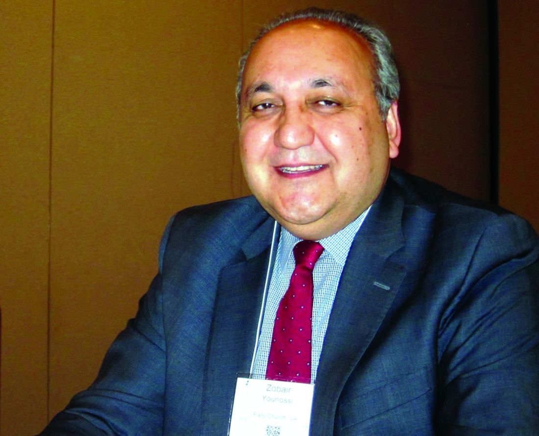
Essentially, value-based care is the evolution of evidence-based medicine, according to Zobair M. Younossi, MD, AGAF, FAASLD, chair of the department of medicine at Inova Fairfax (Va.) Hospital. With value-based care, aligned incentives across all the specialties will lead to more precise accounting and efficiencies of care, said Dr. Younossi, who was also on the panel. “It would be very difficult right now to ask a hospital to tell you exactly what the cost of their liver care would be,” he said.
However, at present there are no true value-based models for hepatology in the United States, according to Dr. Kanwal, so clinicians should start by defining what will “truly constitute value for our patients.”
Because psychosocial support is essential to improved outcomes in patients with cirrhosis, she suggested adding case managers in practice, as they can help coordinate with services in the community at large. Other suggestions she offered included extending office hours, operating an after-hours hotline, and building teams that include general internists, additional nursing staff, and nutritionists.
Even though such changes in clinical practice models now are inevitable, Dr. Kanwal said there are few data at present that support how to innovate care in hospitalized patients with cirrhosis. This matters, as how patients present for outpatient follow-up care will impact reimbursements to the clinicians who treat them.
Some possible ways to improve hospital outcomes for patients with cirrhosis include creating a “best practice alert” that prompts a hepatology consult and triggers the implementation of a standardized set of guidelines for addressing ascites, bleeding, acute kidney injury, encephalopathy, and hepatorenal syndrome. For those with decompensated cirrhosis or for those who need transplants, a similar standardized checklist can be systematized between the hospital and clinic, emphasizing inpatient rifaximin and prophylactic antibiotics in case of spontaneous bacterial peritonitis.
With an overt emphasis on the needs of patients and payers, clinicians now must compete with one another to offer the most comprehensive, cost-effective care supported by information technology structures that can assess real time costs and outcomes, said Dr. Younossi. “Hepatology is lagging behind other fields in all this,” he added.
This worries Dr. Kanwal: “We should be the ones to determine what the value should be. We should be the ones to decide what the model will be and to engage with other fields, and the payers. Otherwise we will not have a seat at the table.”
Dr. Kanwal and Dr. Younossi did not have any relevant financial disclosures.
On Twitter @whitneymcknight
BOSTON – As part of the push to create value-based care across the specialties, hepatologists should consider medical homes for their patients, such as those with cirrhosis, according to experts.
“Cirrhosis is a chronic condition, highly symptomatic, and occurs in highly comorbid individuals. Treating them in a medical home scenario means we can offer services that we don’t otherwise do, but which are associated with better outcomes for our patients,” Fasiha Kanwal, MD, of the department of medicine at Baylor College of Medicine, Houston, said in a panel presentation at the annual meeting of the American Association for the Study of Liver Diseases.
Essentially, value-based care is the evolution of evidence-based medicine, according to Zobair M. Younossi, MD, AGAF, FAASLD, chair of the department of medicine at Inova Fairfax (Va.) Hospital. With value-based care, aligned incentives across all the specialties will lead to more precise accounting and efficiencies of care, said Dr. Younossi, who was also on the panel. “It would be very difficult right now to ask a hospital to tell you exactly what the cost of their liver care would be,” he said.
However, at present there are no true value-based models for hepatology in the United States, according to Dr. Kanwal, so clinicians should start by defining what will “truly constitute value for our patients.”
Because psychosocial support is essential to improved outcomes in patients with cirrhosis, she suggested adding case managers in practice, as they can help coordinate with services in the community at large. Other suggestions she offered included extending office hours, operating an after-hours hotline, and building teams that include general internists, additional nursing staff, and nutritionists.
Even though such changes in clinical practice models now are inevitable, Dr. Kanwal said there are few data at present that support how to innovate care in hospitalized patients with cirrhosis. This matters, as how patients present for outpatient follow-up care will impact reimbursements to the clinicians who treat them.
Some possible ways to improve hospital outcomes for patients with cirrhosis include creating a “best practice alert” that prompts a hepatology consult and triggers the implementation of a standardized set of guidelines for addressing ascites, bleeding, acute kidney injury, encephalopathy, and hepatorenal syndrome. For those with decompensated cirrhosis or for those who need transplants, a similar standardized checklist can be systematized between the hospital and clinic, emphasizing inpatient rifaximin and prophylactic antibiotics in case of spontaneous bacterial peritonitis.
With an overt emphasis on the needs of patients and payers, clinicians now must compete with one another to offer the most comprehensive, cost-effective care supported by information technology structures that can assess real time costs and outcomes, said Dr. Younossi. “Hepatology is lagging behind other fields in all this,” he added.
This worries Dr. Kanwal: “We should be the ones to determine what the value should be. We should be the ones to decide what the model will be and to engage with other fields, and the payers. Otherwise we will not have a seat at the table.”
Dr. Kanwal and Dr. Younossi did not have any relevant financial disclosures.
On Twitter @whitneymcknight
EXPERT ANALYSIS FROM THE LIVER MEETING 2016
Recognizing, addressing giftedness can be challenging
SAN FRANCISCO – Gifted children are far too commonly misunderstood, mislabeled, and misdiagnosed, leading to a mismatch between their needs and others’ perceptions of their needs, Dan Peters, PhD, a licensed psychologist and executive director of the Summit Center in the greater San Francisco and Los Angeles areas, explained at the annual meeting of the American Academy of Pediatrics.
Too often, one or more of these children’s health, developmental, social-emotional or learning needs are overlooked, or they receive an inappropriate mental health, developmental and/or learning disorder diagnosis. In fact, many of the risk factors for giftedness resemble those of other conditions: underachievement, difficulties with peers, social isolation, power struggles, perfectionism, anxiety, and depression.
Further, those who are culturally or linguistically diverse may not be recognized if a non-English first language obscures their performance ability or their socioeconomic status or lack of resources and enrichment opportunities leads them to be overlooked. It’s therefore important that practitioners understand what giftedness actually is and the characteristics gifted children might exhibit.
Understanding giftedness
A simple definition of giftedness is demonstrating a performance or the capacity for performance that significantly exceeds age or grade-level expectations, according to one school district’s gifted and talented education program.
A more involved description provided by the Columbus Group in 1991 defines giftedness as an “asynchronous development in which advanced cognitive abilities and heightened intensity combine to create inner experiences and awareness that are qualitatively different from the norm.” This asynchrony increases with higher intellectual capacity, they wrote. “The uniqueness of the gifted renders them vulnerable and requires modifications in parenting, teaching, and counseling in order for them to develop optimally.”
The level of a child’s giftedness makes a difference in their needs as well; these levels include advanced learners (IQ of 120-129), moderately gifted (130-144), highly gifted (145-159), exceptionally gifted (160-179), and profoundly gifted (180 and greater). Different spheres of giftedness can include intellectual ability, creative or productive thinking, leadership ability, and visual or performing arts. Consider the list of common characteristics of gifted children that Dr. Peters provided:
- Rapid learners.
- Strong memory.
- Large vocabulary.
- Advanced comprehension of nuances.
- Largely self-taught.
- Unusual emotional depth.
- Abstract/complex/logical/insightful thinking.
- Idealism and a sense of justice.
- Intense feelings and reactions.
- Highly sensitive.
- Long attention span and persistence.
- Preoccupied with own thoughts.
- Impatient with self and others’ inabilities and slowness.
- Asks probing questions (able to go beyond what is taught).
- Wide range of interests.
- Highly developed curiosity.
- Interest in experimenting and doing things differently.
- Divergent thinking.
- Keen and unusual sense of humor.
Dr. Peters cited Kazimierz Dabrowski, MD, PhD, a Polish psychiatrist of the mid-20th century, as explaining the sensitivity and intensity experienced by many gifted individuals in terms of overexcitabilities – a “greater capacity to be stimulated by and respond to external and internal stimuli.”
“Overexcitability permeates a gifted person’s existence and gives energy to their intelligence, talents, and personality,” Dr. Peters explained of Dabrowski’s ideas. This enhancement manifests in psychomotor terms as a strong drive, a lot of energy or movement, or extended bouts of activity. Intellectually, gifted children have an “insatiable curiosity, and voracious appetite and capacity for intellectual effort and stimulation,” Dr. Peters said. They may have heightened sensual experience in seeing, smelling, tasting, touching, or hearing, and they have an active imaginary and fantasy life. They also exhibit a capacity for great emotional depth and empathy – they deeply feel their own and others’ emotions.
How giftedness can be misdiagnosed
It is the combination of these very characteristics that can lead gifted children to receive an inappropriate mental or developmental diagnosis instead of being recognized as gifted.
“By current estimates, at any given time, approximately 11%-20% of children in the United States have a behavioral or emotional disorder as defined in the DSM-5,” Dr. Peters cited. Further, one study found that diagnoses of attention-deficit/hyperactivity disorder have increased 66% between 2000 and 2010, with 90% of those children taking psychostimulant medications – yet a study in the Journal of Health Economics estimated that one in five children diagnosed with ADHD are probably misdiagnosed and are receiving those medications.
Other incorrect diagnoses besides ADHD that gifted youth may commonly receive include anger diagnoses, ideational or anxiety disorders, developmental and personality disorders, mood disorders, and learning disorders.
Twice exceptionalism (2e)
Even more challenging are twice exceptional children, or 2e, those who are both gifted and have a learning or emotional disability or challenge. Common dual diagnoses in gifted children include anxiety disorders, depression (or existential depression), sleep disorders (such as nightmares, night terrors, or sleep walking), allergies, asthma, ADHD, oppositional-defiant disorder, obsessive-compulsive personality disorder, autism spectrum disorder, nonverbal learning disability, social/pragmatic communication disorder, and learning disorders such as dyslexia, dyscalculia, central auditory processing disorder, or sensory-motor integration disorder.
“It’s very complex. What happens is, a lot of people think you’re either gifted or not,” Dr. Peters said. “In the classroom, sometimes the advanced ability overshadows the weakness and so we get a lot of readers with an IQ of 130-150 and reading at the 50% percentile, and everyone says they’re fine, but they’re dyslexic.”
Other times, the weakness overshadows the strength, and sometimes they’re right in the middle where neither their giftedness nor their disability is recognized or addressed, Peters said. 2e children are very difficult to diagnose but also at higher risk for difficulties if one or both (or more) of their diagnoses are missed.
Maximizing gifted children’s developmental potential
Pediatricians have an opportunity to support gifted children by recognizing and accepting them for who they are, while also acknowledging that they want to feel “normal,” and therefore need extra reassurance and support from adults. Pediatricians should seek information about giftedness and 2e children from state and national gifted organizations, and, in the office, frame conversations with families and children’s differential diagnoses in terms of a child’s giftedness. If a pediatrician is themself gifted, they may be “a supportive and kindred spirit” to the child, Dr. Peters said.
In daily life, as well, gifted children need to be accepted for who they are, provided opportunities to be with their intellectual and academic peers, and provided challenges in their areas of strength, interests, or passions. Parents and teachers should follow their lead in learning: Keep up the pace for those who want to learn fast, and go deeper for those who want slower, more in-depth learning. Adults also need to understand their intensities and sensitivities and lead with their strengths in discussions.
Dr. Peters reported no disclosures.
EXPERT ANALYSIS FROM AAP 16
Mixed findings on iron repletion in HF
NEW ORLEANS – Oral iron supplementation was declared ineffective for the treatment of iron deficiency in patients with heart failure with reduced ejection fraction in one major randomized trial while intravenous ferric carboxymaltose improved exercise capacity and quality of life in another study presented at the American Heart Association scientific sessions.
Iron deficiency is present in roughly half of patients with heart failure with reduced ejection fraction (HFrEF). In affected patients it’s associated with diminished functional capacity and poor quality of life, and is an independent predictor of mortality. Thus, it has emerged as a potential therapeutic target in HFrEF.
The IRONOUT HF (Oral Iron Repletion Effects on Oxygen UpTake in Heart Failure) trial was a multicenter, randomized, double-blind, placebo-controlled clinical trial conducted by the National Heart, Lung, and Blood Institute’s Clinical Heart Failure Network investigators. It included 225 patients with HFrEF and iron deficiency who were randomized to 16 weeks of oral iron polysaccharide at 150 mg b.i.d. or matching placebo.
The primary endpoint was change from baseline to 16 weeks in exercise capacity, as measured via peak oxygen uptake during cardiopulmonary exercise testing. The results were no better in the iron-supplemented group than in placebo-treated controls. Nor was there any benefit for oral iron therapy in terms of quality of life, as assessed by the Kansas City Cardiomyopathy Questionnaire or any of numerous other secondary endpoints, according to Dr. Lewis, head of the heart failure section and director of the cardiopulmonary exercise testing laboratory at Massachusetts General Hospital, Boston.
Participants’ mean baseline hemoglobin level was 12.6 g/dL, but whether patients were anemic at baseline or not made no difference in terms of outcomes. Rates of venous congestion and bleeding were low during the trial.
The problem, the investigators found, was that high-dose oral iron only minimally repleted iron stores. Transferrin saturation increased by a paltry absolute 3% after 16 weeks of twice-daily therapy. Serum ferritin levels increased only 1/20th as much as after intravenous iron ferric carboxymaltose therapy in the earlier positive FAIR-HF (Ferinject Assessment in Patients With Iron Deficiency and Chronic Heart Failure) trial (N Engl J Med. 2009 Dec 17;361:2436-48).
Levels of hepcidin were elevated in study participants. And the higher the level of hepcidin – which Dr. Lewis called “a massive regulator of iron bioavailability and absorption” – the more refractory patients were to oral iron repletion.
“Hepcidin was invented by nature to protect against iron from stimulating growth of bacteria. And when it’s elevated, taking iron orally just has no chance of success,” explained Dr. Anker, professor of cardiology at the University of Gottingen (Germany).
Dr. Anker, who chaired the positive FAIR-HF trial of intravenous iron, said IRONOUT HF was a very well-conducted and definitive clinical trial of oral iron supplementation in HFrEF.
“It’s very simple: Oral iron does not work in patients with chronic heart failure. That’s the take home message. It’s true for peak VO2 [oxygen consumption], for 6-minute walk distance, for symptoms, for quality of life, and even for surrogate markers like NT-proBNP [N-terminal pro b-type natriuretic peptide]. If iron doesn’t get into the body, it’s really difficult to [imagine] that the iron that doesn’t get into the body can exert an effect,” he said.
The primary endpoint in EFFECT-HF was assessor-blinded change in peak VO2 from baseline to week 24. The control group experienced a decrease in peak VO2 over time such that there was a significant difference of 1.0 mL/kg per minute between the two groups.
The ferric carboxymaltose recipients also did significantly better in terms of secondary endpoints including improvement in New York Heart Association functional class, self-reported Patient Global Assessment score, and quality-of-life measures, reported Dr. van Veldhuisen, professor and chairman of cardiology at University Medical Center Groningen (the Netherlands).
Session moderator Clyde Yancy, MD, commented that intravenous iron is not ready for prime time use in clinical practice for several reasons. The open-label EFFECT-HF trial, like the earlier positive double-blind FAIR-HF and CONFIRM-HF IV ferric carboxymaltose trials, was too modest in size to be convincing, especially since this is an expensive and intrusive therapy.
“The endpoint of peak VO2, although a very powerful endpoint, is still one for which there may be some subjectivity, and so we need a more definitive endpoint to be absolutely certain about the potential benefit of the administration of ferric carboxymaltose,” said Dr. Yancy, professor of medicine and chief of cardiology at Northwestern University in Chicago.
Discussant Adrian Hernandez, MD, of the Duke Clinical Research Institute in Durham, N.C., said he considers peak VO2 an important endpoint.
“When you talk to patients, exercise capacity is an outcome that matters to them,” he said. “They often comment that what matters to them is living longer with a better quality of life, free of worsening heart failure, and having improvement in everyday functional status. So the cardiopulmonary exercise test is not just a surrogate endpoint; it’s a measure of functional outcome that matters to patients,” he said.
Still, like Dr. Yancy, Dr. Hernandez said he thinks it’s time to have larger, longer, definitive trials with clinical endpoints in order to understand the role of intravenous iron. Both cardiologists applauded Dr. Anker’s announcement that such a trial, known as FAIR-HF2, is now getting started.
The IRONOUT HF trial was funded by the National Institutes of Health. Dr. Lewis reported receiving research support from a handful of pharmaceutical and medical device companies. Dr. van Velduisen reported serving as a scientific adviser to Vifor Pharma, which sponsored the EFFECT-HF trial. Dr. Anker, who was an EFFECT-HF investigator, serves as a consultant to Vifor and several other companies. Dr. Hernandez was an IRONOUT HF investigator and reported receiving research grants from a handful of pharmaceutical companies.
EXPERT ANALYSIS FROM THE AHA SCIENTIFIC SESSIONS
VIDEO: Appealing DAA denials is worth it for hepatitis C patients
BOSTON – Though criteria for insurance approval for direct-acting antivirals (DAAs) for hepatitis C virus (HCV) are loosening, many patients are still denied by both public and private carriers. Providers and patients should know that it’s worthwhile to appeal these denials; 78.1% (115 of 145) of initial DAA denials were approved on appeal, according to a recent study.
Jane Giang, PharmD, a clinical pharmacist at the University of North Carolina Health System, Chapel Hill, worked with colleagues to conduct a retrospective study of patients at her facility who were prescribed a DAA for HCV between October 2014 and April 2016.
Dr. Giang, in a video interview at the annual meeting of the American Association for the Study of Liver Diseases, said that she works alongside physicians in her clinic to craft appeals, tailoring appeal letters to individual patients’ circumstances.
The video associated with this article is no longer available on this site. Please view all of our videos on the MDedge YouTube channel
In a poster presentation, Dr. Giang and her colleagues presented results from a total of 1,017 patients who were prescribed DAAs for HCV during the study time frame. Of these, three-quarters (n = 794, 78.1%) received initial approval for their medication, while one in five patients (n = 206, 20.2%) had their initial applications denied. The remaining patients either had their cases closed for a variety of reasons, or had pending applications at the time of data analysis.
About half of the patients (n = 496, 48.8%) had commercial insurance. Medicare was the primary insurance for another 207 (n = 20.4%), and Medicaid for 140 (13.8%).
Of the patients whose DAAs were initially denied, 65 (31.6%) of the denials were for fibrosis less than stage F2. Another quarter of patients (n = 57, 27.7%) did not meet approval criteria, and 41 (19.9%) of denials said that the DAA was nonformulary or excluded by the plan. The remaining denials were for missing information or a variety of other reasons.
Half of the patients (n = 504, 49.6%) had F4 fibrosis at the time of application for DAA treatment; 241 (23.7%) had F0 or F1 fibrosis, and the remainder were approximately evenly split between F3 and F4 fibrosis.
When a subset of these denials went through an appeals process, most were eventually approved. Of the 42 denials for less than F2 fibrosis that were appealed, 20 (47.6%) were eventually approved. Of the 40 patients whose applications for DAAs were based on not meeting criteria, 37 (92.5%) were approved on appeal, and of the 32 denials based on plan exclusions or DAAs being nonformulary, 29 (90.6%) were approved on appeal.
When Dr. Giang and her colleagues broke down the numbers by type of insurance, they found that of the 496 patients who had commercial insurance, only 44 (8.9%) eventually received a final denial. Medicare patients had a 3.9% final denial rate, while just one of the 172 patients (0.6%) without insurance who received medication through programs directly from the manufacturer had a final denial.
The largest number of final denials occurred in the group of patients with Medicaid. Of these 140 patients, 25 (17.9%) had a final denial. Dr. Giang said that in North Carolina, providers cannot file appeals for DAA approval on behalf of patients; the paperwork is sent directly to the patient’s home, and patients have to complete and file the forms. She said that she and her colleagues learned to alert these patients to be on the lookout for paperwork, and counseled them to bring the appeal forms into the clinic so that staff could assist them in completing the appeal.
Though Dr. Giang said that her study showed that payer type matters when it comes to reimbursement for HCV care, the “impact of payer type on access to care remains incompletely explored, especially among noninsured patients,” she said.
Dr. Giang reported no outside sources of funding, and no conflicts of interest.
[email protected]
On Twitter@karioakes
AT THE LIVER MEETING 2016

Surgery for bowel obstruction in cancer patients didn’t increase 90-day mortality
CORONADO, CALIF. – Among advanced cancer patients with bowel obstruction, surgery was not an independent predictor of the ability to eat at discharge or survival within 90 days of consultation, results from a long-term retrospective study showed.
“I think this represents the complexity in treating these patients,” lead study author Brian D. Badgwell, MD, said at the annual meeting of the Western Surgical Association. “We need future studies to identify the optimal outcome measures.”

For the current study, the researchers retrospectively reviewed the medical records of 490 patients who required surgical consultation for bowel obstruction at MD Anderson Cancer Center between January 2000 and May 2014. They set out to determine the incidence of obstruction due to intra-abdominal tumor and to identify variables associated with the ability to eat at hospital discharge and 90-day survival. They excluded patients without clinical or radiologic features of mechanical bowel obstruction. Clinical variables of interest included obstruction site, tumor vs. non-tumor cause, laboratory parameters, radiologic extent of malignancy, and the type of treatment performed (surgical, medical, or interventional, defined as interventional radiology or endoscopy). Overall survival was calculated from the date of first surgical evaluation for bowel obstruction to any cause mortality or last follow-up. Univariate and multivariate analyses were performed for ability to eat and a Cox proportional hazards model for 90-day survival.
Dr. Badgwell reported that the most common obstruction site in the 490 patients was the small bowel (64%), followed by large bowel (20%) and gastric outlet (16%). Obstruction etiology was identified as tumor-related in 68% of cases, followed by adhesion-related (20%) and unclear (12%). Nearly half of patients (46%) received chemotherapy within 6 weeks of their surgical consultation, but only 4% were neutropenic. More than half of patients (52%) had an albumin level of less than 3.5 g/dL, 52% had a hemoglobin of 10 g/dL or greater, 36% had lymphadenopathy, 35% had ascites, 34% had peritoneal disease, and 31% had a primary or recurrent tumor in place. In addition, 53% had an abdominal visceral malignancy, 9% had bone metastases, and 14% had lung metastases.
About half of patients (49%) received medical management as their treatment, followed by surgical and procedural treatment (32% and 17%, respectively). Fifteen percent were discharged to in-home hospice or to an inpatient hospice facility. More than two-thirds (68%) were able to eat at the time of discharge, and 43% died within 90 days of surgical consultation.
Multivariate analysis revealed that the following factors were negatively associated with eating at discharge: an intact/primary local recurrence (odds ratio, 0.46), carcinomatosis (OR, 0.34), and albumin level of less than 3.5 g/dL (OR, 0.55). At the same time, variables associated with death within 90 days of consultation included having an intact primary/local recurrence (hazard ratio, 1.75), carcinomatosis (HR, 1.98), and abdominal visceral metastasis (HR, 1.75). Finally, compared with procedural treatment, both medical management and surgical management were negatively associated with death within 90 days (HR of 0.51 and 0.44, respectively).
“There is a high rate of non-mechanical bowel dysfunction in patients undergoing surgical consultation for bowel obstruction,” Dr. Badgwell concluded. “It’s very difficult to categorize these cases preoperatively. They do require a selective approach. Variables associated with outcome measures support caution in patients with carcinomatosis, hypoalbuminemia, and multiple sites of disease on imaging.”
Dr. Badgwell reported having no financial disclosures.
CORONADO, CALIF. – Among advanced cancer patients with bowel obstruction, surgery was not an independent predictor of the ability to eat at discharge or survival within 90 days of consultation, results from a long-term retrospective study showed.
“I think this represents the complexity in treating these patients,” lead study author Brian D. Badgwell, MD, said at the annual meeting of the Western Surgical Association. “We need future studies to identify the optimal outcome measures.”
For the current study, the researchers retrospectively reviewed the medical records of 490 patients who required surgical consultation for bowel obstruction at MD Anderson Cancer Center between January 2000 and May 2014. They set out to determine the incidence of obstruction due to intra-abdominal tumor and to identify variables associated with the ability to eat at hospital discharge and 90-day survival. They excluded patients without clinical or radiologic features of mechanical bowel obstruction. Clinical variables of interest included obstruction site, tumor vs. non-tumor cause, laboratory parameters, radiologic extent of malignancy, and the type of treatment performed (surgical, medical, or interventional, defined as interventional radiology or endoscopy). Overall survival was calculated from the date of first surgical evaluation for bowel obstruction to any cause mortality or last follow-up. Univariate and multivariate analyses were performed for ability to eat and a Cox proportional hazards model for 90-day survival.
Dr. Badgwell reported that the most common obstruction site in the 490 patients was the small bowel (64%), followed by large bowel (20%) and gastric outlet (16%). Obstruction etiology was identified as tumor-related in 68% of cases, followed by adhesion-related (20%) and unclear (12%). Nearly half of patients (46%) received chemotherapy within 6 weeks of their surgical consultation, but only 4% were neutropenic. More than half of patients (52%) had an albumin level of less than 3.5 g/dL, 52% had a hemoglobin of 10 g/dL or greater, 36% had lymphadenopathy, 35% had ascites, 34% had peritoneal disease, and 31% had a primary or recurrent tumor in place. In addition, 53% had an abdominal visceral malignancy, 9% had bone metastases, and 14% had lung metastases.
About half of patients (49%) received medical management as their treatment, followed by surgical and procedural treatment (32% and 17%, respectively). Fifteen percent were discharged to in-home hospice or to an inpatient hospice facility. More than two-thirds (68%) were able to eat at the time of discharge, and 43% died within 90 days of surgical consultation.
Multivariate analysis revealed that the following factors were negatively associated with eating at discharge: an intact/primary local recurrence (odds ratio, 0.46), carcinomatosis (OR, 0.34), and albumin level of less than 3.5 g/dL (OR, 0.55). At the same time, variables associated with death within 90 days of consultation included having an intact primary/local recurrence (hazard ratio, 1.75), carcinomatosis (HR, 1.98), and abdominal visceral metastasis (HR, 1.75). Finally, compared with procedural treatment, both medical management and surgical management were negatively associated with death within 90 days (HR of 0.51 and 0.44, respectively).
“There is a high rate of non-mechanical bowel dysfunction in patients undergoing surgical consultation for bowel obstruction,” Dr. Badgwell concluded. “It’s very difficult to categorize these cases preoperatively. They do require a selective approach. Variables associated with outcome measures support caution in patients with carcinomatosis, hypoalbuminemia, and multiple sites of disease on imaging.”
Dr. Badgwell reported having no financial disclosures.
AT WSA 2016
Key clinical point:
Major finding: Compared with procedural treatment of bowel obstruction, both medical management and surgical management were negatively associated with death within 90 days (HR of 0.51 and 0.44, respectively).
Data source: A retrospective review of 490 patients with advanced cancer who required surgical consultation for bowel obstruction at MD Anderson Cancer Center, Houston, between January 2000 and May 2014.
Disclosures: Dr. Badgwell reported having no financial disclosures.