User login
VIDEO: NAFLD costing U.S. $290 billion annually
BOSTON – A sophisticated mathematical analysis shows that nonalcoholic fatty liver disease is costing the United States over $290 billion annually. A similar economic burden was seen in several Western European countries as well.
The study, which incorporated available data about current cases of nonalcoholic fatty liver disease (NAFLD), looked at both medical and nonmedical costs, as the disease’s economic and social impact extends far beyond medical bills.
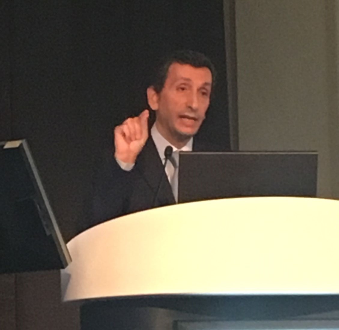
“It impacts patients’ quality of life, their work productivity; there’s a significant fatigue associated with that. In addition to this, there is an economic burden of nonalcoholic fatty liver disease,” said the study’s lead author, Zobair Younossi, MD, in a video interview at the annual meeting of the American Association for the Study of Liver Diseases.
The video associated with this article is no longer available on this site. Please view all of our videos on the MDedge YouTube channel
Dr. Younossi, executive director of the Center for Liver Diseases at Inova Fairfax Hospital, Falls Church, Va., and his coinvestigators constructed a Markov chain to model costs associated with NAFLD. This mathematical technique allows the probability of the occurrence of an event to influence the model’s prediction of the probability of later events, a useful technique when trying to model real-world, dynamic conditions.
They used the Markov chain technique to model the transition of patients along the path of NAFLD progression. States included in the model were nonalcoholic steatohepatitis (NASH), NASH with fibrosis, compensated and decompensated cirrhosis, hepatocellular carcinoma, transplant, posttransplant, and death. The probabilities of progressing through these states were built from a meta-analysis and systematic literature review conducted by the authors, which they then adjusted by incorporating real-world data.
According to the model, annual direct medical costs are projected at $103 billion, or $1,613 for each of the 64 million NAFLD patients in the United States. The four European Union countries included in the model were Germany, France, Italy, and the United Kingdom. In aggregate, these countries are projected to have about 52 million people with NAFLD, for an annual cost of 35 billion euros. The estimated annual direct cost per patient varies by country, ranging from 354 euros to 1,163 euros.
When societal costs are incorporated, the numbers leap higher: to $292.19 billion in the United States, and over 200 billion euros in the four European countries studied.
In recent work, Dr. Younossi and his colleagues have reached an estimate that about a quarter of the world’s population has NAFLD. He said he was surprised to learn that the highest prevalences were in some areas of South America and the Middle East, with rapid increases in Asia as well.
However, the etiology of NAFLD helps explain these increases. “It’s really a phenotype. A number of different pathways lead to this disease; the most common is the one that is associated with obesity, type 2 diabetes, and insulin resistance,” said Dr. Younossi.
The subset of NAFLD patients who have NASH also risks progression to cirrhosis and hepatocellular carcinoma. According to Dr. Younossi’s examination of the UNOS transplant database, NASH is the second leading cause of liver transplantation in the United States, and one of the top three causes of death from liver disease.
These sobering data make the need for medical treatment for NASH an urgent priority, said Dr. Younossi. Currently, lifestyle modification such as weight loss is the only known treatment for NAFLD and NASH, “which is not easy to do,” he noted.
Several candidate drugs are in the pipeline currently. Also, said Dr. Younossi, NASH can be diagnosed only by liver biopsy currently, a risky and expensive procedure. The search is on for accurate and reliable imaging and serum biomarkers for NASH, so physicians can understand who’s most at risk for progression to more serious liver disease.
“The analysis quantifies the enormity of the clinical and economic burden of NAFLD, which will likely increase as incidence of NAFLD continues to rise with the increasing obesity and diabetes rates,” wrote Dr. Younossi and his coauthors.
Dr. Younossi reported having financial relationships with several pharmaceutical companies.
[email protected]
On Twitter @karioakes
BOSTON – A sophisticated mathematical analysis shows that nonalcoholic fatty liver disease is costing the United States over $290 billion annually. A similar economic burden was seen in several Western European countries as well.
The study, which incorporated available data about current cases of nonalcoholic fatty liver disease (NAFLD), looked at both medical and nonmedical costs, as the disease’s economic and social impact extends far beyond medical bills.
“It impacts patients’ quality of life, their work productivity; there’s a significant fatigue associated with that. In addition to this, there is an economic burden of nonalcoholic fatty liver disease,” said the study’s lead author, Zobair Younossi, MD, in a video interview at the annual meeting of the American Association for the Study of Liver Diseases.
The video associated with this article is no longer available on this site. Please view all of our videos on the MDedge YouTube channel
Dr. Younossi, executive director of the Center for Liver Diseases at Inova Fairfax Hospital, Falls Church, Va., and his coinvestigators constructed a Markov chain to model costs associated with NAFLD. This mathematical technique allows the probability of the occurrence of an event to influence the model’s prediction of the probability of later events, a useful technique when trying to model real-world, dynamic conditions.
They used the Markov chain technique to model the transition of patients along the path of NAFLD progression. States included in the model were nonalcoholic steatohepatitis (NASH), NASH with fibrosis, compensated and decompensated cirrhosis, hepatocellular carcinoma, transplant, posttransplant, and death. The probabilities of progressing through these states were built from a meta-analysis and systematic literature review conducted by the authors, which they then adjusted by incorporating real-world data.
According to the model, annual direct medical costs are projected at $103 billion, or $1,613 for each of the 64 million NAFLD patients in the United States. The four European Union countries included in the model were Germany, France, Italy, and the United Kingdom. In aggregate, these countries are projected to have about 52 million people with NAFLD, for an annual cost of 35 billion euros. The estimated annual direct cost per patient varies by country, ranging from 354 euros to 1,163 euros.
When societal costs are incorporated, the numbers leap higher: to $292.19 billion in the United States, and over 200 billion euros in the four European countries studied.
In recent work, Dr. Younossi and his colleagues have reached an estimate that about a quarter of the world’s population has NAFLD. He said he was surprised to learn that the highest prevalences were in some areas of South America and the Middle East, with rapid increases in Asia as well.
However, the etiology of NAFLD helps explain these increases. “It’s really a phenotype. A number of different pathways lead to this disease; the most common is the one that is associated with obesity, type 2 diabetes, and insulin resistance,” said Dr. Younossi.
The subset of NAFLD patients who have NASH also risks progression to cirrhosis and hepatocellular carcinoma. According to Dr. Younossi’s examination of the UNOS transplant database, NASH is the second leading cause of liver transplantation in the United States, and one of the top three causes of death from liver disease.
These sobering data make the need for medical treatment for NASH an urgent priority, said Dr. Younossi. Currently, lifestyle modification such as weight loss is the only known treatment for NAFLD and NASH, “which is not easy to do,” he noted.
Several candidate drugs are in the pipeline currently. Also, said Dr. Younossi, NASH can be diagnosed only by liver biopsy currently, a risky and expensive procedure. The search is on for accurate and reliable imaging and serum biomarkers for NASH, so physicians can understand who’s most at risk for progression to more serious liver disease.
“The analysis quantifies the enormity of the clinical and economic burden of NAFLD, which will likely increase as incidence of NAFLD continues to rise with the increasing obesity and diabetes rates,” wrote Dr. Younossi and his coauthors.
Dr. Younossi reported having financial relationships with several pharmaceutical companies.
[email protected]
On Twitter @karioakes
Key clinical point:
Major finding: Annual direct medical costs alone are $1,613 for each of the estimated 64 million U.S. NAFLD patients.
Data source: Markov-chain modeling of NAFLD prevalence and morbidity in the United States and four European countries.
Disclosures: Dr. Younossi reported financial relationships with several pharmaceutical companies.

Four-part ‘safety bundle’ targets gynecologic surgery infections
In a new consensus safety bundle designed to reduce the frequency of infections related to gynecologic surgery, an expert panel is calling for preoperative evaluation of infection risk in every patient and surgical timeouts in every case.
The bundle, from the Council on Patient Safety in Women & Mother’s Health Care, seeks to compile existing guidelines and evidence-based recommendations in a way that can be easily implemented with whatever resources an individual institution has available.
The safety bundle covers four domains:
1. Readiness (every facility). The statement calls for standardized preoperative and postoperative care instructions and clearly defined roles for each surgical team member.
Standards should also be established regarding skin preparation, use of prophylactic antibiotics (terminate them within 24 hours after surgery completion unless medical indications are present), and temperature, such as ambient operating room temperature.
“Although the effect of temperature maintenance on surgical site infection is not definitive,” the consensus statement says, “there is no denying other benefits of normothermia; foremost among these is overall patient satisfaction and comfort.”
2. Recognition and prevention (every patient). Every patient should undergo a preoperative evaluation of infection risk based on blood glucose level, body mass index, immunodeficiency, methicillin-resistant Staphylococcus aureus (MRSA) status, nutritional status, and smoking status.
3. Response (every case). Develop “timeouts” during operations, as mandated by the Joint Commission, to address antibiotic dosage and timing, and reassess risk for infection following the procedure based on the length of surgery and blood loss.
4. Reporting and systems learning (every facility). Develop “huddles” – brief team meetings of less than 15 minutes – for high-risk patients. Surgeons and hospital officials should also create a system to report and analyze data about surgical site infections and share physician-specific infection data with all surgeons as part of ongoing professional practice evaluation.
The statement appeared in Obstetrics & Gynecology (2017;129:50-61) and was published concurrently in Anesthesia & Analgesia, the Journal of Obstetric, Gynecologic & Neonatal Nursing, and the AANA Journal.
The authors reported having no potential conflicts of interest.
FROM OBSTETRICS & GYNECOLOGY
Autologous stem cell transplantation beat bortezomib regimen in myeloma
SAN DIEGO – Autologous stem cell transplantation outperformed bortezomib-based intensification in fit patients younger than 66 years of age with newly diagnosed multiple myeloma, based on a prespecified interim analysis of 1,192 patients from a randomized phase III trial.
After a median follow-up of 32 months, median progression-free survival (PFS) had not been reached among patients who received high-dose melphalan plus single or double autologous stem cell transplantation, but was 42.5 months among patients who instead received standard-dose bortezomib-melphalan-prednisone (VMP), Michele Cavo, MD, reported at the annual meeting of the American Society of Hematology. Three-year rates of progression free survival were 65% with ASCT and 57% with VMP (hazard ratio, 0.73; 95% confidence interval, 0.61-0.88; P = .001), he reported.
There was a trend toward better outcomes with double ASCT instead of single ASCT, said Dr. Cavo of Bologna (Italy) University. At 3 years, PFS rates were 74% with double ASCT and 62% with single ASCT (HR, 0.7; P = .05).
The effect was stronger among patients with high-risk cytogenetics, for whom 3-year PFS rates were 65% and 41% (HR, 0.49; P = .046). Those patients had median PFS times of 47 months and 27 months, respectively, Dr. Cavo said. In a multivariable analysis, double ASCT also reduced the chances of death or progression by about 35% compared with single ASCT, even after controlling for high-risk cytogenetics, age, and other risk factors for poor prognosis (HR, 0.65; P = .03).
This is the first trial of its type to prospectively compare single and double ASCT with a novel myeloma regimen, according to Dr. Cavo. The data are not yet mature enough to support firm conclusions, but do highlight the role of ASCT in the bortezomib era and the potential for double ASCT to benefit patients with poor prognostic risk factors, particularly high-risk cytogenetics, he said.
The EMN02/HO95 trial enrolled more than 1,500 patients aged 18-65 years with symptomatic, newly diagnosed multiple myeloma. Patients underwent induction therapy with three to four cycles of bortezomib plus cyclophosphamide and dexamethasone (VCD), and then were randomly assigned to either high-dose melphalan (200 mg/m2) plus single or double ASCT, or to four cycles of bortezomib (1.3 mg/m2), melphalan (9 mg/m2), and prednisone (60 mg/m2; VMP). Patients were then re-randomized to receive lenalidomide maintenance alone or after consolidation with two cycles of bortezomib, lenalidomide, and dexamethasone (VRD).
This prespecified analysis was triggered in early November 2016, when 33% of required events occurred. Future analyses will examine the effects of consolidation as well as safety, toxicity, and quality of life, Dr. Cavo noted.
Celgene and Janssen provided funding for the study. Dr. Cavo disclosed ties to Celgene, Janssen, Takeda, Bristol-Myers Squibb, and Amgen.
AT ASH 2016
Key clinical point: Autologous stem cell transplantation outperformed bortezomib-based intensification in patients with newly diagnosed multiple myeloma.
Major finding: Progression-free survival at 3 years was 65% with melphalan plus ASCT and 57% with bortezomib, melphalan, and prednisone (HR, 0.73; P = .001).
Data source: An interim analysis of a phase III study of 1,510 patients with newly diagnosed multiple myeloma.
Disclosures: Celgene and Janssen provided funding. Dr. Cavo disclosed ties to Celgene, Janssen, Takeda, Bristol-Myers Squibb, and Amgen.
Initial data on biosimilars in IBD are encouraging, but more needed
ORLANDO – The rapidly evolving field of biologic and biosimilar agents can leave gastroenterologists reeling in terms of what role biosimilars will play in practice and how best to utilize them for a patient with ulcerative colitis or Crohn’s disease. Limited data so far – primarily case series and limited postmarketing findings – suggest that a biosimilar can be as safe and effective as the innovator biologic. However, more rigorous research and more research overall are needed to confirm this comparative efficacy and to address unanswered questions around switching and cost effectiveness.
The uncertainty stems in part from a lack of prospective, randomized comparison studies to guide evidence-based clinical practice, Miguel D. Regueiro, MD, gastroenterologist and IBD clinical medical director at the University of Pittsburgh Medical Center.

As clinicians await more definitive evidence, a number of limited studies have been published in the past year assessing biosimilars in inflammatory bowel disease (IBD). “The clinical response ranges were high, in most studies more than two-thirds [of patients] had a response, but again there are no head-to-head studies,” Dr. Regueiro said at the meeting sponsored by the Crohn’s & Colitis Foundation of America.
“We don’t have the data yet in IBD.”
A review of the literature on anti-tumor necrosis factor antibody biosimilar Inflectra (infliximab-dyyb, Celltrion) supports its bioequivalence to infliximab (Remicade, Janssen) in ankylosing spondylitis (Aliment Pharmacol Ther. 2015;42:1158-69). However, dosing regimens and use of concomitant medications differ in the IBD population, the investigators noted, so the case series and short-duration postmarketing results published in the literature leave unanswered questions for gastroenterologists.
Just because biosimilars appear bioequivalent so far in IBD, it does not mean this new class of agents is free of controversy. When the U.S. Food and Drug Administration approved Inflectra in April 2016 based on evidence in ankylosing spondylitis and rheumatoid arthritis, it extrapolated the approval to all conditions for which infliximab is approved, including ulcerative colitis and Crohn’s disease. However, this “highly controversial approach has been criticized by various rheumatology and gastroenterology professional societies around the world,” according to authors of a critical review on biosimilars in IBD (Inflamm Bowel Dis. 2016;22:2513-26).
Clinicians can safely switch patients from infliximab to Inflectra, according to the noninferiority phase IV NOR-SWITCH study conducted in Norway. Researchers assessed almost 500 patients on stable infliximab treatment for a minimum of 6 months prior to switching. However, this was a multi-indication study that included inflammatory bowel disease patients and many others, Dr. Regueiro said. “When you read the fine print in these studies, especially with Crohn’s disease, you start to see differences,” he added.
Although the evidence is encouraging so far, “we do not have prospective, comparative studies yet.”
During a Q&A panel session, a meeting attendee asked if clinicians will be able to use the same laboratory assay used to check a biologic’s drug and antibody levels for its biosimilar. “Our understanding is you will not be able to use the same assay as you do for infliximab. There are companies developing assays specifically for the biosimilars, and I think they will be available in addition to the ones for the innovator agents.”
ORLANDO – The rapidly evolving field of biologic and biosimilar agents can leave gastroenterologists reeling in terms of what role biosimilars will play in practice and how best to utilize them for a patient with ulcerative colitis or Crohn’s disease. Limited data so far – primarily case series and limited postmarketing findings – suggest that a biosimilar can be as safe and effective as the innovator biologic. However, more rigorous research and more research overall are needed to confirm this comparative efficacy and to address unanswered questions around switching and cost effectiveness.
The uncertainty stems in part from a lack of prospective, randomized comparison studies to guide evidence-based clinical practice, Miguel D. Regueiro, MD, gastroenterologist and IBD clinical medical director at the University of Pittsburgh Medical Center.
As clinicians await more definitive evidence, a number of limited studies have been published in the past year assessing biosimilars in inflammatory bowel disease (IBD). “The clinical response ranges were high, in most studies more than two-thirds [of patients] had a response, but again there are no head-to-head studies,” Dr. Regueiro said at the meeting sponsored by the Crohn’s & Colitis Foundation of America.
“We don’t have the data yet in IBD.”
A review of the literature on anti-tumor necrosis factor antibody biosimilar Inflectra (infliximab-dyyb, Celltrion) supports its bioequivalence to infliximab (Remicade, Janssen) in ankylosing spondylitis (Aliment Pharmacol Ther. 2015;42:1158-69). However, dosing regimens and use of concomitant medications differ in the IBD population, the investigators noted, so the case series and short-duration postmarketing results published in the literature leave unanswered questions for gastroenterologists.
Just because biosimilars appear bioequivalent so far in IBD, it does not mean this new class of agents is free of controversy. When the U.S. Food and Drug Administration approved Inflectra in April 2016 based on evidence in ankylosing spondylitis and rheumatoid arthritis, it extrapolated the approval to all conditions for which infliximab is approved, including ulcerative colitis and Crohn’s disease. However, this “highly controversial approach has been criticized by various rheumatology and gastroenterology professional societies around the world,” according to authors of a critical review on biosimilars in IBD (Inflamm Bowel Dis. 2016;22:2513-26).
Clinicians can safely switch patients from infliximab to Inflectra, according to the noninferiority phase IV NOR-SWITCH study conducted in Norway. Researchers assessed almost 500 patients on stable infliximab treatment for a minimum of 6 months prior to switching. However, this was a multi-indication study that included inflammatory bowel disease patients and many others, Dr. Regueiro said. “When you read the fine print in these studies, especially with Crohn’s disease, you start to see differences,” he added.
Although the evidence is encouraging so far, “we do not have prospective, comparative studies yet.”
During a Q&A panel session, a meeting attendee asked if clinicians will be able to use the same laboratory assay used to check a biologic’s drug and antibody levels for its biosimilar. “Our understanding is you will not be able to use the same assay as you do for infliximab. There are companies developing assays specifically for the biosimilars, and I think they will be available in addition to the ones for the innovator agents.”
AT AIBD 2016
Key clinical point: Initial research on biosimilars in ulcerative colitis and Crohn’s disease suggests similar efficacy and safety to originator biologic agents, which researchers hope to confirm in larger, prospective studies underway.
Major finding: The clinical response ranges were high – in most studies more than two-thirds of patients had a response to a biosimilar agent.
Data source: Literature review, expert opinion.
Disclosures: Dr. Regueiro reported he is a consultant for AbbVie, Janssen, Pfizer, Takeda, and UCB.
OA drug development needs patient-focused approach to biomarkers and outcome measures
Advances in our understanding of the development and treatment of osteoarthritis have led to renewed interest in leveraging these insights to establish more ways to evaluate potential drug candidates in clinical trials, particularly through the use of qualified biomarkers as meaningful outcome measures.
It is with this goal in mind that the Arthritis Foundation and the Food and Drug Administration held the Accelerating Osteoarthritis Clinical Trials Workshop in Atlanta in February 2016,1 to discuss ways of enhancing the likelihood of successful OA trials, including possible “qualification” of biomarkers that can be used as endpoints in trials of OA treatments. Clinical trial endpoints would ideally be known to be clinically meaningful to the patient (such as pain, fatigue, functional status, etc.)2 and would reflect treatments that enable patients to feel and function better.

Patient engagement in different steps of the development process might be possible through patient registries, social media communities, smart phones, and wearable devices to be used as tools for capturing patient perceptions and mobility. These means for capturing dynamic data about domains of high interest to OA patients may lead to the development of better outcome measures that can be used as clinical trial endpoints.4 Researchers at the workshop presented new ways in which they are beginning to specialize in these techniques and methods that will be useful for turning the patient experience into usable data to aid in precision medicine.

Imaging outcomes and biomarkers are attractive for their potential to demonstrate an effect on the structural and pathophysiologic elements of OA in the time frame of a clinical trial, but rely on well-characterized relationships between those structural and pathophysiologic elements of OA and the clinical outcomes of OA. A recent advance is data showing that semiquantitative knee joint features, including cartilage thickness and surface area, meniscal morphology, and bone marrow lesions, were associated with clinically relevant OA progression and could be potentially useful as measures of efficacy in clinical trials of disease-modifying interventions.5 Being able to describe the clinical benefit to be expected from such changes is essential to use these outcomes in the benefit-risk assessment. How to include structural outcomes in OA trials will depend on the level of information available to characterize clinical benefit. With less information, structural outcomes may still be useful as adjunct or secondary endpoints. To be used as the primary endpoint to support approval, a high level of characterization would be needed about the relationship of the endpoint to anticipated clinical benefit. The FDA recommends that sponsors proposing such a primary endpoint should engage with the agency early in the development program.

Another option discussed was to study an accelerated OA population, that is, subjects (prior to joint replacement) with a history of trauma to a joint and other injuries predictive of early OA. In that setting, the study duration needed to demonstrate the relationship of structural and clinical outcomes may be more feasible.
A third option discussed was an end-stage OA trial that could enroll subjects who meet criteria for joint replacement. Such a study could possibly have outcomes that include delay in time to surgery, reduction in need for surgery, and clinical outcomes, such as need for concomitant analgesics, patient pain, and patient function. In any OA population, demonstrating a treatment’s effectiveness on clinical outcomes could be the sole basis for approval, in the context of an acceptable safety profile.

Dr. Niskar is national scientific director of the Arthritis Foundation. Dr. Yim is supervisory associate director of the division of pulmonary, allergy, and rheumatology products in the Office of New Drugs at the FDA’s Center for Drug Evaluation and Research. Dr. Kraus is professor of medicine, pathology, and orthopedic surgery at Duke University, Durham, N.C., and is a faculty member of the Duke Molecular Physiology Institute. Dr. Tuan is director of the Center for Cellular and Molecular Engineering and Distinguished Professor of Orthopedic Surgery at the University of Pittsburgh.
Dr. Niskar reports that the Arthritis Foundation received a donation from Samumed and a cosponsorship grant from the FDA to implement the Accelerating OA Clinical Trials Workshop. Dr. Kraus reports an Arthritis Foundation Delivering on Discovery Award that is outside of this editorial. Dr. Yim and Dr. Tuan disclosed no conflicts. Dr. Yim is relaying her personal views in this editorial, and they are not intended to convey official FDA policy, and no official support or endorsement by the FDA is provided or should be inferred. Samumed did not have a role in the writing of this editorial.
References
1. Bioworld. 2016;27:1-4.
2. Osteoarthritis Cartilage. 2015 May;23[5]:677-85.
3. Clinical Trials Transformation Initiative. PG engagement across the research & development continuum. 2015.
4. Sci Transl Med. 2016;8:336ps11.
5. Arthritis Rheumatol. 2016 Oct;68[10]:2422-31.
Advances in our understanding of the development and treatment of osteoarthritis have led to renewed interest in leveraging these insights to establish more ways to evaluate potential drug candidates in clinical trials, particularly through the use of qualified biomarkers as meaningful outcome measures.
It is with this goal in mind that the Arthritis Foundation and the Food and Drug Administration held the Accelerating Osteoarthritis Clinical Trials Workshop in Atlanta in February 2016,1 to discuss ways of enhancing the likelihood of successful OA trials, including possible “qualification” of biomarkers that can be used as endpoints in trials of OA treatments. Clinical trial endpoints would ideally be known to be clinically meaningful to the patient (such as pain, fatigue, functional status, etc.)2 and would reflect treatments that enable patients to feel and function better.
Patient engagement in different steps of the development process might be possible through patient registries, social media communities, smart phones, and wearable devices to be used as tools for capturing patient perceptions and mobility. These means for capturing dynamic data about domains of high interest to OA patients may lead to the development of better outcome measures that can be used as clinical trial endpoints.4 Researchers at the workshop presented new ways in which they are beginning to specialize in these techniques and methods that will be useful for turning the patient experience into usable data to aid in precision medicine.
Imaging outcomes and biomarkers are attractive for their potential to demonstrate an effect on the structural and pathophysiologic elements of OA in the time frame of a clinical trial, but rely on well-characterized relationships between those structural and pathophysiologic elements of OA and the clinical outcomes of OA. A recent advance is data showing that semiquantitative knee joint features, including cartilage thickness and surface area, meniscal morphology, and bone marrow lesions, were associated with clinically relevant OA progression and could be potentially useful as measures of efficacy in clinical trials of disease-modifying interventions.5 Being able to describe the clinical benefit to be expected from such changes is essential to use these outcomes in the benefit-risk assessment. How to include structural outcomes in OA trials will depend on the level of information available to characterize clinical benefit. With less information, structural outcomes may still be useful as adjunct or secondary endpoints. To be used as the primary endpoint to support approval, a high level of characterization would be needed about the relationship of the endpoint to anticipated clinical benefit. The FDA recommends that sponsors proposing such a primary endpoint should engage with the agency early in the development program.
Another option discussed was to study an accelerated OA population, that is, subjects (prior to joint replacement) with a history of trauma to a joint and other injuries predictive of early OA. In that setting, the study duration needed to demonstrate the relationship of structural and clinical outcomes may be more feasible.
A third option discussed was an end-stage OA trial that could enroll subjects who meet criteria for joint replacement. Such a study could possibly have outcomes that include delay in time to surgery, reduction in need for surgery, and clinical outcomes, such as need for concomitant analgesics, patient pain, and patient function. In any OA population, demonstrating a treatment’s effectiveness on clinical outcomes could be the sole basis for approval, in the context of an acceptable safety profile.
Dr. Niskar is national scientific director of the Arthritis Foundation. Dr. Yim is supervisory associate director of the division of pulmonary, allergy, and rheumatology products in the Office of New Drugs at the FDA’s Center for Drug Evaluation and Research. Dr. Kraus is professor of medicine, pathology, and orthopedic surgery at Duke University, Durham, N.C., and is a faculty member of the Duke Molecular Physiology Institute. Dr. Tuan is director of the Center for Cellular and Molecular Engineering and Distinguished Professor of Orthopedic Surgery at the University of Pittsburgh.
Dr. Niskar reports that the Arthritis Foundation received a donation from Samumed and a cosponsorship grant from the FDA to implement the Accelerating OA Clinical Trials Workshop. Dr. Kraus reports an Arthritis Foundation Delivering on Discovery Award that is outside of this editorial. Dr. Yim and Dr. Tuan disclosed no conflicts. Dr. Yim is relaying her personal views in this editorial, and they are not intended to convey official FDA policy, and no official support or endorsement by the FDA is provided or should be inferred. Samumed did not have a role in the writing of this editorial.
References
1. Bioworld. 2016;27:1-4.
2. Osteoarthritis Cartilage. 2015 May;23[5]:677-85.
3. Clinical Trials Transformation Initiative. PG engagement across the research & development continuum. 2015.
4. Sci Transl Med. 2016;8:336ps11.
5. Arthritis Rheumatol. 2016 Oct;68[10]:2422-31.
Teen drug use hits lowest levels in decades
, with the exception of marijuana, according to results of the 2016 Monitoring the Future survey released on Dec. 13 by the National Institute on Drug Abuse.
Overall, 14% of 12th graders reported using an illicit drug (other than marijuana), compared with 18% in 2013 (the highest rate in the history of the survey).

The Monitoring the Future survey is a national analysis of 8th, 10th, and 12th graders about drug use and attitudes. The 2016 survey included 45,473 students in 372 schools nationwide. The survey has been conducted since 1975.
The 2016 results showed the lowest levels of use of heroin, methamphetamines, inhalants, and ecstasy across all age groups since the inception of the survey, Dr. Volkow said. The next step is “to try to understand what is driving the decreases so we can strengthen them and sustain them,” she said.
Marijuana use remains high among 12th graders (6%), making it a continuing area of concern, and the notion that marijuana is harmless is becoming more common, she added.
Past-year marijuana use among 8th graders dropped significantly from 12% in 2015 to 9% in 2016. However, past-year marijuana use was stable from last year for 10th and 12th graders, at 24% and 36%, respectively. Daily marijuana use for 10th and 12th graders also remained stable, at 3% and 6%, respectively.
Use of synthetic cannabinoids dropped significantly from last year among 12th graders, according to the survey; from 5% in 2015 to 4% in 2016, and a significant drop from a peak of 11% in 2011. Use of synthetic marijuana declined among 10th graders as well.
Nonmedical use of opioid pain relievers such as Vicodin continued to drop (from 10% to 3% among 12th graders over the past 10 years) and the rate of Vicodin misuse is now lower than that of Oxycontin. Nonmedical use of Adderall remained stable at approximately 6% among 12th graders but misuse of other ADHD medications and tranquilizers declined.
“We all have a role to play in the community” to continue to reduce availability and access of illicit substances for teenagers, said National Drug Control Policy Director Michael Botticelli.
Healthcare providers in particular can drive down the overprescribing of pain medication that can contribute to opioid abuse through excess medicine and the diversion of unused medicine, he said. According to the survey results, most teens say they get their prescription opioids from friends and family.
All physicians should have some education about appropriate opioid prescribing, and should themselves educate patients and families about removing unused medicine from the home, Mr. Botticelli emphasized. In addition, healthcare providers continue to play an important role in identifying teens at increased risk for substance abuse, such as those with diagnosed or undiagnosed mental health issues, to help prevent problems before they start, Dr. Volkow noted.
Factors driving the downward trend in illicit drug use likely include the declines in alcohol and tobacco use, said Lloyd D. Johnston, PhD, principal investigator at the Institute for Social Research, University of Michigan, Ann Arbor, who was involved in the creation of the Monitoring the Future survey in 1975.
Alcohol and tobacco use “are at the lowest levels we have ever recorded,” he said. “What we are seeing is a real decline” that is contributing to fewer teens moving on to other substances; “there’s a connection,” he noted.
Approximately 56% of 12th graders reported past-year alcohol use, compared with a peak of 75% in 1997. Past-year alcohol use was 38% in 10th graders and 18% of 8th graders, also down from past peaks of 65% and 47%, respectively.
Binge drinking continued to decrease among 8th, 10th, and 12th graders, and 37%, 21%, and 6% of 12th, 10th, and 8th graders respectively reported having been drunk, all significant declines.
Smoking both regular cigarettes and e-cigarettes declined across all three age groups, continuing a long-term decrease from peak use 2 decades ago, according to the report. Past-month rates were 12% for e-cigarettes and 11% for regular cigarettes. Approximately 60% of those surveyed said they use e-cigarettes for flavor rather than nicotine, Dr. Volkow noted in a video interview discussing the findings.
Find more details about the 2016 Monitoring the Future survey at the NIDA website, drugabuse.gov.
, with the exception of marijuana, according to results of the 2016 Monitoring the Future survey released on Dec. 13 by the National Institute on Drug Abuse.
Overall, 14% of 12th graders reported using an illicit drug (other than marijuana), compared with 18% in 2013 (the highest rate in the history of the survey).
The Monitoring the Future survey is a national analysis of 8th, 10th, and 12th graders about drug use and attitudes. The 2016 survey included 45,473 students in 372 schools nationwide. The survey has been conducted since 1975.
The 2016 results showed the lowest levels of use of heroin, methamphetamines, inhalants, and ecstasy across all age groups since the inception of the survey, Dr. Volkow said. The next step is “to try to understand what is driving the decreases so we can strengthen them and sustain them,” she said.
Marijuana use remains high among 12th graders (6%), making it a continuing area of concern, and the notion that marijuana is harmless is becoming more common, she added.
Past-year marijuana use among 8th graders dropped significantly from 12% in 2015 to 9% in 2016. However, past-year marijuana use was stable from last year for 10th and 12th graders, at 24% and 36%, respectively. Daily marijuana use for 10th and 12th graders also remained stable, at 3% and 6%, respectively.
Use of synthetic cannabinoids dropped significantly from last year among 12th graders, according to the survey; from 5% in 2015 to 4% in 2016, and a significant drop from a peak of 11% in 2011. Use of synthetic marijuana declined among 10th graders as well.
Nonmedical use of opioid pain relievers such as Vicodin continued to drop (from 10% to 3% among 12th graders over the past 10 years) and the rate of Vicodin misuse is now lower than that of Oxycontin. Nonmedical use of Adderall remained stable at approximately 6% among 12th graders but misuse of other ADHD medications and tranquilizers declined.
“We all have a role to play in the community” to continue to reduce availability and access of illicit substances for teenagers, said National Drug Control Policy Director Michael Botticelli.
Healthcare providers in particular can drive down the overprescribing of pain medication that can contribute to opioid abuse through excess medicine and the diversion of unused medicine, he said. According to the survey results, most teens say they get their prescription opioids from friends and family.
All physicians should have some education about appropriate opioid prescribing, and should themselves educate patients and families about removing unused medicine from the home, Mr. Botticelli emphasized. In addition, healthcare providers continue to play an important role in identifying teens at increased risk for substance abuse, such as those with diagnosed or undiagnosed mental health issues, to help prevent problems before they start, Dr. Volkow noted.
Factors driving the downward trend in illicit drug use likely include the declines in alcohol and tobacco use, said Lloyd D. Johnston, PhD, principal investigator at the Institute for Social Research, University of Michigan, Ann Arbor, who was involved in the creation of the Monitoring the Future survey in 1975.
Alcohol and tobacco use “are at the lowest levels we have ever recorded,” he said. “What we are seeing is a real decline” that is contributing to fewer teens moving on to other substances; “there’s a connection,” he noted.
Approximately 56% of 12th graders reported past-year alcohol use, compared with a peak of 75% in 1997. Past-year alcohol use was 38% in 10th graders and 18% of 8th graders, also down from past peaks of 65% and 47%, respectively.
Binge drinking continued to decrease among 8th, 10th, and 12th graders, and 37%, 21%, and 6% of 12th, 10th, and 8th graders respectively reported having been drunk, all significant declines.
Smoking both regular cigarettes and e-cigarettes declined across all three age groups, continuing a long-term decrease from peak use 2 decades ago, according to the report. Past-month rates were 12% for e-cigarettes and 11% for regular cigarettes. Approximately 60% of those surveyed said they use e-cigarettes for flavor rather than nicotine, Dr. Volkow noted in a video interview discussing the findings.
Find more details about the 2016 Monitoring the Future survey at the NIDA website, drugabuse.gov.
First addiction medicine certification exam slated for fall 2017
The certification exam for the new subspecialty of addiction medicine will be held in fall 2017, the American Board of Preventive Medicine and the Addiction Medicine Foundation announced.
“We recognize the importance of providing certification in addiction medicine for physicians who have expertise and demonstrated knowledge in the prevention and treatment of substance use disorders,” ABPM President Marie Krousel-Wood, MD, said in a statement. “The advances in physician certification and training in addiction medicine set the stage to engage all of medicine as strong partners in addressing this critical public health issue.”
1) Have an appropriate medical degree or its equivalent.
2) Have current certification by at least one American Board of Medical Specialties member board.
3) Complete specified education and training or experience in the subspecialty field through either the Practice Pathway or an Accreditation Council for Graduate Medical Education–accredited fellowship training pathway.
4) Have a current, unrestricted, and valid license to practice medicine.
During the first 5 years the exam is administered, physicians will use the Practice Pathway to meet certification eligibility requirements. The includes a minimum of 1,920 hours engaged in the practice of addiction medicine occurring over at least 24 of the previous 60 months prior to the application, though the 24 months need not be continuous. Credit for completing a non-ACGME–accredited fellowship may be substituted for the time in practice requirements.
Dates and updates to the application process will be posted on ABPM’s website as they become available.
The American Society of Addiction Medicine announced a review course scheduled for July 27-29, 2017, in Dallas, to help prepare physicians for the certification exam.
Mild, moderate hypertriglyceridemia raises pancreatitis risk
Mild to moderate hypertriglyceridemia, not just severe hypertriglyceridemia, is associated with increased risk of acute pancreatitis, according to a report published in JAMA Internal Medicine.
Severe hypertriglyceridemia is a recognized risk factor for acute pancreatitis, but “there is no consensus on a clear threshold above which triglycerides” raise that risk. The American College of Gastroenterology and The Endocrine Society state that levels over 1,000 mg/dL should be considered a risk factor, while the European Society of Cardiology and the European Atherosclerosis Society set the cutoff at 885 mg/dL, said Simon B. Pedersen, MD, of the department of clinical biochemistry, Herlev and Gentofte Hospital, Copenhagen University, Denmark, and his associates.
To examine whether lower triglyceride levels also put patients at risk for acute pancreatitis, the investigators analyzed data from two large prospective longitudinal studies of the general Danish population. They included 116,550 consecutive men and women who provided nonfasting triglyceride measurements and were followed for a median of 6.7 years. During that time, 434 of these participants developed acute pancreatitis.
The risk of developing acute pancreatitis increased with increasing triglyceride levels starting at the mildly elevated level of only 177 mg/dL. Compared with normal triglyceride levels of less than 89 mg/dL, the risk increased with a hazard ratio (HR) of 1.6 at 89-176 mg/dL, an HR of 2.3 at 177-265 mg/dL, an HR of 2.9 at 266-353 mg/dL, an HR of 3.9 at 354-442 mg/dL, and an HR of 8.7 at 443 mg/dL or above, Dr. Pedersen and his associates said (JAMA Intern. Med. 2016;176:1834-42).
This linear association persisted after the data were adjusted to account for potential confounders such as patient age, sex, body mass index, smoking status, alcohol intake, and education level, as well as the presence or absence of hypertension, diabetes, alcohol use, gallstone disease, and statin therapy.
This study was supported by the Herlev and Gentofte Hospital and Copenhagen University Hospital. Dr. Pedersen reported having no relevant financial disclosures; one of his associates reported ties to AstraZeneca, Merck, Omthera, Ionis, and Kowa.
FROM JAMA INTERNAL MEDICINE
Key clinical point: Mild to moderate hypertriglyceridemia is associated with increased risk of acute pancreatitis.
Major finding: Compared with normal triglyceride levels of less than 89 mg/dL, the risk for acute pancreatitis increased with a hazard ratio (HR) of 1.6 at 89-176 mg/dL, an HR of 2.3 at 177-265 mg/dL, an HR of 2.9 at 266-353 mg/dL, an HR of 3.9 at 354-442 mg/dL, and an HR of 8.7 at 443 mg/dL or above.
Data source: A prospective, longitudinal cohort study involving 116,550 adults followed for 6.7 years.
Disclosures: This study was supported by the Herlev and Gentofte Hospital and Copenhagen University Hospital. Dr. Pedersen reported having no relevant financial disclosures; one of his associates reported ties to AstraZeneca, Merck, Omthera, Ionis, and Kowa.
Is better reporting to blame for the rise maternal mortality rates?
Better surveillance could be responsible for increased rates of U.S. maternal mortality, a sign that previous estimates were too low, according to a new study.
For more than 2 decades, rates of maternal deaths have increased significantly, climbing from 7.55 per 100,000 live births in 1993 to 9.88 in 1999, and rising again to 21.5 in 2014. This means the relative risk of maternal death between 2014 and 1993 was 2.84, while it was 2.17 between 2014 and 1999.
Although rising rates of obesity and other chronic conditions are often theorized as possible factors in the spiking maternal death rates K.S. Joseph, MD, PhD, of the University of British Columbia, Vancouver, and his colleagues suggest the more likely causes are improved maternal death surveillance and changes in how these deaths are coded.
This conclusion is based on a retrospective cohort analysis of maternal deaths and live births from 1993-2014 as recorded in the National Center for Health Statistics and the Wide-Ranging Online Data for Epidemiologic Research files of the Centers for Disease Control and Prevention (Obstet Gynecol 2017;129:91-100).
Surveillance and reporting changes have been implemented across the country over the last few decades. During the 1990s, some states began including a separate question regarding pregnancy on death certificates. Starting in 2003, pregnancy was added to the standardized checklist of causes of death on certificates used nationwide, although state-by-state implementation has varied. In addition, in 1999, ICD-10 codes for underlying causes of death were introduced, including O96 and O97 for late maternal deaths, O26.8 for specified pregnancy-related conditions, and O99 for other maternal diseases classifiable elsewhere.
When Dr. Joseph and his colleagues excluded from their analysis maternal deaths coded according to the new ICD-10 codes primarily related to renal disease and other maternal diseases classifiable elsewhere, the increase between 1999 and 2014 was erased, dropping the relative risk to 1.09 (95% CI, 0.94-1.27).
Adjustment for improvements in surveillance also wiped out the temporal increase in maternal mortality rates for a relative risk of 1.06 for 2013 compared to 1993 (95% CI, 0.90-1.25).
“Reports of temporal increases in maternal mortality rates in the United States have led to shock and soul-searching by clinicians,” the investigators wrote. “In fact, maternal deaths from conditions historically associated with high case fatality rates including preeclampsia, eclampsia, complications of labor and delivery, antepartum and postpartum hemorrhage, and abortion either declined substantially or remained stable between 1999 and 2014.”
The investigators did not report having any potential conflicts of interest.
[email protected]
On Twitter @whitneymcknight
Key clinical point:
Major finding: The relative risk for maternal death was 2.17 between 1999 and 2014, but dropped to 1.09 when certain new ICD-10 codes were excluded.
Data source: Retrospective cohort study of maternal deaths recorded in federal health data from 1993-2004.
Disclosures: The investigators did not report any potential conflicts of interest.
Delays in Diagnosis and Treatment of Infantile Spasms Are Common
HOUSTON—Children with infantile spasms commonly endure substantial delays in diagnosis and treatment, according to research presented at the 70th Annual Meeting of the American Epilepsy Society. “A simple lack of awareness of infantile spasms among healthcare providers may be responsible for potentially catastrophic delays in diagnosis and treatment,” said Shaun Hussain, MD, Director of the University of California, Los Angeles Infantile Spasms Program, and colleagues. “There is a desperate need for effective interventions to increase basic familiarity with infantile spasms among healthcare providers.”

Treatment delay among children with infantile spasms is associated with poor long-term developmental outcomes. Dr. Hussain and his colleagues performed a study to measure delays in diagnosis and treatment of infantile spasms and identify barriers to optimal care.
The researchers retrospectively identified children with video-EEG-confirmed infantile spasms in a clinical database. When the children’s parents presented for follow-up, they were surveyed about their experiences with diagnosis and treatment. The investigators asked about medical and sociodemographic factors that could affect the care of infantile spasms. Specifically, the researchers determined the dates of infantile spasms onset, first visit with any healthcare provider, first visit with any neurologist, and first visit with an effective provider. Dr. Hussain and colleagues defined an effective provider as a healthcare provider who identified infantile spasms and prescribed a first-line treatment (ie, ACTH, corticosteroids, vigabatrin, or surgical resection). The investigators reviewed medical records to corroborate parents’ survey responses. They calculated the time to first effective provider using Cox proportional hazards regression.
The parents of 100 children with previous or ongoing infantile spasms were included in the study. Approximately 29% of patients were seen by an effective provider within one week of spasms onset. The median time from spasms onset to the first visit with an effective provider was 24.5 days. In sequential univariate analyses, parental sociodemographic attributes (eg, race, ethnicity, religion, household income, education level, type of healthcare insurance, and distance from patients’ home to the tertiary center) did not predict time to first effective provider. In open-ended discussions, numerous parents reported that their suspicions that “something was wrong” often had been discounted by pediatricians, emergency room physicians, and neurologists. In a qualitative analysis, many parents reported self-diagnosis using Internet resources and self-referral after various diagnostic difficulties and false reassurance by healthcare providers.
“A delay in diagnosis can lead to treatment failure and increase the risk of intellectual disability, autism, lifelong epilepsy, and even death,” said Dr. Hussain. “Some of these children can be cured, but successful treatment often depends on prompt diagnosis. The delays we observed are simply horrifying and represent a failure of our healthcare system to address a preventable cause of mental retardation.”
—Erik Greb
HOUSTON—Children with infantile spasms commonly endure substantial delays in diagnosis and treatment, according to research presented at the 70th Annual Meeting of the American Epilepsy Society. “A simple lack of awareness of infantile spasms among healthcare providers may be responsible for potentially catastrophic delays in diagnosis and treatment,” said Shaun Hussain, MD, Director of the University of California, Los Angeles Infantile Spasms Program, and colleagues. “There is a desperate need for effective interventions to increase basic familiarity with infantile spasms among healthcare providers.”
Treatment delay among children with infantile spasms is associated with poor long-term developmental outcomes. Dr. Hussain and his colleagues performed a study to measure delays in diagnosis and treatment of infantile spasms and identify barriers to optimal care.
The researchers retrospectively identified children with video-EEG-confirmed infantile spasms in a clinical database. When the children’s parents presented for follow-up, they were surveyed about their experiences with diagnosis and treatment. The investigators asked about medical and sociodemographic factors that could affect the care of infantile spasms. Specifically, the researchers determined the dates of infantile spasms onset, first visit with any healthcare provider, first visit with any neurologist, and first visit with an effective provider. Dr. Hussain and colleagues defined an effective provider as a healthcare provider who identified infantile spasms and prescribed a first-line treatment (ie, ACTH, corticosteroids, vigabatrin, or surgical resection). The investigators reviewed medical records to corroborate parents’ survey responses. They calculated the time to first effective provider using Cox proportional hazards regression.
The parents of 100 children with previous or ongoing infantile spasms were included in the study. Approximately 29% of patients were seen by an effective provider within one week of spasms onset. The median time from spasms onset to the first visit with an effective provider was 24.5 days. In sequential univariate analyses, parental sociodemographic attributes (eg, race, ethnicity, religion, household income, education level, type of healthcare insurance, and distance from patients’ home to the tertiary center) did not predict time to first effective provider. In open-ended discussions, numerous parents reported that their suspicions that “something was wrong” often had been discounted by pediatricians, emergency room physicians, and neurologists. In a qualitative analysis, many parents reported self-diagnosis using Internet resources and self-referral after various diagnostic difficulties and false reassurance by healthcare providers.
“A delay in diagnosis can lead to treatment failure and increase the risk of intellectual disability, autism, lifelong epilepsy, and even death,” said Dr. Hussain. “Some of these children can be cured, but successful treatment often depends on prompt diagnosis. The delays we observed are simply horrifying and represent a failure of our healthcare system to address a preventable cause of mental retardation.”
—Erik Greb