User login
Winners-All at CHEST 2016
We all know that, with the great success of CHEST 2016, everyone who shared that event is a winner. But, we would especially like to call out some of the special winners who were recognized during our annual meeting.
CHEST Awards
College Medalist Award
Lewis J. Rubin, MD, FCCP
Distinguished Service Award
Kim D. French, MHSA, CAPPM, FCCP
Alfred Soffer Award for Editorial Excellence
Seth J. Koenig, MD, FCCP
Master Clinician Educator Award
Jack D. Buckley, MD, MPH, FCCP
Distinguished Scientist Honor Lecture
Jay Nadel, MD
Edward C. Rosenow III, MD, Master FCCP/Master Teacher Honor Lecture
Suhail Raoof, MBBS, FCCP
Murray Kornfeld Memorial Founders Lecture
Michael Niederman, MD, FCCP Pasquale Ciaglia Memorial Lecture Kevin L. Kovitz, MD, FCCP
Roger C. Bone Memorial Lecture
Robert A. Berg, MD Thomas L. Petty, MD, Master FCCP Memorial Lecture Nicola A. Hanania, MD, MS, FCCP Margaret Pfrommer Memorial Lecture in Long-term Mechanical Ventilation Thomas G. Keens, MD Om P. Sharma, MD, Master FCCP Memorial Lecture Robert P. Baughman, MD, FCCP
Early Career Educator
Gabriel Bosslet, MD, FCCP
CHEST Challenge Championship 2016
1st Place
The University of Arizona
Huthayfa Ateeli, MBBS Naser Mahmoud, MD
Muna Omar, MD, MBBS
PD: James L. Knepler Jr.
2nd Place
New York Methodist Hospital
Anu R. Jacob, MD
Stephen D. Milan, MD
Jordan Taillon, MD
PD: Anthony G. Saleh, MD, FCCP
3rd Place
Interfaith Medical Center
Chidozie C. Agu, MD
Saroj P. Kandel, MBBS Divya Salhan, MD, MBBS
PD: Marie Frances J. Schmidt, MD, FCCP
CHEST Foundation Grant Winners
GlaxoSmithKline Distinguished Scholar in Respiratory Health
Don Hayes Jr., MD, FCCP The Research Institute at Nationwide Children’s Hospital
Implications of the Lung Allocation Score in Prioritizing Critically Ill Patients for Lung Transplantation
Supported by GlaxoSmithKline.
2016 Research Grantees
Alice Turner, MBChB, MRCP, PhD
University of Birmingham, United Kingdom
CHEST Foundation and the Alpha-1 Foundation Research Grant in Alpha-1 Antitrypsin Deficiency
Improving Access to Augmentation: A Propensity-Matching Study Between the UK AATD Registry and AlphaNet
This grant is jointly supported by the CHEST Foundation and the Alpha-1 Foundation.
Robert Busch, MD
Brigham and Women’s Hospital, Channing Division of Network Medicine
CHEST Foundation Research Grant in Chronic Obstructive Pulmonary Disease
Methylation Quantitative Trait Loci: Markers of Race-Specific Disparities in African Americans With COPD
This grant is supported by AstraZeneca.
Clemens Grassberger, PhD
Massachusetts General Hospital – Harvard University
CHEST Foundation Research Grant in Lung Cancer
Dynamic FLT-PET as Biomarker for Early Response in Locally Advanced Lung Cancer Patients
This grant is supported by Genentech Inc.
Cristina Russo, MD, PhD
Bambino Gesù Children’s Hospital, Rome, Italy
CHEST Foundation Research Grant in Nontuberculous Mycobacteria
A Proteomic-Metaproteomic Analysis Approach Allows Identification of Drug Target Candidates for the Future Design of Preventive, Diagnostic, and Therapeutic Strategies Against Nontuberculous Mycobacteria Diseases
This grant is supported by Insmed.
Peter Leary, MD, MS
University of Washington
CHEST Foundation Research Grant in Pulmonary Arterial Hypertension
Expression Profiling in Pulmonary Arterial Hypertension
This grant is supported by Actelion Pharmaceuticals, US, Inc.
Brett Ley, MD
University of California, San Francisco
CHEST Foundation Research Grant in Pulmonary Fibrosis
Extracellular Circulation RNAs as Predictors of Disease Progression in Idiopathic Pulmonary Fibrosis
This grant is supported by Boehringer Ingelheim Pharmaceuticals & Genentech Inc.
Sydney Montesi, MD
Massachusetts General Hospital
CHEST Foundation Research Grant in Pulmonary Fibrosis
Gadofosveset-Enhanced Lung MRI to Detect Idiopathic Pulmonary Fibrosis Disease Activity
This grant is supported by Boehringer Ingelheim Pharmaceuticals & Genentech Inc.
Farbod Rahaghi, MD, PhD
Brigham and Women’s Hospital
CHEST Foundation Research Grant in Venous Thromboembolism
CT Scan-Based Markers for Prediction of Outcomes in Acute Pulmonary Embolism
This grant is supported by Daiichi Sankyo.
Catherine Oberg, MD
Icahn School of Medicine at Mount Sinai
CHEST Foundation Research Grant in Women’s Lung Health
Effects of Household Air Pollution on Airway Inflammation, Lung Function, and Respiratory Symptoms
This grant is supported in full by the CHEST Foundation.
2016 Community Service Grantee
Ethel Jane Carter, MD, FCCP
Warren Alpert School of Medicine at Brown University
CHEST Foundation Community Service Grant Honoring D. Robert McCaffree, MD, Master FCCP
East African Training Initiative ( EATI) in Pulmonary Medicine
2016 NetWorks Challenge Travel Grantees
Debarsee Banerjee, MS, MD
Women’s Health NetWork
Drew Harris, MD
Occupational and Environmental Health NetWork
Kerry Hena, MD
Occupational and Environmental Health NetWork
Amanpreet Kaur, MD
Women’s Health NetWork
2016 Diversity Travel Grant Winners
John B. Bishara, DO
Renato F. Blanco Jr., MD
Angel Coz-Yataco, MD Sherie A. Gause, MD
Anthony Nebor, MD
James T. Williams, MD
Alfred Soffer Research Award Winners
Kerry Hena, MD
Deepak Pradhan, MD, FCCP
Young Investigator Award Winners
Elizabeth Becker: Clinical Characteristics of Sarcoidosis in World Trade Center (WTC) Exposed Fire Department of the City of New York (FDNY) Firefighters
Daniel Altman, MD : Cost-Effectiveness of Universally Funding Smoking Cessation Pharmacotherapy
Top 3 Poster Winners
Epaminondas Kosmas, MD, PhD, FCCP : Bronchiectasis in Patients With COPD: An Irrelevant Imaging Finding or a Clinically Important Phenotype?
Mark Regala, MD, BS : Evaluation of Outcomes of Post-Extubation Dysphagia in Elderly Patients
Massa Zantah, MD : Correlation of Esophageal Dilatation and Pulmonary Fibrosis in Scleroderma
Runner-up: Alev Gurgun, MD : Pulmonary Rehabilitation Response in Elderly and Younger Patients With COPD
Case Report Slide Winners
John Egan, MD, BA : An Unusual Cause of Tracheal Stenosis Due to a Vascular Anomaly Successfully Managed With Silicone Airway Stenting Prior to Definitive Vascular Repair
Harprett Grewal, MD : Bladder PTLD: First Reported Case of Post-Transplant Lymphoproliferative Disorder (PTLD) in the Bladder in a Lung Transplant Recipient
Michael Fingerhood, MD, MPH : Pulmonary Overlap Histiocytosis: A Rare Case of Interstitial Lung Disease Due to Erdheim Chester Disease in a Patient With Langerhans Cell Histiocytosis and Myelodisplastic Syndrome
Yihenew Negatu, MD : Acute ST Elevation Myocardial Infarction Related to Carbon Monoxide Poisoning in a Young Patient Without Coronary Artery Disease
Stephanie Wappel, MD : False-Negative Pet Imaging in Early Stage Malignant Pleural Mesothelioma
Lina Miyakawa, MD : Restrictive EGFR Mutation
Jeffrey Bonenfant, DO : A Unique Case of Follicular Bronchiolitis
Melissa Myers, MD : Seeing the Forest and Not Just the Trees: A Case of Recurrent Fever, Cough, and Respiratory Failure
Carly Fabrizio, DO : An Unusual Case of Submassive Hemoptysis
Meilinh Thi, DO : A Case to Make Your Skin Crawl
Garrett Harp, MD : Lambertosis: A Lung Cancer Mimic
Malik Khan, MD : Pleural Epithelioid Hemangioendothelioma: A Case Report
Priya Patel, MD : A Troubling Trifecta: Pulmonary Alveolar Proteinosis and Pneumocystis Pneumonia in Acute Myeloid Leukemia
Atul Palkar, MD : SGLT2 Inhibitors: Mind the Gap
Ji Yeon Lee, MD : Making Unusual Connections: Fibrosing Mediastinitis Leading to Bronchoesophageal Fistula
Sailm Daouk, MD : A Rare Form of Invasive Aspergillus Infection in a Severely Immunocompromised Host
Venkata Ravi Kumar Angirekula, MD : Vanishing Lung
Stephen Milan, MD : An Unexpected Mass
Lelia Logue, MD : A Rare Cause of Dysphagia
Daniel Hershberger, MD : Rapidly Progressive Hypoxic Respiratory Failure After a Rash: A Case of Clinically Amyopathic Dermatomyositis (CADM)-Associated ILD
Fellow Case Report Poster Winners
Krishna Siva Sai Kakkera
An Unusual Case of Crypotococcal Pleural Effusion
George Cheng
Use of Laparoscopic Suction Irrigator With Rigid Pleuroscope in Medical Thoracoscopy
Matt Koroscil
Wong Type Dermatomyositis Complicated by Interstitial Lung Disease
Derek Hansen
Acute Fibrinous and Organizing Pneumonia Following Hematopoietic Stem Cell Transplantation Responsive to Corticosteroid Therapy
Ala Eddin Sagar
Pulmonary Embolism Caused by Thrombin-Based Hemostatic Matrix After Discectomy
Sandeep Chennadi
Systemic Lupus Erythematosus (SLE) With Refractory Bilateral Chylothorax and Chylous Ascites
Medical Student/Resident Case Report Poster Winners
Justin Fiala
Pulmonary Presentation Without Concurrent Bone Involvement in Erdheim-Chester Disease: A Report of Two Cases
Navitha Ramesh
A Fatal Migration: A Case of Intra-Cardiac Embolization of a Peripheral Stent
Humna Abid Memon
Use of Extracorporeal Membrane Oxygenation in Postpartum Management of a Patient With PAH
Vanessa Ohleyer
A Case of Unusual Anatomy for an Uncommon Mediastinal Tumor
Tanushree Gahlot
Three Unusual Presentations of Job’s Syndrome (Hyper Immunoglobulin E Syndrome)
NetWorks Challenge Winners
Round 1
Women’s Lung Health NetWork
Round 2
Practice and Operations NetWork-1st place
Home-Based Mechanical Ventilation and Neuromuscular Disease NetWorks – 2nd place
Round 3
Home-Based Mechanical Ventilation, Neuromuscular Disease, and the Women’s Lung Health NetWorks
CHEST Bingo Winners
Youseff Anid, MD, FCCP
Karen Cochran, ACNP
Molly Howsware, DO
Katie Jeans, MD
Genovena Medina, RN
Gregory Eisinger, MD
Saurabh Mittal, MD, MBBS
Navitha Ramesh, MD
Dalvinder Dhillon, MD
Teresita Saylor, MD, FCCP
Carl Kaplan, MD, FCCP
Vishal Patel, MBBS, FCCP
Erin Peterson, CNP
Lilian Pereira, DO
Joint CHEST-SGP Congress 2017
Basel, Switzerland June 7-9
Join leaders in CHEST medicine for a program designed by clinicians for clinicians.
The Joint Congress organized by CHEST and the Swiss Society of Pneumology will be held from June 7-9 in Basel, Switzerland. The program has been designed by more than 140 faculty members from both the United States and Europe, and it aims to provide a robust overview of all aspects of respiratory medicine through interactive sessions, plenary discussions, critical appraisals on controversial topics, and a review of the last year of published works.
The Joint Congress also provides the opportunity to take part in hands-on simulation in areas such as lung function techniques including body plethysmography, N2 washout techniques, and respiratory physiotherapy. Another hands-on opportunity is the interventional pneumology CHEST experience course, which will be held from 8:00 AM-11:00 AM on June 7 and 8 on site. This course will provide an overview of conventional and EBUS-guided TBNA, an anatomy identification of airway nodes, management of airway bleeding, and management of pneumothorax. This course is ideal for clinicians and health-care professionals with specialties in pulmonary, critical care, and intensive care medicine, as well as thoracic surgery.
The program at the Joint CHEST-SGP Congress aims to improve the patient care abilities of every attendee, as well as provide an ideal environment for networking with leaders in your field.
The call for abstracts remains open until January 24, 2017. The abstract topic areas are:
- Airway disease
- Interstitial lung disease
- Sleep/Breathing
- Lung cancer
- Epidemiology/Rehabilitation
- Interventional pneumology
- Pulmonary hypertension
- Basic science
- Thoracic surgery
- Pediatrics
All abstracts must be submitted via the Joint Congress abstracts web portal www.chest-sgp-switzerland2017.org.
CHEST recognizes the value of international outreach, and this Joint Congress advances that initiative. CHEST aims to standardize the patient care across borders and to encourage international collaboration to build the future of chest medicine. To further this mission, an application has been made to the European Accreditation Council for Continuing Medical Education (EACCME®) for CME accreditation of this event. Additionally, an application has been made to the European Board for Accreditation in Pneumology (EBAP) to provide quality assurance and CME for the event.
For more information or to register, visit the CHEST Joint Congress website www.chest-sgp-switzerland2017.org. Early registration ends on March 16, 2017.
Adding epoetin alfa to lenalidomide boosted myelodysplastic syndrome responses
SAN DIEGO – Dual therapy with lenalidomide and epoetin alfa was safe and led to freedom from transfusion significantly more often than lenalidomide alone in patients with erythropoietin-refractory, lower-risk, non-del(5q) myelodysplastic syndromes, according to a randomized phase III head-to-head trial.
After 16 weeks of treatment, 33% of patients who received both lenalidomide and epoetin alfa met International Working Group 2000 criteria for major erythroid response, compared with only 14% of patients receiving lenalidomide monotherapy (P = .03), Alan F. List, MD, reported at the annual meeting of the American Society of Hematology.
Recombinant human erythropoietin improves anemia in some cases of MDS, but salvage options are limited. “Cytokine therapy is generally ineffective in patients with high transfusion burden or elevated serum erythropoietin level,” Dr. List said.
Lenalidomide (Revlimid) promotes the in vitro expansion of primitive erythroid precursors, and in a recent phase III, placebo-controlled trial, the immunomodulator improved erythropoiesis in about 25% of lower-risk, non-del(5q) MDS patients who were azanucleoside-naïve and transfusion-dependent, with effects lasting about 8 months. In another pilot study, adding epoetin alfa to lenalidomide induced erythroid responses in 28% of MDS patients who were not responding to lenalidomide alone. “This suggests that lenalidomide overcomes resistance and augments response to recombinant human erythropoietin,” Dr. List explained.
For their phase III trial, he and his associates randomly assigned erythropoietin-refractory, lower-risk, non-del(5q) MDS patients with hemoglobin levels under 9.5 g/dL to receive lenalidomide (10 mg per day for 21 days every 28 days) with or without epoetin alfa (weekly dose, 60,000 units subcutaneously). A total of 14% of patients had previously received azanucleoside therapy, about 92% had received erythropoietic stimulating agents, and median serum erythropoietin levels were 167 and 143 mU per mL in the monotherapy and dual therapy arms, respectively.
In accordance with International Working Group 2000 criteria, the researchers defined major erythroid response as transfusion independence for least 8 consecutive weeks, with at least a 1 g/dL increase in hemoglobin levels if patients were transfusion-dependent at baseline, and at least a 2 g/dL rise in hemoglobin if they were transfusion-independent.
In an interim analysis of 163 patients, 26% of the dual therapy group and 11% of lenalidomide-only patients met this primary endpoint (P = .02). These results met predefined criteria for stopping the study, after which 34 lenalidomide nonresponders crossed over to dual therapy. In all, 21% of these patients also had a major erythroid response, Dr. List said.
A multivariable analysis that included disease duration, International Prognostic Scoring System low versus intermediate-1 risk status, baseline erythropoietin level, and prior azanucleoside exposure showed that only dual lenalidomide–epoetin alfa therapy predicted major erythroid response. Specifically, dual therapy increased the odds of this outcome by about 63% when compared with lenalidomide monotherapy (P = .03).
Secondary analyses linked major erythroid response to having more low than high molecular weight CD45 isoform. In fact, the median ratio of high to low molecular weight CD45 was 1.5 among responders and 4.2 among nonresponders (P = .04) This finding fits the hypothesis that larger CD45 isoforms keep lenalidomide from enhancing erythropoietin receptor signaling, Dr. List said. Indeed, rates of major erythroid response to lenalidomide–epoetin alfa therapy were 73% when patients had a low isoform ratio, but were only 18% when they had a high isoform ratio (P = .03). The CD45 isoform ratio distinguished responders from nonresponders with a sensitivity and specificity of 80% and 75%, respectively, Dr. List said.
Grade 3 or higher nonhematologic events affected about a quarter of patients in each arm, and rates of individual events were similar. The most common serious adverse event was fatigue (5% of patients), followed by elevated serum creatinine (3.7%). About 10% of patients in each arm died while on study.
The National Institutes of Health supported the study. Dr. List had no relevant financial disclosures.
Key clinical point: Dual therapy with lenalidomide and epoetin alfa was more effective than lenalidomide monotherapy in patients with erythropoietin-refractory, lower-risk, non-del(5q) myelodysplastic syndrome.
Major finding: After 16 weeks of treatment, 33% of patients who received both agents met International Working Group 2000 criteria for major erythroid response, compared with 14% of patients receiving lenalidomide monotherapy (P = .03).
Data source: An interim analysis of 163 patients in the phase III ECOG-ACRIN E2905 Intergroup Study.
Disclosures: The National Institutes of Health supported the study. Dr. List had no relevant financial disclosures.
ACGME seeks to return trainees’ maximum shifts to 24 hours
First-year residents may be permitted to work up to 24 consecutive hours – 8 hours longer than they can now – under a proposal from a task force of the Accreditation Council for Graduate Medical Education (ACGME).
The ACGME Duty Hour Task Force proposes to raise first-year trainees’ work hour limit from 16 hours, reverting to the 24-hour maximum that remained in effect until 2011 – and the existing limit in place for all other residents.
“There is better team continuity of care provided with less micromanagement of resident duty hours,” said Thomas J. Nasca, MD, ACGME chief executive and vice chairman of the task force.
The ACGME instituted the 16-hour cap for first-year residents in the wake of a December 2008 report released by the Institute of Medicine (IOM), “Resident Duty Hours: Enhancing Sleep, Supervision, and Safety.” The ACGME also prohibited 30-hour shifts, which some trainees had been clocking.
“Every 5 years, the ACGME reviews its program requirements,” Dr. Nasca said. “That’s a promise we made to the community and to the public” in July 2003. The most recent changes occurred in July 2011.
Since then, some faculty have contended that shorter work hour limits for the first-year residents are accelerating the frequency of patient transitions and disrupting continuity of care. Many educators also noted that residents would gain more knowledge by monitoring a hospitalized patient during the initial 24 hours.
The American College of Physicians “approves and commends ACGME for explicitly addressing [resident] well being in the Common Program Requirements,” ACP President Nitin S. Damle, MD, said in a statement. “We support ACGME in removing the standard limiting duty hours for PGY-1 residents to no more than 16 hours. By mandating different hours for PGY-residents and more senior residents, the old standard disrupted the continuity of the resident team and resulted in considerable unintended consequences without commensurate improvement in patient safety or educational outcomes.”
The ACP does not support a proposed additional 4-hour extension to the current 4-hour extension to a 24-hour call shift. “We believe that the [current] 24+ rule adequately addresses all clinical and educational needs,” Dr. Damle said in the statement. “The new standard places residents at risk for 28 consecutive hours of duty.”
The proposed revisions to training requirements also include phasing out the term “duty” hours in favor of “clinical experience and education,” highlighting that residents’ responsibility to patients takes precedence over any adherence to a schedule or clock. Working a certain number of hospital hours is only one aspect of delivering safe and quality care, the ACGME Duty Hour Task Force noted in its proposal.
The proposal does not change the following, which all are averaged over a period of 4 weeks: a maximum of 80 hours per week, 1 day free from clinical experience or education in 7, and in-house call no more often than every third night.
“It is important to note that the absence of a common 16-hour limit does not imply that programs may no longer configure their clinical schedules in 16-hour increments if that is the preferred option for a given setting or clinical context,” Dr. Nasca wrote in a Nov. 4 letter to the graduate medical education community.
In its December 2008 report, the IOM noted that revamping residents’ work hours alone offered no guarantee of patient safety. It called for increased supervision by seasoned physicians, restrictions on patient caseloads based on residents’ levels of experience and specialty, overlap in schedules around shift changes to decrease the possibility of error in transitioning patients from one provider to another, and broad-based research to examine the outcomes from these changes.
“Surgical residents, when they’re operating, come under very direct supervision by attending physicians,” said Jay Bhattacharya, MD, professor of medicine at Stanford (Calif.) University, who served on the 2008 IOM committee. “That supervision is generally pretty good. Even for a tired resident, the supervision makes it so that the fatigue the trainee faces doesn’t endanger the patient.”
Acknowledging the emerging evidence that physicians are at heightened risk for burnout and perhaps depression, the proposal underscores the need for residency programs and institutions to prioritize the well-being of residents as well as nurses and other hospital personnel.
“Burnout and depression impair a physician’s ability to provide excellent care. Self-care is an important aspect of professionalism, and a skill that must be learned and nurtured under the guidance and role modeling of faculty members,” Dr. Nasca wrote in his Nov. 4 letter.
If the proposal is approved by the ACGME board of directors in February, the changes will be rolled out in July.
The ACGME accepted comments on the proposal through Dec. 19; comments will be reviewed and considered before a final set of proposed requirements are sent to the ACGME board for approval.
“We’re here to serve the public, and so the focus is on the comments that reflect the most recent and comprehensive evidence and educational outcomes rather than the individual opinions we receive,” Dr. Nasca said.
Transient Benign Neonatal Skin Findings
Review the PDF of the fact sheet on transient benign neonatal skin findings with board-relevant, easy-to-review material. This fact sheet lists benign findings that can be seen in neonates and infants.
Practice Questions
1. The parents of a 2-month-old infant present with their child. They are worried because the infant has “acne” that is not going away. Friends told them to try gentle cleansers and they have avoided using lotions or cream on her face. However, the bumps will not go away. On examination she has papules and pustules. Comedones cannot be identified. What are your next steps?
a. adapalene cream 0.1% every night at bedtime
b. benzoyl peroxide cream 4%
c. benzoyl peroxide wash 2.5%
d. erythromycin gel 2%
e. ketoconazole cream 2% twice daily
2. While in the newborn nursery prior to discharge, the attending pediatrician notices a rash on a 2-day-old neonate who is otherwise completely healthy. The pediatrician consults a dermatologist for his/her opinion. The dermatologist sees erythematous macules with central pustules located predominately on the trunk and proximal extremities. A pustule is unroofed with a blade, the contents smeared on a glass slide, and a Giemsa stain is performed. What is the predominant cell type you would expect to see on histological examination?
a. eosinophils
b. Langerhans cells
c. lymphocytes
d. neutrophils
e. no cells are visualized
3. Shortly after delivery, the pediatricians notice that the baby has numerous hyperpigmented macules on the back. No other primary lesions are seen. The neonate is otherwise normal in appearance and nontoxic appearing. A dermatologist is consulted for a recommendation for further workup or potential biopsy. The dermatologist examines the newborn. He is a well-appearing black boy with skin that is otherwise intact. A few pustules on the back are present that have a collarette of scale. The dermatologist reviews the mother’s prenatal history and the review shows that she was screened for syphilis and had a negative screening test with no other history of infectious diseases. What is the most appropriate next step to confirm your suspicions?
a. do a swab of a pustule and send it for viral culture
b. have his blood drawn and check for signs of neonatal herpes simplex virus infection
c. perform a biopsy of a pustule
d. perform a Giemsa stain on a smear of the pustule
e. start treatment with permethrin
4. Which intraoral cysts occur on the alveolar ridge of a neonate?
a. Bohn nodule
b. branchial cleft cyst
c. Epstein pearls
d. median raphe cyst
e. palatal cysts of the newborn
5. Miliaria rubra is associated with inflammation of the sweat glands in what portion of the skin?
a. basement membrane zone
b. dermis
c. dermoepidermal junction
d. intraepidermal
e. subcutis
Answers to practice questions provided on next page
Practice Question Answers
1. The parents of a 2-month-old infant present with their child. They are worried because the infant has “acne” that is not going away. Friends told them to try gentle cleansers and they have avoided using lotions or cream on her face. However, the bumps will not go away. On examination she has papules and pustules. Comedones cannot be identified. What are your next steps?
a. adapalene cream 0.1% every night at bedtime
b. benzoyl peroxide cream 4%
c. benzoyl peroxide wash 2.5%
d. erythromycin gel 2%
e. ketoconazole cream 2% twice daily
2. While in the newborn nursery prior to discharge, the attending pediatrician notices a rash on a 2-day-old neonate who is otherwise completely healthy. The pediatrician consults a dermatologist for his/her opinion. The dermatologist sees erythematous macules with central pustules located predominately on the trunk and proximal extremities. A pustule is unroofed with a blade, the contents smeared on a glass slide, and a Giemsa stain is performed. What is the predominant cell type you would expect to see on histological examination?
a. eosinophils
b. Langerhans cells
c. lymphocytes
d. neutrophils
e. no cells are visualized
3. Shortly after delivery, the pediatricians notice that the baby has numerous hyperpigmented macules on the back. No other primary lesions are seen. The neonate is otherwise normal in appearance and nontoxic appearing. A dermatologist is consulted for a recommendation for further workup or potential biopsy. The dermatologist examines the newborn. He is a well-appearing black boy with skin that is otherwise intact. A few pustules on the back are present that have a collarette of scale. The dermatologist reviews the mother’s prenatal history and the review shows that she was screened for syphilis and had a negative screening test with no other history of infectious diseases. What is the most appropriate next step to confirm your suspicions?
a. do a swab of a pustule and send it for viral culture
b. have his blood drawn and check for signs of neonatal herpes simplex virus infection
c. perform a biopsy of a pustule
d. perform a Giemsa stain on a smear of the pustule
e. start treatment with permethrin
4. Which intraoral cysts occur on the alveolar ridge of a neonate?
a. Bohn nodule
b. branchial cleft cyst
c. Epstein pearls
d. median raphe cyst
e. palatal cysts of the newborn
5. Miliaria rubra is associated with inflammation of the sweat glands in what portion of the skin?
a. basement membrane zone
b. dermis
c. dermoepidermal junction
d. intraepidermal
e. subcutis

U.S. health care policy: What lies ahead?
The New Year brings new leadership in the United States, with President-elect Donald Trump taking office later this month. With a Republican-controlled Congress, party leaders have the opportunity to shape the nation’s policies around conservative ideals. This includes health care.
Since the Affordable Care Act (ACA) was passed in 2010, Republicans have vowed to repeal and replace it. This could be their opportunity.
However, “there is no clear coalescence around specific policy reforms that would replace the Affordable Care Act,” says Christine Eibner, PhD, a senior economist at Rand and a professor at the Pardee Rand Graduate School.
As a candidate, Trump did little to advance policy ideas around health care. Meanwhile, House Speaker Paul Ryan (R-Wis.) and others have, over the years, proposed reforms with which Trump may or may not agree.
“The Republicans now have a hard issue in their hands,” says Allison Hoffman, JD, professor of law at UCLA School of Law and an expert on health care law and policy. “It was hard before the Affordable Care Act, and it will be hard after. There is not an easy solution.”
By 2016, the ACA had expanded health coverage to 20 million people through Medicaid and private insurance on health care marketplaces. It extended the solvency of the Medicare Hospital Insurance Trust Fund. It accelerated the pace of delivery system and payment reform through creation of the Center for Medicare & Medicaid Innovation.
The law, however, has not been without its challenges.
“It was a strong achievement to get 20 million people insured, but it’s not clear that it bent the cost curve,” says Dr. Eibner. “There are high premiums on the individual market and still 31 million people without coverage. There is still opportunity to improve.”
Where we stand January 2017
Whether the Republicans can or will repeal the ACA in its entirety and improve it remains unknown. But, the experts say, the landmark law has left its mark on the American health care system.
“Everyone is complaining about the uncertainty created by the election, but we have been dealing with a highly uncertain environment for many years,” says Ron Greeno, MD, FCCP, MHM, senior advisor for medical affairs at TeamHealth, chair of the SHM Public Policy Committee, and SHM president-elect. “There will be changes, but things were going to change no matter the outcome of the election. It continues to require tolerance for change and tolerance for uncertainty.”
In an analysis for the Commonwealth Fund, Dr. Eibner investigated the economic implications of aspects of Trump’s plans as a candidate. Using a computer model that incorporates economic theory and data to simulate the effects of health policy changes, Dr. Eibner found that Trump’s plans (full repeal alone or repeal with tax deductions for health care premiums, Medicaid block grants, or selling health insurance across state lines) would increase the number of uninsured people by 16 million to 25 million, disproportionately impact low-income and sicker patients, expose individual market enrollees to higher out-of-pocket costs, and increase the federal deficit by $0.5 billion to $41 billion.1 The Congressional Budget Office (CBO) estimates full repeal could increase the federal deficit by $137 billion to $353 billion by 2025.2 Rep. Ryan’s plan, A Better Way, proposes providing people more control over their health care, giving tax credits instead of subsidies for premiums, capping the employer-sponsored health insurance tax exclusion, and expanding use of health savings accounts.3 However, Rep. Ryan’s plan “doesn’t reduce the cost of health care. It puts more onus on individuals, and their costs go up,” Ms. Hoffman says. “The weight of that will be more on people who have preexisting conditions.”
Joshua Lenchus, DO, RPh, FACP, SFHM, a member of SHM’s Public Policy Committee and hospitalist at the University of Miami/Jackson Memorial Hospital in Florida, is no fan of entitlement programs like Medicaid but says, “The safety-net hospital where I work would rather have people covered with something than nothing.”
Dr. Lenchus is optimistic that economic reforms under Trump will lead to more jobs, increasing the number of people covered by employer plans. “The economy drives health care reform,” he says. “He has to up his ante now and show people that he can stimulate job growth in this country so we don’t have this middle class that is continuously squeezed.”
Dr. Greeno and Ms. Hoffman, who is also a faculty associate at the UCLA Center for Health Policy Research and vice chair of the Insurance Law Section of the Association of American Law Schools, suggest hospitalists get involved as rules are being shaped and written.
“We want to help reform the delivery system, and we want it to be done right and to be done fairly. We want to have say in how our patients are treated,” Dr. Greeno says.
Key provisions: A delicate balance
Many people equate the ACA with the individual mandate, which requires nearly all Americans to purchase health insurance or pay a fine. The federal government provides subsidies to enrollees between 138% and 400% of the federal poverty level so their out-of-pocket costs never exceed a defined threshold even if premiums go up. These could be on the chopping block.
“The last bill Congress passed to repeal the Affordable Care Act, which Obama vetoed, repealed the individual mandate and subsidies for people to buy insurance,” Ms. Hoffman says. “If they do repeal it, private insurance through the exchanges will crumble.”
Mr. Trump’s tax deductions to offset premium costs are based on income, making them more generous for higher-income earners than low-income ones, Hoffman adds.

As a result, she says, people may choose high-deductible plans and face high out-of-pocket costs if they do seek care.
“It’s asking individuals to save by deciding how they’re going to ration care, where someone says they’re not going to go to the doctor today or fill a prescription drug they need,” Ms. Hoffman says.
Meanwhile, Mr. Trump has said he would like to keep the provision of the ACA that bans insurers from denying individuals with preexisting conditions. This, experts agree, may not be possible if other parts of the law are repealed and not replaced with similar protections for insurers.
“If you try to keep the rules about not including preexisting conditions and get rid of subsidies and the individual mandate, it just won’t work,” Ms. Hoffman says. “You end up with extraordinarily expensive health insurance.”
Rep. Ryan’s plan would prohibit insurers from denying patients with preexisting conditions but only if patients maintain continuous coverage, with a single open-enrollment period. He has promised to provide at least $25 billion in federal funding for state high-risk pools.
Prior to the passage of the ACA, 35 states offered high-risk pools to people excluded from the individual market. The Kaiser Family Foundation shows the net annual losses in these states averaged $5,510 per enrollee in 2011. Premiums ranged from 100% to 200% higher than non–high-risk group coverage. Government subsidies to cover losses amounted to $1 billion in each state.4
Meanwhile, both Mr. Trump and Rep. Ryan have proposed profound changes for Medicaid. Dr. Greeno calls this a “massive political challenge” unless they can provide an alternative way to cover people who currently rely on the federal-state entitlement, as well as those who gained coverage through ACA expansion. Currently, 70 million people are enrolled in Medicaid and the Children’s Health Insurance Program.5 Through Mr. Trump’s suggested block grants, states would receive a fixed amount of money to administer their program with increased flexibility. Rep. Ryan’s plan calls for enrollment caps that would distribute a dollar amount to each participant in the program with no limit on the number of enrollees. Either would be adjusted for inflation.
States could implement work requirements for beneficiaries or ask them to pay a small amount toward their premiums. Expansion states could also lower the Medicaid threshold below 138%.
Some states will struggle to provide for all their enrollees, Ms. Hoffman says, particularly since health spending generally outpaces inflation. Dr. Lenchus is more optimistic. “I believe states that didn’t expand Medicaid, one way or another, will figure out a way to deal with that population,” he says.
And … Medicare
The other entitlement program facing abrupt change is Medicare, typically considered the third rail of American politics.
“This is the hot political moment,” Ms. Hoffman says. “This is the point where the Republicans think they can tick off their wish list. For many Republicans, this kind of entitlement program is the opposite of what they believe in.”
Though Mr. Trump has said before he would not alter Medicare, he remained quiet on this point in the aftermath of the election. Repealing the ACA would affect Medicare by potentially reopening the Part D prescription drug doughnut hole and eliminating some of the savings provisions in the law. In fact, the CBO estimates Medicare’s direct spending would increase $802 billion between 2016 and 2025.1 Rep. Ryan has talked about privatizing Medicare by offering seniors who rely on it vouchers to apply toward private insurance.
“At the highest level, it’s moving Medicare from a defined benefit to a defined contribution program,” Ms. Hoffman says. “It shifts financial risk from the federal government onto beneficiaries. If Medicare spending continues to grow faster than the rest of the economy, Medicare beneficiaries will pay more and more.”
Seniors may also find themselves rationing or skimping on care.
Despite Rep. Ryan’s statements to the contrary, Medicare is not broken because of the ACA, Ms. Hoffman says. Its solvency has been prolonged, and though the reasons are not clear, Medicare spending has slowed since the passage of the ACA.6
MACRA launch
Another key factor in the health care policy landscape is MACRA, the Medicare Access and CHIP Reauthorization Act, which fundamentally shifts the way the government administrates and reimburses physicians for health care. MACRA begins in 2017. Dr. Greeno is concerned that changes to the ACA will impact the testing of payment models CMS is testing.
“There are hundreds of hospitals and thousands of physicians already invested in different models, so I don’t expect anybody has any desire to pull the rug from under physicians who are testing alternative payment models [APMs],” he says. “MACRA was passed on a strong bipartisan vote, and it created an APM track. Obviously, Congress intended APM models to continue to expand.”
Dr. Greeno says hospitalists are helping “shape these models,” working with the CMS and the Physician-Focused Payment Model Technical Advisory Committee (PTAC) “to ensure physicians participate in APMs and feel engaged rather than being a worker in a model someone else controls.”
On the campaign trail, Mr. Trump spoke of importing pharmaceuticals from overseas in an effort to control high prices. This policy is no longer part of his online plan. He also proposes allowing the sale of health insurance across state lines.
“It would be giving enrollees in states with stricter regulations the opportunity to circumvent to a looser state, which undermines the state with the stricter regulations,” Dr. Eibner says. “That would really create winners and losers. People who are healthy can buy a policy in a state with looser regulations, and their costs would likely fall. But someone sicker and older, it would be harder.”
Ms. Hoffman defines such a plan as a “race to the bottom.” Without well-established networks of physicians and hospitals, startup costs in new states are prohibitive, and many insurers may not wish to compete across state lines, she adds.
Repeal of the ACA could also limit some of the health benefits it required of plans on the individual market. For example, policymakers might be allowed to strip the contraceptive coverage regulation, which provides for free birth control.
“The reality is a lot of things changing in health care now were changing before the Affordable Care Act passed – PQRS, value-based purchasing, hospital-acquired infections,” Dr. Greeno says. “MACRA will continue the journey away from fee-for-service toward outcome-based models.”
At such a pivotal time, he strongly encourages hospitalists to join SHM if they are not already members and to get involved in SHM’s Grassroots Network.
“For a society of our age – young – and size, we’ve been tremendously impactful in helping with delivery system reform,” Dr. Greeno says. “I think it’s because we’re supporting change, not trying to stop it. We just want it to be intelligent change.”
He also is “convinced” hospitalists will be “critical to the redesign of the health care system. Since we are going to be taking care of the majority of hospitalized adult patients in hospitals, hospitalists want to have our say.”
Kelly April Tyrrell is a freelance writer in Madison, Wis.
References
1. Eibner C. Donald Trump’s health care reform proposals: Anticipated effects on insurance coverage, out-of-pocket costs, and the federal deficit. The Commonweath Fund website. Available at: http://www.commonwealthfund.org/publications/issue-briefs/2016/sep/trump-presidential-health-care-proposal. Accessed Nov. 17, 2016.
2. Budgetary and economic effects of repealing the Affordable Care Act. Congressional Budget Office website. Available at: https://www.cbo.gov/sites/default/files/114th-congress-2015-2016/reports/50252-Effects_of_ACA_Repeal.pdf. Accessed Nov. 15, 2016.
3. Our vision for a confident America. A Better Way website. Available at: http://abetterway.speaker.gov. Accessed Nov. 17, 2016.
4. Pollitz K. High-risk pools for uninsurable individuals. Kaiser Family Foundation website. Available at: http://kff.org/health-reform/issue-brief/high-risk-pools-for-uninsurable-individuals/. Accessed Nov. 17, 2016.
5. How accessible is individual health insurance for consumers in less-than-ideal health? Kaiser Family Foundation website. Available at: https://kaiserfamilyfoundation.files.wordpress.com/2013/01/how-accessible-is-individual-health-insurance-for-consumer-in-less-than-perfect-health-report.pdf. Accessed Nov. 17, 2016.
6. The Affordable Care Act and Medicare. The Commonwealth Fund website. Available at: http://www.commonwealthfund.org/publications/fund-reports/2015/jun/medicare-affordable-care-act Accessed Nov. 17, 2016.
The New Year brings new leadership in the United States, with President-elect Donald Trump taking office later this month. With a Republican-controlled Congress, party leaders have the opportunity to shape the nation’s policies around conservative ideals. This includes health care.
Since the Affordable Care Act (ACA) was passed in 2010, Republicans have vowed to repeal and replace it. This could be their opportunity.
However, “there is no clear coalescence around specific policy reforms that would replace the Affordable Care Act,” says Christine Eibner, PhD, a senior economist at Rand and a professor at the Pardee Rand Graduate School.
As a candidate, Trump did little to advance policy ideas around health care. Meanwhile, House Speaker Paul Ryan (R-Wis.) and others have, over the years, proposed reforms with which Trump may or may not agree.
“The Republicans now have a hard issue in their hands,” says Allison Hoffman, JD, professor of law at UCLA School of Law and an expert on health care law and policy. “It was hard before the Affordable Care Act, and it will be hard after. There is not an easy solution.”
By 2016, the ACA had expanded health coverage to 20 million people through Medicaid and private insurance on health care marketplaces. It extended the solvency of the Medicare Hospital Insurance Trust Fund. It accelerated the pace of delivery system and payment reform through creation of the Center for Medicare & Medicaid Innovation.
The law, however, has not been without its challenges.
“It was a strong achievement to get 20 million people insured, but it’s not clear that it bent the cost curve,” says Dr. Eibner. “There are high premiums on the individual market and still 31 million people without coverage. There is still opportunity to improve.”
Where we stand January 2017
Whether the Republicans can or will repeal the ACA in its entirety and improve it remains unknown. But, the experts say, the landmark law has left its mark on the American health care system.
“Everyone is complaining about the uncertainty created by the election, but we have been dealing with a highly uncertain environment for many years,” says Ron Greeno, MD, FCCP, MHM, senior advisor for medical affairs at TeamHealth, chair of the SHM Public Policy Committee, and SHM president-elect. “There will be changes, but things were going to change no matter the outcome of the election. It continues to require tolerance for change and tolerance for uncertainty.”
In an analysis for the Commonwealth Fund, Dr. Eibner investigated the economic implications of aspects of Trump’s plans as a candidate. Using a computer model that incorporates economic theory and data to simulate the effects of health policy changes, Dr. Eibner found that Trump’s plans (full repeal alone or repeal with tax deductions for health care premiums, Medicaid block grants, or selling health insurance across state lines) would increase the number of uninsured people by 16 million to 25 million, disproportionately impact low-income and sicker patients, expose individual market enrollees to higher out-of-pocket costs, and increase the federal deficit by $0.5 billion to $41 billion.1 The Congressional Budget Office (CBO) estimates full repeal could increase the federal deficit by $137 billion to $353 billion by 2025.2 Rep. Ryan’s plan, A Better Way, proposes providing people more control over their health care, giving tax credits instead of subsidies for premiums, capping the employer-sponsored health insurance tax exclusion, and expanding use of health savings accounts.3 However, Rep. Ryan’s plan “doesn’t reduce the cost of health care. It puts more onus on individuals, and their costs go up,” Ms. Hoffman says. “The weight of that will be more on people who have preexisting conditions.”
Joshua Lenchus, DO, RPh, FACP, SFHM, a member of SHM’s Public Policy Committee and hospitalist at the University of Miami/Jackson Memorial Hospital in Florida, is no fan of entitlement programs like Medicaid but says, “The safety-net hospital where I work would rather have people covered with something than nothing.”
Dr. Lenchus is optimistic that economic reforms under Trump will lead to more jobs, increasing the number of people covered by employer plans. “The economy drives health care reform,” he says. “He has to up his ante now and show people that he can stimulate job growth in this country so we don’t have this middle class that is continuously squeezed.”
Dr. Greeno and Ms. Hoffman, who is also a faculty associate at the UCLA Center for Health Policy Research and vice chair of the Insurance Law Section of the Association of American Law Schools, suggest hospitalists get involved as rules are being shaped and written.
“We want to help reform the delivery system, and we want it to be done right and to be done fairly. We want to have say in how our patients are treated,” Dr. Greeno says.
Key provisions: A delicate balance
Many people equate the ACA with the individual mandate, which requires nearly all Americans to purchase health insurance or pay a fine. The federal government provides subsidies to enrollees between 138% and 400% of the federal poverty level so their out-of-pocket costs never exceed a defined threshold even if premiums go up. These could be on the chopping block.
“The last bill Congress passed to repeal the Affordable Care Act, which Obama vetoed, repealed the individual mandate and subsidies for people to buy insurance,” Ms. Hoffman says. “If they do repeal it, private insurance through the exchanges will crumble.”
Mr. Trump’s tax deductions to offset premium costs are based on income, making them more generous for higher-income earners than low-income ones, Hoffman adds.
As a result, she says, people may choose high-deductible plans and face high out-of-pocket costs if they do seek care.
“It’s asking individuals to save by deciding how they’re going to ration care, where someone says they’re not going to go to the doctor today or fill a prescription drug they need,” Ms. Hoffman says.
Meanwhile, Mr. Trump has said he would like to keep the provision of the ACA that bans insurers from denying individuals with preexisting conditions. This, experts agree, may not be possible if other parts of the law are repealed and not replaced with similar protections for insurers.
“If you try to keep the rules about not including preexisting conditions and get rid of subsidies and the individual mandate, it just won’t work,” Ms. Hoffman says. “You end up with extraordinarily expensive health insurance.”
Rep. Ryan’s plan would prohibit insurers from denying patients with preexisting conditions but only if patients maintain continuous coverage, with a single open-enrollment period. He has promised to provide at least $25 billion in federal funding for state high-risk pools.
Prior to the passage of the ACA, 35 states offered high-risk pools to people excluded from the individual market. The Kaiser Family Foundation shows the net annual losses in these states averaged $5,510 per enrollee in 2011. Premiums ranged from 100% to 200% higher than non–high-risk group coverage. Government subsidies to cover losses amounted to $1 billion in each state.4
Meanwhile, both Mr. Trump and Rep. Ryan have proposed profound changes for Medicaid. Dr. Greeno calls this a “massive political challenge” unless they can provide an alternative way to cover people who currently rely on the federal-state entitlement, as well as those who gained coverage through ACA expansion. Currently, 70 million people are enrolled in Medicaid and the Children’s Health Insurance Program.5 Through Mr. Trump’s suggested block grants, states would receive a fixed amount of money to administer their program with increased flexibility. Rep. Ryan’s plan calls for enrollment caps that would distribute a dollar amount to each participant in the program with no limit on the number of enrollees. Either would be adjusted for inflation.
States could implement work requirements for beneficiaries or ask them to pay a small amount toward their premiums. Expansion states could also lower the Medicaid threshold below 138%.
Some states will struggle to provide for all their enrollees, Ms. Hoffman says, particularly since health spending generally outpaces inflation. Dr. Lenchus is more optimistic. “I believe states that didn’t expand Medicaid, one way or another, will figure out a way to deal with that population,” he says.
And … Medicare
The other entitlement program facing abrupt change is Medicare, typically considered the third rail of American politics.
“This is the hot political moment,” Ms. Hoffman says. “This is the point where the Republicans think they can tick off their wish list. For many Republicans, this kind of entitlement program is the opposite of what they believe in.”
Though Mr. Trump has said before he would not alter Medicare, he remained quiet on this point in the aftermath of the election. Repealing the ACA would affect Medicare by potentially reopening the Part D prescription drug doughnut hole and eliminating some of the savings provisions in the law. In fact, the CBO estimates Medicare’s direct spending would increase $802 billion between 2016 and 2025.1 Rep. Ryan has talked about privatizing Medicare by offering seniors who rely on it vouchers to apply toward private insurance.
“At the highest level, it’s moving Medicare from a defined benefit to a defined contribution program,” Ms. Hoffman says. “It shifts financial risk from the federal government onto beneficiaries. If Medicare spending continues to grow faster than the rest of the economy, Medicare beneficiaries will pay more and more.”
Seniors may also find themselves rationing or skimping on care.
Despite Rep. Ryan’s statements to the contrary, Medicare is not broken because of the ACA, Ms. Hoffman says. Its solvency has been prolonged, and though the reasons are not clear, Medicare spending has slowed since the passage of the ACA.6
MACRA launch
Another key factor in the health care policy landscape is MACRA, the Medicare Access and CHIP Reauthorization Act, which fundamentally shifts the way the government administrates and reimburses physicians for health care. MACRA begins in 2017. Dr. Greeno is concerned that changes to the ACA will impact the testing of payment models CMS is testing.
“There are hundreds of hospitals and thousands of physicians already invested in different models, so I don’t expect anybody has any desire to pull the rug from under physicians who are testing alternative payment models [APMs],” he says. “MACRA was passed on a strong bipartisan vote, and it created an APM track. Obviously, Congress intended APM models to continue to expand.”
Dr. Greeno says hospitalists are helping “shape these models,” working with the CMS and the Physician-Focused Payment Model Technical Advisory Committee (PTAC) “to ensure physicians participate in APMs and feel engaged rather than being a worker in a model someone else controls.”
On the campaign trail, Mr. Trump spoke of importing pharmaceuticals from overseas in an effort to control high prices. This policy is no longer part of his online plan. He also proposes allowing the sale of health insurance across state lines.
“It would be giving enrollees in states with stricter regulations the opportunity to circumvent to a looser state, which undermines the state with the stricter regulations,” Dr. Eibner says. “That would really create winners and losers. People who are healthy can buy a policy in a state with looser regulations, and their costs would likely fall. But someone sicker and older, it would be harder.”
Ms. Hoffman defines such a plan as a “race to the bottom.” Without well-established networks of physicians and hospitals, startup costs in new states are prohibitive, and many insurers may not wish to compete across state lines, she adds.
Repeal of the ACA could also limit some of the health benefits it required of plans on the individual market. For example, policymakers might be allowed to strip the contraceptive coverage regulation, which provides for free birth control.
“The reality is a lot of things changing in health care now were changing before the Affordable Care Act passed – PQRS, value-based purchasing, hospital-acquired infections,” Dr. Greeno says. “MACRA will continue the journey away from fee-for-service toward outcome-based models.”
At such a pivotal time, he strongly encourages hospitalists to join SHM if they are not already members and to get involved in SHM’s Grassroots Network.
“For a society of our age – young – and size, we’ve been tremendously impactful in helping with delivery system reform,” Dr. Greeno says. “I think it’s because we’re supporting change, not trying to stop it. We just want it to be intelligent change.”
He also is “convinced” hospitalists will be “critical to the redesign of the health care system. Since we are going to be taking care of the majority of hospitalized adult patients in hospitals, hospitalists want to have our say.”
Kelly April Tyrrell is a freelance writer in Madison, Wis.
References
1. Eibner C. Donald Trump’s health care reform proposals: Anticipated effects on insurance coverage, out-of-pocket costs, and the federal deficit. The Commonweath Fund website. Available at: http://www.commonwealthfund.org/publications/issue-briefs/2016/sep/trump-presidential-health-care-proposal. Accessed Nov. 17, 2016.
2. Budgetary and economic effects of repealing the Affordable Care Act. Congressional Budget Office website. Available at: https://www.cbo.gov/sites/default/files/114th-congress-2015-2016/reports/50252-Effects_of_ACA_Repeal.pdf. Accessed Nov. 15, 2016.
3. Our vision for a confident America. A Better Way website. Available at: http://abetterway.speaker.gov. Accessed Nov. 17, 2016.
4. Pollitz K. High-risk pools for uninsurable individuals. Kaiser Family Foundation website. Available at: http://kff.org/health-reform/issue-brief/high-risk-pools-for-uninsurable-individuals/. Accessed Nov. 17, 2016.
5. How accessible is individual health insurance for consumers in less-than-ideal health? Kaiser Family Foundation website. Available at: https://kaiserfamilyfoundation.files.wordpress.com/2013/01/how-accessible-is-individual-health-insurance-for-consumer-in-less-than-perfect-health-report.pdf. Accessed Nov. 17, 2016.
6. The Affordable Care Act and Medicare. The Commonwealth Fund website. Available at: http://www.commonwealthfund.org/publications/fund-reports/2015/jun/medicare-affordable-care-act Accessed Nov. 17, 2016.
ACL injuries: Why are teen females at greater risk?
Over the last 2 decades, the number of teen females involved in competitive sports has skyrocketed. Research studies continue to show that girls involved in sports have better self-esteem and reduce the risk of obesity.But this involvement in competitive sports has not come without a cost. Recent studies have shown that females are four to six times more likely to tear their anterior cruciate ligament (ACL) than are their male counterparts playing the same sport.1,2 But why is this? And can it be prevented?
Researchers have spent countless hours studying videos to better determine why females are at greater risk. Their findings showed that there were many contributing factors for the difference between the sexes. The general mechanics that put the ACL at risk are landing on an extended knee, the center of mass being off the base, and internal rotation and adduction of the knee. These combined movements put the ACL at its most vulnerable position.

Hormones are another contributing factor. Estrogen and relaxin give strength and flexibility to the ligaments. But during surges of these hormones – such as during menses or a growth spurt – there is laxity within the ligament, putting it at further risk of injury.1 Testosterone increases muscle mass and strength. For males, this is protective because they rely less on their ligaments during jumping and deceleration. Puberty also contributes to the increased risk by accelerating the body mass index quickly over a short period, and therefore, greater strength is needed.1,3
Core strength, which helps with balance, is another contributing factor. Females tend to have lower core strength, which puts them at greater risk for rotational forces, especially when landing on one foot.1,4 As core strength improves, athletes can change direction more efficiently and hold their bodies upright on a single limb, which prevents the torsion that contributes to injury.
Prevention for ACL injuries occurs through very specific training in repetitive jumping and balance exercises are known as plyometric exercises. Neuromuscular training (NMT) includes plyometric training along with strengthening exercises such as lunges, squats, and plank exercises.4 One can reduce the risk of injury by 72% with 6-8 weeks of training by a professional experienced in NMT.5
Educating parents on the increased risk of injury to the ACL in females and importance of proper training can reduce injury and the risk of degenerative joint disease in later years.6 The Institute for Sports Medicine has a website that offers a knee injury prevention program you can refer families to for more information to prevent knee injuries. Sportsmetrics can help locate an NMT professional near them. Knowledge is power!
Reference
1. Pediatrics. 2014 May;133(5):e1437-50
2. Bull Hosp Jt Dis. 2000;59(4):217-26
3. Clin Biomech (Bristol, Avon). 2006 Dec;21(10):1060-6
4. Am J Phys Med Rehabil. 2005 Feb;84(2):122-30
5. Am J Sports Med. 2008 Aug;36(8):1476-83
6. Curr Womens Health Rep. 2001 Dec;1(3):218-24
Dr. Pearce is a pediatrician in Frankfort, Ill. Email her at [email protected].
Over the last 2 decades, the number of teen females involved in competitive sports has skyrocketed. Research studies continue to show that girls involved in sports have better self-esteem and reduce the risk of obesity.But this involvement in competitive sports has not come without a cost. Recent studies have shown that females are four to six times more likely to tear their anterior cruciate ligament (ACL) than are their male counterparts playing the same sport.1,2 But why is this? And can it be prevented?
Researchers have spent countless hours studying videos to better determine why females are at greater risk. Their findings showed that there were many contributing factors for the difference between the sexes. The general mechanics that put the ACL at risk are landing on an extended knee, the center of mass being off the base, and internal rotation and adduction of the knee. These combined movements put the ACL at its most vulnerable position.
Hormones are another contributing factor. Estrogen and relaxin give strength and flexibility to the ligaments. But during surges of these hormones – such as during menses or a growth spurt – there is laxity within the ligament, putting it at further risk of injury.1 Testosterone increases muscle mass and strength. For males, this is protective because they rely less on their ligaments during jumping and deceleration. Puberty also contributes to the increased risk by accelerating the body mass index quickly over a short period, and therefore, greater strength is needed.1,3
Core strength, which helps with balance, is another contributing factor. Females tend to have lower core strength, which puts them at greater risk for rotational forces, especially when landing on one foot.1,4 As core strength improves, athletes can change direction more efficiently and hold their bodies upright on a single limb, which prevents the torsion that contributes to injury.
Prevention for ACL injuries occurs through very specific training in repetitive jumping and balance exercises are known as plyometric exercises. Neuromuscular training (NMT) includes plyometric training along with strengthening exercises such as lunges, squats, and plank exercises.4 One can reduce the risk of injury by 72% with 6-8 weeks of training by a professional experienced in NMT.5
Educating parents on the increased risk of injury to the ACL in females and importance of proper training can reduce injury and the risk of degenerative joint disease in later years.6 The Institute for Sports Medicine has a website that offers a knee injury prevention program you can refer families to for more information to prevent knee injuries. Sportsmetrics can help locate an NMT professional near them. Knowledge is power!
Reference
1. Pediatrics. 2014 May;133(5):e1437-50
2. Bull Hosp Jt Dis. 2000;59(4):217-26
3. Clin Biomech (Bristol, Avon). 2006 Dec;21(10):1060-6
4. Am J Phys Med Rehabil. 2005 Feb;84(2):122-30
5. Am J Sports Med. 2008 Aug;36(8):1476-83
6. Curr Womens Health Rep. 2001 Dec;1(3):218-24
Dr. Pearce is a pediatrician in Frankfort, Ill. Email her at [email protected].
Depression further boosts stroke risk in A-fib
NEW ORLEANS – Depression increases the risk of stroke in patients with atrial fibrillation, Bruno B. Lima, MD, reported at the American Heart Association scientific sessions.
This novel finding from what’s believed to be the first prospective observational study to examine the association between depression and stroke risk in patients with atrial fibrillation (AF) raises the exciting possibility that systematic screening for depression and provision of effective antidepressant therapy in affected AF patients could reduce their risk of stroke. But clinical trials will be necessary to answer that question, noted Dr. Lima of the University of Pittsburgh. 
He reported on 5,976 consecutive patients diagnosed with paroxysmal or persistent AF and placed on rhythm control medication at the University of Pittsburgh Medical Center and its affiliated clinics. Physician-diagnosed depression was present in 11.4% of the patients’ charts.
During a median follow-up of 39 months, 200 strokes occurred, including 35 in patients with depression. The incidence was 7.5% in AF patients with depression and 4.7% in those without diagnosed depression.
The depressed AF patients had more comorbid conditions, including significantly higher rates of hypertension, diabetes, chronic kidney disease, chronic obstructive pulmonary disease, obesity, cancer, and heart failure. They were also more likely to be smokers. But in a multivariate analysis controlling for comorbidities, age, gender, previous stroke, and use of anticoagulant therapy, the depressed AF patients remained at 45% greater risk of stroke compared with AF patients without diagnosed depression.
Strengths of this study include its large size and reasonable duration of follow-up. Limitations include the fact that investigators had to rely upon physician documentation of depressive episodes in the medical record, with no standardized measure of depression severity. Clinical trials addressing the issue of whether effective antidepressant therapy prevents strokes in depressed AF patients will need to use systematic screening for depression and serial measurements of depression severity, according to Dr. Lima.
He reported having no financial conflicts of interest regarding this study, which was conducted free of commercial support.
NEW ORLEANS – Depression increases the risk of stroke in patients with atrial fibrillation, Bruno B. Lima, MD, reported at the American Heart Association scientific sessions.
This novel finding from what’s believed to be the first prospective observational study to examine the association between depression and stroke risk in patients with atrial fibrillation (AF) raises the exciting possibility that systematic screening for depression and provision of effective antidepressant therapy in affected AF patients could reduce their risk of stroke. But clinical trials will be necessary to answer that question, noted Dr. Lima of the University of Pittsburgh.
He reported on 5,976 consecutive patients diagnosed with paroxysmal or persistent AF and placed on rhythm control medication at the University of Pittsburgh Medical Center and its affiliated clinics. Physician-diagnosed depression was present in 11.4% of the patients’ charts.
During a median follow-up of 39 months, 200 strokes occurred, including 35 in patients with depression. The incidence was 7.5% in AF patients with depression and 4.7% in those without diagnosed depression.
The depressed AF patients had more comorbid conditions, including significantly higher rates of hypertension, diabetes, chronic kidney disease, chronic obstructive pulmonary disease, obesity, cancer, and heart failure. They were also more likely to be smokers. But in a multivariate analysis controlling for comorbidities, age, gender, previous stroke, and use of anticoagulant therapy, the depressed AF patients remained at 45% greater risk of stroke compared with AF patients without diagnosed depression.
Strengths of this study include its large size and reasonable duration of follow-up. Limitations include the fact that investigators had to rely upon physician documentation of depressive episodes in the medical record, with no standardized measure of depression severity. Clinical trials addressing the issue of whether effective antidepressant therapy prevents strokes in depressed AF patients will need to use systematic screening for depression and serial measurements of depression severity, according to Dr. Lima.
He reported having no financial conflicts of interest regarding this study, which was conducted free of commercial support.
AT THE AHA SCIENTIFIC SESSIONS
Key clinical point:
Major finding: After adjustment for numerous potential confounders, depressed patients with atrial fibrillation were 45% more likely to have a stroke during follow-up than were nondepressed patients with the arrhythmia.
Data source: A single-center prospective observational study of nearly 6,000 consecutive AF patients followed for a median of 39 months, 11.4% of whom carried a diagnosis of depression.
Disclosures: The presenter reported having no financial conflicts of interest regarding this study, which was conducted free of commercial support.
VIDEO: ECG screen for cardiac disease in all youths is cost effective
NEW ORLEANS – Results of a first-of-its-kind nationwide U.K. cardiac disease screening program in the general population of teens and young adults indicate that incorporating a 12-lead ECG alongside history and physical examination not only increases the diagnostic yield for conditions predisposing to sudden cardiac death, but it’s actually cost saving, compared with screening by history and physical exam alone, Harshil Dhutia, MD, said at the American Heart Association scientific sessions.
Current practice in the United Kingdom and most other western countries, including the United States, is to screen for cardiac disease in the general population of young people by history and physical exam alone. Only those with symptoms or a positive family history on the initial screen go on to a more comprehensive evaluation including an ECG. The ECG isn’t part of the initial screen primarily because of cost concerns. However, those concerns are based upon conjecture. The diagnostic and financial implications of routine screening by 12-lead ECG in the general population of young people hadn’t been examined prior to the nationwide U.K. screening program – and the results of the U.K. project indicate it’s time for a change in policy, according to Dr. Dhutia, a cardiology research fellow at St. George’s University in London.
The screening project, known as the Cardiac Risk in the Young Program, included 26,900 subjects aged 14-35 years without known cardiovascular disease who responded to public service announcements and voluntarily presented for screening at schools, community centers, and health centers across the U.K. during 2011-2013. The mean age of the subjects was 19.4 years, roughly two-thirds were male, and 90% were white. All screening was performed by cardiologists who followed the AHA protocol for history-taking, performed the physical exam, and followed the European Society of Cardiology 2010 recommendations for ECG interpretation. Individuals with a positive screen were referred to their local hospital for further investigation. Participants were prospectively followed for 2 years.
“This was the first study of comprehensive cardiovascular screening of young individuals outside competitive sport,” Dr. Dhutia noted.
Among the key findings: 3.5% of subjects were deemed by their screening cardiologist to have an abnormal history and/or physical exam warranting further investigation, 8.1% had an abnormal 12-lead ECG, and 0.5% had both. Overall, 11.7% of subjects underwent echocardiography to confirm or refute a diagnosis of cardiac disease, 1.7% underwent Holter monitoring, 1.7% had an exercise stress test, and 0.9% underwent cardiac MRI.
At 2 years of follow-up, 87 individuals, or 0.3% of the overall study cohort, had been diagnosed with a serious cardiac disease predisposing to sudden cardiac death.
“The vast majority of these individuals were asymptomatic and diagnosed on the basis of an ECG abnormality,” Dr. Dhutia observed.
Indeed, 72 of the 87 patients with serious cardiac disease were diagnosed on the basis on their abnormal ECG. This tool proved particularly helpful in identifying individuals with cardiomyopathies or congenital accessory pathways, such as Wolff-Parkinson-White syndrome or long QT syndrome. At 2 years of follow-up, 42 of these 72 patients were on disease-modifying therapies beyond lifestyle interventions, most commonly ablation procedures or antiarrhythmic medication.
The history and physical exam was useful in identifying young people with channelopathies or Marfan syndrome. Roughly half of individuals identified by history and physical exam as having serious cardiac disease were on antiarrhythmic medication – or, less commonly, pacemaker therapy – at 2 years follow-up.
The overall cost per individual screened using history, physical exam, and ECG amounted to $110 on the basis of U.K. National Health Service rates. The cost per serious cardiac condition identified was $33,927. In contrast, the average cost per serious cardiac diagnosis under the current U.K. protocol of screening youth by history and physical exam only is 67% greater, at $56,597.
“Inclusion of ECG to history and physical examination is associated with a significantly improved diagnostic yield and superior economic profile. These findings have the potential to influence health care policy and certainly suggest that the National Health Service framework in the United Kingdom is counterintuitive and may warrant revision,” he concluded.
Session moderator Nisha Parikh, MD, of the University of California, San Francisco, asked if Dr. Dhutia and his coinvestigators had identified any characteristics associated with serious occult cardiac disease in adolescents and young adults that would permit a more refined, selective approach to screening.
Not in the general population of nearly 27,000 subjects in the U.K. study, he replied. Competitive athletes are a different story, though.
“In competitive athletes, certainly individuals who play stop/start sports such as basketball or football [soccer], appear to be at higher risk, especially those of Afro-Caribbean origin. The incidence of sudden cardiac death in Afro-Caribbean basketball players is 1 in 3,000,” according to Dr. Dhutia, who discussed his findings in a video interview.
He reported receiving a small research grant for his study from the Cardiac Risk in the Young Program, a U.K. charitable organization.
The video associated with this article is no longer available on this site. Please view all of our videos on the MDedge YouTube channel
Key clinical point:
Major finding: The cost of identifying one case of serious cardiac disease predisposing to sudden cardiac death in young people was reduced by 40% when initial screening of the general population ages 14-35 years was conducted by history, physical examination, and 12-lead ECG, compared with no ECG.
Data source: A prospective observational study of nearly 27,000 young people ages 14-35 years in the general population across the United Kingdom who presented for cardiac screening conducted by cardiologists using history, physical examination, and a 12-lead ECG.
Disclosures: The study was funded by the Cardiac Risk in the Young Program, a U.K. charitable organization.
Daratumumab combo holds up across POLLUX myeloma subgroups
SAN DIEGO – Adding daratumumab (D) to lenalidomide and dexamethasone (Rd) significantly improved outcomes in relapsed and refractory multiple myeloma, even when patients had previously received lenalidomide, were refractory to bortezomib, or had high-risk tumor cytogenetics, based on updated analyses from the multicenter, randomized, phase III, open-label POLLUX trial.
The findings underscore the “significant benefit of combining daratumumab with lenalidomide and dexamethasone for relapsed or refractory multiple myeloma,” said lead investigator Philippe Moreau, MD, of University Hospital Hotel-Dieu in Nantes, France.
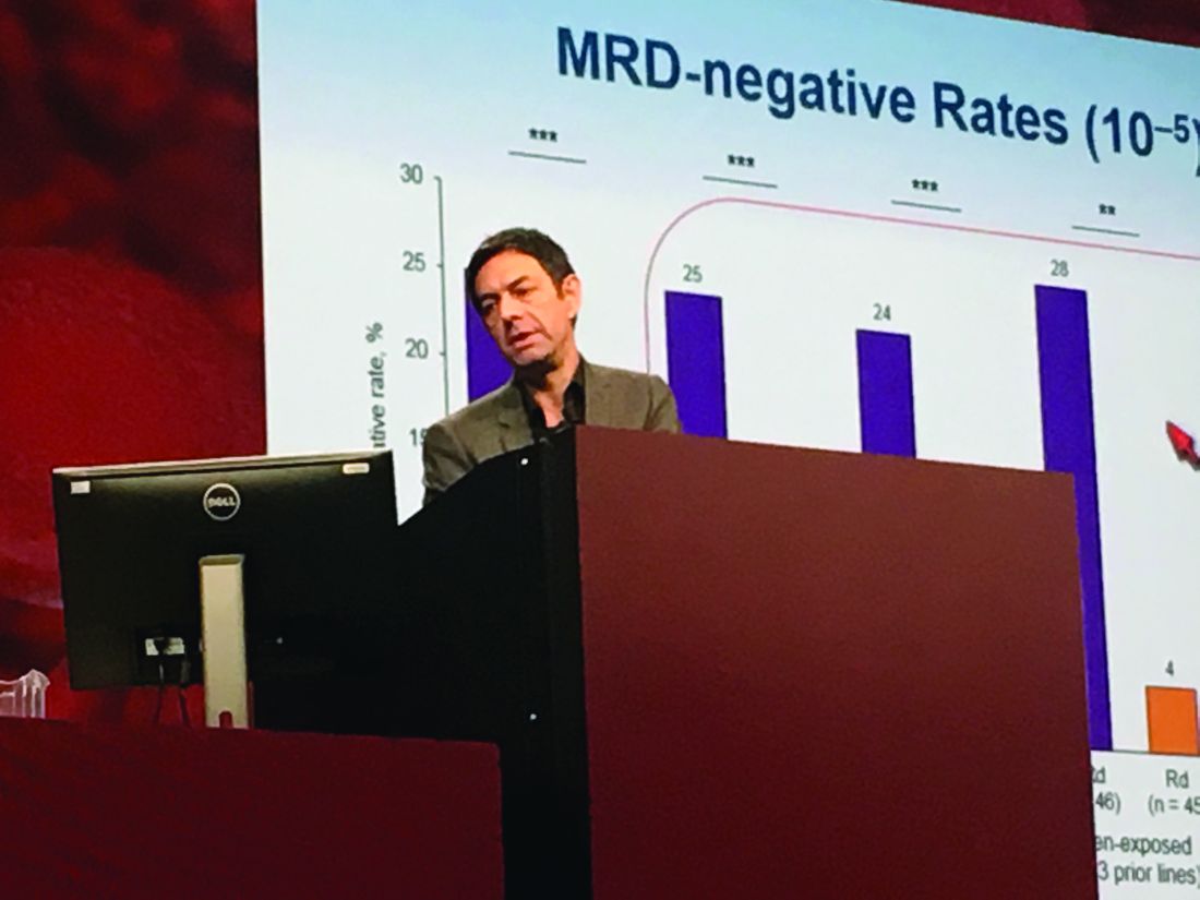
Among a large subgroup of 524 POLLUX patients who had received one to three prior lines of therapy, estimated median progression-free survival (PFS) has not been reached in the daratumumab, lenalidomide, and dexamethasone (DRd) arm, versus 18.4 months in the lenalidomide and dexamethasone (Rd) arm (hazard ratio, 0.36; 95% CI: 0.26 to 0.49; P less than .0001), Dr. Moreau said at the annual meeting of the American Society of Hematology.
That means adding daratumumab to Rd led to a 64% reduction in the risk of disease progression or death among patients with relapsed or refractory multiple myeloma, he noted. Fully 77% of DRd patients were alive without having progressed at 18 months, and responses “continued to deepen in the DRd group with longer follow-up,” he added.
Additional analyses supported the use of DRd in relapsed or refractory multiple myeloma, “irrespective of prior lenalidomide treatment or bortezomib refractoriness,” Dr. Moreau continued. He reported that DRd significantly improved PFS over Rd alone not only among 445 lenalidomide-naive patients (HR, 0.37; P less than .0001), but also among 91 lenalidomide-exposed patients (HR, 0.45; P = .04), 140 patients who were refractory to their most recent line of therapy (HR, 0.45; P = .001), and 99 bortezomib- refractory patients (HR 0.51; P = .02).
Daratumumab (Darzalex), a human CD38 IgG1k monoclonal antibody, was first approved as monotherapy for multiple myeloma in patients who had received at least three prior lines of therapy or had double-refractory disease. In 2016, results from the twin POLLUX and CASTOR studies won daratumumab a Food and Drug Administration breakthrough designation status for use with Rd in patients who had received at least one prior line.
The POLLUX trial included 569 patients with multiple myeloma who had received a median of 1 and up to 11 prior lines of therapy. Patients were randomized to either Rd alone or to Rd plus intravenous daratumumab (16 mg/kg) once a week during the first two 28-day treatment cycles, every 2 weeks during cycles 3-6, and once only on day 1 of subsequent cycles.
POLLUX patients were fairly heavily pretreated, Dr. Moreau noted. Thirteen percent had received three prior lines of therapy, 86% had received a proteasome inhibitor, 18% had received lenalidomide, 21% were refractory to bortezomib, and 28% were refractory to their most recent line of therapy.
Researchers performed “stringent, unbiased” assessments of minimal residual disease (MRD) negativity not only when a complete response was suspected, but also 3 and 6 months later, Dr. Moreau said. He emphasized that rates of MRD negativity in lenalidomide-exposed, bortezomib-refractory subgroups in POLLUX almost exactly matched those in the intent-to-treat population (25% on DRd vs. 6% on Rd; P less than .0001).
A total of 17% of DRd patients and 25% of Rd patients had high-risk cytogenetic profiles, and DRd performed well in these individuals, Dr. Moreau reported. Fully 85% of all evaluable high-risk patients had at least a partial response to DRd, and 33% had a complete response, versus only 67% and 6% of high-risk Rd patients, respectively. Among patients with standard-risk cytogenetics, rates of best overall response were 95% on DRd and 82% on Rd, and rates of complete response were 52% on DRd and 24% on Rd.
POLLUX yielded no new safety signals for DRd, Dr. Moreau said. Rates of primary and secondary malignancies were less than 2%. Neutropenia, the most common adverse effect, was managed by interrupting treatment, reducing the dose of lenalidomide, and administering growth factor.
Janssen Research & Development funded the study. Dr. Moreau had no relevant financial disclosures.
SAN DIEGO – Adding daratumumab (D) to lenalidomide and dexamethasone (Rd) significantly improved outcomes in relapsed and refractory multiple myeloma, even when patients had previously received lenalidomide, were refractory to bortezomib, or had high-risk tumor cytogenetics, based on updated analyses from the multicenter, randomized, phase III, open-label POLLUX trial.
The findings underscore the “significant benefit of combining daratumumab with lenalidomide and dexamethasone for relapsed or refractory multiple myeloma,” said lead investigator Philippe Moreau, MD, of University Hospital Hotel-Dieu in Nantes, France.
Among a large subgroup of 524 POLLUX patients who had received one to three prior lines of therapy, estimated median progression-free survival (PFS) has not been reached in the daratumumab, lenalidomide, and dexamethasone (DRd) arm, versus 18.4 months in the lenalidomide and dexamethasone (Rd) arm (hazard ratio, 0.36; 95% CI: 0.26 to 0.49; P less than .0001), Dr. Moreau said at the annual meeting of the American Society of Hematology.
That means adding daratumumab to Rd led to a 64% reduction in the risk of disease progression or death among patients with relapsed or refractory multiple myeloma, he noted. Fully 77% of DRd patients were alive without having progressed at 18 months, and responses “continued to deepen in the DRd group with longer follow-up,” he added.
Additional analyses supported the use of DRd in relapsed or refractory multiple myeloma, “irrespective of prior lenalidomide treatment or bortezomib refractoriness,” Dr. Moreau continued. He reported that DRd significantly improved PFS over Rd alone not only among 445 lenalidomide-naive patients (HR, 0.37; P less than .0001), but also among 91 lenalidomide-exposed patients (HR, 0.45; P = .04), 140 patients who were refractory to their most recent line of therapy (HR, 0.45; P = .001), and 99 bortezomib- refractory patients (HR 0.51; P = .02).
Daratumumab (Darzalex), a human CD38 IgG1k monoclonal antibody, was first approved as monotherapy for multiple myeloma in patients who had received at least three prior lines of therapy or had double-refractory disease. In 2016, results from the twin POLLUX and CASTOR studies won daratumumab a Food and Drug Administration breakthrough designation status for use with Rd in patients who had received at least one prior line.
The POLLUX trial included 569 patients with multiple myeloma who had received a median of 1 and up to 11 prior lines of therapy. Patients were randomized to either Rd alone or to Rd plus intravenous daratumumab (16 mg/kg) once a week during the first two 28-day treatment cycles, every 2 weeks during cycles 3-6, and once only on day 1 of subsequent cycles.
POLLUX patients were fairly heavily pretreated, Dr. Moreau noted. Thirteen percent had received three prior lines of therapy, 86% had received a proteasome inhibitor, 18% had received lenalidomide, 21% were refractory to bortezomib, and 28% were refractory to their most recent line of therapy.
Researchers performed “stringent, unbiased” assessments of minimal residual disease (MRD) negativity not only when a complete response was suspected, but also 3 and 6 months later, Dr. Moreau said. He emphasized that rates of MRD negativity in lenalidomide-exposed, bortezomib-refractory subgroups in POLLUX almost exactly matched those in the intent-to-treat population (25% on DRd vs. 6% on Rd; P less than .0001).
A total of 17% of DRd patients and 25% of Rd patients had high-risk cytogenetic profiles, and DRd performed well in these individuals, Dr. Moreau reported. Fully 85% of all evaluable high-risk patients had at least a partial response to DRd, and 33% had a complete response, versus only 67% and 6% of high-risk Rd patients, respectively. Among patients with standard-risk cytogenetics, rates of best overall response were 95% on DRd and 82% on Rd, and rates of complete response were 52% on DRd and 24% on Rd.
POLLUX yielded no new safety signals for DRd, Dr. Moreau said. Rates of primary and secondary malignancies were less than 2%. Neutropenia, the most common adverse effect, was managed by interrupting treatment, reducing the dose of lenalidomide, and administering growth factor.
Janssen Research & Development funded the study. Dr. Moreau had no relevant financial disclosures.
AT ASH 2016
Key clinical point: Adding daratumumab (D) to lenalidomide and dexamethasone (Rd) significantly improved outcomes in patients with relapsed and refractory multiple myeloma, regardless of factors such as bortezomib refractoriness, lenalidomide exposure, or high-risk tumor cytogenetics.
Major finding: DRd significantly improved PFS over Rd not only among 445 lenalidomide-naive patients (HR, 0.37; P less than .0001), but also among 91 lenalidomide-exposed patients (HR, 0.45; P = .04), 140 patients who were refractory to their most recent line of therapy (HR, 0.45; P = .001), and 99 bortezomib-refractory patients (HR 0.51; P = .02).
Data source: POLLUX, a multicenter, randomized, phase III, open-label trial of 569 patients with multiple myeloma who had received one or more previous lines of therapy.
Disclosures: Janssen Research & Development funded the study. Dr. Moreau had no relevant financial disclosures.