User login
Combo prevents GVHD, prolongs survival in monkeys

ORLANDO, FL—A 2-drug combination is “an exceptional candidate for clinical translation” as prophylaxis for graft-vs-host disease (GVHD), according to a presenter at the 2017 BMT Tandem Meetings.
The combination consists of sirolimus and KY1005, a monoclonal antibody that binds to OX40L and stops it from activating OX40, a protein that induces prolonged responses in T cells.
Experiments in rhesus macaques showed that KY1005 alone can have a modest effect on GVHD, but the combination of KY1005 and sirolimus can provide long-term, GVHD-free survival.
Victor Tkachev, PhD, of Seattle Children’s Research Institute in Washington, presented these results as one of the “Best Abstracts” at the recent BMT Tandem Meetings (abstract 3). This research was supported by Kymab, the company developing KY1005.
Dr Tkachev and his colleagues tested KY1005 alone and in combination with sirolimus in a previously described model of GVHD. In this model, rhesus macaques that do not receive prophylaxis develop severe GVHD after haploidentical hematopoietic stem cell transplant (HSCT).
For the current study, the animals received no prophylaxis, KY1005 alone, sirolimus alone, or KY1005 plus sirolimus.
When compared to no prophylaxis, KY1005 delayed the progression of acute GVHD and significantly prolonged the survival of HSCT recipients. However, all KY1005-treated animals eventually developed lethal GVHD.
Dr Tkachev noted that KY1005 provided partial control of T-cell activation, decreasing CD4 T-cell proliferation but having no significant effect on CD8 T-cell expansion.
As with KY1005 alone, sirolimus alone delayed GVHD progression and prolonged survival when compared to no GVHD prophylaxis.
However, all animals treated with sirolimus monotherapy eventually developed GVHD and died, and sirolimus alone wasn’t able to control T-cell proliferation.
On the other hand, the combination of KY1005 and sirolimus provided long-term, GVHD-free survival. All of the animals that received this combination survived, without developing GVHD, through day 100 after HSCT.
Dr Tkachev noted that, when given together, KY1005 and sirolimus synergistically controlled both CD4 and CD8 T-cell proliferation. However, this effect did not result in a lack of engraftment. In fact, animals that received the combination “displayed robust hematopoietic reconstitution” and maintained a high number of donor T cells.
Further investigation revealed that combination treatment with KY1005 and sirolimus preserves the reconstitution of regulatory T cells after HSCT and prevents both Th1- and Th17-driven alloimmunity.
Dr Tkachev and his colleagues also found that KY1005 plus sirolimus demonstrates an “unprecedented capacity” to protect against acute GVHD. Results with this combination were superior to those observed with tacrolimus and methotrexate in combination as well as abatacept and sirolimus in combination.
“Taken together, these data suggest that combined prophylaxis with KY1005 plus sirolimus represents an exceptional candidate for clinical translation,” Dr Tkachev concluded.
Kymab has said it will begin testing KY1005 in clinical trials this year. ![]()
Study sheds light on genetic landscape of HSTL
SAN FRANCISCO—Researchers say they have identified new driver genes and oncogenic pathways in hepatosplenic T-cell lymphoma (HSTL).
The team found that SETD2, a known tumor suppressor, was the most frequently silenced gene in HSTL.
The researchers also found evidence suggesting the JAK-STAT and PI3K pathways could be therapeutic targets in HSTL.
Sandeep Dave, MD, of Duke University in Durham, North Carolina, presented these findings at the 9th Annual T-cell Lymphoma Forum. Results from this research were also published in Cancer Discovery.
The researchers collected complete clinical data on 68 HSTL cases, including 20 with normal DNA. The team performed whole-genome sequencing, bioinformatics analysis, and biological characterization of these cases.
“This is the largest group of HSTL cases ever described, and the data implicate new driver genes and oncogenic pathways for the first time in HSTL,” Dr Dave said.
The data revealed that the most commonly mutated group of genes in HSTL are chromatin modifiers (SETD2, INO80, ARID1B, TET3, and SMARCA2) and signaling pathway genes (STAT5B, STAT3, and PIK3CD). Among these, STAT3, PIK3CD, and SETD2 showed the highest proportion of clonal events.
On the other hand, mutations in EZH2, KRAS, and TP53 were less frequently observed.
Common genetic abnormalities in HSTL include copy number alterations in chromosome 7, trisomy 8, loss of 10p, and gain of 1q.
A comparison of the frequencies of recurrently mutated genes in HSTL with other lymphomas demonstrated the genetically distinct profile of HSTL, wherein mutations in SETD2, INO80, TET3, and STAT5B occurred exclusively in HSTL.
SETD2, a histone lysine methyltransferase and a known tumor suppressor, was identified as the most frequently silenced gene in HSTL.
So the researchers investigated the biological effects of SETD2 loss in HSTL cells and a knockout mouse model.
While loss of SETD2 in HSTL cells resulted in increased cell proliferation, in vivo knockdown of SETD2 led to expansion of γ-δ T cells and a reduction in α-β T cells. A majority of HSTLs are known to arise predominantly from γ-δ T cells.
“These results implicate SETD2 in HSTL oncogenesis and T-cell development,” Dr Dave said.
He and his colleagues also found that constitutive activation of the JAK-STAT and PI3K pathways in HSTL cells was associated with increased proliferation, and inhibition of these pathways led to reduced survival of HSTL cells. This suggests that agents targeting these pathways might be effective in treating HSTL. ![]()
Strong Reaction to Chagas Disease
Having experienced the devastating effects of the “kissing bug” in September 2016, “Chagas Disease: Creeping into Family Practice” (2016;26[11]:38-45) was a timely article for me. I am writing to thank the authors for giving my case more credence and to issue a note of caution: People may have a strong reaction to the bite of this bug and therefore assume it to be from a spider. In some cases, including my own, bite-related anaphylaxis can send a person to the emergency department. After being bitten, I lost consciousness, fell down a flight of stairs, and sustained a bimalleolar fracture of my right ankle that required surgery. I later found the bug and submitted it to the CDC for testing; it was infected with Trypanosoma cruzi, the parasite that causes Chagas disease.
I want others to be aware that severe allergic reactions can result from the bite of a beetle-like bug, such as the kissing bug. I am taking it upon myself to educate my patients and colleagues and have also employed an exterminator for my home. If a person is bitten by this cone-nosed, prehistoric-looking bug, it should not be touched, but it should be collected and kept in the freezer until it can be sent to the CDC. If the bug is found to be positive for the parasite, an antibody test should then be performed on the affected person. I am lucky that my antibody test came back negative for the disease.
Constance Wylie, RN, MSN, FNP-C
Sacramento, CA
A New Model for PA Practice
Throughout my 12 years of practice, I have struggled with my delegated authority as a PA in relation to the autonomy of my NP colleagues. Most of the time, I enjoy the PA-physician relationship and see it as a mentoring program, not unlike residency. But after a decade of practice, I find myself occasionally wishing for the ability to hang out a shingle and run my own practice. I often feel taken advantage of by physician employers who reap surplus value from my labor without much appreciation.
So, what could a new model look like? Upon completion of a 5- to 8-year residency, PAs would be eligible to take a post-residency board exam and gain independent practice status. This would be limited to nonsurgical practice and might include family medicine, urgent care, dermatology, and emergency medicine. After passing the board, PAs would gain the ability to practice as an independent entity and treat patients without supervision.
Personally, I would appreciate this flexibility. After 10 years of working in orthopedics, I am now solely in urgent care and locum tenens. Independent status would give PAs the ability to compete with NPs for locum positions where independent practitioners are needed, as well as increase their negotiating/earning potential and range of volunteer opportunities.
Robert Feinberg, PA-C
Bryn Mawr, PA
PA Autonomy Levels the Field
I can’t tell you how refreshing it is to read an editorial that portrays PA autonomy in a positive light (2017;27[2]:12-14). I live in Michigan, and the physician I worked with for years is relieved to finally see laws changing for PAs. Physicians want less accountability, as they are carrying so much already. The phrase “supervising physician” can feel burdensome, particularly because of its implications in a court of law. When I took time off to spend with my kids, the physician I worked with hired two NPs; he said that their increased drive for autonomy made him feel less legally responsible. The new bill passed here has altered his thinking about hiring PAs versus NPs.
I believe eventually, with increased practice authority, a title change is inevitable for the PA profession. According to my 14–year-old niece, “PAs obviously can’t do anything by themselves, or their title wouldn’t be ‘assistant.’” This is truly a misnomer that doesn’t reflect the PA scope of practice.
Diana Burmeister,
Southfield, MI
When Did I Start Consulting for Dr. Google?
I have witnessed firsthand the evolution of patient satisfaction (2017;27[1]:13-14). When I became an NP in 1986, I worked in rural areas as the primary provider. My patients trusted that I cared for them and in the best way possible. A lot changed with the internet—Google became the primary care provider, and I became the consultant. Expressions of gratitude and respect from patients have been replaced by entitlement to services mandated by them. I have had patients bring in requests for diagnostic studies and further work-up for which there is no clinical evidence of need. They have their minds made up; if I am noncompliant, there is something wrong with me.
I have been reported to administration for being rude, insensitive, and incompetent. Now, I am not perfect, but most of these complaints were a result of me saying “no.” I was initially shocked by these demands and complaints, but they have become the new normal. Precious care time has been replaced by explanations and discussions about why I can’t do what patients want me to do.
Many providers give in to patients’ desires just to keep them happy and coming back. Unnecessary antibiotics, misuse of controlled pain medication, and pointless diagnostic studies have drained insurance and kept providers from just doing what is right. Big corporations run many medical practices, and unfortunately their main goal is to keep the doors open—even if that means compromising evidence-based medicine.
We have a generation of entitled people who become offended when you disagree with them. Many administrators transfer patient complaints to providers so that they will toe the line but fail to include pertinent information, such as what the complaint was and who filed it. This is a control technique; the provider is now automatically guilty, without a trial or defense.
I am blessed to work for a company that agrees that if concern is expressed, I have the right, as the provider, to know the details. If I am perceived as rude or abrupt, then knowing who feels that way can help me improve. Upon that patient’s next visit, I will adopt a gentler attitude and make an extra effort to pick up on cues that I may have missed.
Sometimes, patients express dissatisfaction because I will not dispense a controlled substance for pain upon request. My administration allows me to respond to situations like this in writing, and they can then verify that the complaint is unwarranted, and it will not be held against me or used as a control tactic. This approach has been so helpful. The company I work for also informs providers when patients express gratitude and thankfulness, which creates a great balance.
Surveys can be useful, but only if they are used as a tool to help the provider excel at his/her job—not create compliance for maintaining the patient head count. Providers should not have to worry about pleasing administration; they need to give quality care without fear.
Sue Beebe, APRN
Wichita, KA
MACRA advice: Do the MIPS bare minimum this year
ORLANDO – Confused about what MACRA, MIPS, and APMs mean for you and your practice this year?
Clifford Lober, MD, offered some very simple advice at the annual meeting of the American Academy of Dermatology: “If you take nothing else from this annual meeting – not this session, but the whole meeting – submit one quality measure or one improvement activity or the required advancing care information base measures – one time, on one patient” to the MIPS program, Dr. Lober said. “If you do that, you will save 4% of your Medicare payments in 2019.”*
MIPS is a system of bonuses and penalties based on data the Medicare program started collecting on Jan. 1, 2017; it incorporates the legacy quality measure reporting systems including the Physician Quality Reporting System, meaningful use, and the value-based modifier program, said Dr. Lober, a dermatologist in private practice in Kissimmee, Fla.
Based on feedback from physicians and other stakeholders, Medicare announced last fall that physicians could “pick their pace” as to how they wanted to report data to MIPS in 2017, the first baseline year of the program. They could:
- Choose to not participate. These physicians will see an automatic 4% reduction in their Medicare payments in 2019.
- Test QPP. Report on one quality measure or improvement activity for one patient. Of note: This information does not have to be submitted electronically; paper submission is okay. These physicians will avoid the 4% Medicare pay cut.
- Participate for part of the year. These physicians will report for 90 consecutive days of their choice on more than one quality measure, more than one improvement activity, or more than the required measures in the advancing care information performance category. Reporting must start before Oct. 2, 2017. These physicians may earn a pay increase.*
- Participate for the full year. Submit a full year of data to Medicare. These physicians may get a pay increase.
Dr. Lober said pointedly, “The first choice – doing nothing – is the dumbest move you could make.”
He also noted that the CMS regulations say verbatim that “positive adjustments are based on data on the performance information submitted, not the amount of information or the length of time submitted.”
A few physicians and other clinicians are exempted from the QPP for 2017. Those who are in their first year of participating in Medicare Part B are exempt, as are those who bill less than $30,000 or have fewer than 100 Medicare patients.*
“The low-value exemption alone, CMS actuaries think, will exempt 32.5% of clinicians in the United States – a huge exemption,” Dr. Lober said, adding that when it comes to dermatologists, it’s estimated that about 18.3% will be exempt from MIPS.
Despite the new political landscape in Washington, Dr. Lober said that he does not think MACRA will go away.
“One of the things I want to say out front is, what is the likelihood of MACRA being repealed?” he said. “Somewhere between slim and none or even none and none. It was supported by both Democrats and Republicans and I think it’s here to stay. It might be modified, but it’s here to stay.”
He urged physicians to reach out to CMS if they have comments about the MIPS, APMs, and the Quality Payment Program, noting that despite the formal comment period being closed, agency officials have been receptive to comments and suggestions.
*This article was updated on March 13, 2017 at 3:30 p.m.
[email protected]
On Twitter @denisefulton
AT AAD 17
Physicians need to take hyperhidrosis in teens seriously
ORLANDO – Not quite a fifth of teens experience excessive, uncontrollable sweating, according to the results of an online survey presented during this year’s annual American Academy of Dermatology.
Because nearly 70% of teens who reported the condition said it interferes with their activities of daily living, late-breaking research presenter, Adelaide A. Hebert, MD, chief of pediatric dermatology at the University of Texas, Houston, said it was time medical schools paid more attention to it.
For the study, Dr. Hebert and her colleagues online-surveyed 1,000 adolescents between 12 and 17 years who meet the accepted diagnostic criteria for primary focal hyperhidrosis. An analysis of the 981 surveys that were complete showed that 17.1% of respondents experienced excessive, uncontrollable sweating and that 68.6% of these reported the sweating was moderate or major, impairing their normal functioning.
The average age of onset for the condition was 11 years, although more than a quarter of respondents said their sweating began at age 10 years. Nearly all those surveyed said they sweat from at least two focal areas, with five areas being the average number of focal areas.
Adolescence is when hyperhidrosis begins for many adults with the condition, yet few if any data exist regarding the condition in this age group, according to Dr. Hebert. “We have to figure out what is going on so maybe we can make a difference later.”
[email protected]
On Twitter @whitneymcknight
AT AAD 17
Key clinical point:
Major finding: Hyperhidrosis is common in 17% of teens surveyed; 69% of these reported hyperhidrosis interferes with activities of daily living.
Data source: Online survey of 1,000 U.S. teens between 12 and 17 years who reported excessive, uncontrollable sweating.
Disclosures: Dr. Hebert received a grant from GlaxoSmithKline for this study. She is also a board member of the International Hyperhidrosis Society.

Teletriage app reduced skin specialty care wait times
ORLANDO – A teletriage app significantly reduced the time between triage and evaluation by a dermatologist, helping a volunteer-staffed clinic for the uninsured see more patients and improve rates of accurate diagnosis and treatment.
Peter Chansky, a medical student at the University of Pennsylvania, Philadelphia, presented the data on a study of the app during a late-breaking research session at this year’s annual meeting of the American Academy of Dermatology.
The teletriage app AccessDerm, which is free to all AAD members, uses a photo of the dermatological condition, along with basic clinical and demographic information. The investigators found that with the app, the mean time from referral to teledermatology response was 1.4 days vs. 13.4 days for waiting to be seen in the next dermatology clinic (P less than .0001). Additionally, because the app requires the referring physicians to enter a differential diagnosis and a suggested treatment plan, it was established that diagnoses improved with teledermatology. Compared with self-reported primary care provider plans, the app led to an altered course of treatment in 95% of cases.
In all, 70% of cases were triaged via teledermatology without the need for an in-person evaluation, resulting in an average of 1.4 out of 8 appointments being saved monthly. This allowed the clinic to treat 18% more patients who needed in-person care, according to Mr. Chansky.
“Teletriage can be easily implemented in an underserved clinical setting, and provides an opportunity for dermatologists to make a significant and meaningful impact on the health care and outcomes of underserved populations,” Mr. Chansky said.
[email protected]
On Twitter @whitneymcknight
AT AAD 17
VIDEO: Don’t overlook psychosocial concerns of vitiligo patients
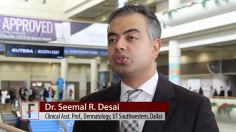
ORLANDO – There are ways to assess the psychosocial needs of your patients with vitiligo, even if you don’t believe you have the necessary skills to do a complete mental health work-up, according to Seemal R. Desai, MD.
In an interview recorded at this year’s annual meeting of the American Academy of Dermatology, Dr. Desai, an assistant clinical professor of dermatology at the University of Texas, Dallas, shares his ideas for how to have casual conversations with patients that can help reveal important clues to psychosocial stress patients with this serious medical skin condition might be facing.
“There are subtle clues to look for to know that these patients are uncomfortable with others seeing their skin,” says Dr. Desai.
In the video interview, he also covers taking a multidisciplinary approach to caring for these patients, what treatments are available to those who are suffering psychosocial stress after having failed several interventions, and how patients from many Asian and African counties are especially at risk for ostracization.
“It’s important to let your patients know that you understand this is really affecting them psychosocially, and that you care,” says Dr. Desai.
The video associated with this article is no longer available on this site. Please view all of our videos on the MDedge YouTube channel
[email protected]
On Twitter @whitneymcknight
EXPERT ANALYSIS FROM AAD 17