User login
FDA approves new treatment for episodic cluster headaches in adults
The Food and Drug Administration announced April 18 the approval of the gammaCore device to treat pain associated with episodic cluster headache in adult patients.
The agency based its approval of the noninvasive vagus nerve stimulator device on subgroup analyses of episodic cluster headache patients in the ACT1 and ACT2 trials, which were double-blind, placebo-controlled, randomized studies.
In the ACT1 trial of 85 patients, 34.2% experienced a reduction in pain (defined as the percentage of patients who reported mild or no pain 15 minutes after treatment initiation with gammaCore), compared with 10.6% of patients treated with placebo (P = .008). Of 27 patients in the ACT2 trial, 47.5% of patients using the device were pain free at 15 minutes after the onset of pain from cluster headache, with no use of rescue medication through the 30-minute treatment period, which was significantly greater than for placebo (6.2%; P = .003).
“Cluster headache is a rare, debilitating, and difficult to treat disorder with few effective acute therapies,” Stephen Silberstein, MD, director of the Headache Center at Jefferson University, Philadelphia, said in a statement from the manufacturer, electroCore. “The FDA release of gammaCore is an important advance in the treatment of the pain associated with cluster headache. It is a way for patients to treat their symptoms as often as they need to use the device. It does not have the side effects or dose limitations of commonly prescribed treatments or the need for invasive implantation procedures, which can be inconvenient, costly, and high-risk.”
The gammaCore device works by transmitting a mild electrical stimulation to the vagus nerve through the skin, resulting in a reduction of pain. Currently, gammaCore is in use only outside of the United States, including in the European Union. Commercial availability of gammaCore in the United States is expected to begin early in the third quarter of 2017, the company said.
Infantile hemangiomas: Calculating dose for propranolol is tricky
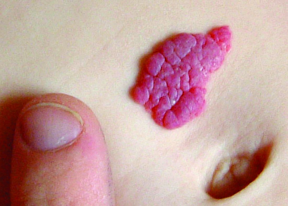
Use of Hemangeol was associated with fewer dosing errors than use of generic propranolol hydrochloride oral solutions in treating infantile hemangiomas, according to an online questionnaire survey.
Anastasia O. Kurta, DO, of Saint Louis University and her associates emailed a questionnaire to 531 physicians who were members of the Society for Pediatric Dermatology and physicians known to treat infantile hemangiomas. Most of the 220 physicians who responded were pediatric dermatologists. Of those, 90% had prescribed generic propranolol available in concentrations of 4 mg/mL and 8 mg/mL, and 58.6% had prescribed Hemangeol, the Food and Drug Administration-approved formulation of propranolol hydrochloride available in a concentration of 4.28 mg/mL.
“A dosing chart accompanies propranolol 4.28 mg/mL [Hemangeol] using mL/kg doses, which eliminates conversion from milligrams to milliliters and could potentially explain the lower reported dose calculation error reported with propranolol 4.28 mg/mL,” Dr. Kurta and her associates said. “The risk of dispensing errors is increased for medications that are commercially available in different concentrations. Liquid medications prescribed for pediatric patients require additional computation for weight-based dosing and conversion, increasing the possibility of miscalculation.”
Daisy Dai, PhD, is employed by Pierre Fabre Pharmaceuticals and Elaine C. Siegfried, PhD, is a consultant for the company. Dr. Kurta and Eric S. Ambrecht, PhD, have no relevant financial disclosures.
Read more at J Am Acad Dermatol. 2017 May;76(5):999-1000.
Dr. Kurta reported no relevant financial disclosures.
FROM THE JOURNAL OF THE AMERICAN ACADEMY OF DERMATOLOGY
Three-strategy combo reduces pancreatoduodenectomy fistula risk
MIAMI BEACH – Pancreaticojejunostomy reconstruction, use of stents, and avoidance of prophylactic octreotide, especially in combination, could reduce the fistula rate associated with pancreatoduodenectomy.
Failure of the anastomosis is “of greatest concern” to surgeons performing a pancreatoduodenectomy, said Brett L. Ecker, MD, a surgical resident at the University of Pennsylvania, Philadelphia.
“There is no shortage of high-quality data to help guide the use of [fistula reduction] strategies,” he added. However, “the utility of these strategies in patients most vulnerable to fistula … has rarely been particularly explored.”
Dr. Ecker and his colleagues conducted a study with 62 surgeons at 17 institutions to compare various fistula mitigation strategies in this higher-risk population. They assessed surgical reconstruction, dunking, tissue patches, intraperitoneal drains, stents, prophylactic octreotide, and use of tissue sealants.
“Ultimately, we want to know the best way to deal with this high-stakes situation, and whether outcomes might be optimized by bundling these proactive strategies,” Dr. Ecker explained.
“We found the combination of externalized stents and PJ [pancreaticojejunostomy] reconstruction with the omission of prophylactic octreotide was associated with significant improvements in fistula that exceeded the benefit of any individual mitigation approach or any other combination of strategies,” he said at the annual meeting of the Americas Hepato-Pancreato-Biliary Association.
“The mitigation of risk in real-life practice is often the result of multiple moving parts,” Dr. Ecker said. “The best outcomes [may result from] the synergistic effects of multiple strategies.”
Of the approximately 10% of patients with a Fistula Risk Score of 7-10, 152 ended up with clinically relevant postoperative pancreatic fistula (CR-POPF). “An FRS score of 7 or higher is associated with worse outcomes, including a fistula rate approaching 30%,” Dr. Ecker said. Grade B or C fistula based on International Study Group on Pancreatic Surgery criteria were considered clinically relevant. All patients had surgery from 2003 to 2016 in the retrospective, multinational study.
“This represents the only series of high-risk cases where current international standards were used to define both the risk and the outcome,” he noted.
“Almost all [of the 522] patients had a soft gland and a small duct, a median of 2 mm,” Dr. Ecker added. “High-risk pathology was common, in 86%.”
Surgeons contributing to the series were at high-volume centers and had performed more than 200 Whipple procedures in their careers. “Both institutional and surgeon volume were associated with improved fistula outcomes,” Dr. Ecker said. “We found that intraperitoneal drains were not associated with improved fistula outcomes, but that is limited by the fact that drains were rarely omitted in these cases.”
Four strategies compared
The investigators compared the outcomes of four fistula strategies among the patients considered high risk prior to surgery. When they combined pancreaticogastrostomy, prophylactic octreotide, and no stent, the CR-POPF rate was 47%. “This was associated with an alarming fistula rate approaching 50%,” Dr. Ecker said.
When surgeons combined pancreaticojejunostomy, octreotide, and no stent, the CR-POPF rate declined to 34%. Furthermore, pancreaticojejunostomy without octreotide or a stent yielded a 26% CR-POPF rate.
Ultimately, the most effective strategy to avoid clinically relevant fistula was pancreaticojejunostomy with an external stent and no octreotide.
“The use of PJ reconstruction with an external stent and omission of octreotide was associated with a fistula rate of about 13%, which was a greater than 50% risk reduction from the overall cohort,” Dr. Ecker said.
The researchers also performed propensity score matching to reduce bias associated with surgeon or patient factors. They matched 167 participants in the study with 155 controls. Dr. Ecker said, “Still, we observed that patients managed this way had significantly lower fistula rates.”
“This is an excellent paper and an important topic,” said study discussant Michael L. Kendrick, MD, a general surgeon at the Mayo Clinic in Rochester, Minn.
“At our institution, we’ve used the same Fistula Risk Score and found it very helpful for a mitigation strategy in a separate protocol, and we found that reduced our leak rates as well,” Dr. Kendrick noted.
Dr. Ecker and Dr. Kendrick had no relevant disclosures.
AT AHPBA 2017
Key clinical point: Reconstruction, use of stents, and avoidance of octreotide, especially in combination, could reduce the fistula rate associated with pancreatoduodenectomy.
Major finding: The incidence of fistula decreased from 33% to 13% by combining the three strategies.
Data source: Multicenter retrospective study from 2003 to 2016 with 522 patients undergoing pancreatoduodenectomy.
Disclosures: Dr. Ecker and Dr. Kendrick had no relevant disclosures.
The psychiatric care system of the future
Yogi Berra once said, “It’s tough to make predictions, especially about the future.” It is particularly difficult to talk about the future of psychiatric care and the profession of psychiatry given the current state of affairs and the dysfunction of the mental health services system in America today.
The current system is broken. Needs are not being met. Care is underfunded and uncoordinated. Patients fall through the cracks and are criminalized or homeless. And although there are some early signs of reform, such as the push for integration of psychiatry in medical care systems, it is unclear with the potential repeal of the Affordable Care Act and the undoing of parity whether the situation for our patients and the profession might get even worse.
In the year 2067, most physicians will be employees of one out of four major health care nonprofit corporations that are vertically or horizontally integrated systems of care. All Americans will be enrolled through a government-financed universal single payer plan of care, as employer-based health insurance will have disappeared for the last 25 years. Americans will choose which of the four health systems they wish to join during an annual open season and be able to select their primary and specialty care physicians. Many of the services provided will be in the home or workplace through broad and interactive computing and telemedicine capacity and high-tech centers. Hospitals will provide sophisticated gene therapy, organ transplantation, and biomedical engineering. Approximately 30% of the gross national product will be spent on health care.
Americans will live to an average age of 125 years, but it would not be unusual to find some individuals living to age 150. These Americans will have had many of their organs replaced by either genetically programmed animal organs or harvested organs from special banks. However, the brain is the only irreplaceable organ, and psychiatrists will be prominently involved in the interface of brain and behavior as they have been for the past 200 years.
In the year 2067, an expanded specialty of psychiatric physicians will be certified in one of four major categories of practice. Those certifications will be in neuroscience, medical psychiatry, psychotherapy, and social psychiatry.
The neuroscience psychiatrists will combine an MD with a PhD, and will be the most highly technical and specialized and the most highly compensated psychiatrists. They will be the clinician scientists. The neuroscience psychiatrist will be an expert on the human genome, sophisticated brain imaging and mapping, and the differential use of a variety of neurochemicals, as well as the application of technology such as magnetic fields for the treatment of mental illness and direct intervention into the brain with psychosurgery.
The medical psychiatrists will most resemble the early 21st century psychiatrists with subspecialties in geriatrics, adult, child and adolescent, and substance use. The medical psychiatrists will be integrated with other medical colleagues in many ambulatory as well as residential settings. Geriatrics will be the specialty for the treatment of the very old working with the neuroscience psychiatrist in the treatment of dementias and similarly, the child psychiatrist will work with the neuroscience psychiatrists in early preventive interventions at the intrauterine level with genetic abnormalities being corrected before birth. The medical psychiatrist will be a very popular area for all physicians, with more than 20% of all medical graduates specializing in medical psychiatry.
The psychotherapy psychiatrists will combine the MD degree with psychology education, religion, and the humanities. This psychotherapist will work one to one and in group settings on the age-old problems of individuation, separation, grief, loss, insight, and self-actualization.
The social psychiatrists will combine the MD degree with a degree in sociology or criminology and/or a law degree. They will focus on the social control issues of the day. Forensic prisons will be an area of government-sponsored treatment and will dominate the criminal justice system with interventions in an effort to reduce criminal behavior.
Managed care will not exist 50 years from now. It will be perceived as a regrettable experiment of the late 20th century ending in the first part of the 21st century. With the enactment of a universal single payer system of care, the high-cost intrusive middle management of carve-out behavioral health care companies will become moot.
Human progress comes in many forms. By the year 2067, psychiatry will have made significant advances that will make the prior 200 years of psychiatric care seem crude, quaint, and absurd.
Dr. Sharfstein, a past president of the American Psychiatric Association, is president emeritus of the Sheppard Pratt Health System, Baltimore. This essay is based on a presentation he made in February 2017 at the annual meeting of the American College of Psychiatrists in Scottsdale, Ariz.
Waldenström macroglobulinemia panel advises on IgM paraproteinemic neuropathies
With optimal approaches still evolving for the diagnosis and management of peripheral neuropathies associated with Waldenström macroglobulinemia and other IgM paraproteinemias, new consensus recommendations from a multidisciplinary panel were recently published in the British Journal of Haematology.
Up to half of patients with IgM monoclonal gammopathies develop peripheral neuropathy, according to the 11-member panel (Br J Haematol. 2017 Mar;176[5]:728-42). The panel began deliberations at the eighth International Workshop on Waldenström Macroglobulinemia in London and was led by Shirley D’Sa, MD, of the Waldenström Clinic, Cancer Division, University College London Hospitals NHS Foundation Trust.
• Diagnostic evaluation: “The indications for invasive investigations such as cerebrospinal fluid analysis, nerve conduction tests, and sensory nerve biopsies are unclear,” according to the panelists.
When clinical examination identifies a neuropathy, neurophysiologic testing can ascertain its nature and inform additional work-up. Cerebrospinal fluid examination is not mandatory in cases of demyelinating neuropathy, but it is indicated when clinical evaluation is inconclusive and malignancy or CNS invasion is suspected.
Nerve biopsy carries substantial risk and is rarely indicated. It may be warranted when a comprehensive systemic work-up has not identified a cause and clinicians still suspect amyloid, vasculitis, or direct cellular invasion; in atypical cases not responding to treatment; or when the neuropathy is progressive and debilitating.
When it comes to imaging, “MRI of the neuraxis should be performed prior to lumbar puncture to avoid false positive meningeal enhancement,” they advised. “Prior discussion of likely sites of involvement with an experienced neuroradiologist will ensure that the correct sequences of the correct anatomical area are performed with appropriate gadolinium enhancement.”
• Clinical phenotypes and their treatment: IgM-associated neuropathies vary with respect to specific antibodies present and the likelihood that they are causally associated with the neuropathy, Dr. D’Sa and her colleagues noted. They provided a decision tree to help guide the work-up to determine the specific etiology.
“The presence of a neuropathy alone is not a justification for treatment, but steady progression with accumulating disability should prompt action,” they maintained.
Patients with antibody-negative peripheral neuropathy associated with IgM monoclonal gammopathies of undetermined significance who have mild disease and no hematologic reason for treatment can be managed with surveillance, according to the panelists.
However, immunosuppressive or immunomodulatory treatment should be considered when there is substantial or progressive disability associated with demyelination.
Patients with anti-MAG (myelin-associated glycoprotein) demyelinating neuropathy may benefit from rituximab (Rituxan). In those with more advanced disease, clinicians should consider immunosuppressive or immunomodulatory treatment instead.
Surveillance is also an option for Waldenström macroglobulinemia–associated peripheral neuropathy that is progressing slowly. When used, treatment should be tailored to severity of both systemic and neurologic disease.
• Treatment response assessment: “The optimum way to measure clinical response to treatment unknown,” Dr. D’Sa and her fellow panelists noted. A variety of measures of muscle strength, sensory function, and disability are used.
“The I-RODS [Inflammatory Rasch-Built Overall Disability Scale] more often captures clinically meaningful changes over time, with a greater magnitude of change, compared with the INCAT-ONLS [Inflammatory Neuropathy Cause and Treatment–Overall Neuropathy Limitation Scale] disability scale and its use is therefore suggested in future trials involving patients with inflammatory neuropathies,” they wrote.
• Model of care: Management of patients with IgM-associated neuropathies requires multidisciplinary care with good collaboration to optimize patient outcomes, the consensus panel said.
“A suggested model of care is a combined neurological and hematological clinic, in which patients are seen jointly by a specialist neurologist and hematologist and a decision can be made about the sequence of investigations, interventions, and the formulation of a treatment plan,” they proposed. “Appropriate and timely referral to physical, occupational, and orthotic professionals is recommended in order to maximize safety and function.”
• Future perspectives: “There is much to be done to improve outcomes for patients with IgM and [Waldenström macroglobulinemia]-associated peripheral neuropathies,” the panelists concluded.
Key areas of focus are “early recognition of the problem, appropriate causal attribution achieved through sensitive diagnostics that are not overly invasive, timely therapeutic intervention with effective and nonneurotoxic therapies, achievement of an appropriate degree of clonal reduction for optimum clinical outcomes, and the use of reproducible and readily applicable tools to measure outcomes.”
Dr. D’Sa disclosed that she receives honoraria from Janssen.
FROM THE BRITISH JOURNAL OF HAEMATOLOGY
Key clinical point:
Major finding: The indications for invasive testing and definitive answers about when and how to treat peripheral neuropathies due to Waldenström macroglobulinemia and other IgM paraproteinemias are unclear.
Data source: Recommendations from the eighth International Workshop on Waldenström Macroglobulinemia (IWWM-8) consensus panel.
Disclosures: Dr. D’Sa disclosed that she receives honoraria from Janssen.
SHM receives Eisenberg Award as part of I-PASS Study Group
The Society of Hospital Medicine is part of a patient safety research group that received the prestigious 2016 John M. Eisenberg Award for Innovation in Patient Safety and Quality presented annually by The Joint Commission and the National Quality Forum, two leading organizations that set standards in patient care as part of the I-PASS Study Group.
I-PASS comprises a suite of educational materials and interventions dedicated to improving patient safety by reducing miscommunication during patient handoffs that can lead to harmful medical errors. The team in SHM’s Center for Quality Improvement has been instrumental in supporting the I-PASS Study Group, which represents more than 50 hospitals from across North America.
“The Eisenberg Award for Innovation represents the highest patient safety and quality award in the country, and we are honored to be recognized for our role in this important program,” said Jenna Goldstein, director of SHM’s Center for Quality Improvement. “Our team’s participation in developing and sustaining the SHM I-PASS mentored implementation demonstrates our commitment to ensure safe and high-quality care for hospitalized patients.”
SHM previously won the 2011 Eisenberg Award at the national level for its mentored implementation program model. Through its mentored implementation framework and project management, SHM has supported the I-PASS program across the country at 32 hospitals of varying types, including pediatric and adult hospitals, academic medical centers, and community-based hospitals. SHM has offered both an I-PASS mentored implementation program, in which a physician mentor coaches hospital team members on evidence-based best practices in process improvement and culture change for safe patient handoffs, and an implementation guide, which contains strategies and tools needed to lead the quality improvement effort in the hospital.
In a large multicenter study published in the New England Journal of Medicine, implementation of I-PASS was associated with a 30% reduction in medical errors that harm patients. An estimated 80% of the most serious medical errors can be linked to communication failures, particularly during patient handoffs.
In addition to its work with I-PASS, SHM’s Center for Quality Improvement plays a prominent role in developing tools that empower clinicians to lead quality improvement efforts in their institutions.
Brett Radler is SHM’s communications specialist.
VA to Review Caregiver Program Following Funding Concerns
Reacting quickly to complaints of caregivers who had their eligibility for Program of Comprehensive Assistance for Family Caregivers (PCAFC) funding revoked, the VA has announced that it will pause revocations while reviewing the program’s implementation. “VA is taking immediate action to review the National Caregiver Support Program to ensure we honor our commitment to enhance the health and well-being of veterans,” said Secretary of Veterans Affairs David J. Shulkin, MD, in a prepared statement. “I have instructed an internal review to evaluate consistency of revocations in the program and standardize communication with veterans and caregivers nationwide.”
An NPR report documented a number of cases of PCAFC support that had been changed recently despite little evidence for change in the veterans’ need for care. According to the NPR analysis, some VA facilities saw significant drops in the number of caregivers who received support; whereas others saw equally significant increases.
According to the VA, veterans who need a VA designated family caregiver to assist with the management of personal care functions that are required for everyday living and are in conjunction with standard care provided by the VA are eligible for the program. A clinical support team evaluates the veteran for eligibility, and the caregiver receives training. The program provides a monthly stipend based on the veterans’ “level of need and required assistance.”
During the review, the VA will continue accepting PCAFC applications, approving applicants based on current eligibility criteria, processing appeals, and monitoring eligible veterans’ well-being at least every 90 days unless otherwise clinically indicated.
“Caregivers play a critically important role in the health and well-being of veterans, and caring for an injured veteran is a labor of love,” said Dr. Poonam Alaigh, acting VA Under Secretary for Health. “We remain focused on process improvements and support services for our family caregivers so they can take care of our veterans.”
Not Getting Enough Sleep? NIOSH Wants to Help
This should not come as much of a surprise, but health care made the top-5 list of occupations whose workers are getting too little sleep, according to a study by The National Institute for Occupational Safety and Health (NIOSH) researchers.
The researchers analyzed data from 179,621 working adults who responded to the 2013 or 2014 Behavior Risk Factor Surveillance System annual surveys. Among the 22 major occupation groups, health care support (40.1%) and health care practitioners and technical (40.0%) ranked second and third in “short sleep duration,” after production (42.9%). Among the occupational subgroups, nursing, psychiatric, and home health aides had a high adjusted prevalence of short sleep duration.
Workers in occupations where alternative shiftwork is common were more likely to have a higher adjusted prevalence of short sleep duration. More than 35% of health care practitioners work shifts. Workers in other occupation groups such as teachers, farmers, or pilots, were more likely to report getting enough sleep.
Time at work also is on the rise in the US where workers have the longest annual working hours among workers in all wealthy industrialized countries, which reduces the time available for sleep, NIOSH says. The researchers point out that lack of sleep has been linked to negative health outcomes including cardiovascular disease, obesity, and depression, as well as safety issues related to drowsy driving and injuries.
To help people get more sleep or improve the quality of the sleep they get, NIOSH offers training and resources about sleep, shiftwork, and fatigue for a variety of audiences including health care workers and emergency responders. Free downloadable materials are available at www.cdc.gov/niosh/topics/workschedules/education.html.
Method could prevent GVHD while preserving GVL effect
Researchers believe they have found a way to prevent graft-versus-host disease (GVHD) after hematopoietic stem cell transplant (HSCT) while preserving a strong graft-versus-leukemia (GVL) effect.
In experiments with mice, the team found that temporary in vivo depletion of CD4+ T cells soon after HSCT prevented GVHD without inhibiting GVL effects.
The depletion of CD4+ cells essentially caused CD8+ cells to become exhausted in their quest to destroy normal tissue but strengthened in their fight against leukemia, which meant the CD8+ cells could eliminate leukemic cells without causing GVHD.
Defu Zeng, MD, of City of Hope in Duarte, California, and his colleagues recounted these findings in the Journal of Clinical Investigation.
The researchers were able to achieve temporary in vivo depletion of CD4+ T cells by injecting mice with an anti-CD4 monoclonal antibody (mAb).
The team found that a single injection of the mAb given immediately after HSCT prevented acute but not chronic GVHD. Three injections of the mAb—given on days 0, 14, and 28—prevented both types of GVHD.
The researchers said their results suggest GVHD is more effectively prevented by temporary in vivo depletion of donor CD4+ T cells early after HSCT than by ex vivo depletion of donor CD4+ T cells.
This is because treatment with an anti-CD4 mAb temporarily depletes both the injected mature CD4+ T cells and the CD4+ T cells generated de novo from the marrow progenitors early after HSCT.
In explaining the mechanism behind their observations, the researchers noted that the interaction between PD-L1 and CD80 has been shown to exacerbate GVHD, but costimulation of CD80 and PD-1 ameliorates GVHD.
In their experiments, the team found that depleting CD4+ T cells increased serum IFN-γ and reduced IL-2 concentrations. And this led to upregulation of PD-L1 expression by recipient tissues and donor CD8+ T cells.
The researchers said that, in GVHD target tissues, the interactions of PD-L1 and PD-1 on donor CD8+ T cells caused anergy, exhaustion, and apoptosis. These effects prevented the development of GVHD.
On the other hand, in lymphoid tissues, the interactions of PD-L1 and CD80 augmented CD8+ T-cell expansion without increasing anergy, exhaustion, or apoptosis. This allowed for the preservation of GVL effects.
“If successfully translated into clinical application, this [CD4+ T-cell-depleting] regimen may represent one of the novel approaches that allow strong GVL effects without causing GVHD,” Dr Zeng said.
“This kind of regimen has the potential to promote widespread application of allogenic [HSCT] as a curative therapy for hematological malignancies.” ![]()
CAR T-cell therapy receives breakthrough designation
The US Food and Drug Administration (FDA) has granted another breakthrough therapy designation to CTL019 (tisagenlecleucel-T), an investigational chimeric antigen receptor (CAR) T-cell therapy.
CTL019 now has breakthrough designation as a treatment for adults with relapsed or refractory diffuse large B-cell lymphoma (DLBCL) who have failed 2 or more prior therapies.
The FDA previously granted CTL019 breakthrough designation as a treatment for relapsed/refractory B-cell acute lymphoblastic leukemia (ALL) in pediatric and young adult patients.
Breakthrough designation is intended to expedite the development and review of new medicines that treat serious or life-threatening conditions, if those medicines have demonstrated substantial improvement over available therapies.
The breakthrough designation for CTL019 in DLBCL is based on data from the phase 2 JULIET trial (NCT02445248), in which researchers are evaluating the efficacy and safety of CTL019 in adults with relapsed/refractory DLBCL.
Findings from JULIET are expected to be presented at an upcoming medical meeting.
About CTL019
CTL019 consists of autologous T cells expressing a CD19-specific CAR. The therapy was first developed by the University of Pennsylvania.
In 2012, the university and Novartis entered into a global collaboration to further research, develop, and commercialize CAR-T cell therapies, including CTL019. Novartis holds the worldwide rights to CARs developed through the collaboration.
According to Novartis, regulatory submissions for CTL019 in the treatment of relapsed/refractory DLBCL are expected to be filed by the end of the year.
The company has already filed a biologics license application with the FDA for CTL019 as a treatment for patients with relapsed/refractory B-cell ALL. The FDA granted that application priority review. ![]()
