User login
Home induction viewed as OK with Suboxone
NEW ORLEANS – About half the patients who arrive for opioid addiction treatment at Community Mental Health Affiliates in New Britain, Conn., already have buprenorphine in their urine, according to staff psychiatrist Margaret Chaplin, MD.
“There’s a huge black market for Suboxone right now, not so much because people want to abuse it to get high, but because there’s recognition” on the street that it helps with addiction. “It seems kind of silly to do” a witnessed induction “when people are already on the medication, and there’s lot of data that show home induction is just as safe,” she said at the American Psychiatric Association’s Institute on Psychiatric Services.

Plus, “if somebody comes in and they have an opiate” in their pocket, “I am afraid that they won’t make it through the night. I’ll send them home with a script and see them the next day,” she said.
Witnessed inductions were recommended when Suboxone was approved in 2002 in case of side effects, but fatal overdose is unlikely. Other addiction specialists in Dr. Chaplin’s audience said they now feel comfortable with home induction, as well.
To prevent problems, patients need to be either completely opioid free or in withdrawal, which usually starts about 12 hours after the last heroin dose. Most will need to be titrated up to about 16 mg/d. Some will need more, but Dr. Chaplin caps it at 24 mg, because of the diversion risk and the dearth of evidence showing additional benefit with higher doses.
The clinic checks urine norbuprenorphine, a metabolite of buprenorphine, for adherence. “All you have to do to get a buprenorphine positive urine is dip a Suboxone strip in [it], but only the human liver makes norbuprenorphine. If it’s there, we have a pretty good sense that they are taking their Suboxone,” she said.
The clinic doesn’t use the new buprenorphine implant (Probuphine), because it’s limited to people who are stable on just 8 mg buprenorphine daily, and the requirement for surgical implant and removal is a problem for clinic patients, who are sometimes homeless.
Naltrexone isn’t used much, either; the risk of fatal overdose is too high when patients come off it, and there’s not much incentive to stay on it. Patients can’t feel it work, like with methadone and Suboxone, and there’s no continuity with doing heroin. When naltrexone is used, Dr. Chaplin opts for the monthly IM formulation (Vivitrol) instead of daily tablets. With the shot, “you don’t need resolve as long as you come back and get a second injection.” IM naltrexone does help with the cravings, she noted, but not until about the third or fourth shot.
A lot of people hope to eventually come off medications such as Suboxone, but that’s dangerous thinking, Dr. Chaplin said.
One of her patients was buprenorphine free for several years. His wife went to the ED for an injury and returned with a bottle of Percocet, his drug of choice. “He heard the pills jiggling around in the bottle,” and that was all it took; he downed the whole thing. “He’s back on Suboxone now, and we don’t have any intention to take him off,” she said.
“I don’t think we should have as our goal to be off treatment. I think we should have as our goal to be alive and well, and managing our lives,” she said. “I’d rather see someone continue on 1 or 2 mg of Suboxone and be protected and feel normal” than come off it.
Also, people in treatment should have naloxone nasal spray (Narcan) on hand, just in case, she said.
Dr. Chaplin had no disclosures.
NEW ORLEANS – About half the patients who arrive for opioid addiction treatment at Community Mental Health Affiliates in New Britain, Conn., already have buprenorphine in their urine, according to staff psychiatrist Margaret Chaplin, MD.
“There’s a huge black market for Suboxone right now, not so much because people want to abuse it to get high, but because there’s recognition” on the street that it helps with addiction. “It seems kind of silly to do” a witnessed induction “when people are already on the medication, and there’s lot of data that show home induction is just as safe,” she said at the American Psychiatric Association’s Institute on Psychiatric Services.
Plus, “if somebody comes in and they have an opiate” in their pocket, “I am afraid that they won’t make it through the night. I’ll send them home with a script and see them the next day,” she said.
Witnessed inductions were recommended when Suboxone was approved in 2002 in case of side effects, but fatal overdose is unlikely. Other addiction specialists in Dr. Chaplin’s audience said they now feel comfortable with home induction, as well.
To prevent problems, patients need to be either completely opioid free or in withdrawal, which usually starts about 12 hours after the last heroin dose. Most will need to be titrated up to about 16 mg/d. Some will need more, but Dr. Chaplin caps it at 24 mg, because of the diversion risk and the dearth of evidence showing additional benefit with higher doses.
The clinic checks urine norbuprenorphine, a metabolite of buprenorphine, for adherence. “All you have to do to get a buprenorphine positive urine is dip a Suboxone strip in [it], but only the human liver makes norbuprenorphine. If it’s there, we have a pretty good sense that they are taking their Suboxone,” she said.
The clinic doesn’t use the new buprenorphine implant (Probuphine), because it’s limited to people who are stable on just 8 mg buprenorphine daily, and the requirement for surgical implant and removal is a problem for clinic patients, who are sometimes homeless.
Naltrexone isn’t used much, either; the risk of fatal overdose is too high when patients come off it, and there’s not much incentive to stay on it. Patients can’t feel it work, like with methadone and Suboxone, and there’s no continuity with doing heroin. When naltrexone is used, Dr. Chaplin opts for the monthly IM formulation (Vivitrol) instead of daily tablets. With the shot, “you don’t need resolve as long as you come back and get a second injection.” IM naltrexone does help with the cravings, she noted, but not until about the third or fourth shot.
A lot of people hope to eventually come off medications such as Suboxone, but that’s dangerous thinking, Dr. Chaplin said.
One of her patients was buprenorphine free for several years. His wife went to the ED for an injury and returned with a bottle of Percocet, his drug of choice. “He heard the pills jiggling around in the bottle,” and that was all it took; he downed the whole thing. “He’s back on Suboxone now, and we don’t have any intention to take him off,” she said.
“I don’t think we should have as our goal to be off treatment. I think we should have as our goal to be alive and well, and managing our lives,” she said. “I’d rather see someone continue on 1 or 2 mg of Suboxone and be protected and feel normal” than come off it.
Also, people in treatment should have naloxone nasal spray (Narcan) on hand, just in case, she said.
Dr. Chaplin had no disclosures.
NEW ORLEANS – About half the patients who arrive for opioid addiction treatment at Community Mental Health Affiliates in New Britain, Conn., already have buprenorphine in their urine, according to staff psychiatrist Margaret Chaplin, MD.
“There’s a huge black market for Suboxone right now, not so much because people want to abuse it to get high, but because there’s recognition” on the street that it helps with addiction. “It seems kind of silly to do” a witnessed induction “when people are already on the medication, and there’s lot of data that show home induction is just as safe,” she said at the American Psychiatric Association’s Institute on Psychiatric Services.
Plus, “if somebody comes in and they have an opiate” in their pocket, “I am afraid that they won’t make it through the night. I’ll send them home with a script and see them the next day,” she said.
Witnessed inductions were recommended when Suboxone was approved in 2002 in case of side effects, but fatal overdose is unlikely. Other addiction specialists in Dr. Chaplin’s audience said they now feel comfortable with home induction, as well.
To prevent problems, patients need to be either completely opioid free or in withdrawal, which usually starts about 12 hours after the last heroin dose. Most will need to be titrated up to about 16 mg/d. Some will need more, but Dr. Chaplin caps it at 24 mg, because of the diversion risk and the dearth of evidence showing additional benefit with higher doses.
The clinic checks urine norbuprenorphine, a metabolite of buprenorphine, for adherence. “All you have to do to get a buprenorphine positive urine is dip a Suboxone strip in [it], but only the human liver makes norbuprenorphine. If it’s there, we have a pretty good sense that they are taking their Suboxone,” she said.
The clinic doesn’t use the new buprenorphine implant (Probuphine), because it’s limited to people who are stable on just 8 mg buprenorphine daily, and the requirement for surgical implant and removal is a problem for clinic patients, who are sometimes homeless.
Naltrexone isn’t used much, either; the risk of fatal overdose is too high when patients come off it, and there’s not much incentive to stay on it. Patients can’t feel it work, like with methadone and Suboxone, and there’s no continuity with doing heroin. When naltrexone is used, Dr. Chaplin opts for the monthly IM formulation (Vivitrol) instead of daily tablets. With the shot, “you don’t need resolve as long as you come back and get a second injection.” IM naltrexone does help with the cravings, she noted, but not until about the third or fourth shot.
A lot of people hope to eventually come off medications such as Suboxone, but that’s dangerous thinking, Dr. Chaplin said.
One of her patients was buprenorphine free for several years. His wife went to the ED for an injury and returned with a bottle of Percocet, his drug of choice. “He heard the pills jiggling around in the bottle,” and that was all it took; he downed the whole thing. “He’s back on Suboxone now, and we don’t have any intention to take him off,” she said.
“I don’t think we should have as our goal to be off treatment. I think we should have as our goal to be alive and well, and managing our lives,” she said. “I’d rather see someone continue on 1 or 2 mg of Suboxone and be protected and feel normal” than come off it.
Also, people in treatment should have naloxone nasal spray (Narcan) on hand, just in case, she said.
Dr. Chaplin had no disclosures.
EXPERT ANALYSIS FROM IPS 2017

Miscarriages after IUD is missing
Miscarriages after IUD is missing: $488,157 verdict
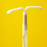
After the patient had miscarriages in 2009 and 2011, she asked her ObGyn if the missing IUD, which she had never seen leave her body, might have contributed to the miscarriages. She was told that the device was not present. No further testing was performed.
After the patient switched providers in 2013, an abdominal x-ray located the IUD. The patient became pregnant after the IUD was successfully removed.
PATIENT'S CLAIM:
The patient sued the ObGyn and his clinic for personal injury and wrongful death of the unborn fetuses. She claimed that it is below the standard of care not to perform an abdominal x-ray when a patient's IUD is missing.
The patient's attorney objected to the tests of the miscarried fetuses, contending that the tissues were sent without consent and that misinformation was conveyed on the pathology requisition.
DEFENDANTS' DEFENSE:
The standard of care set by the American College of Obstetricians and Gynecologists (ACOG) did not specify the use of an abdominal x-ray until Practice Bulletin No. 121 in July 2011 stated that the location of a lost IUD should be confirmed by x-ray.1
The IUD did not produce the miscarriages; testing of remains of 2 miscarried fetuses showed trisomic abnormalities that could not be attributed to the IUD.
The defense countered the patient's attorney that the testing forms had the patient's signed consent and that nothing was misrepresented on the forms.
VERDICT:
A Missouri defense verdict was returned for the wrongful death counts, but the jury awarded the patient $488,157 on the injury claim.
Reference
- Espey E, Singh RH; Committee on Practice Bulletins--Gynecology. American College of Gynecologists and Obstetricians Practice Bulletin No. 121: Long-acting reversible contraception: Implants and intrauterine devices. Obstet Gynecol. 2011;118(1):184-196.
Related article:
Pregnancy test missed before IUD placement? Your liability.
Needle left behind during mastectomy reconstruction
After a woman was diagnosed with invasive ductal cancer in her left breast, she underwent a double mastectomy with simple mastectomy reconstruction on the left breast. During the operation, the surgical count could not account for 1 pop-off needle. The surgeon searched for the missing needle and ordered an x-ray but the needle could not be located. The surgeon did not tell the patient that the needle count was incomplete.
The patient underwent several other breast reconstruction procedures and the needle was never found.
Five years after the initial surgery, the needle was discovered and surgically removed.
PATIENT'S CLAIM:
The surgeon was negligent in not finding the needle on the x-ray and not notifying her that the needle was missing.
PHYSICIAN'S DEFENSE:
The surgeon contended that his search for the needle and reliance on x-ray were in line with the standard of care. Since his actions were within the standard of care, he was not required to inform the patient.
VERDICT:
A Mississippi defense verdict was returned.
Failure to diagnose breast cancer on mammography
A 62-year-old woman started having routine mammographies in 2003. From 2006 to 2010, her annual mammographies were read by the same radiologist (Dr. A), who reported them as normal. Her 2011 mammography was read by a second radiologist (Dr. B), who reported it as normal. A year later, the patient was found to have several breast masses. Testing revealed that the cancer had metastasized. She underwent radical mastectomy and aggressive radiation treatment, but her cancer was deemed incurable.
PATIENT'S CLAIM:
Dr. A misread her mammograms from 2006 to 2010. There was evidence of asymmetric density suggestive of cancer on the 2006 mammography film.
PHYSICIAN'S DEFENSE:
There was no negligence. His reading of the mammographies was reasonable.
VERDICT:
A Kentucky defense verdict was returned.
These cases were selected by the editors of OBG Management from Medical Malpractice Verdicts, Settlements & Experts, with permission of the editor, Lewis Laska (www.verdictslaska.com). The information available to the editors about the cases presented here is sometimes incomplete. Moreover, the cases may or may not have merit. Nevertheless, these cases represent the types of clinical situations that typically result in litigation and are meant to illustrate nationwide variation in jury verdicts and awards.
Share your thoughts! Send your Letter to the Editor to [email protected]. Please include your name and the city and state in which you practice.
Miscarriages after IUD is missing: $488,157 verdict
After the patient had miscarriages in 2009 and 2011, she asked her ObGyn if the missing IUD, which she had never seen leave her body, might have contributed to the miscarriages. She was told that the device was not present. No further testing was performed.
After the patient switched providers in 2013, an abdominal x-ray located the IUD. The patient became pregnant after the IUD was successfully removed.
PATIENT'S CLAIM:
The patient sued the ObGyn and his clinic for personal injury and wrongful death of the unborn fetuses. She claimed that it is below the standard of care not to perform an abdominal x-ray when a patient's IUD is missing.
The patient's attorney objected to the tests of the miscarried fetuses, contending that the tissues were sent without consent and that misinformation was conveyed on the pathology requisition.
DEFENDANTS' DEFENSE:
The standard of care set by the American College of Obstetricians and Gynecologists (ACOG) did not specify the use of an abdominal x-ray until Practice Bulletin No. 121 in July 2011 stated that the location of a lost IUD should be confirmed by x-ray.1
The IUD did not produce the miscarriages; testing of remains of 2 miscarried fetuses showed trisomic abnormalities that could not be attributed to the IUD.
The defense countered the patient's attorney that the testing forms had the patient's signed consent and that nothing was misrepresented on the forms.
VERDICT:
A Missouri defense verdict was returned for the wrongful death counts, but the jury awarded the patient $488,157 on the injury claim.
Reference
- Espey E, Singh RH; Committee on Practice Bulletins--Gynecology. American College of Gynecologists and Obstetricians Practice Bulletin No. 121: Long-acting reversible contraception: Implants and intrauterine devices. Obstet Gynecol. 2011;118(1):184-196.
Related article:
Pregnancy test missed before IUD placement? Your liability.
Needle left behind during mastectomy reconstruction
After a woman was diagnosed with invasive ductal cancer in her left breast, she underwent a double mastectomy with simple mastectomy reconstruction on the left breast. During the operation, the surgical count could not account for 1 pop-off needle. The surgeon searched for the missing needle and ordered an x-ray but the needle could not be located. The surgeon did not tell the patient that the needle count was incomplete.
The patient underwent several other breast reconstruction procedures and the needle was never found.
Five years after the initial surgery, the needle was discovered and surgically removed.
PATIENT'S CLAIM:
The surgeon was negligent in not finding the needle on the x-ray and not notifying her that the needle was missing.
PHYSICIAN'S DEFENSE:
The surgeon contended that his search for the needle and reliance on x-ray were in line with the standard of care. Since his actions were within the standard of care, he was not required to inform the patient.
VERDICT:
A Mississippi defense verdict was returned.
Failure to diagnose breast cancer on mammography
A 62-year-old woman started having routine mammographies in 2003. From 2006 to 2010, her annual mammographies were read by the same radiologist (Dr. A), who reported them as normal. Her 2011 mammography was read by a second radiologist (Dr. B), who reported it as normal. A year later, the patient was found to have several breast masses. Testing revealed that the cancer had metastasized. She underwent radical mastectomy and aggressive radiation treatment, but her cancer was deemed incurable.
PATIENT'S CLAIM:
Dr. A misread her mammograms from 2006 to 2010. There was evidence of asymmetric density suggestive of cancer on the 2006 mammography film.
PHYSICIAN'S DEFENSE:
There was no negligence. His reading of the mammographies was reasonable.
VERDICT:
A Kentucky defense verdict was returned.
These cases were selected by the editors of OBG Management from Medical Malpractice Verdicts, Settlements & Experts, with permission of the editor, Lewis Laska (www.verdictslaska.com). The information available to the editors about the cases presented here is sometimes incomplete. Moreover, the cases may or may not have merit. Nevertheless, these cases represent the types of clinical situations that typically result in litigation and are meant to illustrate nationwide variation in jury verdicts and awards.
Share your thoughts! Send your Letter to the Editor to [email protected]. Please include your name and the city and state in which you practice.
Miscarriages after IUD is missing: $488,157 verdict
After the patient had miscarriages in 2009 and 2011, she asked her ObGyn if the missing IUD, which she had never seen leave her body, might have contributed to the miscarriages. She was told that the device was not present. No further testing was performed.
After the patient switched providers in 2013, an abdominal x-ray located the IUD. The patient became pregnant after the IUD was successfully removed.
PATIENT'S CLAIM:
The patient sued the ObGyn and his clinic for personal injury and wrongful death of the unborn fetuses. She claimed that it is below the standard of care not to perform an abdominal x-ray when a patient's IUD is missing.
The patient's attorney objected to the tests of the miscarried fetuses, contending that the tissues were sent without consent and that misinformation was conveyed on the pathology requisition.
DEFENDANTS' DEFENSE:
The standard of care set by the American College of Obstetricians and Gynecologists (ACOG) did not specify the use of an abdominal x-ray until Practice Bulletin No. 121 in July 2011 stated that the location of a lost IUD should be confirmed by x-ray.1
The IUD did not produce the miscarriages; testing of remains of 2 miscarried fetuses showed trisomic abnormalities that could not be attributed to the IUD.
The defense countered the patient's attorney that the testing forms had the patient's signed consent and that nothing was misrepresented on the forms.
VERDICT:
A Missouri defense verdict was returned for the wrongful death counts, but the jury awarded the patient $488,157 on the injury claim.
Reference
- Espey E, Singh RH; Committee on Practice Bulletins--Gynecology. American College of Gynecologists and Obstetricians Practice Bulletin No. 121: Long-acting reversible contraception: Implants and intrauterine devices. Obstet Gynecol. 2011;118(1):184-196.
Related article:
Pregnancy test missed before IUD placement? Your liability.
Needle left behind during mastectomy reconstruction
After a woman was diagnosed with invasive ductal cancer in her left breast, she underwent a double mastectomy with simple mastectomy reconstruction on the left breast. During the operation, the surgical count could not account for 1 pop-off needle. The surgeon searched for the missing needle and ordered an x-ray but the needle could not be located. The surgeon did not tell the patient that the needle count was incomplete.
The patient underwent several other breast reconstruction procedures and the needle was never found.
Five years after the initial surgery, the needle was discovered and surgically removed.
PATIENT'S CLAIM:
The surgeon was negligent in not finding the needle on the x-ray and not notifying her that the needle was missing.
PHYSICIAN'S DEFENSE:
The surgeon contended that his search for the needle and reliance on x-ray were in line with the standard of care. Since his actions were within the standard of care, he was not required to inform the patient.
VERDICT:
A Mississippi defense verdict was returned.
Failure to diagnose breast cancer on mammography
A 62-year-old woman started having routine mammographies in 2003. From 2006 to 2010, her annual mammographies were read by the same radiologist (Dr. A), who reported them as normal. Her 2011 mammography was read by a second radiologist (Dr. B), who reported it as normal. A year later, the patient was found to have several breast masses. Testing revealed that the cancer had metastasized. She underwent radical mastectomy and aggressive radiation treatment, but her cancer was deemed incurable.
PATIENT'S CLAIM:
Dr. A misread her mammograms from 2006 to 2010. There was evidence of asymmetric density suggestive of cancer on the 2006 mammography film.
PHYSICIAN'S DEFENSE:
There was no negligence. His reading of the mammographies was reasonable.
VERDICT:
A Kentucky defense verdict was returned.
These cases were selected by the editors of OBG Management from Medical Malpractice Verdicts, Settlements & Experts, with permission of the editor, Lewis Laska (www.verdictslaska.com). The information available to the editors about the cases presented here is sometimes incomplete. Moreover, the cases may or may not have merit. Nevertheless, these cases represent the types of clinical situations that typically result in litigation and are meant to illustrate nationwide variation in jury verdicts and awards.
Share your thoughts! Send your Letter to the Editor to [email protected]. Please include your name and the city and state in which you practice.
Questions value of ACOG/SMFM guidelines
“FOR THE MANAGEMENT OF LABOR, PATIENCE IS A VIRTUE”
ROBERT L. BARBIERI, MD (EDITORIAL; AUGUST 2017)
Questions value of ACOG/SMFM guidelines
The labor management guidelines recommended by the American College of Obstetricians and Gynecologists (ACOG) and the Society of Maternal-Fetal Medicine (SMFM) are terrible. Now retired, I trained in 1959–1963. In my career as an obstetrician, my primary cesarean delivery rate was 10% or less, and part of that was external pressure from people who did not know how to deliver a baby. Persistent occiput posterior position is a problem of inadequate flexion of the head, often due to ineffective contractions earlier. In such a situation, “pit” early! Rotate the head if you must, and teach residents how, please. The guidelines do not discuss the exhausted mother who goes home after a long labor or hours of pushing. I have interviewed new obstetricians in my community as early as 1980 who did not know what deep transverse arrest was. There, I am done voicing my disgust with obstetrics as it is practiced today.
James Honig, MD
Merritt Island, Florida
Managing difficult labor scenarios
I concur with Dr. Barbieri’s views on labor management that watchful waiting and giving the patient adequate time to progress naturally is the key to increase the chances of vaginal delivery. After all, labor is a physiologic process and should progress naturally. Having said that, I would like to know Dr. Barbieri’s views on handling certain circumstances in which patients these days land in the labor room, including 1) postdated pregnancy with reduced fetal movements and not in labor; 2) full-term/post-term pregnancy with free-floating head and poor Bishop score; 3) full-term pregnancy with niggling pains for more than 1 week; and many such conditions that place you in the dilemma of whether to induce, knowing that chances of failure are high.
Manju Hotchandani, MD
New Delhi, India
Midwives always use patience to guide labor
As a Certified Nurse-Midwife since 1985 (now retired), “patience” in managing labor has always been my guide, as it has been for my midwifery colleagues. This is another example of ACOG finally acknowledging the truths we women have always known, without crediting the wisdom of midwives over the centuries. Lamaze International’s 6 Healthy Birth Practices also must have been their guide. “Evolving concepts of normal labor progress,” as though this was new information, would be humorous if it were not so frustrating!
Marsha Kelly, CNM
Charlotte, North Carolina
Dr. Barbieri responds
The readers of OBG Management have vast clinical experience, and we can all learn from their insights and guidance. On behalf of all our readers, I thank Drs. Honig and Hotchandani and Ms. Kelly for taking the time to share their expert advice.
Every clinician involved in the birth process is deeply committed to a safe delivery for both mother and baby. Clinicians guide the birth process based on the unique characteristics and needs of each woman. Dr. Honig advocates for the active management of the labor process, while Ms. Kelly advocates for less intervention. Both approaches to labor management may be optimal depending on the unique clinical needs of each woman. Dr. Hotchandani inquires about managing common obstetrical presentations. In my practice, induction is recommended for all women post-term who report consistently reduced fetal movement with the goal of reducing the risk of sudden intrauterine fetal demise. For healthy women at term with painful contractions and reassuring fetal status, but no cervical change, we support and counsel the patient and offer therapeutic rest with morphine. For women at term with a floating head and poor Bishop score, we would not intervene, until 41 weeks’ gestation when we would initiate gentle cervical ripening with mechanical or pharmacologic treatment.
Share your thoughts! Send your Letter to the Editor to [email protected]. Please include your name and the city and state in which you practice.
“FOR THE MANAGEMENT OF LABOR, PATIENCE IS A VIRTUE”
ROBERT L. BARBIERI, MD (EDITORIAL; AUGUST 2017)
Questions value of ACOG/SMFM guidelines
The labor management guidelines recommended by the American College of Obstetricians and Gynecologists (ACOG) and the Society of Maternal-Fetal Medicine (SMFM) are terrible. Now retired, I trained in 1959–1963. In my career as an obstetrician, my primary cesarean delivery rate was 10% or less, and part of that was external pressure from people who did not know how to deliver a baby. Persistent occiput posterior position is a problem of inadequate flexion of the head, often due to ineffective contractions earlier. In such a situation, “pit” early! Rotate the head if you must, and teach residents how, please. The guidelines do not discuss the exhausted mother who goes home after a long labor or hours of pushing. I have interviewed new obstetricians in my community as early as 1980 who did not know what deep transverse arrest was. There, I am done voicing my disgust with obstetrics as it is practiced today.
James Honig, MD
Merritt Island, Florida
Managing difficult labor scenarios
I concur with Dr. Barbieri’s views on labor management that watchful waiting and giving the patient adequate time to progress naturally is the key to increase the chances of vaginal delivery. After all, labor is a physiologic process and should progress naturally. Having said that, I would like to know Dr. Barbieri’s views on handling certain circumstances in which patients these days land in the labor room, including 1) postdated pregnancy with reduced fetal movements and not in labor; 2) full-term/post-term pregnancy with free-floating head and poor Bishop score; 3) full-term pregnancy with niggling pains for more than 1 week; and many such conditions that place you in the dilemma of whether to induce, knowing that chances of failure are high.
Manju Hotchandani, MD
New Delhi, India
Midwives always use patience to guide labor
As a Certified Nurse-Midwife since 1985 (now retired), “patience” in managing labor has always been my guide, as it has been for my midwifery colleagues. This is another example of ACOG finally acknowledging the truths we women have always known, without crediting the wisdom of midwives over the centuries. Lamaze International’s 6 Healthy Birth Practices also must have been their guide. “Evolving concepts of normal labor progress,” as though this was new information, would be humorous if it were not so frustrating!
Marsha Kelly, CNM
Charlotte, North Carolina
Dr. Barbieri responds
The readers of OBG Management have vast clinical experience, and we can all learn from their insights and guidance. On behalf of all our readers, I thank Drs. Honig and Hotchandani and Ms. Kelly for taking the time to share their expert advice.
Every clinician involved in the birth process is deeply committed to a safe delivery for both mother and baby. Clinicians guide the birth process based on the unique characteristics and needs of each woman. Dr. Honig advocates for the active management of the labor process, while Ms. Kelly advocates for less intervention. Both approaches to labor management may be optimal depending on the unique clinical needs of each woman. Dr. Hotchandani inquires about managing common obstetrical presentations. In my practice, induction is recommended for all women post-term who report consistently reduced fetal movement with the goal of reducing the risk of sudden intrauterine fetal demise. For healthy women at term with painful contractions and reassuring fetal status, but no cervical change, we support and counsel the patient and offer therapeutic rest with morphine. For women at term with a floating head and poor Bishop score, we would not intervene, until 41 weeks’ gestation when we would initiate gentle cervical ripening with mechanical or pharmacologic treatment.
Share your thoughts! Send your Letter to the Editor to [email protected]. Please include your name and the city and state in which you practice.
“FOR THE MANAGEMENT OF LABOR, PATIENCE IS A VIRTUE”
ROBERT L. BARBIERI, MD (EDITORIAL; AUGUST 2017)
Questions value of ACOG/SMFM guidelines
The labor management guidelines recommended by the American College of Obstetricians and Gynecologists (ACOG) and the Society of Maternal-Fetal Medicine (SMFM) are terrible. Now retired, I trained in 1959–1963. In my career as an obstetrician, my primary cesarean delivery rate was 10% or less, and part of that was external pressure from people who did not know how to deliver a baby. Persistent occiput posterior position is a problem of inadequate flexion of the head, often due to ineffective contractions earlier. In such a situation, “pit” early! Rotate the head if you must, and teach residents how, please. The guidelines do not discuss the exhausted mother who goes home after a long labor or hours of pushing. I have interviewed new obstetricians in my community as early as 1980 who did not know what deep transverse arrest was. There, I am done voicing my disgust with obstetrics as it is practiced today.
James Honig, MD
Merritt Island, Florida
Managing difficult labor scenarios
I concur with Dr. Barbieri’s views on labor management that watchful waiting and giving the patient adequate time to progress naturally is the key to increase the chances of vaginal delivery. After all, labor is a physiologic process and should progress naturally. Having said that, I would like to know Dr. Barbieri’s views on handling certain circumstances in which patients these days land in the labor room, including 1) postdated pregnancy with reduced fetal movements and not in labor; 2) full-term/post-term pregnancy with free-floating head and poor Bishop score; 3) full-term pregnancy with niggling pains for more than 1 week; and many such conditions that place you in the dilemma of whether to induce, knowing that chances of failure are high.
Manju Hotchandani, MD
New Delhi, India
Midwives always use patience to guide labor
As a Certified Nurse-Midwife since 1985 (now retired), “patience” in managing labor has always been my guide, as it has been for my midwifery colleagues. This is another example of ACOG finally acknowledging the truths we women have always known, without crediting the wisdom of midwives over the centuries. Lamaze International’s 6 Healthy Birth Practices also must have been their guide. “Evolving concepts of normal labor progress,” as though this was new information, would be humorous if it were not so frustrating!
Marsha Kelly, CNM
Charlotte, North Carolina
Dr. Barbieri responds
The readers of OBG Management have vast clinical experience, and we can all learn from their insights and guidance. On behalf of all our readers, I thank Drs. Honig and Hotchandani and Ms. Kelly for taking the time to share their expert advice.
Every clinician involved in the birth process is deeply committed to a safe delivery for both mother and baby. Clinicians guide the birth process based on the unique characteristics and needs of each woman. Dr. Honig advocates for the active management of the labor process, while Ms. Kelly advocates for less intervention. Both approaches to labor management may be optimal depending on the unique clinical needs of each woman. Dr. Hotchandani inquires about managing common obstetrical presentations. In my practice, induction is recommended for all women post-term who report consistently reduced fetal movement with the goal of reducing the risk of sudden intrauterine fetal demise. For healthy women at term with painful contractions and reassuring fetal status, but no cervical change, we support and counsel the patient and offer therapeutic rest with morphine. For women at term with a floating head and poor Bishop score, we would not intervene, until 41 weeks’ gestation when we would initiate gentle cervical ripening with mechanical or pharmacologic treatment.
Share your thoughts! Send your Letter to the Editor to [email protected]. Please include your name and the city and state in which you practice.

Adverse vaginal environment can trigger vaginosis
“EFFECTIVE TREATMENT OF RECURRENT BACTERIAL VAGINOSIS”
ROBERT L. BARBIERI, MD (EDITORIAL; JULY 2017)
Adverse vaginal environment can trigger vaginosis
I truly appreciated the formulaic presentation of specific regimens to attempt to eradicate recurrent bacterial vaginosis (BV), and in the future I will probably try one for a confounding case. However, although not the focus of the editorial, I found it disturbing that BV was presented as such a recalcitrant “medical” condition without emphasizing a simple understanding and approach that I have employed for the last 20 years with impressive curative results.
I have “cured” many women who have come to me after having bounced from physician to physician. Understanding that BV is not transmitted but results from an ecosystem imbalance—specifically, the lack of Lactobacillus bacteria and the overgrowth of anaerobes—any environmental manipulation that decreases the resting aerobic bacterial population can trigger the condition of vaginosis (not vaginitis).
My standard checklist, which reflects the multitude of products that pamper the modern vagina but are in fact detrimental, includes: bubble baths, which can leave a film in the vagina similar to that left in the bathtub; all forms of commercial and home-prepared douches; use of tampons extended beyond the heavy menstrual days, which can dry up the resting bacteria; repetitive immersion into a chlorinated (bactericidal) body of water (pool or hot tub); condoms that contain spermicides that are bactericidal as well; any antibacterial soap, especially fragrant liquid variants (great for the hands, awful for the vagina); fabrics like Spandex, pantyhose, and polyester that do not allow the aerobic bacteria to survive; noncotton underwear that does not let the vagina “breathe”; popular brands of scented and unscented winged pantyliners that suffocate the vaginal outlet; prolonged compression by the devoted long-distance cyclist and spa spinner; vaginal atrophy; and, anatomically, closely opposed labia, which can contribute to a chronically anaerobic vaginal environment through obstruction. When these factors are discussed and addressed, you would be surprised how much “recurrent” BV can be avoided, and therefore effectively treated.
Michael Abrahams, MD
New York, New York
Dr. Barbieri responds
I thank Dr. Abrahams for sharing his expert advice. I agree that reducing environmental exposures that inhibit the growth of vaginal lactobacilli is important in treating recurrent BV.
Share your thoughts! Send your Letter to the Editor to [email protected]. Please include your name and the city and state in which you practice.
“EFFECTIVE TREATMENT OF RECURRENT BACTERIAL VAGINOSIS”
ROBERT L. BARBIERI, MD (EDITORIAL; JULY 2017)
Adverse vaginal environment can trigger vaginosis
I truly appreciated the formulaic presentation of specific regimens to attempt to eradicate recurrent bacterial vaginosis (BV), and in the future I will probably try one for a confounding case. However, although not the focus of the editorial, I found it disturbing that BV was presented as such a recalcitrant “medical” condition without emphasizing a simple understanding and approach that I have employed for the last 20 years with impressive curative results.
I have “cured” many women who have come to me after having bounced from physician to physician. Understanding that BV is not transmitted but results from an ecosystem imbalance—specifically, the lack of Lactobacillus bacteria and the overgrowth of anaerobes—any environmental manipulation that decreases the resting aerobic bacterial population can trigger the condition of vaginosis (not vaginitis).
My standard checklist, which reflects the multitude of products that pamper the modern vagina but are in fact detrimental, includes: bubble baths, which can leave a film in the vagina similar to that left in the bathtub; all forms of commercial and home-prepared douches; use of tampons extended beyond the heavy menstrual days, which can dry up the resting bacteria; repetitive immersion into a chlorinated (bactericidal) body of water (pool or hot tub); condoms that contain spermicides that are bactericidal as well; any antibacterial soap, especially fragrant liquid variants (great for the hands, awful for the vagina); fabrics like Spandex, pantyhose, and polyester that do not allow the aerobic bacteria to survive; noncotton underwear that does not let the vagina “breathe”; popular brands of scented and unscented winged pantyliners that suffocate the vaginal outlet; prolonged compression by the devoted long-distance cyclist and spa spinner; vaginal atrophy; and, anatomically, closely opposed labia, which can contribute to a chronically anaerobic vaginal environment through obstruction. When these factors are discussed and addressed, you would be surprised how much “recurrent” BV can be avoided, and therefore effectively treated.
Michael Abrahams, MD
New York, New York
Dr. Barbieri responds
I thank Dr. Abrahams for sharing his expert advice. I agree that reducing environmental exposures that inhibit the growth of vaginal lactobacilli is important in treating recurrent BV.
Share your thoughts! Send your Letter to the Editor to [email protected]. Please include your name and the city and state in which you practice.
“EFFECTIVE TREATMENT OF RECURRENT BACTERIAL VAGINOSIS”
ROBERT L. BARBIERI, MD (EDITORIAL; JULY 2017)
Adverse vaginal environment can trigger vaginosis
I truly appreciated the formulaic presentation of specific regimens to attempt to eradicate recurrent bacterial vaginosis (BV), and in the future I will probably try one for a confounding case. However, although not the focus of the editorial, I found it disturbing that BV was presented as such a recalcitrant “medical” condition without emphasizing a simple understanding and approach that I have employed for the last 20 years with impressive curative results.
I have “cured” many women who have come to me after having bounced from physician to physician. Understanding that BV is not transmitted but results from an ecosystem imbalance—specifically, the lack of Lactobacillus bacteria and the overgrowth of anaerobes—any environmental manipulation that decreases the resting aerobic bacterial population can trigger the condition of vaginosis (not vaginitis).
My standard checklist, which reflects the multitude of products that pamper the modern vagina but are in fact detrimental, includes: bubble baths, which can leave a film in the vagina similar to that left in the bathtub; all forms of commercial and home-prepared douches; use of tampons extended beyond the heavy menstrual days, which can dry up the resting bacteria; repetitive immersion into a chlorinated (bactericidal) body of water (pool or hot tub); condoms that contain spermicides that are bactericidal as well; any antibacterial soap, especially fragrant liquid variants (great for the hands, awful for the vagina); fabrics like Spandex, pantyhose, and polyester that do not allow the aerobic bacteria to survive; noncotton underwear that does not let the vagina “breathe”; popular brands of scented and unscented winged pantyliners that suffocate the vaginal outlet; prolonged compression by the devoted long-distance cyclist and spa spinner; vaginal atrophy; and, anatomically, closely opposed labia, which can contribute to a chronically anaerobic vaginal environment through obstruction. When these factors are discussed and addressed, you would be surprised how much “recurrent” BV can be avoided, and therefore effectively treated.
Michael Abrahams, MD
New York, New York
Dr. Barbieri responds
I thank Dr. Abrahams for sharing his expert advice. I agree that reducing environmental exposures that inhibit the growth of vaginal lactobacilli is important in treating recurrent BV.
Share your thoughts! Send your Letter to the Editor to [email protected]. Please include your name and the city and state in which you practice.
Approach for removing cervical fibroids
“LAPAROSCOPIC MYOMECTOMY: TIPS FOR PATIENT SELECTION AND TECHNIQUE”
WILLIAM H. PARKER, MD (JULY 2017)
Approach for removing cervical fibroids
I thank Dr. Parker for his tips on laparoscopic myomectomy. I have one question: Should large cervical fibroids be tackled laparoscopically? If yes, then please provide some tips. Cervical fibroids are sometimes difficult to enucleate, and nothing can catch the fibroid, as the consistency is such that everything cuts through.
Manju Hotchandani, MD
New Delhi, India
Dr. Parker responds
Magnetic resonance imaging is the best imaging approach for helping to evaluate the position and size of a cervical fibroid. Fibroids that are intracervical are best removed through a vaginal approach. With the patient under adequate anesthesia, the cervix is dilated or, if necessary, incised (Dührssen incisions), and the fibroid grasped with a tenaculum. The fibroid is finger dissected away from the cervix until the pedicle is palpated. The pedicle is either clamped or ligated with suture and then cut, and the cervix is repaired.
If the fibroid is intramural/subserosal and coming off the lower uterine segment or cervix, we identify the ipsilateral ureter and follow its course near the fibroid. An incision is made over the fibroid and directed away from the ureter. It is important to incise down through the fibroid pseudocapsule and to dissect the fibroid underneath the pseudocapsule, decreasing the risk of injury to the ureter and uterine vessels. Depending on the size and position of the fibroid and the experience of the surgeon, this technique can be performed laparoscopically.
Share your thoughts! Send your Letter to the Editor to [email protected]. Please include your name and the city and state in which you practice.
“LAPAROSCOPIC MYOMECTOMY: TIPS FOR PATIENT SELECTION AND TECHNIQUE”
WILLIAM H. PARKER, MD (JULY 2017)
Approach for removing cervical fibroids
I thank Dr. Parker for his tips on laparoscopic myomectomy. I have one question: Should large cervical fibroids be tackled laparoscopically? If yes, then please provide some tips. Cervical fibroids are sometimes difficult to enucleate, and nothing can catch the fibroid, as the consistency is such that everything cuts through.
Manju Hotchandani, MD
New Delhi, India
Dr. Parker responds
Magnetic resonance imaging is the best imaging approach for helping to evaluate the position and size of a cervical fibroid. Fibroids that are intracervical are best removed through a vaginal approach. With the patient under adequate anesthesia, the cervix is dilated or, if necessary, incised (Dührssen incisions), and the fibroid grasped with a tenaculum. The fibroid is finger dissected away from the cervix until the pedicle is palpated. The pedicle is either clamped or ligated with suture and then cut, and the cervix is repaired.
If the fibroid is intramural/subserosal and coming off the lower uterine segment or cervix, we identify the ipsilateral ureter and follow its course near the fibroid. An incision is made over the fibroid and directed away from the ureter. It is important to incise down through the fibroid pseudocapsule and to dissect the fibroid underneath the pseudocapsule, decreasing the risk of injury to the ureter and uterine vessels. Depending on the size and position of the fibroid and the experience of the surgeon, this technique can be performed laparoscopically.
Share your thoughts! Send your Letter to the Editor to [email protected]. Please include your name and the city and state in which you practice.
“LAPAROSCOPIC MYOMECTOMY: TIPS FOR PATIENT SELECTION AND TECHNIQUE”
WILLIAM H. PARKER, MD (JULY 2017)
Approach for removing cervical fibroids
I thank Dr. Parker for his tips on laparoscopic myomectomy. I have one question: Should large cervical fibroids be tackled laparoscopically? If yes, then please provide some tips. Cervical fibroids are sometimes difficult to enucleate, and nothing can catch the fibroid, as the consistency is such that everything cuts through.
Manju Hotchandani, MD
New Delhi, India
Dr. Parker responds
Magnetic resonance imaging is the best imaging approach for helping to evaluate the position and size of a cervical fibroid. Fibroids that are intracervical are best removed through a vaginal approach. With the patient under adequate anesthesia, the cervix is dilated or, if necessary, incised (Dührssen incisions), and the fibroid grasped with a tenaculum. The fibroid is finger dissected away from the cervix until the pedicle is palpated. The pedicle is either clamped or ligated with suture and then cut, and the cervix is repaired.
If the fibroid is intramural/subserosal and coming off the lower uterine segment or cervix, we identify the ipsilateral ureter and follow its course near the fibroid. An incision is made over the fibroid and directed away from the ureter. It is important to incise down through the fibroid pseudocapsule and to dissect the fibroid underneath the pseudocapsule, decreasing the risk of injury to the ureter and uterine vessels. Depending on the size and position of the fibroid and the experience of the surgeon, this technique can be performed laparoscopically.
Share your thoughts! Send your Letter to the Editor to [email protected]. Please include your name and the city and state in which you practice.
Late delivery of macrosomic baby
Late delivery of macrosomic baby: $5.5M settlement

After a woman was admitted to the hospital, her labor was complicated by very slow progress, maternal fever, and multiple fetal heart-rate (FHR) monitor strip abnormalities. A baby boy was born by cesarean delivery 26 hours after the mother's admission, weighing almost 11 lb. At birth, the baby was asphyxiated, with an arterial cord blood pH of 7.01 and a base excess of -14.4. He was resuscitated and sent to the neonatal intensive care unit (NICU). Nine days after birth, he began to have seizures. The placental pathology report revealed infection; placental cultures grew Group B strep. The infant was diagnosed 6 days later with hypoxic ischemic encephalopathy after magnetic resonance imaging.
PARENTS' CLAIM:
The ObGyn and hospital failed to recognize that the baby was macrosomic. The ObGyn should have delivered the baby when the FHR monitor first showed fetal distress.
DEFENDANTS' DEFENSE:
The case was settled during the trial.
VERDICT:
A $5.5 million Washington settlement was reached.
Size of perineal tear and its consequences disputed: $1.8M verdict
A 34-year-old woman gave birth vaginally. During forceps delivery, she suffered a 4th-degree perineal tear, running from her vagina to her rectum, which was misidentified by the ObGyn as a 2nd-degree tear. The perineal tear lead to complications including infection and a persistent rectovaginal fistula. She underwent several operations over the next 5 years.
PARENTS' CLAIM:
The ObGyn did not appropriately address the wound. Had a cesarean delivery been performed, the wound would not have occurred.
DEFENDANTS' DEFENSE:
The decisions to allow labor to continue and to use forceps were reasonable. The ObGyn contended that the patient sustained only a 2nd-degree tear in the delivery room, with the 4th-degree tear occurring the next day due to attenuated tissue.
VERDICT:
A $1.8 million Illinois verdict was returned.
Related article:
Develop and use a checklist for 3rd- and 4th-degree perineal lacerations
Macrosomic baby, cerebral palsy: $5.5M settlement
At 42 weeks' gestation, a mother was sent to the hospital in labor. During delivery, thick meconium was encountered. At birth, the baby weighed more than 9 lb. At age 16 years, the child has cerebral palsy, a seizure disorder, and developmental and cognitive deficits. She functions at the level of an 8-year-old child.
PARENTS' CLAIM:
The ObGyn and nurses did not deliver the baby in a timely manner by cesarean delivery. The estimated weight and a deteriorating in utero environment, as evidenced by the thick meconium, caused the child's brain injury.
DEFENDANTS' DEFENSE:
The case settled during trial.
VERDICT:
A $5.5 million Illinois settlement was reached.
Did baby get Group B strep from her mother?
During prenatal care by an ObGyn, a woman underwent a routine test for Group B strep, the results of which were negative. The child, with APGAR scores of 9 at 1 and 5 minutes after birth, was admitted to the well-baby nursery. Four hours after birth, the baby appeared to be in distress and was moved to the NICU. She was found to have respiratory distress, metabolic acidosis, and sepsis, allegedly caused by Group B strep, and was placed on a ventilator and prescribed medications. When the baby's condition worsened, she was transferred to a children's hospital. She was discharged 3 months later with bronchopulmonary dysplasia and a chronic lung disease that requires continuous treatment.
PARENTS' CLAIM:
The baby's injuries were attributable to Group B strep. The ObGyn's prenatal treatment did not meet the standard of care.
PHYSICIAN'S DEFENSE:
There was no negligence on the part of the ObGyn. The standard of care was met.
VERDICT:
An Alabama defense verdict was returned.
These cases were selected by the editors of OBG Management from Medical Malpractice Verdicts, Settlements & Experts, with permission of the editor, Lewis Laska (www.verdictslaska.com). The information available to the editors about the cases presented here is sometimes incomplete. Moreover, the cases may or may not have merit. Nevertheless, these cases represent the types of clinical situations that typically result in litigation and are meant to illustrate nationwide variation in jury verdicts and awards.
Share your thoughts! Send your Letter to the Editor to [email protected]. Please include your name and the city and state in which you practice.
Late delivery of macrosomic baby: $5.5M settlement
After a woman was admitted to the hospital, her labor was complicated by very slow progress, maternal fever, and multiple fetal heart-rate (FHR) monitor strip abnormalities. A baby boy was born by cesarean delivery 26 hours after the mother's admission, weighing almost 11 lb. At birth, the baby was asphyxiated, with an arterial cord blood pH of 7.01 and a base excess of -14.4. He was resuscitated and sent to the neonatal intensive care unit (NICU). Nine days after birth, he began to have seizures. The placental pathology report revealed infection; placental cultures grew Group B strep. The infant was diagnosed 6 days later with hypoxic ischemic encephalopathy after magnetic resonance imaging.
PARENTS' CLAIM:
The ObGyn and hospital failed to recognize that the baby was macrosomic. The ObGyn should have delivered the baby when the FHR monitor first showed fetal distress.
DEFENDANTS' DEFENSE:
The case was settled during the trial.
VERDICT:
A $5.5 million Washington settlement was reached.
Size of perineal tear and its consequences disputed: $1.8M verdict
A 34-year-old woman gave birth vaginally. During forceps delivery, she suffered a 4th-degree perineal tear, running from her vagina to her rectum, which was misidentified by the ObGyn as a 2nd-degree tear. The perineal tear lead to complications including infection and a persistent rectovaginal fistula. She underwent several operations over the next 5 years.
PARENTS' CLAIM:
The ObGyn did not appropriately address the wound. Had a cesarean delivery been performed, the wound would not have occurred.
DEFENDANTS' DEFENSE:
The decisions to allow labor to continue and to use forceps were reasonable. The ObGyn contended that the patient sustained only a 2nd-degree tear in the delivery room, with the 4th-degree tear occurring the next day due to attenuated tissue.
VERDICT:
A $1.8 million Illinois verdict was returned.
Related article:
Develop and use a checklist for 3rd- and 4th-degree perineal lacerations
Macrosomic baby, cerebral palsy: $5.5M settlement
At 42 weeks' gestation, a mother was sent to the hospital in labor. During delivery, thick meconium was encountered. At birth, the baby weighed more than 9 lb. At age 16 years, the child has cerebral palsy, a seizure disorder, and developmental and cognitive deficits. She functions at the level of an 8-year-old child.
PARENTS' CLAIM:
The ObGyn and nurses did not deliver the baby in a timely manner by cesarean delivery. The estimated weight and a deteriorating in utero environment, as evidenced by the thick meconium, caused the child's brain injury.
DEFENDANTS' DEFENSE:
The case settled during trial.
VERDICT:
A $5.5 million Illinois settlement was reached.
Did baby get Group B strep from her mother?
During prenatal care by an ObGyn, a woman underwent a routine test for Group B strep, the results of which were negative. The child, with APGAR scores of 9 at 1 and 5 minutes after birth, was admitted to the well-baby nursery. Four hours after birth, the baby appeared to be in distress and was moved to the NICU. She was found to have respiratory distress, metabolic acidosis, and sepsis, allegedly caused by Group B strep, and was placed on a ventilator and prescribed medications. When the baby's condition worsened, she was transferred to a children's hospital. She was discharged 3 months later with bronchopulmonary dysplasia and a chronic lung disease that requires continuous treatment.
PARENTS' CLAIM:
The baby's injuries were attributable to Group B strep. The ObGyn's prenatal treatment did not meet the standard of care.
PHYSICIAN'S DEFENSE:
There was no negligence on the part of the ObGyn. The standard of care was met.
VERDICT:
An Alabama defense verdict was returned.
These cases were selected by the editors of OBG Management from Medical Malpractice Verdicts, Settlements & Experts, with permission of the editor, Lewis Laska (www.verdictslaska.com). The information available to the editors about the cases presented here is sometimes incomplete. Moreover, the cases may or may not have merit. Nevertheless, these cases represent the types of clinical situations that typically result in litigation and are meant to illustrate nationwide variation in jury verdicts and awards.
Share your thoughts! Send your Letter to the Editor to [email protected]. Please include your name and the city and state in which you practice.
Late delivery of macrosomic baby: $5.5M settlement
After a woman was admitted to the hospital, her labor was complicated by very slow progress, maternal fever, and multiple fetal heart-rate (FHR) monitor strip abnormalities. A baby boy was born by cesarean delivery 26 hours after the mother's admission, weighing almost 11 lb. At birth, the baby was asphyxiated, with an arterial cord blood pH of 7.01 and a base excess of -14.4. He was resuscitated and sent to the neonatal intensive care unit (NICU). Nine days after birth, he began to have seizures. The placental pathology report revealed infection; placental cultures grew Group B strep. The infant was diagnosed 6 days later with hypoxic ischemic encephalopathy after magnetic resonance imaging.
PARENTS' CLAIM:
The ObGyn and hospital failed to recognize that the baby was macrosomic. The ObGyn should have delivered the baby when the FHR monitor first showed fetal distress.
DEFENDANTS' DEFENSE:
The case was settled during the trial.
VERDICT:
A $5.5 million Washington settlement was reached.
Size of perineal tear and its consequences disputed: $1.8M verdict
A 34-year-old woman gave birth vaginally. During forceps delivery, she suffered a 4th-degree perineal tear, running from her vagina to her rectum, which was misidentified by the ObGyn as a 2nd-degree tear. The perineal tear lead to complications including infection and a persistent rectovaginal fistula. She underwent several operations over the next 5 years.
PARENTS' CLAIM:
The ObGyn did not appropriately address the wound. Had a cesarean delivery been performed, the wound would not have occurred.
DEFENDANTS' DEFENSE:
The decisions to allow labor to continue and to use forceps were reasonable. The ObGyn contended that the patient sustained only a 2nd-degree tear in the delivery room, with the 4th-degree tear occurring the next day due to attenuated tissue.
VERDICT:
A $1.8 million Illinois verdict was returned.
Related article:
Develop and use a checklist for 3rd- and 4th-degree perineal lacerations
Macrosomic baby, cerebral palsy: $5.5M settlement
At 42 weeks' gestation, a mother was sent to the hospital in labor. During delivery, thick meconium was encountered. At birth, the baby weighed more than 9 lb. At age 16 years, the child has cerebral palsy, a seizure disorder, and developmental and cognitive deficits. She functions at the level of an 8-year-old child.
PARENTS' CLAIM:
The ObGyn and nurses did not deliver the baby in a timely manner by cesarean delivery. The estimated weight and a deteriorating in utero environment, as evidenced by the thick meconium, caused the child's brain injury.
DEFENDANTS' DEFENSE:
The case settled during trial.
VERDICT:
A $5.5 million Illinois settlement was reached.
Did baby get Group B strep from her mother?
During prenatal care by an ObGyn, a woman underwent a routine test for Group B strep, the results of which were negative. The child, with APGAR scores of 9 at 1 and 5 minutes after birth, was admitted to the well-baby nursery. Four hours after birth, the baby appeared to be in distress and was moved to the NICU. She was found to have respiratory distress, metabolic acidosis, and sepsis, allegedly caused by Group B strep, and was placed on a ventilator and prescribed medications. When the baby's condition worsened, she was transferred to a children's hospital. She was discharged 3 months later with bronchopulmonary dysplasia and a chronic lung disease that requires continuous treatment.
PARENTS' CLAIM:
The baby's injuries were attributable to Group B strep. The ObGyn's prenatal treatment did not meet the standard of care.
PHYSICIAN'S DEFENSE:
There was no negligence on the part of the ObGyn. The standard of care was met.
VERDICT:
An Alabama defense verdict was returned.
These cases were selected by the editors of OBG Management from Medical Malpractice Verdicts, Settlements & Experts, with permission of the editor, Lewis Laska (www.verdictslaska.com). The information available to the editors about the cases presented here is sometimes incomplete. Moreover, the cases may or may not have merit. Nevertheless, these cases represent the types of clinical situations that typically result in litigation and are meant to illustrate nationwide variation in jury verdicts and awards.
Share your thoughts! Send your Letter to the Editor to [email protected]. Please include your name and the city and state in which you practice.
Hypothyroidism carries higher surgical risk not captured by calculator
VICTORIA, B.C. – Even with contemporary anesthesia and surgical techniques, patients who are overtly hypothyroid at the time of major surgery have a rockier course, suggests a retrospective cohort study of 58 patients in a poster presentation at the annual meeting of the American Thyroid Association.
Actual length of stay for hypothyroid patients was twice that predicted by a commonly used risk calculator, whereas actual and predicted stays aligned well for euthyroid patients. The hypothyroid group had more cases of postoperative atrial fibrillation, ileus, reintubation, and death, although numbers were too small for statistical comparison.
“This will have an impact on how we look at patients, especially from a hospital standpoint and management. That’s quite a bit longer stay and quite a bit more cost. And the longer you stay, the more complications you have, too, so it could be riskier for the patient as well,” said first author Raquel Villavicencio, MD, a fellow at Indiana University at the time of the study, and now a clinical endocrinologist at Community Hospital in Indianapolis.

“Although we don’t consider hypothyroidism an absolute contraindication to surgery, especially if it’s necessary surgery, certainly anybody who is having elective surgery should have it postponed, in our opinion, until they are rendered euthyroid,” she said. “More studies are needed to look at this a little bit closer.”
Explaining the study’s rationale, Dr. Villavicencio noted, “This was a question that came up maybe three or four times a year, where we would get a hypothyroid patient and had to decide whether or not to clear them for surgery.”
Previous studies conducted at large institutions, the Mayo Clinic and Massachusetts General Hospital, had conflicting findings and were done about 30 years ago, she said. Anesthesia and surgical care have improved substantially since then, leading the investigators to hypothesize that hypothyroidism would not carry higher surgical risk today.
Dr. Villavicencio and her coinvestigator, Cary Mariash, MD, used their institutional database to identify 29 adult patients with a thyroid-stimulating hormone (TSH) level of greater than 10 mcU/mL alone or with a TSH level exceeding the upper limit of normal along with a free thyroxine (T4) level of less than 0.6 ng/dL who underwent surgery during 2010-2015. They matched each patient on age, sex, and surgical procedure with a control euthyroid patient.
The mean TSH level in the hypothyroid group was 29.2 mcU/mL. The majority of patients in each group – 59% of the hypothyroid group and 62% of the euthyroid group – had an American Surgical Association class of 3, denoting that this was a fairly sick population. The groups were generally similar on rates of comorbidity, except that the euthyroid patients had a slightly higher prevalence of obstructive sleep apnea.
In both groups, the majority of procedures were laparotomy and/or bowel resection; pharyngolaryngectomy and esophagectomy/esophagoplasty; and wound or bone debridement.
Main results showed that in the hypothyroid group, hospital length of stay predicted with the American College of Surgeons National Surgical Quality Improvement Program surgical risk calculator was 6.9 days, but actual length of stay was 14.4 days (P = .0004). In contrast, in the euthyroid group, predicted length of stay was a similar at 7.1 days, and actual length of stay was statistically indistinguishable at 9.2 days (P = .1).
“Hypothyroidism is not taken into account with this calculator,” Dr. Villavicencio noted, adding that she was unaware of any surgical calculators that do.
One patient in the hypothyroid group died, compared with none in the euthyroid group. In terms of postoperative cardiac complications, two patients in the hypothyroid group experienced atrial fibrillation, and there was one case of pulseless electrical–activity arrest in each group.
The groups did not differ on incidence of hypothermia, bradycardia, hyponatremia, time to extubation, and hypotension. However, mean arterial pressure tended to be lower in the hypothyroid group (51 mm Hg) than in the euthyroid group (56 mm Hg), and the former more often needed vasopressors. Furthermore, postoperative ileus and reintubation were more common in the hypothyroid group.
“I think that there are kind of a lot of little things that add up to explain [the longer stay],” said Dr. Villavicencio, who disclosed that she had no relevant conflicts of interest.
VICTORIA, B.C. – Even with contemporary anesthesia and surgical techniques, patients who are overtly hypothyroid at the time of major surgery have a rockier course, suggests a retrospective cohort study of 58 patients in a poster presentation at the annual meeting of the American Thyroid Association.
Actual length of stay for hypothyroid patients was twice that predicted by a commonly used risk calculator, whereas actual and predicted stays aligned well for euthyroid patients. The hypothyroid group had more cases of postoperative atrial fibrillation, ileus, reintubation, and death, although numbers were too small for statistical comparison.
“This will have an impact on how we look at patients, especially from a hospital standpoint and management. That’s quite a bit longer stay and quite a bit more cost. And the longer you stay, the more complications you have, too, so it could be riskier for the patient as well,” said first author Raquel Villavicencio, MD, a fellow at Indiana University at the time of the study, and now a clinical endocrinologist at Community Hospital in Indianapolis.
“Although we don’t consider hypothyroidism an absolute contraindication to surgery, especially if it’s necessary surgery, certainly anybody who is having elective surgery should have it postponed, in our opinion, until they are rendered euthyroid,” she said. “More studies are needed to look at this a little bit closer.”
Explaining the study’s rationale, Dr. Villavicencio noted, “This was a question that came up maybe three or four times a year, where we would get a hypothyroid patient and had to decide whether or not to clear them for surgery.”
Previous studies conducted at large institutions, the Mayo Clinic and Massachusetts General Hospital, had conflicting findings and were done about 30 years ago, she said. Anesthesia and surgical care have improved substantially since then, leading the investigators to hypothesize that hypothyroidism would not carry higher surgical risk today.
Dr. Villavicencio and her coinvestigator, Cary Mariash, MD, used their institutional database to identify 29 adult patients with a thyroid-stimulating hormone (TSH) level of greater than 10 mcU/mL alone or with a TSH level exceeding the upper limit of normal along with a free thyroxine (T4) level of less than 0.6 ng/dL who underwent surgery during 2010-2015. They matched each patient on age, sex, and surgical procedure with a control euthyroid patient.
The mean TSH level in the hypothyroid group was 29.2 mcU/mL. The majority of patients in each group – 59% of the hypothyroid group and 62% of the euthyroid group – had an American Surgical Association class of 3, denoting that this was a fairly sick population. The groups were generally similar on rates of comorbidity, except that the euthyroid patients had a slightly higher prevalence of obstructive sleep apnea.
In both groups, the majority of procedures were laparotomy and/or bowel resection; pharyngolaryngectomy and esophagectomy/esophagoplasty; and wound or bone debridement.
Main results showed that in the hypothyroid group, hospital length of stay predicted with the American College of Surgeons National Surgical Quality Improvement Program surgical risk calculator was 6.9 days, but actual length of stay was 14.4 days (P = .0004). In contrast, in the euthyroid group, predicted length of stay was a similar at 7.1 days, and actual length of stay was statistically indistinguishable at 9.2 days (P = .1).
“Hypothyroidism is not taken into account with this calculator,” Dr. Villavicencio noted, adding that she was unaware of any surgical calculators that do.
One patient in the hypothyroid group died, compared with none in the euthyroid group. In terms of postoperative cardiac complications, two patients in the hypothyroid group experienced atrial fibrillation, and there was one case of pulseless electrical–activity arrest in each group.
The groups did not differ on incidence of hypothermia, bradycardia, hyponatremia, time to extubation, and hypotension. However, mean arterial pressure tended to be lower in the hypothyroid group (51 mm Hg) than in the euthyroid group (56 mm Hg), and the former more often needed vasopressors. Furthermore, postoperative ileus and reintubation were more common in the hypothyroid group.
“I think that there are kind of a lot of little things that add up to explain [the longer stay],” said Dr. Villavicencio, who disclosed that she had no relevant conflicts of interest.
VICTORIA, B.C. – Even with contemporary anesthesia and surgical techniques, patients who are overtly hypothyroid at the time of major surgery have a rockier course, suggests a retrospective cohort study of 58 patients in a poster presentation at the annual meeting of the American Thyroid Association.
Actual length of stay for hypothyroid patients was twice that predicted by a commonly used risk calculator, whereas actual and predicted stays aligned well for euthyroid patients. The hypothyroid group had more cases of postoperative atrial fibrillation, ileus, reintubation, and death, although numbers were too small for statistical comparison.
“This will have an impact on how we look at patients, especially from a hospital standpoint and management. That’s quite a bit longer stay and quite a bit more cost. And the longer you stay, the more complications you have, too, so it could be riskier for the patient as well,” said first author Raquel Villavicencio, MD, a fellow at Indiana University at the time of the study, and now a clinical endocrinologist at Community Hospital in Indianapolis.
“Although we don’t consider hypothyroidism an absolute contraindication to surgery, especially if it’s necessary surgery, certainly anybody who is having elective surgery should have it postponed, in our opinion, until they are rendered euthyroid,” she said. “More studies are needed to look at this a little bit closer.”
Explaining the study’s rationale, Dr. Villavicencio noted, “This was a question that came up maybe three or four times a year, where we would get a hypothyroid patient and had to decide whether or not to clear them for surgery.”
Previous studies conducted at large institutions, the Mayo Clinic and Massachusetts General Hospital, had conflicting findings and were done about 30 years ago, she said. Anesthesia and surgical care have improved substantially since then, leading the investigators to hypothesize that hypothyroidism would not carry higher surgical risk today.
Dr. Villavicencio and her coinvestigator, Cary Mariash, MD, used their institutional database to identify 29 adult patients with a thyroid-stimulating hormone (TSH) level of greater than 10 mcU/mL alone or with a TSH level exceeding the upper limit of normal along with a free thyroxine (T4) level of less than 0.6 ng/dL who underwent surgery during 2010-2015. They matched each patient on age, sex, and surgical procedure with a control euthyroid patient.
The mean TSH level in the hypothyroid group was 29.2 mcU/mL. The majority of patients in each group – 59% of the hypothyroid group and 62% of the euthyroid group – had an American Surgical Association class of 3, denoting that this was a fairly sick population. The groups were generally similar on rates of comorbidity, except that the euthyroid patients had a slightly higher prevalence of obstructive sleep apnea.
In both groups, the majority of procedures were laparotomy and/or bowel resection; pharyngolaryngectomy and esophagectomy/esophagoplasty; and wound or bone debridement.
Main results showed that in the hypothyroid group, hospital length of stay predicted with the American College of Surgeons National Surgical Quality Improvement Program surgical risk calculator was 6.9 days, but actual length of stay was 14.4 days (P = .0004). In contrast, in the euthyroid group, predicted length of stay was a similar at 7.1 days, and actual length of stay was statistically indistinguishable at 9.2 days (P = .1).
“Hypothyroidism is not taken into account with this calculator,” Dr. Villavicencio noted, adding that she was unaware of any surgical calculators that do.
One patient in the hypothyroid group died, compared with none in the euthyroid group. In terms of postoperative cardiac complications, two patients in the hypothyroid group experienced atrial fibrillation, and there was one case of pulseless electrical–activity arrest in each group.
The groups did not differ on incidence of hypothermia, bradycardia, hyponatremia, time to extubation, and hypotension. However, mean arterial pressure tended to be lower in the hypothyroid group (51 mm Hg) than in the euthyroid group (56 mm Hg), and the former more often needed vasopressors. Furthermore, postoperative ileus and reintubation were more common in the hypothyroid group.
“I think that there are kind of a lot of little things that add up to explain [the longer stay],” said Dr. Villavicencio, who disclosed that she had no relevant conflicts of interest.
AT ATA 2017
Key clinical point:
Major finding: Actual length of stay was significantly longer than calculator-predicted length of stay among hypothyroid patients (14.4 vs. 6.9 days, P = .0004) but not among euthyroid patients (9.2 vs. 7.1 days; P = .1).
Data source: A retrospective cohort study of 29 hypothyroid patients and 29 matched euthyroid patients undergoing major surgery.
Disclosures: Dr. Villavicencio disclosed that she had no relevant conflicts of interest.
Barrett’s esophagus length predicts disease progression
ORLANDO – Barrett’s esophagus length is a readily accessible endoscopic marker for disease progression, and it could aid in risk stratification and decision making about patient management, according to a review of records at a tertiary care center.
Of 301 patients who were diagnosed with Barrett’s esophagus and who underwent radiofrequency ablation (RFA) between March 2006 and 2016, 106 met a standardized definition of Barrett’s esophagus and were included in the study on the basis of the remaining criteria, including having nondysplastic Barrett’s esophagus and at least 1 year of follow-up from the time of initial diagnosis.
Of those 106 patients, 53 progressed to high-grade dysplasia/esophageal adenocarcinoma (HGD/EAC). The overall annual risk of EAC and combined HGD/EAC for the entire cohort was 1.23%/year and 5.94%/year, respectively. Those who progressed had significantly longer Barrett’s esophagus length, compared with 53 nonprogressors (6.37 cm vs. 4.3 cm).

In fact, of all characteristics assessed, including Barrett’s esophagus length, age, sex, race, mean body mass index, family history of esophageal cancer, proton pump inhibitor use, and total duration of follow-up, only the first was a significant predictor of progression.
“For every 1-cm increase in length of BE [Barrett’s esophagus], the risk of progression to EAC increases by 16%,” Dr. Spataro said.
Although this work, which was awarded a “Presidential Poster” ribbon, is limited by the retrospective design, lack of standardization of surveillance intervals and biopsy protocols, and by the possibility of elevated progression rates due to the nature of the center (a referral center with ablative therapy options), the study included a “decent sample and follow-up,” and has important implications for patient care, he noted, explaining that the incidence of EAC has increased faster than any other malignancy in the Western world.
Despite therapeutic advances, the prognosis for patients with EAC remains poor; the annual risk of progression from Barrett’s esophagus to HGD is 0.38%, he added.
Currently, the most commonly used risk-stratification tool for determining surveillance intervals and management of patients with Barrett’s esophagus is the degree of dysplasia. Prior studies have evaluated Barrett’s esophagus length as a predictor of progression to HGD/EAC, but findings have been conflicting, he said.
The current findings suggest that until molecular biomarkers are identified and validated as adjunctive tools for risk stratification, Barrett’s esophagus length could be used to identify patients with nondysplastic Barrett’s esophagus at risk for disease progression.
This could facilitate more rational tailoring of endoscopic surveillance, explained lead author Christina Tofani, MD.
Currently, Barrett’s esophagus patients at the center who have dysplasia generally undergo ablation, while those without dysplasia generally undergo surveillance. Barrett’s esophagus length could be used to adjust surveillance intervals, or to lower the bar for ablation in some cases, she said.
The authors reported having no disclosures.
ORLANDO – Barrett’s esophagus length is a readily accessible endoscopic marker for disease progression, and it could aid in risk stratification and decision making about patient management, according to a review of records at a tertiary care center.
Of 301 patients who were diagnosed with Barrett’s esophagus and who underwent radiofrequency ablation (RFA) between March 2006 and 2016, 106 met a standardized definition of Barrett’s esophagus and were included in the study on the basis of the remaining criteria, including having nondysplastic Barrett’s esophagus and at least 1 year of follow-up from the time of initial diagnosis.
Of those 106 patients, 53 progressed to high-grade dysplasia/esophageal adenocarcinoma (HGD/EAC). The overall annual risk of EAC and combined HGD/EAC for the entire cohort was 1.23%/year and 5.94%/year, respectively. Those who progressed had significantly longer Barrett’s esophagus length, compared with 53 nonprogressors (6.37 cm vs. 4.3 cm).
In fact, of all characteristics assessed, including Barrett’s esophagus length, age, sex, race, mean body mass index, family history of esophageal cancer, proton pump inhibitor use, and total duration of follow-up, only the first was a significant predictor of progression.
“For every 1-cm increase in length of BE [Barrett’s esophagus], the risk of progression to EAC increases by 16%,” Dr. Spataro said.
Although this work, which was awarded a “Presidential Poster” ribbon, is limited by the retrospective design, lack of standardization of surveillance intervals and biopsy protocols, and by the possibility of elevated progression rates due to the nature of the center (a referral center with ablative therapy options), the study included a “decent sample and follow-up,” and has important implications for patient care, he noted, explaining that the incidence of EAC has increased faster than any other malignancy in the Western world.
Despite therapeutic advances, the prognosis for patients with EAC remains poor; the annual risk of progression from Barrett’s esophagus to HGD is 0.38%, he added.
Currently, the most commonly used risk-stratification tool for determining surveillance intervals and management of patients with Barrett’s esophagus is the degree of dysplasia. Prior studies have evaluated Barrett’s esophagus length as a predictor of progression to HGD/EAC, but findings have been conflicting, he said.
The current findings suggest that until molecular biomarkers are identified and validated as adjunctive tools for risk stratification, Barrett’s esophagus length could be used to identify patients with nondysplastic Barrett’s esophagus at risk for disease progression.
This could facilitate more rational tailoring of endoscopic surveillance, explained lead author Christina Tofani, MD.
Currently, Barrett’s esophagus patients at the center who have dysplasia generally undergo ablation, while those without dysplasia generally undergo surveillance. Barrett’s esophagus length could be used to adjust surveillance intervals, or to lower the bar for ablation in some cases, she said.
The authors reported having no disclosures.
ORLANDO – Barrett’s esophagus length is a readily accessible endoscopic marker for disease progression, and it could aid in risk stratification and decision making about patient management, according to a review of records at a tertiary care center.
Of 301 patients who were diagnosed with Barrett’s esophagus and who underwent radiofrequency ablation (RFA) between March 2006 and 2016, 106 met a standardized definition of Barrett’s esophagus and were included in the study on the basis of the remaining criteria, including having nondysplastic Barrett’s esophagus and at least 1 year of follow-up from the time of initial diagnosis.
Of those 106 patients, 53 progressed to high-grade dysplasia/esophageal adenocarcinoma (HGD/EAC). The overall annual risk of EAC and combined HGD/EAC for the entire cohort was 1.23%/year and 5.94%/year, respectively. Those who progressed had significantly longer Barrett’s esophagus length, compared with 53 nonprogressors (6.37 cm vs. 4.3 cm).
In fact, of all characteristics assessed, including Barrett’s esophagus length, age, sex, race, mean body mass index, family history of esophageal cancer, proton pump inhibitor use, and total duration of follow-up, only the first was a significant predictor of progression.
“For every 1-cm increase in length of BE [Barrett’s esophagus], the risk of progression to EAC increases by 16%,” Dr. Spataro said.
Although this work, which was awarded a “Presidential Poster” ribbon, is limited by the retrospective design, lack of standardization of surveillance intervals and biopsy protocols, and by the possibility of elevated progression rates due to the nature of the center (a referral center with ablative therapy options), the study included a “decent sample and follow-up,” and has important implications for patient care, he noted, explaining that the incidence of EAC has increased faster than any other malignancy in the Western world.
Despite therapeutic advances, the prognosis for patients with EAC remains poor; the annual risk of progression from Barrett’s esophagus to HGD is 0.38%, he added.
Currently, the most commonly used risk-stratification tool for determining surveillance intervals and management of patients with Barrett’s esophagus is the degree of dysplasia. Prior studies have evaluated Barrett’s esophagus length as a predictor of progression to HGD/EAC, but findings have been conflicting, he said.
The current findings suggest that until molecular biomarkers are identified and validated as adjunctive tools for risk stratification, Barrett’s esophagus length could be used to identify patients with nondysplastic Barrett’s esophagus at risk for disease progression.
This could facilitate more rational tailoring of endoscopic surveillance, explained lead author Christina Tofani, MD.
Currently, Barrett’s esophagus patients at the center who have dysplasia generally undergo ablation, while those without dysplasia generally undergo surveillance. Barrett’s esophagus length could be used to adjust surveillance intervals, or to lower the bar for ablation in some cases, she said.
The authors reported having no disclosures.
AT THE 13TH WORLD CONGRESS OF GASTROENTEROLOGY
Key clinical point:
Major finding: Barrett’s esophagus length was found to be a significant independent predictor of progression to adenocarcinoma (odds ratio, 1.16).
Data source: A retrospective review of 106 cases.
Disclosures: The authors reported having no disclosures.
Scheduling patterns in hospital medicine
For years, the Society of Hospital Medicine has been asking hospital medicine programs about operational metrics in order to understand and catalog how they are functioning and evolving. After compensation, the scheduling patterns that hospital medicine groups (HMGs) are using is the most reviewed item in the report.
When hospital medicine first started, 7 days working followed by 7 days off (7-on-7-off) quickly became vogue. No one really knows how this happened, but it was most likely due to the fact that hospital medicine most closely resembled emergency medicine and scheduling similar to emergency medicine seemed to make sense (that is, 14 shifts per month). That along with the assumption that continuity of care was critical in inpatient care and would improve quality most likely resulted in the popularity of the 7-on-7-off schedule.

In the most recent survey in 2016, HMGs were once again asked to comment on how they schedule. Groups were able to choose from five scheduling options:
1. Seven days on followed by 7 days off
2. Other fixed rotation block schedules (such as 5-on 5-off; or 10-on 5-off)
3. Monday to Friday with rotating weekend coverage
4. Variable schedule
5. Other
Looking at HMG programs that serve only adult populations, a majority of them (48%) follow a fixed rotating schedule either 7 days on followed by 7 days off, or some other fixed schedule, while 31% of programs that responded stated that they used a Monday to Friday schedule. Looking at the programs as a whole, it would seem that the 7-on-7-off schedule was quickly losing popularity while the Monday to Friday schedule was increasingly being used. However, this broad generalization doesn’t really give you the full picture.
Upon analyzing the data further, we see some distinct differences arise based on program size. Small programs (fewer than 10 full-time employees [FTEs]) are much more likely to schedule a Monday to Friday schedule than any other model, whereas only a handful of large programs (greater than 20 FTEs) schedule in this way, rather choosing to use a 7-on-7-off schedule.
The last survey was done in 2014 and a lot has changed since then. Significantly more programs responded in 2016, compared with 2014 (530 vs. 355) and the majority of this increase was made of up smaller programs (fewer than 10 FTEs). Programs with four or fewer FTEs, compared with the prior survey, increased by over 400% (37 programs in 2014 vs. 151 programs in 2016). Overall, programs with fewer than 10 FTEs constituted over 50% of the total programs that responded in 2016 (whereas they made up only a third in 2014). This was particularly significant since size of the program was the one variable that determined how a program might schedule – other factors like geographic region, academic status, or primary hospital GME status did not show significant variance in how groups scheduled.
The second major change that occurred is that these same small programs (those with fewer than 10 FTEs) moved overwhelmingly to a Monday to Friday schedule. In 2014, only 3% of small programs scheduled using a Monday to Friday pattern, but in 2016 almost 50% of small programs reported scheduling in this way. This change in the overall composition of programs, with small programs now making up over 50% of the programs that reported, and the specific change in how small programs schedule results in a noteworthy decrease of programs using a 7 days on followed by 7 days off (7-on-7-off) schedule (53.8% in 2014 and only 38.1% in 2016), and a corresponding increase in the number of programs that schedule using a Monday to Friday schedule (4% in 2014 to 31% in 2016).
In distinct contrast to programs with fewer than 10 FTEs, a very similar number of programs with greater than 20 FTEs reported in 2016 as in 2014 – there was no increase in this subgroup. I’m not clear at this time if this is because there is truly no increase in the number of large programs nationally, or if there is another factor causing larger programs to under-report. The large programs that did report data in 2016 continue to utilize a 7-on-7-off schedule or another fixed rotating block schedule more than 50% of the time. In fact, the utilization of one of these two scheduling patterns increased slightly from 2014 to 2016 (from 52% to 58%). Those that did not use one of the prior mentioned scheduling patterns were most likely to schedule with a variable schedule. A Monday to Friday schedule was almost never used in programs of this size and showed no significant change from 2014 to 2016.
This snapshot highlights the changing landscape in hospital medicine. Hospital medicine is penetrating more and more into smaller and smaller hospitals, and has even made it into critical access hospitals. As recently as 5-10 years ago, it was felt that these hospitals were too small to have a hospital medicine program. This is likely one of the reasons for the increase in programs with four or fewer FTEs. There has also been increasing discontent with the 7-on-7-off schedule, which many feel is leading to burnout. Dr. Bob Wachter famously said during the closing plenary of the 2016 Society of Hospital Medicine Annual Meeting that the 7-on-7-off schedule was “a mistake.” Despite this brewing discontent, larger programs have not changed their scheduling patterns, likely because finding a another scheduling pattern that is effective, supports high-quality care, and is sustainable for such a large group is challenging.
Many people will say that there are as many different types of hospital medicine programs as there are hospital medicine programs. This is true for scheduling as for other aspects of hospital medicine operations. As we continue to grow and evolve as an industry, scheduling patterns will continue to change and evolve as well. For now, two patterns are emerging – smaller programs are utilizing a Monday to Friday schedule and larger programs are utilizing a 7-on-7-off schedule. Only time will tell if these scheduling patterns persist or continue to evolve.
Dr. George is a board certified internal medicine physician and practicing hospitalist with over 15 years of experience in hospital medicine. She has been actively involved in the Society of Hospital Medicine and has participated in and chaired multiple committees and task forces. She is currently executive vice president and chief medical officer of Hospital Medicine at Schumacher Clinical Partners, a national provider of emergency medicine and hospital medicine services. She lives in the northwest suburbs of Chicago with her family.
For years, the Society of Hospital Medicine has been asking hospital medicine programs about operational metrics in order to understand and catalog how they are functioning and evolving. After compensation, the scheduling patterns that hospital medicine groups (HMGs) are using is the most reviewed item in the report.
When hospital medicine first started, 7 days working followed by 7 days off (7-on-7-off) quickly became vogue. No one really knows how this happened, but it was most likely due to the fact that hospital medicine most closely resembled emergency medicine and scheduling similar to emergency medicine seemed to make sense (that is, 14 shifts per month). That along with the assumption that continuity of care was critical in inpatient care and would improve quality most likely resulted in the popularity of the 7-on-7-off schedule.
In the most recent survey in 2016, HMGs were once again asked to comment on how they schedule. Groups were able to choose from five scheduling options:
1. Seven days on followed by 7 days off
2. Other fixed rotation block schedules (such as 5-on 5-off; or 10-on 5-off)
3. Monday to Friday with rotating weekend coverage
4. Variable schedule
5. Other
Looking at HMG programs that serve only adult populations, a majority of them (48%) follow a fixed rotating schedule either 7 days on followed by 7 days off, or some other fixed schedule, while 31% of programs that responded stated that they used a Monday to Friday schedule. Looking at the programs as a whole, it would seem that the 7-on-7-off schedule was quickly losing popularity while the Monday to Friday schedule was increasingly being used. However, this broad generalization doesn’t really give you the full picture.
Upon analyzing the data further, we see some distinct differences arise based on program size. Small programs (fewer than 10 full-time employees [FTEs]) are much more likely to schedule a Monday to Friday schedule than any other model, whereas only a handful of large programs (greater than 20 FTEs) schedule in this way, rather choosing to use a 7-on-7-off schedule.
The last survey was done in 2014 and a lot has changed since then. Significantly more programs responded in 2016, compared with 2014 (530 vs. 355) and the majority of this increase was made of up smaller programs (fewer than 10 FTEs). Programs with four or fewer FTEs, compared with the prior survey, increased by over 400% (37 programs in 2014 vs. 151 programs in 2016). Overall, programs with fewer than 10 FTEs constituted over 50% of the total programs that responded in 2016 (whereas they made up only a third in 2014). This was particularly significant since size of the program was the one variable that determined how a program might schedule – other factors like geographic region, academic status, or primary hospital GME status did not show significant variance in how groups scheduled.
The second major change that occurred is that these same small programs (those with fewer than 10 FTEs) moved overwhelmingly to a Monday to Friday schedule. In 2014, only 3% of small programs scheduled using a Monday to Friday pattern, but in 2016 almost 50% of small programs reported scheduling in this way. This change in the overall composition of programs, with small programs now making up over 50% of the programs that reported, and the specific change in how small programs schedule results in a noteworthy decrease of programs using a 7 days on followed by 7 days off (7-on-7-off) schedule (53.8% in 2014 and only 38.1% in 2016), and a corresponding increase in the number of programs that schedule using a Monday to Friday schedule (4% in 2014 to 31% in 2016).
In distinct contrast to programs with fewer than 10 FTEs, a very similar number of programs with greater than 20 FTEs reported in 2016 as in 2014 – there was no increase in this subgroup. I’m not clear at this time if this is because there is truly no increase in the number of large programs nationally, or if there is another factor causing larger programs to under-report. The large programs that did report data in 2016 continue to utilize a 7-on-7-off schedule or another fixed rotating block schedule more than 50% of the time. In fact, the utilization of one of these two scheduling patterns increased slightly from 2014 to 2016 (from 52% to 58%). Those that did not use one of the prior mentioned scheduling patterns were most likely to schedule with a variable schedule. A Monday to Friday schedule was almost never used in programs of this size and showed no significant change from 2014 to 2016.
This snapshot highlights the changing landscape in hospital medicine. Hospital medicine is penetrating more and more into smaller and smaller hospitals, and has even made it into critical access hospitals. As recently as 5-10 years ago, it was felt that these hospitals were too small to have a hospital medicine program. This is likely one of the reasons for the increase in programs with four or fewer FTEs. There has also been increasing discontent with the 7-on-7-off schedule, which many feel is leading to burnout. Dr. Bob Wachter famously said during the closing plenary of the 2016 Society of Hospital Medicine Annual Meeting that the 7-on-7-off schedule was “a mistake.” Despite this brewing discontent, larger programs have not changed their scheduling patterns, likely because finding a another scheduling pattern that is effective, supports high-quality care, and is sustainable for such a large group is challenging.
Many people will say that there are as many different types of hospital medicine programs as there are hospital medicine programs. This is true for scheduling as for other aspects of hospital medicine operations. As we continue to grow and evolve as an industry, scheduling patterns will continue to change and evolve as well. For now, two patterns are emerging – smaller programs are utilizing a Monday to Friday schedule and larger programs are utilizing a 7-on-7-off schedule. Only time will tell if these scheduling patterns persist or continue to evolve.
Dr. George is a board certified internal medicine physician and practicing hospitalist with over 15 years of experience in hospital medicine. She has been actively involved in the Society of Hospital Medicine and has participated in and chaired multiple committees and task forces. She is currently executive vice president and chief medical officer of Hospital Medicine at Schumacher Clinical Partners, a national provider of emergency medicine and hospital medicine services. She lives in the northwest suburbs of Chicago with her family.
For years, the Society of Hospital Medicine has been asking hospital medicine programs about operational metrics in order to understand and catalog how they are functioning and evolving. After compensation, the scheduling patterns that hospital medicine groups (HMGs) are using is the most reviewed item in the report.
When hospital medicine first started, 7 days working followed by 7 days off (7-on-7-off) quickly became vogue. No one really knows how this happened, but it was most likely due to the fact that hospital medicine most closely resembled emergency medicine and scheduling similar to emergency medicine seemed to make sense (that is, 14 shifts per month). That along with the assumption that continuity of care was critical in inpatient care and would improve quality most likely resulted in the popularity of the 7-on-7-off schedule.
In the most recent survey in 2016, HMGs were once again asked to comment on how they schedule. Groups were able to choose from five scheduling options:
1. Seven days on followed by 7 days off
2. Other fixed rotation block schedules (such as 5-on 5-off; or 10-on 5-off)
3. Monday to Friday with rotating weekend coverage
4. Variable schedule
5. Other
Looking at HMG programs that serve only adult populations, a majority of them (48%) follow a fixed rotating schedule either 7 days on followed by 7 days off, or some other fixed schedule, while 31% of programs that responded stated that they used a Monday to Friday schedule. Looking at the programs as a whole, it would seem that the 7-on-7-off schedule was quickly losing popularity while the Monday to Friday schedule was increasingly being used. However, this broad generalization doesn’t really give you the full picture.
Upon analyzing the data further, we see some distinct differences arise based on program size. Small programs (fewer than 10 full-time employees [FTEs]) are much more likely to schedule a Monday to Friday schedule than any other model, whereas only a handful of large programs (greater than 20 FTEs) schedule in this way, rather choosing to use a 7-on-7-off schedule.
The last survey was done in 2014 and a lot has changed since then. Significantly more programs responded in 2016, compared with 2014 (530 vs. 355) and the majority of this increase was made of up smaller programs (fewer than 10 FTEs). Programs with four or fewer FTEs, compared with the prior survey, increased by over 400% (37 programs in 2014 vs. 151 programs in 2016). Overall, programs with fewer than 10 FTEs constituted over 50% of the total programs that responded in 2016 (whereas they made up only a third in 2014). This was particularly significant since size of the program was the one variable that determined how a program might schedule – other factors like geographic region, academic status, or primary hospital GME status did not show significant variance in how groups scheduled.
The second major change that occurred is that these same small programs (those with fewer than 10 FTEs) moved overwhelmingly to a Monday to Friday schedule. In 2014, only 3% of small programs scheduled using a Monday to Friday pattern, but in 2016 almost 50% of small programs reported scheduling in this way. This change in the overall composition of programs, with small programs now making up over 50% of the programs that reported, and the specific change in how small programs schedule results in a noteworthy decrease of programs using a 7 days on followed by 7 days off (7-on-7-off) schedule (53.8% in 2014 and only 38.1% in 2016), and a corresponding increase in the number of programs that schedule using a Monday to Friday schedule (4% in 2014 to 31% in 2016).
In distinct contrast to programs with fewer than 10 FTEs, a very similar number of programs with greater than 20 FTEs reported in 2016 as in 2014 – there was no increase in this subgroup. I’m not clear at this time if this is because there is truly no increase in the number of large programs nationally, or if there is another factor causing larger programs to under-report. The large programs that did report data in 2016 continue to utilize a 7-on-7-off schedule or another fixed rotating block schedule more than 50% of the time. In fact, the utilization of one of these two scheduling patterns increased slightly from 2014 to 2016 (from 52% to 58%). Those that did not use one of the prior mentioned scheduling patterns were most likely to schedule with a variable schedule. A Monday to Friday schedule was almost never used in programs of this size and showed no significant change from 2014 to 2016.
This snapshot highlights the changing landscape in hospital medicine. Hospital medicine is penetrating more and more into smaller and smaller hospitals, and has even made it into critical access hospitals. As recently as 5-10 years ago, it was felt that these hospitals were too small to have a hospital medicine program. This is likely one of the reasons for the increase in programs with four or fewer FTEs. There has also been increasing discontent with the 7-on-7-off schedule, which many feel is leading to burnout. Dr. Bob Wachter famously said during the closing plenary of the 2016 Society of Hospital Medicine Annual Meeting that the 7-on-7-off schedule was “a mistake.” Despite this brewing discontent, larger programs have not changed their scheduling patterns, likely because finding a another scheduling pattern that is effective, supports high-quality care, and is sustainable for such a large group is challenging.
Many people will say that there are as many different types of hospital medicine programs as there are hospital medicine programs. This is true for scheduling as for other aspects of hospital medicine operations. As we continue to grow and evolve as an industry, scheduling patterns will continue to change and evolve as well. For now, two patterns are emerging – smaller programs are utilizing a Monday to Friday schedule and larger programs are utilizing a 7-on-7-off schedule. Only time will tell if these scheduling patterns persist or continue to evolve.
Dr. George is a board certified internal medicine physician and practicing hospitalist with over 15 years of experience in hospital medicine. She has been actively involved in the Society of Hospital Medicine and has participated in and chaired multiple committees and task forces. She is currently executive vice president and chief medical officer of Hospital Medicine at Schumacher Clinical Partners, a national provider of emergency medicine and hospital medicine services. She lives in the northwest suburbs of Chicago with her family.
Skills training improves psychosocial outcomes for young cancer patients
Compared with standard psychosocial care, a one-on-one skills-based intervention improved psychosocial outcomes in adolescents and young adults with cancer, according to results of a pilot randomized study presented at the Palliative and Supportive Care in Oncology Symposium.
The novel intervention was associated with improved patient resilience, cancer-specific quality of life, and hope, plus fewer cases of depression, said lead study author Abby R. Rosenberg, MD, director of palliative care and resilience research at Seattle Children’s Research Institute.
Brief, developmentally-targeted psychosocial interventions are promising for this population of adolescents and young adults with cancer, Dr. Rosenberg said in a press conference at the symposium, which was cosponsored by AAHPM, ASCO, ASTRO, and MASCC.
Adolescents and young adults with cancer tend to have poor psychosocial outcomes, possibly because they have not yet developed skills that would help them manage hardships they encounter as a result of having cancer, according to Dr. Rosenberg.
She and her colleagues previously designed and tested the intervention, called Promoting Resilience in Stress Management (PRISM). The intervention is brief and focuses on helping patients develop skills in stress management, goal setting, positive reframing, and benefit finding.
The clinical evaluation of PRISM presented at the symposium included 100 English-speaking patients aged 12-25 who had new or recently recurrent cancer. They were randomized to the skills-based intervention or standard psychosocial care.
In the PRISM group, the adolescents and young adults participated in four in-person one-on-one training sessions lasting 30-60 minutes, plus a facilitated family meeting. Patients were surveyed at baseline and again at 6 months to measure the impact of the intervention.
A total of 36 patients in the PRISM arm and 38 in the usual-care arm completed the study. Most attrition was due to medical complications or death, the investigators said.
Results showed that, compared with standard psychosocial care, the skills-based intervention was associated with significant improvements in resilience (+2.3; 95% confidence interval, 0.7-4.0), hope (+2.8; 95% CI, 0.5-5.1), quality of life (+6.3; 95% CI, –0.8-13.5), and a trend toward less distress (–1.6; 95% CI –3.3-0.0).
Fewer cases of depression occurred in the PRISM group compared with the standard care group (two versus eight cases), Dr. Rosenberg added.
The psychosocial toll of cancer can be significant, especially in a vulnerable population such as adolescents and young adults, according to Andrew S. Epstein, MD, of Memorial Sloan Kettering Cancer Center, New York. “The intervention by Rosenberg and her coauthors represents an important beacon of hope for improving the cancer experience for this population,” Dr. Epstein said.
Compared with standard psychosocial care, a one-on-one skills-based intervention improved psychosocial outcomes in adolescents and young adults with cancer, according to results of a pilot randomized study presented at the Palliative and Supportive Care in Oncology Symposium.
The novel intervention was associated with improved patient resilience, cancer-specific quality of life, and hope, plus fewer cases of depression, said lead study author Abby R. Rosenberg, MD, director of palliative care and resilience research at Seattle Children’s Research Institute.
Brief, developmentally-targeted psychosocial interventions are promising for this population of adolescents and young adults with cancer, Dr. Rosenberg said in a press conference at the symposium, which was cosponsored by AAHPM, ASCO, ASTRO, and MASCC.
Adolescents and young adults with cancer tend to have poor psychosocial outcomes, possibly because they have not yet developed skills that would help them manage hardships they encounter as a result of having cancer, according to Dr. Rosenberg.
She and her colleagues previously designed and tested the intervention, called Promoting Resilience in Stress Management (PRISM). The intervention is brief and focuses on helping patients develop skills in stress management, goal setting, positive reframing, and benefit finding.
The clinical evaluation of PRISM presented at the symposium included 100 English-speaking patients aged 12-25 who had new or recently recurrent cancer. They were randomized to the skills-based intervention or standard psychosocial care.
In the PRISM group, the adolescents and young adults participated in four in-person one-on-one training sessions lasting 30-60 minutes, plus a facilitated family meeting. Patients were surveyed at baseline and again at 6 months to measure the impact of the intervention.
A total of 36 patients in the PRISM arm and 38 in the usual-care arm completed the study. Most attrition was due to medical complications or death, the investigators said.
Results showed that, compared with standard psychosocial care, the skills-based intervention was associated with significant improvements in resilience (+2.3; 95% confidence interval, 0.7-4.0), hope (+2.8; 95% CI, 0.5-5.1), quality of life (+6.3; 95% CI, –0.8-13.5), and a trend toward less distress (–1.6; 95% CI –3.3-0.0).
Fewer cases of depression occurred in the PRISM group compared with the standard care group (two versus eight cases), Dr. Rosenberg added.
The psychosocial toll of cancer can be significant, especially in a vulnerable population such as adolescents and young adults, according to Andrew S. Epstein, MD, of Memorial Sloan Kettering Cancer Center, New York. “The intervention by Rosenberg and her coauthors represents an important beacon of hope for improving the cancer experience for this population,” Dr. Epstein said.
Compared with standard psychosocial care, a one-on-one skills-based intervention improved psychosocial outcomes in adolescents and young adults with cancer, according to results of a pilot randomized study presented at the Palliative and Supportive Care in Oncology Symposium.
The novel intervention was associated with improved patient resilience, cancer-specific quality of life, and hope, plus fewer cases of depression, said lead study author Abby R. Rosenberg, MD, director of palliative care and resilience research at Seattle Children’s Research Institute.
Brief, developmentally-targeted psychosocial interventions are promising for this population of adolescents and young adults with cancer, Dr. Rosenberg said in a press conference at the symposium, which was cosponsored by AAHPM, ASCO, ASTRO, and MASCC.
Adolescents and young adults with cancer tend to have poor psychosocial outcomes, possibly because they have not yet developed skills that would help them manage hardships they encounter as a result of having cancer, according to Dr. Rosenberg.
She and her colleagues previously designed and tested the intervention, called Promoting Resilience in Stress Management (PRISM). The intervention is brief and focuses on helping patients develop skills in stress management, goal setting, positive reframing, and benefit finding.
The clinical evaluation of PRISM presented at the symposium included 100 English-speaking patients aged 12-25 who had new or recently recurrent cancer. They were randomized to the skills-based intervention or standard psychosocial care.
In the PRISM group, the adolescents and young adults participated in four in-person one-on-one training sessions lasting 30-60 minutes, plus a facilitated family meeting. Patients were surveyed at baseline and again at 6 months to measure the impact of the intervention.
A total of 36 patients in the PRISM arm and 38 in the usual-care arm completed the study. Most attrition was due to medical complications or death, the investigators said.
Results showed that, compared with standard psychosocial care, the skills-based intervention was associated with significant improvements in resilience (+2.3; 95% confidence interval, 0.7-4.0), hope (+2.8; 95% CI, 0.5-5.1), quality of life (+6.3; 95% CI, –0.8-13.5), and a trend toward less distress (–1.6; 95% CI –3.3-0.0).
Fewer cases of depression occurred in the PRISM group compared with the standard care group (two versus eight cases), Dr. Rosenberg added.
The psychosocial toll of cancer can be significant, especially in a vulnerable population such as adolescents and young adults, according to Andrew S. Epstein, MD, of Memorial Sloan Kettering Cancer Center, New York. “The intervention by Rosenberg and her coauthors represents an important beacon of hope for improving the cancer experience for this population,” Dr. Epstein said.
FROM PALLONC 2017
Key clinical point: A one-on-one skills-based intervention improved psychosocial outcomes, compared with standard psychosocial care, in adolescents and young adults with cancer.
Major finding: The skills-based intervention was associated with improvements in resilience (+2.3; 95% CI, 0.7-4.0), hope (+2.8; 95% CI, 0.5-5.1), quality of life (+6.3; 95% CI, –0.8-13.5), and distress (–1.6; 95% CI –3.3-0.0).
Data source: A pilot study of 100 English-speaking cancer patients aged 12-25 who were randomly assigned to the skills-based intervention or standard psychosocial care.
Disclosures: The study was partly funded by the National Institutes of Health. The authors reported having no financial disclosures.