User login
Polymerization inhibitor improves hemoglobin levels in adolescents with SCD
ATLANTA – Voxelotor, an investigational inhibitor of hemoglobin polymerization, demonstrated consistent and sustained improvements in hemoglobin levels in adolescents with sickle cell disease (SCD), according to first results of a phase 2b study.
A hemoglobin response of at least 1 g/dL was achieved in 55% of patients (6 of 11) at 16 weeks, in results of the ongoing open-label, 24-week study presented at the annual meeting of the American Society of Hematology.

The treatment was well tolerated, associated with mostly grade 1-2 adverse events, and also improved patient-reported symptoms, according to Carolyn C. Hoppe, MD, of University of California, San Francisco, Benioff Children’s Hospital.
at a press conference. “We need therapies to treat this disease and prevent the complications.”
Dr. Hoppe presented data on the first 12 patients in a study designed to assess the safety, pharmacokinetics, and efficacy of voxelotor in adolescent patients with homozygous hemoglobin SS or hemoglobin S beta-0 thalassemia.
All patients except one were already receiving standard care with hydroxyurea, suggesting that the two drugs may provide additive effects, according to investigators.
Treatment with voxelotor was associated with improvements in hemoglobin at 16 weeks, along with improvements in clinical measures of hemolysis – a 34% median reductions in reticulocytes and a 27% reduction in indirect bilirubin, Dr. Hoppe reported.
Patient-reported symptoms improved in 10 of 12 patients, with a median reduction of 94% in total symptom scores (TSS) at week 16, including 5 subjects with a mean TSS of 0.
Voxelotor is designed to address the cause of sickle cell disease, according to Dr. Hoppe, by modulating hemoglobin’s affinity for oxygen, which in turn inhibits hemoglobin polymerization.
“If we can interrupt that early on, we can hopefully disrupt or interrupt the biologic pathways that lead to the consequences of the disease,” she said.
The study is ongoing, as is a phase 3 clinical trial evaluating voxelotor in patients aged 12 years and older with SCD.
Voxelotor was previously known as GBT440
The study was sponsored by Global Blood Therapeutics. Dr. Hoppe reported research funding from and advisory relationships with Global Blood Therapeutics. Several coauthors are employees of the company.
SOURCE: Hoppe C et al. ASH 2017. Abstract 689
ATLANTA – Voxelotor, an investigational inhibitor of hemoglobin polymerization, demonstrated consistent and sustained improvements in hemoglobin levels in adolescents with sickle cell disease (SCD), according to first results of a phase 2b study.
A hemoglobin response of at least 1 g/dL was achieved in 55% of patients (6 of 11) at 16 weeks, in results of the ongoing open-label, 24-week study presented at the annual meeting of the American Society of Hematology.
The treatment was well tolerated, associated with mostly grade 1-2 adverse events, and also improved patient-reported symptoms, according to Carolyn C. Hoppe, MD, of University of California, San Francisco, Benioff Children’s Hospital.
at a press conference. “We need therapies to treat this disease and prevent the complications.”
Dr. Hoppe presented data on the first 12 patients in a study designed to assess the safety, pharmacokinetics, and efficacy of voxelotor in adolescent patients with homozygous hemoglobin SS or hemoglobin S beta-0 thalassemia.
All patients except one were already receiving standard care with hydroxyurea, suggesting that the two drugs may provide additive effects, according to investigators.
Treatment with voxelotor was associated with improvements in hemoglobin at 16 weeks, along with improvements in clinical measures of hemolysis – a 34% median reductions in reticulocytes and a 27% reduction in indirect bilirubin, Dr. Hoppe reported.
Patient-reported symptoms improved in 10 of 12 patients, with a median reduction of 94% in total symptom scores (TSS) at week 16, including 5 subjects with a mean TSS of 0.
Voxelotor is designed to address the cause of sickle cell disease, according to Dr. Hoppe, by modulating hemoglobin’s affinity for oxygen, which in turn inhibits hemoglobin polymerization.
“If we can interrupt that early on, we can hopefully disrupt or interrupt the biologic pathways that lead to the consequences of the disease,” she said.
The study is ongoing, as is a phase 3 clinical trial evaluating voxelotor in patients aged 12 years and older with SCD.
Voxelotor was previously known as GBT440
The study was sponsored by Global Blood Therapeutics. Dr. Hoppe reported research funding from and advisory relationships with Global Blood Therapeutics. Several coauthors are employees of the company.
SOURCE: Hoppe C et al. ASH 2017. Abstract 689
REPORTING FROM ASH 2017
Key clinical point: Voxelotor was well tolerated and demonstrated consistent and sustained improvements in hemoglobin levels in adolescents with sickle cell disease (SCD).
Major finding: A hemoglobin response of at least 1 g/dL was achieved in 55% of subjects at 16 weeks.
Data source: A phase 2a open-label, interventional study of 12 adolescent patients with sickle cell disease.
Disclosures: The study was sponsored by Global Blood Therapeutics. Dr. Hoppe reported research funding from and advisory relationships with Global Blood Therapeutics. Several coauthors are employees of the company.
Source: Hoppe C et al. ASH 2017. Abstract #689
Expert discusses the role of salt and fructose in diabetes
LOS ANGELES – Sugar consumption has been implicated as a risk factor for the development of diabetes since at least the 1920s, but high salt intake may also increase the risk for obesity and prediabetes by stimulating fructose production in the liver.
“We think about high-salt diets as being associated with hypertension, but if you put people on a high-salt diet, you can induce insulin resistance within 5 or 10 days,” Richard J. Johnson, MD, said at the World Congress on Insulin Resistance, Diabetes & Cardiovascular Disease. “There is a fair amount of published data suggesting that a high salt intake, defined as greater than 150 mmol per day, is associated with hypertension, insulin resistance, obesity, and metabolic syndrome. In fact, it’s been reported that obese people are slightly hyperosmolar and tend to have elevated vasopressin levels. Interestingly, sugar stimulates vasopressin production.”

Furthermore, the mice on high-salt diets became insulin resistant. “Their fasting glucose went up, their fasting insulin went up, and they developed marked fatty liver, obesity, abdominal fat, and hypertension,” Dr. Johnson added. The discovery supports the notion that osmolality is the mechanism by which salt drives blood pressure, not volume expansion. “And I think that’s going to turn out to be important in obesity and metabolic syndrome as well,” he said.
Dr. Johnson, a professor of medicine in the department of renal diseases and hypertension at the University of Colorado at Denver, Aurora, discussed the role of role of sugar and fructose in the development of diabetes as well. He noted that annual sugar consumption in the United States rose from about 4 pounds per person in 1700 to about 150 pounds per person today. About one-third of current sugar intake comes from soft drinks.
“The reason why we think that fructose is a good candidate for playing a role in the diabetes epidemic is because, when you give an animal water drinking combined with fructose, they will rapidly start increasing their energy intake,” he said. “They become lethargic and less active, and they gain weight.”
He and his colleagues have demonstrated that when rats are fed fructose over time, they become leptin resistant (Am J Physiol. 2008 Nov;295:R1370-5). “Not only that, it’s been shown in animals and humans that, if you feed people fructose over time, fructose will decrease resting energy expenditure,” he said. “Our work and that of others has shown that fructose stimulates weight gain by stimulating energy intake. It does so by inducing leptin resistance. It also works in the brain to stimulate dopamine and to drive food intake that way as well.”
Fructose also impairs fatty acid oxidation and reduces energy expenditure, he continued. In one human trial, in which men consumed 200g of fructose for 2 weeks, Dr. Johnson and his associates found that 25% of them developed features of metabolic syndrome (Int J Obes. 2010;34[3]:454-61). “Just think about how long it takes to get obese or metabolic syndrome,” he said. “We think in terms of years, but this was a 2-week study! This means there’s something special about fructose that seems to drive metabolic syndrome.”
A key player in the process appears to be an enzyme in the liver known as fructokinase, which metabolizes fructose so rapidly that it causes ATP depletion. “Normally, when glucose is metabolized, ATP levels stay normal in the cell because if you start to consume too much ATP in the initial phosphorylation, there’s a feedback mechanism,” Dr. Johnson said. “But not so for fructose; it’s a runaway train, and it activates a nucleotide degradation pathway, which we call the energy depletion pathway. This seems to be what is critical for fructose effects.”
Further evaluation of that pathway led him and his associates to discover that lowering uric acid reduced fatty liver formation in fructose-fed rats (PLOS One 2012 Oct. 24. doi:10.1371/journal.pone.0047948). “We started looking at how this worked, and we found that, when we put uric acid on liver cells, that they actually stimulated fat accumulation,” he said. “We showed that in an in vitro system, and we found that both fructose and uric acid stimulate oxidative stress in the mitochondria. It’s very specific. You can actually block the production of oxidative stress with allopurinol, a drug that lowers uric acid. We’ve been building a case that this pathway is involved in a lot of mechanisms that lead to obesity, insulin resistance, and hypertension.”
Dr. Johnson concluded by noting that not all calories are created equal. “Some additives, like salt, might be playing a role in metabolic syndrome, obesity, and diabetes,” he said. “We think that sugar and high fructose corn sugar are the major causes driving metabolic syndrome. High-glycemic carbs are working primarily through fructose to induce [insulin resistance]. We think that salt may accelerate this pathway as well.”
He disclosed that he holds patents and patent applications related to this work and that he has launched a start-up company trying to develop inhibitors of fructose metabolism.
Dr. Johnson reported having no conflicts of interest related to this article.
LOS ANGELES – Sugar consumption has been implicated as a risk factor for the development of diabetes since at least the 1920s, but high salt intake may also increase the risk for obesity and prediabetes by stimulating fructose production in the liver.
“We think about high-salt diets as being associated with hypertension, but if you put people on a high-salt diet, you can induce insulin resistance within 5 or 10 days,” Richard J. Johnson, MD, said at the World Congress on Insulin Resistance, Diabetes & Cardiovascular Disease. “There is a fair amount of published data suggesting that a high salt intake, defined as greater than 150 mmol per day, is associated with hypertension, insulin resistance, obesity, and metabolic syndrome. In fact, it’s been reported that obese people are slightly hyperosmolar and tend to have elevated vasopressin levels. Interestingly, sugar stimulates vasopressin production.”
Furthermore, the mice on high-salt diets became insulin resistant. “Their fasting glucose went up, their fasting insulin went up, and they developed marked fatty liver, obesity, abdominal fat, and hypertension,” Dr. Johnson added. The discovery supports the notion that osmolality is the mechanism by which salt drives blood pressure, not volume expansion. “And I think that’s going to turn out to be important in obesity and metabolic syndrome as well,” he said.
Dr. Johnson, a professor of medicine in the department of renal diseases and hypertension at the University of Colorado at Denver, Aurora, discussed the role of role of sugar and fructose in the development of diabetes as well. He noted that annual sugar consumption in the United States rose from about 4 pounds per person in 1700 to about 150 pounds per person today. About one-third of current sugar intake comes from soft drinks.
“The reason why we think that fructose is a good candidate for playing a role in the diabetes epidemic is because, when you give an animal water drinking combined with fructose, they will rapidly start increasing their energy intake,” he said. “They become lethargic and less active, and they gain weight.”
He and his colleagues have demonstrated that when rats are fed fructose over time, they become leptin resistant (Am J Physiol. 2008 Nov;295:R1370-5). “Not only that, it’s been shown in animals and humans that, if you feed people fructose over time, fructose will decrease resting energy expenditure,” he said. “Our work and that of others has shown that fructose stimulates weight gain by stimulating energy intake. It does so by inducing leptin resistance. It also works in the brain to stimulate dopamine and to drive food intake that way as well.”
Fructose also impairs fatty acid oxidation and reduces energy expenditure, he continued. In one human trial, in which men consumed 200g of fructose for 2 weeks, Dr. Johnson and his associates found that 25% of them developed features of metabolic syndrome (Int J Obes. 2010;34[3]:454-61). “Just think about how long it takes to get obese or metabolic syndrome,” he said. “We think in terms of years, but this was a 2-week study! This means there’s something special about fructose that seems to drive metabolic syndrome.”
A key player in the process appears to be an enzyme in the liver known as fructokinase, which metabolizes fructose so rapidly that it causes ATP depletion. “Normally, when glucose is metabolized, ATP levels stay normal in the cell because if you start to consume too much ATP in the initial phosphorylation, there’s a feedback mechanism,” Dr. Johnson said. “But not so for fructose; it’s a runaway train, and it activates a nucleotide degradation pathway, which we call the energy depletion pathway. This seems to be what is critical for fructose effects.”
Further evaluation of that pathway led him and his associates to discover that lowering uric acid reduced fatty liver formation in fructose-fed rats (PLOS One 2012 Oct. 24. doi:10.1371/journal.pone.0047948). “We started looking at how this worked, and we found that, when we put uric acid on liver cells, that they actually stimulated fat accumulation,” he said. “We showed that in an in vitro system, and we found that both fructose and uric acid stimulate oxidative stress in the mitochondria. It’s very specific. You can actually block the production of oxidative stress with allopurinol, a drug that lowers uric acid. We’ve been building a case that this pathway is involved in a lot of mechanisms that lead to obesity, insulin resistance, and hypertension.”
Dr. Johnson concluded by noting that not all calories are created equal. “Some additives, like salt, might be playing a role in metabolic syndrome, obesity, and diabetes,” he said. “We think that sugar and high fructose corn sugar are the major causes driving metabolic syndrome. High-glycemic carbs are working primarily through fructose to induce [insulin resistance]. We think that salt may accelerate this pathway as well.”
He disclosed that he holds patents and patent applications related to this work and that he has launched a start-up company trying to develop inhibitors of fructose metabolism.
Dr. Johnson reported having no conflicts of interest related to this article.
EXPERT ANALYSIS AT WCIRDC 2017
Nearly all rheumatology slots filled on Specialty Match Day
Once again, rheumatology filled almost every slot offered in the annual Specialty Match.
The specialty had 221 open positions to be filled, with only 3 left open when the 2017 results were announced on Dec. 6.

The number of available positions was up from the 217 slots in 2016, with 7 positions unfilled last year. The number of applicants naming rheumatology as the preferred choice was virtually unchanged year over year, with 313 this year vs. 312 last year.
Of the 313 applicants who identified rheumatology as the preferred choice, 217 were matched to rheumatology, 3 were matched to a different specialty, and 93 did not match.
“Rheumatology did very, very well this year, even better than last year, although it was comparable,” Anne Bass, MD, rheumatology fellowship program director at the Hospital for Special Surgery, New York, said in an interview.
Dr. Bass, who also serves as chair of the American College of Rheumatology Committee on Rheumatology and Training and Workforce Issues, said she expects the remaining open slots to be filled.
“Usually what happens is something called the scramble, which is after the match is over, [the National Resident Matching Program] has a clearinghouse for fellows who haven’t matched and programs that still have slots,” she said in an interview. “It connects them together. I would guess the positions will either be filled or that they are positions that are really targeted, say, for research applicants and there wasn’t anybody appropriate for that position.”
“You do want some excess because not all applicants are necessarily qualified, so you don’t want to have just the right amount of spots if you want to have the best people filling them,” she said, noting that this year, like last, there was around 100 applicants who listed rheumatology as their first choice that did not get assigned as fellowship, about double the number from a few years ago. “At the same time, with the anticipated workforce shortage, what this is telling us is if we created more fellowship slots, we have lots and lots of applicants to fill those slots.”
Dr. Bass said funding issues were the primary stumbling block to training more fellows. She also said more needs to be done to address geographic disparities, as these programs tend to be at major academic institutions in dense urban areas and many fellows practice at sights where they are trained, magnifying shortages in rural areas.
The similarities between last year’s rheumatology fellowship match results and this year’s are in line with what is happening in other specialties.
“Overall, the percentages of programs and positions filled were almost identical to last year,” Mona Singer, president and CEO of the National Resident Matching Program, said. “The same fellowships were competitive this year when compared to last year.”
Specialties filling more than 90% of positions included cardiovascular disease, endocrinology, gastroenterology, hematology/oncology, pulmonary/critical care, and rheumatology.
Ms. Singer noted that infectious diseases and nephrology “have implemented the ‘all-in policy,’ which requires any program participating in the match to place all positions. This was the first year infectious diseases used the policy, and there was a modest improvement in the number of positions filled. Nephrology was unchanged.”
Overall, 4,831 fellowships were available and 4,242 filled. The 87.8% of positions filled is a slight uptick over the 87.5% of positions filled in 2016. A total of 1,249 applicants did not match.
Once again, rheumatology filled almost every slot offered in the annual Specialty Match.
The specialty had 221 open positions to be filled, with only 3 left open when the 2017 results were announced on Dec. 6.
The number of available positions was up from the 217 slots in 2016, with 7 positions unfilled last year. The number of applicants naming rheumatology as the preferred choice was virtually unchanged year over year, with 313 this year vs. 312 last year.
Of the 313 applicants who identified rheumatology as the preferred choice, 217 were matched to rheumatology, 3 were matched to a different specialty, and 93 did not match.
“Rheumatology did very, very well this year, even better than last year, although it was comparable,” Anne Bass, MD, rheumatology fellowship program director at the Hospital for Special Surgery, New York, said in an interview.
Dr. Bass, who also serves as chair of the American College of Rheumatology Committee on Rheumatology and Training and Workforce Issues, said she expects the remaining open slots to be filled.
“Usually what happens is something called the scramble, which is after the match is over, [the National Resident Matching Program] has a clearinghouse for fellows who haven’t matched and programs that still have slots,” she said in an interview. “It connects them together. I would guess the positions will either be filled or that they are positions that are really targeted, say, for research applicants and there wasn’t anybody appropriate for that position.”
“You do want some excess because not all applicants are necessarily qualified, so you don’t want to have just the right amount of spots if you want to have the best people filling them,” she said, noting that this year, like last, there was around 100 applicants who listed rheumatology as their first choice that did not get assigned as fellowship, about double the number from a few years ago. “At the same time, with the anticipated workforce shortage, what this is telling us is if we created more fellowship slots, we have lots and lots of applicants to fill those slots.”
Dr. Bass said funding issues were the primary stumbling block to training more fellows. She also said more needs to be done to address geographic disparities, as these programs tend to be at major academic institutions in dense urban areas and many fellows practice at sights where they are trained, magnifying shortages in rural areas.
The similarities between last year’s rheumatology fellowship match results and this year’s are in line with what is happening in other specialties.
“Overall, the percentages of programs and positions filled were almost identical to last year,” Mona Singer, president and CEO of the National Resident Matching Program, said. “The same fellowships were competitive this year when compared to last year.”
Specialties filling more than 90% of positions included cardiovascular disease, endocrinology, gastroenterology, hematology/oncology, pulmonary/critical care, and rheumatology.
Ms. Singer noted that infectious diseases and nephrology “have implemented the ‘all-in policy,’ which requires any program participating in the match to place all positions. This was the first year infectious diseases used the policy, and there was a modest improvement in the number of positions filled. Nephrology was unchanged.”
Overall, 4,831 fellowships were available and 4,242 filled. The 87.8% of positions filled is a slight uptick over the 87.5% of positions filled in 2016. A total of 1,249 applicants did not match.
Sickle cell: Customized stem cells may improve transfusion efficacy, safety
ATLANTA – Matching red cell products to sickle-cell disease (SCD) patients with rare Rh antibodies can be accomplished with a little fancy stem cell footwork and some genetic legerdemain, investigators report.
to produce a renewable source of red cell reagents and potentially universal infusion products for patients with SCD.
“It has been more than 10 years since iPSCs have been available, but we have yet to see applications for blood diseases that improve how we care for our patients,” Dr. Chou said in a statement. “Using this panel, we would be able to more quickly identify what antibodies the patient has made, informing us what kind of blood we have to give them. It means we’ll be able to improve their ability to be transfused safely and reduce delays in their care.”
Up to half of all patients with SCD who require chronic transfusions may develop antibodies to allogeneic blood products, but identifying inactivating antibodies in a given patient can be both challenging and time consuming, due to a paucity of the appropriate reagent red cells, Dr. Chou and her colleagues reported.
The investigators previously reported that despite receiving transfusions from Rh-matched minority donors, patients with SCD have a high prevalence of red blood cell alloimmunization, and that Rh antibodies are the most common type of antibody in SCD patients, with about 33% of Rh antibodies associated with delayed transfusion reactions (Blood. 2013 Aug 8;122:1062-71; doi: 10.1182/blood-2013-03-490623).
It is both costly and time consuming to genotype donated blood for Rh antigens in order to match a low-antigen product to a specific patient. Instead, the investigators identified a workaround involving reprogramming or genetically engineering iPSCs from donors with rare antigen profiles, with the goal of creating a standard panel of red cell reagents that could reliably and quickly identify SCD patients with complex antibodies.
They used CRISPR/Cas9 gene-editing techniques to modify existing iPSCs to include Rh null cells, other cells lacking all or part of certain high-prevalence Rh antigens, and low prevalence novel antigens.
The investigators then showed that they could induce hematopoietic differentiation of their customized iPSCs through a three-step process and demonstrated that, as they had expected, the engineered antibodies were able to more accurately type Rh in gel card assays than did off-the-shelf commercial assays.
“We have designed a panel of customized iPSCs reprogrammed from rare donors or genetically engineered to express rare blood group antigen phenotypes or combinations that are difficult or impossible to find as donor red cells. Any number of combinations not found in natural populations can be produced and generated in quantities sufficient for reagents,” Dr. Chou said.
They also asserted that iPSC-derived red blood cells (iRBCs) produced from their customized iPSCs could be used with standard blood bank assays as a potential means of streamlining and standardizing the antibody identification process in alloimmunized patients with complex antibody specificities.
“In the future, when technology for scale-up is available, Rh null iRBCs could be used as ‘universal’ donor cells for future therapeutic applications,” the reseachers wrote.
The study was supported by the National Institutes of Health/National Heart Lung and Blood Institute. The authors reported having no conflicts of interest.
SOURCE: Chou S et al. ASH 2017 Abstract 3
REPORTING FROM ASH 2017
Key clinical point:. Customized induced pluripotent stem cells (iPSCs) could help to better and more rapidly match sickle-cell disease patients with appropriate transfusion products.
Major finding: Engineered iPSCs accurately typed Rh in iPSC-derived red blood cells in gel card assays.
Data source: Proof of concept in vitro study.
Disclosures: The study was supported by the National Institutes of Health/National Heart Lung and Blood Institute. The authors reported having no conflicts of interest.
Source: Chou S et al. ASH 2017 Abstract 3
Viral failure lower in dolutegravir treated HIV patients
SAN DIEGO – The proportion of viral failure was lower in people living with HIV who took dolutegravir (DTG), compared with patients on other integrase strand transfer inhibitor (INSTI) regimens, according to a study presented at ID Week 2017, an infectious diseases meeting.
Investigators studied records of 6,636 HIV patients who started regimens between August 2013 and August 2016, gathered from the Center for Aids Research (CFAR) Network of Integrated Clinical Systems. The information was collected from eight centers across the United States.
Patients were split among three ART regimen groups: dolutegravir based, other INSTI based, and darunavir (DRV) based. Among the 6,636 people living with HIV studied, those using a DTG-based regimen were less than half as likely to exhibit viral failure as were those in the DRV-based group (hazard ratio, 0.41; 95% confidence interval, 0.37-0.86), according to Heidi Crane, MD, MPH, associate director of CFAR Clinical Epidemiology and Health Services Research at the University of Washington, Seattle.
In a comparison of DTG-based therapy compared to other INSTI-based therapies, patients in the DTG groups showed significantly lower risk for viral failure in a crude hazard ratio (HR, 0.57; 95% CI, 0.46-0.70), although, when adjusted, the investigators found the odds did not hold the same level of significance, despite still being at a lower risk (adjusted HR, 0.82; 95% CI, 0.65-1.03).
In a sensitivity analysis of ART-naive patients, investigators found that patients taking DTG regimens were at nearly one-third the risk of viral failure, compared with those taking a darunavir-based regimen (adjusted HR, 0.32; 95% CI, 0.14-0.75). However, when comparing DTG with other INSTI therapies among the ART-naive population, there was no significant difference reported, Dr. Crane said in her presentation.
Investigators found this difference significant when comparing DTG with DRV and DTG with other INSTIs to be a trend across all testing models, according to Dr. Crane.
“In the various sensitivity models, the association for dolutegravir versus other integrase inhibitors ranged from 0.8 to 0.98 depending on censoring, sometimes significant and sometimes not,” said Dr. Crane. “In contrast, the hazard ratio for the dolutegravir versus the darunavir regimens ranged from 0.4 to 0.5, depending on censoring definitions, and we could not break that binding.”
The changing levels of significance are becuase of the different definitions of censoring for end of follow-up, according to Dr. Crane.
In addition to improved viral failure rates, patients taking DRV-based regimens had a higher proportion of patients lost to follow-up (20%), compared with those assigned DTG regimens (5%), which Dr. Crane hypothesized could be a result of the difference in time on the market between the two drugs.
This study was limited to studying only those patients receiving treatment after 2013,
Funding was provided by ViiV and the National Institutes of Health, which funds the CFAR Network of Integrated Clinical Systems.
[email protected]
SOURCE: Nance R et al. Abstract 1688.
REPORTING FROM ID WEEK 2017
Key clinical point:
Major finding: Patients on DTG-based regiments were half as likely to experience viral failure than those on darunavir-based treatment (HR, 0.41; 95% CI, 0.37-0.86).
Study details: Retrospective study of data of 6,636 HIV patients who started regimens during August 2013-August 2016, gathered from eight CFAR Network of Integrated Clinical Systems centers.
Disclosures: Study funded by ViiV Healthcare, CFAR Network of Integrated Clinical Systems is funded by NIAID/NHLBI R24 A1067039.
Source: Nance R et al. Abstract 1688.
VIDEO: Novel PARP inhibitor boosts PFS in HER2– breast cancer with BRCA mutations
SAN ANTONIO – In women with advanced HER2-negative breast cancer with germline BRCA mutations, talazoparib, an investigational oral PARP inhibitor, was associated with a near doubling in progression-free survival, compared with single-agent chemotherapy in the phase 3 EMBRACA trial.
After a median follow-up of 11.2 months, median progression-free survival by blinded central review was 8.6 months for patients assigned to receive talazoparib, compared with 5.6 months for patients randomized to receive the physician’s choice of either capecitabine, eribulin, gemcitabine, or vinorelbine.
In this video interview from the San Antonio Breast Cancer Symposium, Jennifer K. Litton, MD, from the University of Texas MD Anderson Cancer Center in Houston discusses the comparative efficacy of the drug relative to standard chemotherapy agents in this population, and the association of the PARP inhibitor with improved patient-reported quality of life outcomes.
The EMBRACA study was funded by Pfizer. Dr. Litton has disclosed research funding with EMD Serono, AstraZeneca, Pfizer, Genentech, and GlaxoSmithKline, and serves on advisory boards for Pfizer and AstraZeneca, all uncompensated.
The video associated with this article is no longer available on this site. Please view all of our videos on the MDedge YouTube channel
REPORTING FROM SABCS 2017
DNA vaccine + PD-1 blockade shows promise in mCRPC
NATIONAL HARBOR, MD. – Combining programmed death (PD)-1 blockade with tumor-targeted T-cell activation by a novel DNA vaccine safely enhanced antitumor immune responses in metastatic castration-resistant prostate cancer (mCRPC) patients in a randomized clinical study.

Of 26 patients with mCRPC who were evaluable for response, 13 received treatment with an investigational DNA vaccine (pTVG-HP) that encodes prostatic acid phosphatase (PAP) and concurrent PD-1 blockade, and 13 received sequential vaccination and PD-1 blockade. No difference was seen between the groups with respect to progression-free survival at 6 months, but of eight patients in the concurrent therapy arm who had measurable disease, one experienced a partial response and two experienced a reduction in tumor volume, Douglas G. McNeel, MD, PhD, reported at the annual meeting of the Society for Immunotherapy of Cancer.
“We did not see objective responses in [six patients with measurable disease in the sequential treatment arm], said Dr. McNeel, a professor at the University of Wisconsin, Madison.
Prostate specific antigen (PSA) responses, which may be a more sensitive marker, were also more common in the concurrent treatment group; 8 of 13 patients in that group had a PSA decline from baseline, and 4 of those had a decline of greater than 25% from baseline, whereas only 1 of the 13 patients in the sequential treatment arm experienced any decline in PSA vs. baseline, Dr. McNeel said.
Responses to the vaccine’s target antigen, prostatic acid phosphatase, were seen in both arms, but only those who received the combined treatment and who had evidence of immune response experienced PSA decline, he added.
Pre- and postvaccination biopsies of metastatic sites showed that concurrent treatment, compared with sequential treatment, elicited tumor-infiltrating CD8+ T cells, PD-L1 expression in tumors, and changes associated with CD8+ T-cell activation, he said, adding that immunization with concurrent PD-1 blockade also elicits changes in proliferation detected by (18F) fluorothymidine PET/CT.
“We’ve been interested in vaccines for cancer, because we know that having the right kind of T cells in the tumor microenvironment is associated with better long-term outcomes,” Dr. McNeel said, noting that the ability of vaccines to activate T cells and augment cytolytic T cells, in particular, should have anticancer activity.
However, the clinical activity of single-agent tumor vaccines has been underwhelming, he noted.
PAP has been a focus in vaccine development, because it is essentially restricted to prostate tissue in humans. A nearly identical prostate-specific rat homologue was used in early studies, and PAP permits evaluation of serum PSA as an independent assessment of response in human trials, he explained.
“It’s the same target as the sipuleucel-T vaccine,” he said, referring to a Food and Drug Administration–approved vaccine for prostate cancer(Provenge).
Two prior phase 1/2 trials looking at DNA vaccine encoding PAP in patients with early biochemically recurrent prostate cancer showed that PAP-specific T-cell immune responses were elicited and that no significant adverse events occurred.
In both trials, the development of persistent PAP-specific, interferon-gamma–secreting T cells was associated with favorable change in PSA doubling time (suggesting a possible impact on the disease), and with PD-L1 expression in circulating tumor cells (suggesting a potential mechanism of resistance), he said.
Laboratory studies helped identify mechanisms of immune resistance following DNA immunization, he said, explaining that immunization elicits T cells secreting interferon-gamma, which leads to an increase in PD-L1 expression on tumor cells.
Encoding epitopes with increased major histocompatibility complex class 1 affinity elicited CD+ t cells with increased and persistent PD-1 expression, and blockade of PD-1 or PD-L1 with vaccination led to improved antitumor responses, he said.
The findings led to the new model focused on timing of PD-1 blockade with vaccine T-cell activation studied in the current trial.
It was hypothesized that PD-1 blockade at the time of T-cell activation with vaccination would be more effective than was blockade of PD-1-regulated T cells previously elicited with vaccination.
Study subjects had mCRPC and evidence of disease progression. Previous treatment with abiraterone(Zytiga), enzalutamide(Xtandi), or chemotherapy, was allowed, but patients with prior sipuleucel-T vaccine exposure were excluded.
Patients in the concurrent treatment arm received both the vaccine and PD-1 blockade with pembrolizumab (Keytruda)over 12 weeks, and those in the sequential therapy arm received vaccination first followed by PD-1 blockade, each for 12 weeks.
Both approaches were well tolerated.
“Essentially, we saw nothing that was unexpected,” Dr. McNeel said.
Adverse events greater than grade 2 included fatigue in one patient, diarrhea in one patient, and autoimmune hepatitis in one patient. No patients discontinued treatment from toxicity, he noted.
One death occurred during follow-up in a patient who had evidence of progression and refused further follow-up, therefore it could not be determined if the death was related to treatment.
The current findings, which are notable in part because PD-1 pathway inhibitors have demonstrated little clinical activity when used as single agents for prostate cancer and which expand upon data presented in a scientific poster at SITC 2016, demonstrate that combining this blockade with tumor-targeted T-cell activation by a DNA vaccine is safe and can augment tumor-specific T cells – as detectable within the peripheral blood and by imaging – and can result in objective antitumor changes.
“To summarize, plasmid DNA vaccines can elicit antigen-specific CD8+ T cells; immunization can increase PD-L1 expression on tumor cells – and we’ve demonstrated, in mice anyway, that this is mediated by the T cells elicited with immunization; PD-1 expression increases on CD8+ T cells following vaccination; and we think this is an opportunity to use checkpoint blockade at the point of vaccination to improve antitumor responses,” Dr. McNeel said.
“So we can look at this as PD-1 blockade to improve the effect of vaccination, but we can also look at it the other way around, and that is that anti-tumor vaccines can elicit the tumor-specific CD8+ T cells needed to enable PD-1 blockade to work,” he said. “ I think this has implications for the choice of vaccine approach, the antigen, and the timing of PD-1 blockade.”
Based on these results, an expansion arm has been opened to evaluate the safety and clinical efficacy of combination treatment beyond 12 weeks, and future studies will look at the combination of two different vaccines to improve antitumor response, he said.
Dr. McNeel disclosed financial relationships (intellectual property rights/patent holder, consultant, ownership interest) with Madison Vaccines Inc. The study is funded by a 2014 Movember PCF Challenge Award and Madison Vaccines.
SOURCE: McNeel D et al., J Immunother Cancer. 2017 5(Suppl 2):86 Abstract O11.
NATIONAL HARBOR, MD. – Combining programmed death (PD)-1 blockade with tumor-targeted T-cell activation by a novel DNA vaccine safely enhanced antitumor immune responses in metastatic castration-resistant prostate cancer (mCRPC) patients in a randomized clinical study.
Of 26 patients with mCRPC who were evaluable for response, 13 received treatment with an investigational DNA vaccine (pTVG-HP) that encodes prostatic acid phosphatase (PAP) and concurrent PD-1 blockade, and 13 received sequential vaccination and PD-1 blockade. No difference was seen between the groups with respect to progression-free survival at 6 months, but of eight patients in the concurrent therapy arm who had measurable disease, one experienced a partial response and two experienced a reduction in tumor volume, Douglas G. McNeel, MD, PhD, reported at the annual meeting of the Society for Immunotherapy of Cancer.
“We did not see objective responses in [six patients with measurable disease in the sequential treatment arm], said Dr. McNeel, a professor at the University of Wisconsin, Madison.
Prostate specific antigen (PSA) responses, which may be a more sensitive marker, were also more common in the concurrent treatment group; 8 of 13 patients in that group had a PSA decline from baseline, and 4 of those had a decline of greater than 25% from baseline, whereas only 1 of the 13 patients in the sequential treatment arm experienced any decline in PSA vs. baseline, Dr. McNeel said.
Responses to the vaccine’s target antigen, prostatic acid phosphatase, were seen in both arms, but only those who received the combined treatment and who had evidence of immune response experienced PSA decline, he added.
Pre- and postvaccination biopsies of metastatic sites showed that concurrent treatment, compared with sequential treatment, elicited tumor-infiltrating CD8+ T cells, PD-L1 expression in tumors, and changes associated with CD8+ T-cell activation, he said, adding that immunization with concurrent PD-1 blockade also elicits changes in proliferation detected by (18F) fluorothymidine PET/CT.
“We’ve been interested in vaccines for cancer, because we know that having the right kind of T cells in the tumor microenvironment is associated with better long-term outcomes,” Dr. McNeel said, noting that the ability of vaccines to activate T cells and augment cytolytic T cells, in particular, should have anticancer activity.
However, the clinical activity of single-agent tumor vaccines has been underwhelming, he noted.
PAP has been a focus in vaccine development, because it is essentially restricted to prostate tissue in humans. A nearly identical prostate-specific rat homologue was used in early studies, and PAP permits evaluation of serum PSA as an independent assessment of response in human trials, he explained.
“It’s the same target as the sipuleucel-T vaccine,” he said, referring to a Food and Drug Administration–approved vaccine for prostate cancer(Provenge).
Two prior phase 1/2 trials looking at DNA vaccine encoding PAP in patients with early biochemically recurrent prostate cancer showed that PAP-specific T-cell immune responses were elicited and that no significant adverse events occurred.
In both trials, the development of persistent PAP-specific, interferon-gamma–secreting T cells was associated with favorable change in PSA doubling time (suggesting a possible impact on the disease), and with PD-L1 expression in circulating tumor cells (suggesting a potential mechanism of resistance), he said.
Laboratory studies helped identify mechanisms of immune resistance following DNA immunization, he said, explaining that immunization elicits T cells secreting interferon-gamma, which leads to an increase in PD-L1 expression on tumor cells.
Encoding epitopes with increased major histocompatibility complex class 1 affinity elicited CD+ t cells with increased and persistent PD-1 expression, and blockade of PD-1 or PD-L1 with vaccination led to improved antitumor responses, he said.
The findings led to the new model focused on timing of PD-1 blockade with vaccine T-cell activation studied in the current trial.
It was hypothesized that PD-1 blockade at the time of T-cell activation with vaccination would be more effective than was blockade of PD-1-regulated T cells previously elicited with vaccination.
Study subjects had mCRPC and evidence of disease progression. Previous treatment with abiraterone(Zytiga), enzalutamide(Xtandi), or chemotherapy, was allowed, but patients with prior sipuleucel-T vaccine exposure were excluded.
Patients in the concurrent treatment arm received both the vaccine and PD-1 blockade with pembrolizumab (Keytruda)over 12 weeks, and those in the sequential therapy arm received vaccination first followed by PD-1 blockade, each for 12 weeks.
Both approaches were well tolerated.
“Essentially, we saw nothing that was unexpected,” Dr. McNeel said.
Adverse events greater than grade 2 included fatigue in one patient, diarrhea in one patient, and autoimmune hepatitis in one patient. No patients discontinued treatment from toxicity, he noted.
One death occurred during follow-up in a patient who had evidence of progression and refused further follow-up, therefore it could not be determined if the death was related to treatment.
The current findings, which are notable in part because PD-1 pathway inhibitors have demonstrated little clinical activity when used as single agents for prostate cancer and which expand upon data presented in a scientific poster at SITC 2016, demonstrate that combining this blockade with tumor-targeted T-cell activation by a DNA vaccine is safe and can augment tumor-specific T cells – as detectable within the peripheral blood and by imaging – and can result in objective antitumor changes.
“To summarize, plasmid DNA vaccines can elicit antigen-specific CD8+ T cells; immunization can increase PD-L1 expression on tumor cells – and we’ve demonstrated, in mice anyway, that this is mediated by the T cells elicited with immunization; PD-1 expression increases on CD8+ T cells following vaccination; and we think this is an opportunity to use checkpoint blockade at the point of vaccination to improve antitumor responses,” Dr. McNeel said.
“So we can look at this as PD-1 blockade to improve the effect of vaccination, but we can also look at it the other way around, and that is that anti-tumor vaccines can elicit the tumor-specific CD8+ T cells needed to enable PD-1 blockade to work,” he said. “ I think this has implications for the choice of vaccine approach, the antigen, and the timing of PD-1 blockade.”
Based on these results, an expansion arm has been opened to evaluate the safety and clinical efficacy of combination treatment beyond 12 weeks, and future studies will look at the combination of two different vaccines to improve antitumor response, he said.
Dr. McNeel disclosed financial relationships (intellectual property rights/patent holder, consultant, ownership interest) with Madison Vaccines Inc. The study is funded by a 2014 Movember PCF Challenge Award and Madison Vaccines.
SOURCE: McNeel D et al., J Immunother Cancer. 2017 5(Suppl 2):86 Abstract O11.
REPORTING FROM SITC 2017
Key clinical point: Concurrent PD-1 blockade enhanced DNA vaccine activity in mCRPC.
Major finding: A partial response and tumor volume reduction occurred in one and two patients, respectively.
Study details: A randomized clinical study of 26 patients.
Disclosures: Dr. McNeel disclosed financial relationships (intellectual property rights/patent holder, consultant, ownership interest) with Madison Vaccines. The study is funded by a 2014 Movember PCF Challenge Award and Madison Vaccines.
Source: Douglas McNeel D et al. J Immunother Cancer. 2017 Nov; 5(Suppl 2):86 Abstract O11.
Ovarian suppression during breast cancer chemo helps stave off early menopause, preserve fertility
SAN ANTONIO – Premenopausal women with early breast cancer should be offered temporary ovarian suppression during chemotherapy if they wish to remain fertile or avoid early menopause, suggests a meta-analysis of five randomized controlled trials among 873 women reported at the San Antonio Breast Cancer Symposium.
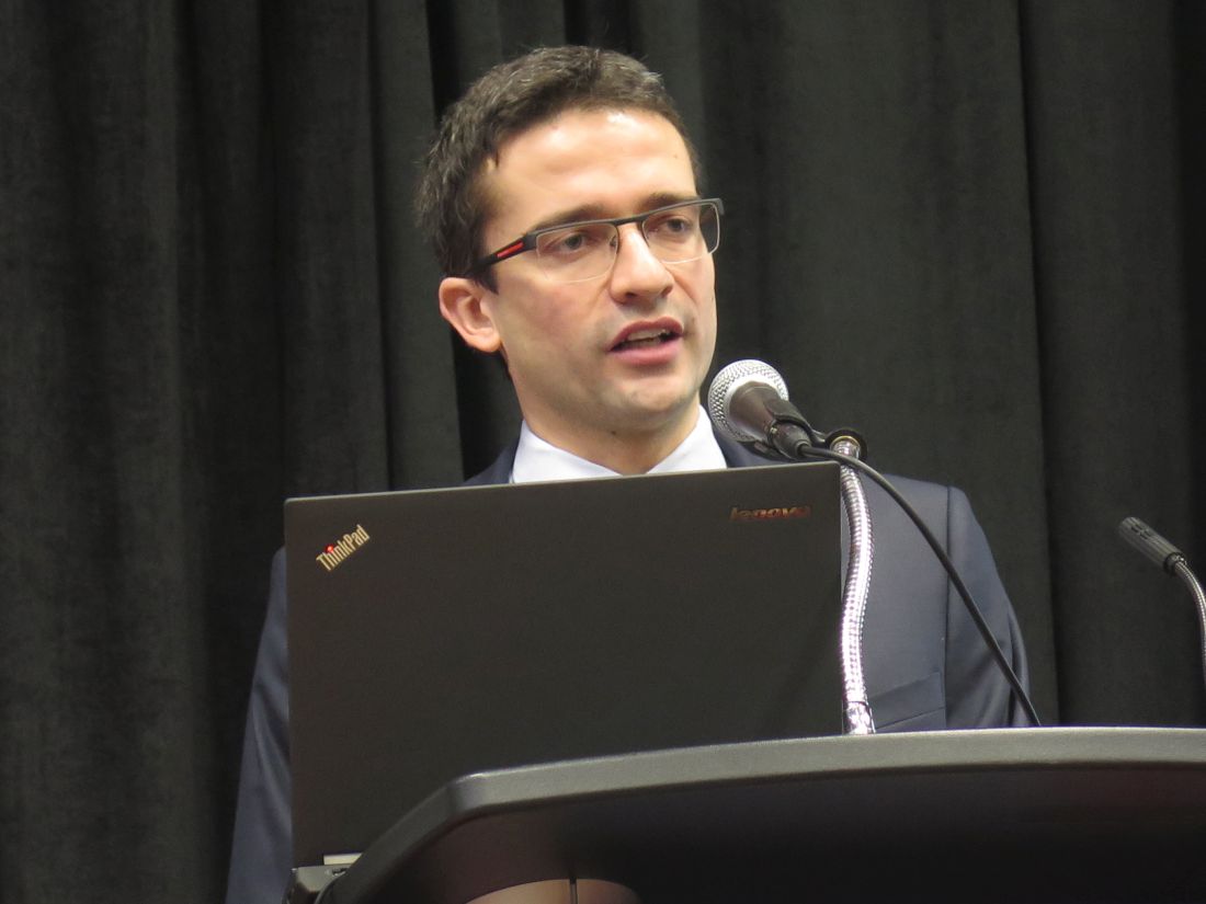
“Oocyte and embryo cryopreservation are standard strategies for fertility preservation in these patients, meaning increasing the chance of pregnancy after the end of treatment,” said lead author Matteo Lambertini, MD, medical oncologist and ESMO fellow at the Institut Jules Bordet in Brussels. “However, they do not prevent the risk of developing chemotherapy-induced premature ovarian insufficiency, and so patients are still at risk of developing early menopause.”
Data from individual trials of ovarian suppression have been mixed, and its use remains controversial, he further noted. As a result, guidelines from ASCO and ESMO for fertility preservation in cancer patients still consider ovarian suppression to be investigational.
Results of the new meta-analysis, reported in a press briefing and session, showed that, compared with control peers, premenopausal women given a gonadotropin-releasing hormone analog (GnRHa) to suppress ovarian function during breast cancer chemotherapy had a more than one-half reduction in odds of premature ovarian insufficiency and were almost twice as likely to become pregnant after completing their treatment.
“We believe that this strategy should now be considered a standard option to reduce the likelihood of chemotherapy-induced premature ovarian insufficiency and potentially improve future fertility in premenopausal early breast cancer patients who undergo adjuvant or neoadjuvant chemotherapy,” Dr. Lambertini maintained. The analysis and trial participants “remind us that there is a life after cancer and to cure the disease should not be considered enough any more.”
These new data are sufficient to put the controversy to rest, for several reasons, he contended. “First, this is an individual-patient meta-analysis of the five major randomized controlled trials in this setting, so it’s kind of the highest level of evidence that we can reach. Second, it’s very unlikely that we will have new randomized, controlled trials in this setting, and I would say also that, based on the results, it would probably be unethical to run randomized, controlled trials in this setting.”
Press briefing moderator Carlos L. Arteaga, MD, director of the Harold C. Simmons Comprehensive Cancer Center at UT Southwestern Medical Center, Dallas, wondered how trials were selected for inclusion in the meta-analysis.
Thirteen trials have been conducted on ovarian suppression in premenopausal patients with breast cancer, and the investigators included the five for which they were able to obtain patient-level data, a subset that contained both positive and negative trials, Dr. Lambertini replied. But findings would likely be the same had all trials been included, given that a 2015 analysis using trial-level data from the 12 trials completed at that time showed very similar results (Ann Oncol. 2015 Dec;26:2408-19).
“Societally, this is a hugely significant issue, but the difference you are showing I have to admit is rather modest,” commented Dr. Arteaga, who is also SABCS codirector and AACR past president. “So what kind of conversation do you have with the patient? Who are the ones who would be the best candidates for this approach?”
“The main message is that giving a GnRHa does not avoid the risk of early menopause in all patients, but still, it decreases significantly the number of patients who have this side effect,” Dr. Lambertini replied.
Two groups are optimal candidates for ovarian suppression, he proposed. “First, the patients who are concerned about developing early menopause and its related side effects, who are not interested per se in having a baby after the end of treatment, but may be preserving ovarian function. [Second], for patients interested in having a baby, so interested in fertility preservation, this strategy can be used after cryopreservation procedures or in patients who have no access, for different reasons, to cryopreservation strategies.”
Another point of view
In the session, attendee Kutluk Oktay, MD, PhD, professor of obstetrics & gynecology and reproductive sciences at Yale University, New Haven, and cochair of ASCO’s guideline committee on fertility preservation said, “I cannot agree with your conclusions based on what you presented to us.”
In particular, he took issue with the exclusion of additional trials in breast cancer as well as trials among patients with other types of cancers. “I’m wondering what the rationale is to limit this to breast cancer because chemotherapy is chemotherapy and ovary is ovary, so underlying disease should not matter. By limiting it to breast cancer, you are leaving out three important studies, all in hematological cancer, with better designs … three negative studies,” he commented.
From a clinical point of view, patients with lymphoma and patients with breast cancer differ greatly, Dr. Lambertini countered: The former are about 20 years younger, on average, and often receive less-granulotoxic chemotherapy. “For these reasons, I don’t believe that mixing these two populations would have been [appropriate] for analysis,” he said. “From a methodological point of view, the studies you have mentioned include overall [fewer] than 150 patients, so it’s a very small proportion in comparison to the data we have in breast cancer.”
Study details
For their meta-analysis, Dr. Lambertini and coinvestigators pooled individual patient data from five trials (PROMISE-GIM6, POEMS/SWOG S0230, Anglo-Celtic Group OPTION, GBG-37 ZORO, and a trial led by the Moffitt Cancer Center) that randomized premenopausal women with early breast cancer to adjuvant or neoadjuvant chemotherapy either with or without concurrent GnRHa therapy.
Two of the trials restricted enrollment to women with estrogen receptor (ER)-negative disease. The GnRHa agents used were triptorelin (Trelstar, Triptodur)and goserelin(Zoladex).
Main results showed that the rate of premature ovarian insufficiency, defined differently across trials, was 14.1% among women given a GnRHa and 30.9% among control women (adjusted odds ratio, 0.38; P less than .001), Dr. Lambertini reported. Findings were similar in subgroups stratified by age, ER status, and type and duration of chemotherapy.
The rate of amenorrhea, used as a standardized definition of premature ovarian insufficiency, was similar in the GnRHa and control groups at 1 year (36.8% and 40.4%) but sharply lower in the former at 2 years (18.2% vs. 30.0%; adjusted odds ratio, 0.51; P = .009).
Overall, 10.3% of women in the GnRHa group and 5.5% in the control group had at least one pregnancy after completing their breast cancer treatment (incidence rate ratio, 1.83; P = .030). “All of the randomized trials except for the POEMS study actually did not have fertility outcomes as a preplanned endpoint, and so the patients’ wish to have a pregnancy was not collected,” he noted; therefore, it was not possible to calculate pregnancy rates in the subset who actually wanted to conceive.
All pregnancies occurred among women aged 40 years or younger, and 86% occurred among women who had had ER-negative disease, likely reflecting use of adjuvant endocrine therapy in patients with ER-positive disease, he said. Of the 57 total pregnancies, 50 resulted in live births, and none of the infants had malformations; the other pregnancies ended in spontaneous or induced abortion.
With a median follow-up of 5 years, the groups did not differ significantly on rates of disease-free survival and overall survival, suggesting that ovarian suppression was safe, according to Dr. Lambertini. Findings were similar when patients were stratified by ER status.
“What I think researchers should do in the next year is to better understand how this strategy [of ovarian suppression] works because this is probably the main controversy right now, because it’s still not very clear how this strategy actually works,” he concluded.
SOURCE: Lambertini M et al., SABCS 2017 Abstract GS4-01.
SAN ANTONIO – Premenopausal women with early breast cancer should be offered temporary ovarian suppression during chemotherapy if they wish to remain fertile or avoid early menopause, suggests a meta-analysis of five randomized controlled trials among 873 women reported at the San Antonio Breast Cancer Symposium.
“Oocyte and embryo cryopreservation are standard strategies for fertility preservation in these patients, meaning increasing the chance of pregnancy after the end of treatment,” said lead author Matteo Lambertini, MD, medical oncologist and ESMO fellow at the Institut Jules Bordet in Brussels. “However, they do not prevent the risk of developing chemotherapy-induced premature ovarian insufficiency, and so patients are still at risk of developing early menopause.”
Data from individual trials of ovarian suppression have been mixed, and its use remains controversial, he further noted. As a result, guidelines from ASCO and ESMO for fertility preservation in cancer patients still consider ovarian suppression to be investigational.
Results of the new meta-analysis, reported in a press briefing and session, showed that, compared with control peers, premenopausal women given a gonadotropin-releasing hormone analog (GnRHa) to suppress ovarian function during breast cancer chemotherapy had a more than one-half reduction in odds of premature ovarian insufficiency and were almost twice as likely to become pregnant after completing their treatment.
“We believe that this strategy should now be considered a standard option to reduce the likelihood of chemotherapy-induced premature ovarian insufficiency and potentially improve future fertility in premenopausal early breast cancer patients who undergo adjuvant or neoadjuvant chemotherapy,” Dr. Lambertini maintained. The analysis and trial participants “remind us that there is a life after cancer and to cure the disease should not be considered enough any more.”
These new data are sufficient to put the controversy to rest, for several reasons, he contended. “First, this is an individual-patient meta-analysis of the five major randomized controlled trials in this setting, so it’s kind of the highest level of evidence that we can reach. Second, it’s very unlikely that we will have new randomized, controlled trials in this setting, and I would say also that, based on the results, it would probably be unethical to run randomized, controlled trials in this setting.”
Press briefing moderator Carlos L. Arteaga, MD, director of the Harold C. Simmons Comprehensive Cancer Center at UT Southwestern Medical Center, Dallas, wondered how trials were selected for inclusion in the meta-analysis.
Thirteen trials have been conducted on ovarian suppression in premenopausal patients with breast cancer, and the investigators included the five for which they were able to obtain patient-level data, a subset that contained both positive and negative trials, Dr. Lambertini replied. But findings would likely be the same had all trials been included, given that a 2015 analysis using trial-level data from the 12 trials completed at that time showed very similar results (Ann Oncol. 2015 Dec;26:2408-19).
“Societally, this is a hugely significant issue, but the difference you are showing I have to admit is rather modest,” commented Dr. Arteaga, who is also SABCS codirector and AACR past president. “So what kind of conversation do you have with the patient? Who are the ones who would be the best candidates for this approach?”
“The main message is that giving a GnRHa does not avoid the risk of early menopause in all patients, but still, it decreases significantly the number of patients who have this side effect,” Dr. Lambertini replied.
Two groups are optimal candidates for ovarian suppression, he proposed. “First, the patients who are concerned about developing early menopause and its related side effects, who are not interested per se in having a baby after the end of treatment, but may be preserving ovarian function. [Second], for patients interested in having a baby, so interested in fertility preservation, this strategy can be used after cryopreservation procedures or in patients who have no access, for different reasons, to cryopreservation strategies.”
Another point of view
In the session, attendee Kutluk Oktay, MD, PhD, professor of obstetrics & gynecology and reproductive sciences at Yale University, New Haven, and cochair of ASCO’s guideline committee on fertility preservation said, “I cannot agree with your conclusions based on what you presented to us.”
In particular, he took issue with the exclusion of additional trials in breast cancer as well as trials among patients with other types of cancers. “I’m wondering what the rationale is to limit this to breast cancer because chemotherapy is chemotherapy and ovary is ovary, so underlying disease should not matter. By limiting it to breast cancer, you are leaving out three important studies, all in hematological cancer, with better designs … three negative studies,” he commented.
From a clinical point of view, patients with lymphoma and patients with breast cancer differ greatly, Dr. Lambertini countered: The former are about 20 years younger, on average, and often receive less-granulotoxic chemotherapy. “For these reasons, I don’t believe that mixing these two populations would have been [appropriate] for analysis,” he said. “From a methodological point of view, the studies you have mentioned include overall [fewer] than 150 patients, so it’s a very small proportion in comparison to the data we have in breast cancer.”
Study details
For their meta-analysis, Dr. Lambertini and coinvestigators pooled individual patient data from five trials (PROMISE-GIM6, POEMS/SWOG S0230, Anglo-Celtic Group OPTION, GBG-37 ZORO, and a trial led by the Moffitt Cancer Center) that randomized premenopausal women with early breast cancer to adjuvant or neoadjuvant chemotherapy either with or without concurrent GnRHa therapy.
Two of the trials restricted enrollment to women with estrogen receptor (ER)-negative disease. The GnRHa agents used were triptorelin (Trelstar, Triptodur)and goserelin(Zoladex).
Main results showed that the rate of premature ovarian insufficiency, defined differently across trials, was 14.1% among women given a GnRHa and 30.9% among control women (adjusted odds ratio, 0.38; P less than .001), Dr. Lambertini reported. Findings were similar in subgroups stratified by age, ER status, and type and duration of chemotherapy.
The rate of amenorrhea, used as a standardized definition of premature ovarian insufficiency, was similar in the GnRHa and control groups at 1 year (36.8% and 40.4%) but sharply lower in the former at 2 years (18.2% vs. 30.0%; adjusted odds ratio, 0.51; P = .009).
Overall, 10.3% of women in the GnRHa group and 5.5% in the control group had at least one pregnancy after completing their breast cancer treatment (incidence rate ratio, 1.83; P = .030). “All of the randomized trials except for the POEMS study actually did not have fertility outcomes as a preplanned endpoint, and so the patients’ wish to have a pregnancy was not collected,” he noted; therefore, it was not possible to calculate pregnancy rates in the subset who actually wanted to conceive.
All pregnancies occurred among women aged 40 years or younger, and 86% occurred among women who had had ER-negative disease, likely reflecting use of adjuvant endocrine therapy in patients with ER-positive disease, he said. Of the 57 total pregnancies, 50 resulted in live births, and none of the infants had malformations; the other pregnancies ended in spontaneous or induced abortion.
With a median follow-up of 5 years, the groups did not differ significantly on rates of disease-free survival and overall survival, suggesting that ovarian suppression was safe, according to Dr. Lambertini. Findings were similar when patients were stratified by ER status.
“What I think researchers should do in the next year is to better understand how this strategy [of ovarian suppression] works because this is probably the main controversy right now, because it’s still not very clear how this strategy actually works,” he concluded.
SOURCE: Lambertini M et al., SABCS 2017 Abstract GS4-01.
REPORTING FROM SABCS 2017
Key clinical point:
Major finding: Compared with controls, women given a GnRHa during chemotherapy were less likely to develop premature ovarian insufficiency (adjusted OR, 0.38; P less than .001) and more likely to become pregnant after treatment (IRR, 1.83; P = .030).
Data source: A meta-analysis of individual patient data from five trials among 873 premenopausal women with early breast cancer.
Disclosures: Dr. Lambertini disclosed that he had no relevant conflicts of interest.
Source: Lambertini M et al., SABCS 2017 Abstract GS4-01.
Submit Research to VAM
Research done? Submit it as an abstract for the 2018 Vascular Annual Meeting, set for June 20-23, 2018, in Boston. Plenaries are June 21-23 and exhibits are June 21-22.
The submission site opened Monday. The deadline for submitting abstracts is Wednesday, Jan. 17, 2018.
Abstracts may be submitted in the following categories:
- Plenary
- Vascular and Endovascular Surgical Society
- International Forum
- International Fast Talk
- Poster Competition
- Interactive Poster
Guidelines, submission policies and general information on VAM are available online.
Obesity an independent risk factor for rosacea in cohort study of U.S. women
Obesity was associated with a significantly higher risk of rosacea, compared with a healthy weight, particularly when the weight was gained after age 18 years, according to an analysis of data from the Nurses Health Study II.
Investigators evaluated data on 89,886 women in the U.S. study from 1991 through 2005 – 5,249 diagnosed with rosacea – over 14 years to determine what, if any, relationship existed between body mass index (BMI) and rosacea risk.
As BMI increased, the risk of rosacea increased: Compared with participants with a BMI of 21.0 to 22.9 kg/m2 (considered a healthy weight), the risk of rosacea for those with a BMI of 25.0-29.9 (overweight) was 11% greater (95% confidence interval, 1.02-1.21), and among those with a BMI of 30.0-34.9, was 21% greater (95% CI, 1.09-1.34). The risk was 48% greater among those with a BMI of 35 or above (95% CI, 1.33-1.64). (A BMI over 30 is considered obese). The results were published in the December issue of the Journal of the American Academy of Dermatology.
The association between weight and rosacea risk was only significant after age 18 years, when the risk of rosacea increased 4% for every 10 pounds of weight gain, even after adjusting for BMI.
The investigators also found a significant relationship between waist circumference and rosacea risk that was independent of BMI, with the highest quintile of waist circumference associated with a 32% greater risk of rosacea, compared with the lowest quintile. Similarly, the highest quintile of hip circumference was associated with a 38% higher risk of rosacea, also independent of BMI. However there was no association between waist-to-hip ratio and rosacea risk.
While there were associations between the risk of rosacea and smoking, alcohol intake, and physical activity, the relationship between BMI and rosacea risk was not modified by these other risk factors.
Suyun Li, PhD, of the Guangzhou (China) Medical University School of Public Health and coauthors from the department of dermatology, Brown University, Providence, R.I.; and Brigham and Women’s Hospital, Boston, wrote that previous epidemiologic studies examining the interaction between obesity and rosacea have shown inconsistent results. Longitudinal studies on the issue had focused only on BMI and ignored other measures of central obesity.
“To our knowledge, this is the first cohort study on the association between obesity and risk for incident rosacea,” they wrote. “The study contributes to the understanding of rosacea etiology and informs clinical practice related to rosacea prevention and patient care.”
They suggested a number of different mechanisms that might explain how obesity increases the risk of rosacea, including the chronic, low-grade inflammatory state associated with obesity. “Adiposity can augment proinflammatory cytokine expression, such as interleukin 6 and tumor necrosis factor–alpha, both relevant to rosacea pathophysiology,” they noted. “Vascular changes associated with obesity might be another mechanism, considering obesity can lead to abnormalities of vascular function and structure, which might lead to the vasodilatation in rosacea.”
In the NHS II study, data are collected every 2 years, which the authors said ensured they had the most up-to-date information. While rosacea diagnoses relied on self-report, the authors said the study participants – nurses – were likely to have a high validity of self-reporting of rosacea. They acknowledged, however, that the lack of information on rosacea subtypes was a limitation of the study.
The study was supported by the department of dermatology, Brown University and a Nurses’ Health Study II grant. Dr. Li was supported by a research grant from the National Rosacea Society and the Dermatology Foundation. Another author declared having served as an investigator and receiving research funds from Sanofi and Regeneron; serving as a consultant for Sanofi and RTI Health Solutions, and having received honoraria from Astellas Canada, Prime, and Spire Learning. The remaining three authors had no disclosures.
SOURCE: Li S et al. J Am Acad Dermatol. 2017 Dec;77(6):1083-7.E5.
FROM JAAD
Key clinical point: independent of other risk factors such as smoking and alcohol intake.
Major finding: A BMI greater than 35 is associated with a 48% higher risk of rosacea, compared with a BMI of 21.
Data source: Information on rosacea diagnoses among 89,886 Nurses’ Health Study II participants.
Disclosures: The study was supported by the Warren Alpert Medical School of Brown University and a Nurses’ Health Study II grant. One author was supported by a research grant from the National Rosacea Society and the Dermatology Foundation; another author declared research funding, consultancies, and honoraria from the pharmaceutical industry.
Source: Li S et al. J Am Acad Dermatol. 2017 Dec;77(6):1083-7.E5.