User login
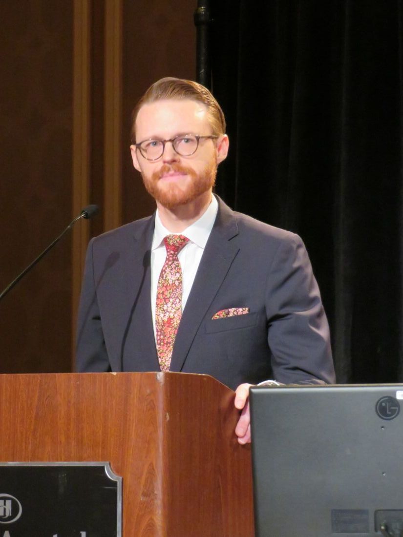
VIDEO: Managing the alemtuzumab paradox
SAN DIEGO – A paradox of treating people with the monoclonal antibody alemtuzumab is that the agent can be an effective therapy for many people living with multiple sclerosis, but in some patients is associated with the development of other autoimmune diseases.
“I counsel patients with multiple sclerosis that this is a high risk, high gain drug,” Alasdair Coles, MD, of the University of Cambridge (England), said at the meeting, held by the Americas Committee for Treatment and Research in Multiple Sclerosis.
Patients can make it safer through strict compliance with the drug’s risk monitoring program. But without patient compliance, alemtuzumab becomes a dangerous drug, he said in a video interview.
Knowing in advance which patients are at an elevated risk for subsequent autoimmune diseases has been difficult to predict. But researchers are getting closer, Dr. Coles said. In the future, measuring a serum factor, potentially interleukin 21, could produce a pretreatment risk assessment for each individual.
REPORTING FROM ACTRIMS FORUM 2018

Ibuprofen appears safe with preeclampsia, study says
DALLAS – Compared to acetaminophen, ibuprofen does not prolong the time needed to control postpartum hypertension in women who experience preeclampsia with severe features, Nathan Blue MD, reported at the Pregnancy Meeting, sponsored by the Society for Maternal-Fetal Medicine.
Ibuprofen did not exacerbate postpartum hypertension, as some studies have suggested, and women randomized to it for postpartum pain control reached their target blood pressure at a mean of 35 hours after delivery, similar to the 38 hours needed among women receiving acetaminophen.
Dr. Blue’s findings contradict the recommendation by the American College of Obstetricians and Gynecologists to avoid the use of ibuprofen and other nonsteroidal anti-inflammatory drugs (NSAIDs) for pain control in women who experience preeclampsia or other hypertensive disorders of pregnancy.
“While our study was not meant to answer all questions about the potential problems of nonsteroidal anti-inflammatories [in this population], I would conclude from these results that providers and patients can make a decision together and feel good about the use of NSAIDs,” in the presence of postpartum hypertension, said Dr. Blue, a fellow at the University of New Mexico, Albuquerque.
These concerns led ACOG to issue its recommendation in 2013 against the use of NSAIDs in pregnant women with hypertension.
“This is problematic, because NSAIDs are particularly well-suited to address obstetric pain,” Dr. Blue said at the meeting. “They have been shown to be better than acetaminophen for perineal injury and they are associated with a reduced use of opioids after cesarean section.”
To investigate the effect of ibuprofen on postpartum hypertension, Dr. Blue and his colleagues conducted a double-blind randomized controlled trial of 100 women with preeclampsia with severe features. The study population also included women who had chronic hypertension complicated by preeclampsia with severe features, and women with HELLP syndrome – a constellation of hemolysis, elevated liver enzymes, and low platelet count.
Women were randomized to either 600 mg ibuprofen or 650 mg acetaminophen every 6 hours, around the clock, with the first dose delivered within 6 hours of delivery. All patients received at least eight doses of their assigned medication. The primary endpoint was the time required to achieve blood pressure control. “We defined blood pressure control as the number of hours from delivery to the last reading of at least 160/110 mm Hg before discharge,” Dr. Blue said. “Our rationale here was that persistence of a blood pressure of that level would require a delay in discharge of at least 24 hours.”
The study had a number of secondary outcomes, including time from delivery to last blood pressure reading of at least 150/100 mm Hg; postpartum mean arterial pressure; any blood pressure reading of 160/110 mm Hg or higher; need for antihypertensive drugs at discharge; prolongation of hospital stay due to hypertension; and the need for postpartum opioids.
There was a 6-week follow-up assessment, at which time women reported any continued antihypertensive or opioid use, obstetrical triage visits after discharge, and hospital readmission.
The study cohort was well-balanced at baseline. Women were a mean of 30 years old; about a third were nulliparous, and half had a vaginal delivery. Chronic hypertension requiring medication was present in about 15%. The maximum blood pressure before delivery was about 180/107.
There was no significant difference between the ibuprofen group and the acetaminophen group in the primary endpoint of time to blood pressure of 160/110 mm Hg or below (35.3 vs. 38 hours). Nor were there significant differences in any of the secondary endpoints, including time to achieve a blood pressure of less than 150/100 mm Hg (58 vs. 57 hours), postpartum mean arterial pressure, maximum systolic and diastolic blood pressures, or the number of women who needed a short-acting antihypertensive (30 vs. 26) and who went home on an antihypertensive (33 vs. 31).
There were also no significant between-group differences in opioid use, either on postpartum days 0, 1, or 2. The total morphine equivalent dose for each group was likewise not significantly different (77 vs. 88 mg).
Dr. Blue was able to contact 77 women at 6 weeks’ postpartum. He found that 6-week outcomes were also similar. There were no significant differences in the number who required continuing antihypertensive or opioids, no difference in obstetric triage visits, and no difference in hospital readmission.
“Our study does not support the hypothesis that NSAIDs adversely affect blood pressure control in patients with preeclampsia,” he said. “Not only did we not find a difference in the primary outcome, we found not even a suggestion of difference in any measure of blood pressure control.”
The University of New Mexico sponsored the study. Dr. Blue reported having no financial disclosures.
SOURCE: Blue et al. The Pregnancy Meeting 2018 Abstract LB04.
REPORTING FROM THE PREGNANCY MEETING
Key clinical point:
Major finding: The mean time to achieve postpartum blood pressure control was 35 hours in the ibuprofen group and 38 hours in the acetaminophen group.
Study details: The randomized study comprised 100 women.
Disclosures: The University of New Mexico sponsored the study. Dr. Blue reported having no financial disclosures.
Source: Blue N et al. The Pregnancy Meeting 2018 Abstract LB04.
Third course of alemtuzumab can improve MS outcomes
SAN DIEGO – Approximately 30% of people with active multiple sclerosis who initially responded well to two courses of alemtuzumab in the CARE-MS II trial experience relapse or MRI activity over time. But investigators set out to determine whether retreatment with a subsequent course of alemtuzumab is worthwhile.
“What we found is, after the third course, they continued to do well again – at this point for an average of another 3-4 years,” Ann D. Bass, MD, said in an interview at the meeting, held by the Americas Committee for Treatment and Research in Multiple Sclerosis.
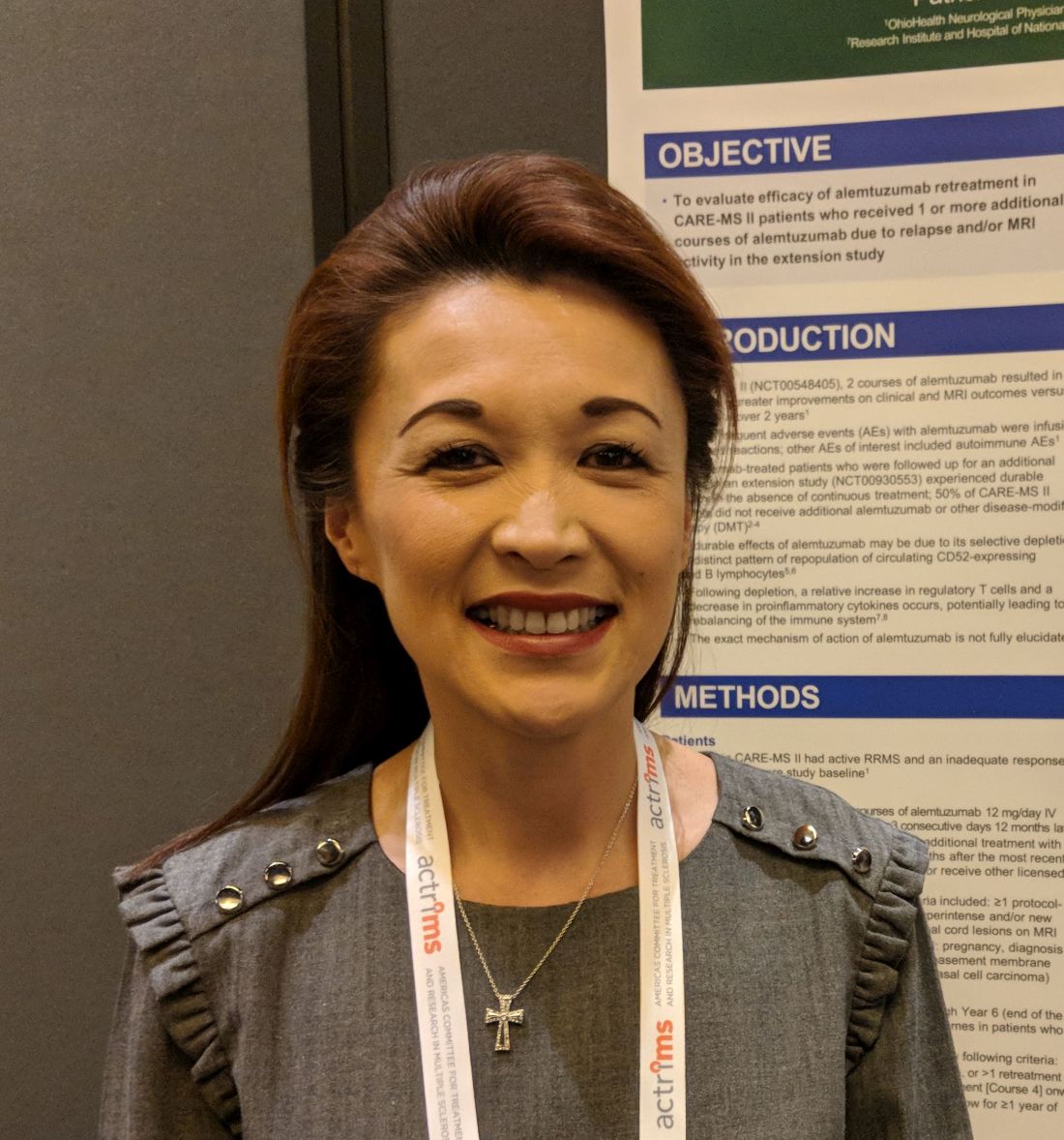
“The take-home message is if they do need to be retreated after those two courses, it doesn’t [necessarily] mean they are a treatment failure. Give them another chance and see if they will do well with a third course, which is what they did,” said Dr. Bass, of the Neurology Center of San Antonio.
Through year 6 of an extension study with 393 of the original CARE-MS II trial participants, 45% received one or more additional courses of alemtuzumab. Participants were required to wait 12 months or more after completion of the two initial courses of therapy. This figure includes 30% who received a third course of alemtuzumab, 12% who received a fourth course, and 2% and 1% who received a fifth or sixth course, respectively.
“The average time until they needed a third course was 2.5 years after the second course; so they didn’t need it right away,” Dr. Bass said. “The majority, even at 6 years, did not need to have a third course.”
When patients did require a subsequent course, “about half needed it because of clinical relapse; one-quarter needed it because of MRI relapse; and about one-quarter needed it because of both,” Dr. Bass said during a poster presentation.
In terms of effectiveness, the annual relapse rate significantly decreased following a third course of alemtuzumab, from 0.85 in the 12 months prior to the third course to 0.20 in the 12 months after (P less than .0001). In these patients, the annual relapse rate remained low, at 0.17, up to 3 years later.
Investigators also tracked disability using the Expanded Disability Status Scale. They found that more than two-thirds, 68%, maintained stable or had improved scores after administration of a third alemtuzumab course. In addition, the percentage of patients with confirmed disability improvement increased from 4.4% in the 12 months prior to a third course to 14.4% in the year following pretreatment.
Retreatment was at the patient’s discretion. “The patients have the right to say ‘No, I’m doing great. I don’t want to be retreated’ or ‘I want to explore other options’ for whatever reason,” Dr. Bass said. “That’s rare though; most patients actually say yes.”
To qualify for retreatment based on MRI findings, patients had to have at least two lesions – one enlarging and one enhancing, two enlarging, or two new.
Only patients who opted for a subsequent course of alemtuzumab were included in the current analysis; those who chose a different disease-modifying therapy were excluded.
“Many achieve clinical and MRI remission. I never say cure – you don’t want to say that word,” Dr. Bass said.
Sanofi and Bayer HealthCare Pharmaceuticals supported the study. Dr. Bass reported that she is a principal investigator, speaker, and member of the advisory board for Sanofi Genzyme.
SOURCE: Bass A et al. ACTRIMS Forum 2018 Poster P035.
SAN DIEGO – Approximately 30% of people with active multiple sclerosis who initially responded well to two courses of alemtuzumab in the CARE-MS II trial experience relapse or MRI activity over time. But investigators set out to determine whether retreatment with a subsequent course of alemtuzumab is worthwhile.
“What we found is, after the third course, they continued to do well again – at this point for an average of another 3-4 years,” Ann D. Bass, MD, said in an interview at the meeting, held by the Americas Committee for Treatment and Research in Multiple Sclerosis.
“The take-home message is if they do need to be retreated after those two courses, it doesn’t [necessarily] mean they are a treatment failure. Give them another chance and see if they will do well with a third course, which is what they did,” said Dr. Bass, of the Neurology Center of San Antonio.
Through year 6 of an extension study with 393 of the original CARE-MS II trial participants, 45% received one or more additional courses of alemtuzumab. Participants were required to wait 12 months or more after completion of the two initial courses of therapy. This figure includes 30% who received a third course of alemtuzumab, 12% who received a fourth course, and 2% and 1% who received a fifth or sixth course, respectively.
“The average time until they needed a third course was 2.5 years after the second course; so they didn’t need it right away,” Dr. Bass said. “The majority, even at 6 years, did not need to have a third course.”
When patients did require a subsequent course, “about half needed it because of clinical relapse; one-quarter needed it because of MRI relapse; and about one-quarter needed it because of both,” Dr. Bass said during a poster presentation.
In terms of effectiveness, the annual relapse rate significantly decreased following a third course of alemtuzumab, from 0.85 in the 12 months prior to the third course to 0.20 in the 12 months after (P less than .0001). In these patients, the annual relapse rate remained low, at 0.17, up to 3 years later.
Investigators also tracked disability using the Expanded Disability Status Scale. They found that more than two-thirds, 68%, maintained stable or had improved scores after administration of a third alemtuzumab course. In addition, the percentage of patients with confirmed disability improvement increased from 4.4% in the 12 months prior to a third course to 14.4% in the year following pretreatment.
Retreatment was at the patient’s discretion. “The patients have the right to say ‘No, I’m doing great. I don’t want to be retreated’ or ‘I want to explore other options’ for whatever reason,” Dr. Bass said. “That’s rare though; most patients actually say yes.”
To qualify for retreatment based on MRI findings, patients had to have at least two lesions – one enlarging and one enhancing, two enlarging, or two new.
Only patients who opted for a subsequent course of alemtuzumab were included in the current analysis; those who chose a different disease-modifying therapy were excluded.
“Many achieve clinical and MRI remission. I never say cure – you don’t want to say that word,” Dr. Bass said.
Sanofi and Bayer HealthCare Pharmaceuticals supported the study. Dr. Bass reported that she is a principal investigator, speaker, and member of the advisory board for Sanofi Genzyme.
SOURCE: Bass A et al. ACTRIMS Forum 2018 Poster P035.
REPORTING FROM ACTRIMS FORUM 2018
Key clinical point:
Major finding: The annual relapse rate significantly decreased following a third course of alemtuzumab, from 0.85 in the 12 months prior to the third course to 0.20 in the 12 months after (P less than .0001).
Study details: An extension study of the CARE-MS II trial involving 393 of the original study participants.
Disclosures: Sanofi and Bayer HealthCare Pharmaceuticals supported the study. Dr. Bass reported that she is a principal investigator, speaker, and member of the advisory board for Sanofi Genzyme.
Source: Bass A et al. ACTRIMS Forum 2018 Poster P035.
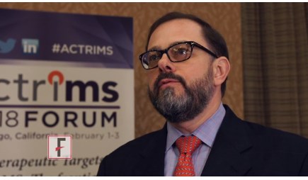
Switching RRMS patients to daclizumab beta appears safe
SAN DIEGO – Switching relapsing-remitting multiple sclerosis patients from glatiramer acetate to daclizumab beta resulted in no increase in the adverse event profile and was associated with superior efficacy, a post hoc analysis of data from the DECIDE study showed.
“There is always a challenge in transitioning patients from one therapeutic agent to another, with concerns for adequate efficacy to justify the switch, and heightened risks of toxicity or adverse events,” lead study author Stanley L. Cohan, MD, PhD, said in an interview prior to the meeting held by the Americas Committee for Treatment and Research in Multiple Sclerosis. “Daclizumab is not a first-line or platform therapeutic agent, but has clearly superior efficacy to first-line medication, and, based upon the current data presented, safety and efficacy are not adversely influenced by prior treatment history with a first-line agent.”

In the phase 3 DECIDE trial, daclizumab beta 150 mg demonstrated greater efficacy versus intramuscular (IM) interferon (IFN) beta-1a 30 mcg on several clinical, radiographic, and patient-centered outcomes in patients with relapsing-remitting multiple sclerosis (N Engl J Med 2015; 373:1418-28). The purpose of the current study was to examine the efficacy and safety of daclizumab beta vs. IM IFN beta-1a in the subgroup of RRMS patients treated with glatiramer acetate (GA) as their only previous disease-modifying therapy (DMT) before entering DECIDE.
Dr. Cohan, a neurologist who directs the Providence MS Center at the Providence Brain and Spine Institute in Portland, Ore., and his associates reported data from 42 of 922 (5%) IM IFN beta-1a and 50 of 919 (5%) daclizumab beta patients who had received treatment with GA only prior to DECIDE. Baseline characteristics were balanced between treatment groups, including duration of prior treatment with GA, reasons for discontinuing GA, and time between GA discontinuation and start of treatment in DECIDE. The annualized relapse rate was 42% lower in patients treated with daclizumab beta vs. IM IFN beta-1a (rate ratio of 0.58). Daclizumab beta also reduced risk of relapse by 53% (hazard ratio of 0.47; P = .048) and the mean number of new or newly-enlarging T2-hyperintense lesions at week 96 by 58% (lesion mean ratio, 0.42; P = .021) vs. IM IFN beta-1a.
In patients treated with GA only before DECIDE, 98% of IM IFN beta-1a patients and 94% of daclizumab beta patients reported any adverse event (AE). In all, 2% of IM IFN beta-1a patients and 16% of daclizumab beta patients had a serious AE (excluding MS relapse), and 10% of IM IFN beta-1a and 12% of daclizumab beta patients discontinued treatment because of an AE (excluding MS relapse).
The incidence of elevations of alanine aminotransferase or aspartate aminotransferase three times the upper limit of normal or greater was 10% in the IM IFN beta-1a group and 8% in the daclizumab beta, while the ALT and AST elevations were greater than five times the ULN in 0% and 4% of patients, respectively.
Serious adverse events were reported in eight participants in the daclizumab beta group (including abortion induced, ankle fracture, anal fistula, anxiety, appendicitis perforated, convulsion, pelvic abscess, inguinal hernia, abnormal cervix smear), and one participant in the IM IFN beta-1a group (ligament rupture).
“This post hoc analysis demonstrates that in switching from glatiramer to daclizumab there was no unanticipated or increase in the AE profile, and that a switch from glatiramer to daclizumab was associated with superior efficacy, again in line with overall efficacy observed for daclizumab in this study,” Dr. Cohan said.
He acknowledged certain limitations of the study, including the post hoc nature of the analysis. “The small glatiramer cohort size, and the large percentage of former glatiramer patients who entered DECIDE because of lack of glatiramer efficacy may have introduced a selection bias which would magnify the seeming efficacy of daclizumab, and interferon-beta,” he added.
Dr. Cohan reported that he receives research support from Biogen, Novartis, Roche, Sanofi, and Mallinckrodt, and speaking honoraria from Acorda, Biogen, Roche, and Sanofi. He has served on advisory boards for Biogen, Sanofi, and Novartis.
SOURCE: Cohan et al. ACTRIMS Forum 2018, Poster 42.
SAN DIEGO – Switching relapsing-remitting multiple sclerosis patients from glatiramer acetate to daclizumab beta resulted in no increase in the adverse event profile and was associated with superior efficacy, a post hoc analysis of data from the DECIDE study showed.
“There is always a challenge in transitioning patients from one therapeutic agent to another, with concerns for adequate efficacy to justify the switch, and heightened risks of toxicity or adverse events,” lead study author Stanley L. Cohan, MD, PhD, said in an interview prior to the meeting held by the Americas Committee for Treatment and Research in Multiple Sclerosis. “Daclizumab is not a first-line or platform therapeutic agent, but has clearly superior efficacy to first-line medication, and, based upon the current data presented, safety and efficacy are not adversely influenced by prior treatment history with a first-line agent.”
In the phase 3 DECIDE trial, daclizumab beta 150 mg demonstrated greater efficacy versus intramuscular (IM) interferon (IFN) beta-1a 30 mcg on several clinical, radiographic, and patient-centered outcomes in patients with relapsing-remitting multiple sclerosis (N Engl J Med 2015; 373:1418-28). The purpose of the current study was to examine the efficacy and safety of daclizumab beta vs. IM IFN beta-1a in the subgroup of RRMS patients treated with glatiramer acetate (GA) as their only previous disease-modifying therapy (DMT) before entering DECIDE.
Dr. Cohan, a neurologist who directs the Providence MS Center at the Providence Brain and Spine Institute in Portland, Ore., and his associates reported data from 42 of 922 (5%) IM IFN beta-1a and 50 of 919 (5%) daclizumab beta patients who had received treatment with GA only prior to DECIDE. Baseline characteristics were balanced between treatment groups, including duration of prior treatment with GA, reasons for discontinuing GA, and time between GA discontinuation and start of treatment in DECIDE. The annualized relapse rate was 42% lower in patients treated with daclizumab beta vs. IM IFN beta-1a (rate ratio of 0.58). Daclizumab beta also reduced risk of relapse by 53% (hazard ratio of 0.47; P = .048) and the mean number of new or newly-enlarging T2-hyperintense lesions at week 96 by 58% (lesion mean ratio, 0.42; P = .021) vs. IM IFN beta-1a.
In patients treated with GA only before DECIDE, 98% of IM IFN beta-1a patients and 94% of daclizumab beta patients reported any adverse event (AE). In all, 2% of IM IFN beta-1a patients and 16% of daclizumab beta patients had a serious AE (excluding MS relapse), and 10% of IM IFN beta-1a and 12% of daclizumab beta patients discontinued treatment because of an AE (excluding MS relapse).
The incidence of elevations of alanine aminotransferase or aspartate aminotransferase three times the upper limit of normal or greater was 10% in the IM IFN beta-1a group and 8% in the daclizumab beta, while the ALT and AST elevations were greater than five times the ULN in 0% and 4% of patients, respectively.
Serious adverse events were reported in eight participants in the daclizumab beta group (including abortion induced, ankle fracture, anal fistula, anxiety, appendicitis perforated, convulsion, pelvic abscess, inguinal hernia, abnormal cervix smear), and one participant in the IM IFN beta-1a group (ligament rupture).
“This post hoc analysis demonstrates that in switching from glatiramer to daclizumab there was no unanticipated or increase in the AE profile, and that a switch from glatiramer to daclizumab was associated with superior efficacy, again in line with overall efficacy observed for daclizumab in this study,” Dr. Cohan said.
He acknowledged certain limitations of the study, including the post hoc nature of the analysis. “The small glatiramer cohort size, and the large percentage of former glatiramer patients who entered DECIDE because of lack of glatiramer efficacy may have introduced a selection bias which would magnify the seeming efficacy of daclizumab, and interferon-beta,” he added.
Dr. Cohan reported that he receives research support from Biogen, Novartis, Roche, Sanofi, and Mallinckrodt, and speaking honoraria from Acorda, Biogen, Roche, and Sanofi. He has served on advisory boards for Biogen, Sanofi, and Novartis.
SOURCE: Cohan et al. ACTRIMS Forum 2018, Poster 42.
REPORTING FROM ACTRIMS FORUM 2018
Key clinical point:
Major finding: The annualized relapse rate was 42% lower in patients treated with daclizumab beta vs. IM IFN beta-1a (rate ratio of 0.58).
Study details: A post hoc analysis of 42 of 922 (5%) IM IFN beta-1a and 50 of 919 (5%) daclizumab beta patients who had received treatment with glatiramer acetate only prior to the DECIDE trial.
Disclosures: Dr. Cohan reported that he receives research support from Biogen, Novartis, Roche, Sanofi, and Mallinckrodt, and speaking honoraria from Acorda, Biogen, Roche, and Sanofi. He has served on advisory boards for Biogen, Sanofi, and Novartis.
Source: Cohan S et al. ACTRIMS Forum 2018 Poster 42.
VIDEO: Oral ozanimod shows promise for relapsing MS
SAN DIEGO – A pair of phase 3 studies offer promising results regarding the safety and efficacy of ozanimod, an experimental immunomodulator, in the treatment of relapsing multiple sclerosis (RMS).
The medication targets sphingosine 1-phosphate 1 and 5 receptors. Industry-funded researchers tested it in two studies against interferon beta-1a.
One study, called SUNBEAM, tested once-daily oral ozanimod (1 mg or 0.5 mg with 7-day dose escalation) against interferon beta-1a (via 30 mcg weekly intramuscular injection) for at least 12 months in 1,346 patients with RMS. The annualized relapse rate, the primary endpoint, was lower in the ozanimod groups versus interferon. For the 1-mg dose, it was 0.181 (P less than .0001), and for 0.5-mg dose, 0.241 (P = .0013).
The number of serious treatment-emergent adverse events in the three groups was low, ranging from 2.5% to 3.5%.
The other study, called RADIANCE, was a similar trial that lasted 24 months. In it, the rate of serious treatment-emergent adverse events in the three groups were similar, ranging from 6.4% to 7.1%.
Ozanimod offers “an excellent therapeutic benefit for patients and a very clean safety profile,” said Bruce Cree, MD, PhD, clinical research director at the University of California, San Francisco, Multiple Sclerosis Center. He is an author on both studies and spoke in a video interview at ACTRIMS Forum 2018, held by the Americas Committee for Treatment and Research in Multiple Sclerosis.
He said the ozanimod should be especially useful as a first-line treatment for MS. The drug is currently being evaluated by the Food and Drug Administration for an RMS indication, and it is also being developed for Crohn’s disease and ulcerative colitis, he said.
The study was funded by Receptos, a wholly owned subsidiary of Celgene. Dr. Cree reported that he has been a consultant to AbbVie, Biogen, EMD Serono, Genzyme, Novartis, and Shire.
SOURCE: Cree B et al. ACTRIMS Forum 2018, abstract P030, and Comi G et al. ACTRIMS Forum 2018, abstract P023
REPORTING FROM ACTRIMS FORUM 2018
VIDEO: Efficacy of DMTs decreases with age
San Diego – , and high-efficacy drugs do a better job of inhibiting MS disability compared with low-efficacy drugs only in patients younger than 40.5 years.
Those are the key conclusions from a meta-analysis of the age-dependent efficacy of MS treatments that was published in the November 2017 issue of Frontiers in Neurology. In a video interview, Ann Marie Weideman, lead study author, discussed highlights from the meta-analysis at the meeting held by the Americas Committee for Treatment and Research in Multiple Sclerosis. The meta-analysis drew from more than 28,000 individuals with MS participating in 38 trials of 13 categories of immunomodulatory drugs.
Ms. Weideman is an IRTA Fellow at the National Institute of Neurological Disorders and Stroke, Bethesda, Md. She reported that study coauthor Bibiana Bielekova, MD, is coinventor of several patents related to daclizumab.
REPORTING FROM ACTRIMS FORUM 2018

VIDEO: New MS ambulatory measure could fill clinical gap
REPORTING FROM ACTRIMS FORUM 2018
SAN DIEGO – Although clinical tools to assess ambulatory function among people with multiple sclerosis exist, some measure it as part of a comprehensive assessment while others require the patient to answer many questions and then clinicians to calculate a score.
To devise a more targeted, simpler instrument, Emily Evans, MD, and her colleagues developed the PDAS or Patient Derived Ambulation Scale. They evaluated the correlation of this single-item scale to assess ambulation – an important measure of patient function – and evaluated how the results correlated with existing tools such as the Patient Determined Disease Steps and 12-item MS Walking Scale. Dr. Evans presented preliminary findings at the ACTRIMS Forum 2018, held by the Americas Committee for Treatment and Research in Multiple Sclerosis.
“We feel this is a quick test that can be readily implemented into clinical practice,” Dr. Evans, a neurologist at the John L. Trotter MS Center at Washington University in St. Louis, said in a video interview.

VIDEO: Alemtuzumab associated with long-term MS control in TOPAZ study
SAN DIEGO – A majority of patients with active relapsing-remitting multiple sclerosis and inadequate response to previous therapy achieved a durable response after treatment with alemtuzumab in the TOPAZ trial, a 5-year extension to the CARE-MS II study.
The video associated with this article is no longer available on this site. Please view all of our videos on the MDedge YouTube channel
Almost half of the 317 participants in TOPAZ received no further therapy beyond their initial two courses of alemtuzumab infusion therapy that they received as part of the CARE-MS II study.
“If you follow patients over time ... you’re seeing a significant group of patients who have improvement. It’s very unexpected, especially when you look at the patients who entered the clinical trial who had a fair amount of active disease,” said Barry A. Singer, MD, director of The MS Center for Innovations in Care at Missouri Baptist Medical Center in St. Louis.
At the 7-year evaluation of patients in TOPAZ, the annualized relapse rate was 0.14. In addition, 87% of patients remained relapse-free in year 7. Dr. Singer and his colleagues also reported that 73% of TOPAZ participants were stable or improved based on their Expanded Disability Status Scale (EDSS) scores.
“As we follow the data out and follow these patients out, we’re seeing how the clinical course for these patients is dramatically improving for the majority of patients,” Dr. Singer said in a video interview at ACTRIMS Forum 2018, the meeting held by the Americas Committee for Treatment and Research in Multiple Sclerosis.
The TOPAZ study also revealed that 69% of patients were free of clinical disease worsening and 44% experienced clinical disease improvement in the 6 months before year 7. The majority also had no evidence of disease activity, Dr. Singer reported.
“One of the attributes that makes alemtuzumab so attractive as a clinician and for patients is you can go through a couple of series of medication [treatments] ... and really alter your disease course – that is the exciting thing,” he said.
The Food and Drug Administration approved alemtuzumab (Lemtrada) in November 2014 for the treatment of patients with relapsing forms of multiple sclerosis. Use of alemtuzumab is generally reserved for patients who have had an inadequate response to two or more previous drugs indicated for the treatment of multiple sclerosis.
In CARE-MS II, participants received two annual courses of alemtuzumab: intravenous infusion of 12 mg/day for 5 days at baseline and again for 3 days at 12 months. Additional treatment in TOPAZ for relapse or MRI evidence of disease was at the discretion of the investigator and could include alemtuzumab retreatment 12 mg/day on 3 consecutive days 12 months or more after a previous course, or another disease-modifying therapy at any time. Annual follow-up exams included an MRI scan.
A durable treatment effect was achieved by a majority of patients, even though 47% received no further treatment with alemtuzumab or another disease-modifying therapy after the initial two alemtuzumab courses.
The incidence of most adverse events, including infusion-associated reactions and infections, decreased over the course of the TOPAZ study and were lower than the incidence reported in the 2-year CARE-MS II trial. Of note, the incidence of thyroid-related adverse events peaked in the third year of the follow-up and continued to decline out to 7 years, Dr. Singer said. “We’re not seeing any new safety issues.”
Dr. Singer and his coinvestigators plan to continue the research, monitoring and scoring patients over time.
The TOPAZ trial was funded by Sanofi Genzyme, which markets alemtuzumab. Dr. Singer disclosed that he receives clinical research support and is a speaker for Sanofi Genzyme.
SOURCE: Singer B et al. ACTRIMS Forum 2018, abstract P026.
REPORTING FROM ACTRIMS FORUM 2018
Key clinical point:
Major finding: The annualized relapse rate was 0.14 at year 7 among the 87% of participants who remained in the TOPAZ study.
Study details: A 5-year extension study of 317 participants from the initial CARE-MS II trial.
Disclosures: The TOPAZ trial was funded by Sanofi Genzyme, which markets alemtuzumab. Dr. Singer disclosed that he receives clinical research support and is a speaker for Sanofi Genzyme.
Source: Singer B et al. ACTRIMS Forum 2018, abstract P026.

In situ vaccination eradicates lymphoma, other cancers

Experiments in mice have shown that injecting immune-stimulating agents directly into a tumor can help the immune system eradicate tumors in other areas of the body.
The approach worked for several cancers, including lymphomas.
The researchers believe the local application of the agents could serve as a rapid and relatively inexpensive cancer therapy that is unlikely to cause the adverse effects often seen with more widespread immune stimulation.
“Our approach uses a one-time application of very small amounts of two agents to stimulate the immune cells only within the tumor itself,” said Ronald Levy, MD, of Stanford University Medical Center in California.
“In the mice, we saw amazing, body-wide effects, including the elimination of tumors all over the animal. This approach bypasses the need to identify tumor-specific immune targets and doesn’t require wholesale activation of the immune system or customization of a patient’s immune cells.”
Dr Levy and his colleagues described this approach in Science Translational Medicine.
The method involves reactivating cancer-specific T cells by injecting microgram amounts of two agents directly into the tumor site.
One of the agents is an unmethylated CG–enriched oligodeoxynucleotide (CpG)—a Toll-like receptor 9 (TLR9) ligand. It works with nearby immune cells to amplify the expression of OX40 on the surface of T cells.
The other agent is an antibody that binds to OX40. It activates the T cells to lead the charge against the cancer cells.
Because the agents are injected directly into the tumor, only T cells that have infiltrated it are activated. In effect, these T cells are “prescreened” by the body to recognize only cancer-specific proteins.
Some of these tumor-specific, activated T cells then leave the original tumor to find and destroy other identical tumors throughout the body.
The researchers found this approach worked well in mice with A20 B-cell lymphoma tumors transplanted in two sites on their bodies.
Injecting one tumor site with the agents caused regression of the untreated tumor as well as the treated one. In this way, 87 of 90 mice were cured.
Although lymphoma recurred in 3 of the mice, the tumors again regressed after a second treatment with CpG and anti-OX40.
The researchers saw similar results in mice with melanoma as well as breast and colon cancer.
Mice genetically engineered to spontaneously develop breast cancers in all 10 of their mammary pads also responded to the treatment. Treating the first tumor that arose often prevented the occurrence of future tumors and significantly increased the animals’ life span, the researchers found.
Finally, the team explored the specificity of the T cells by transplanting two types of tumors into mice.
They transplanted A20 lymphoma cells in two locations and a colon cancer cell line in a third location. Treatment of one of the lymphoma sites caused the regression of both lymphoma tumors but did not affect the colon cancer cells.
“This is a very targeted approach,” Dr Levy said. “Only the tumor that shares the protein targets displayed by the treated site is affected. We’re attacking specific targets without having to identify exactly what proteins the T cells are recognizing.”
Dr Levy and his colleagues have launched a clinical trial (NCT03410901) to test this treatment approach. The researchers hope to determine the adverse effects and optimal dose of the TLR9 agonist SD-101, the anti-OX40 antibody BMS 986178, and radiation therapy in patients with low-grade B-cell non-Hodgkin lymphomas.
If the trial is successful, Dr Levy believes the treatment could be useful for many tumor types.
“I don’t think there’s a limit to the type of tumor we could potentially treat,” he said, “as long as it has been infiltrated by the immune system.” ![]()
Canagliflozin falls short for primary CV prevention in T2DM
ANAHEIM, CALIF. – The first large randomized trial to report results on a sodium glucose cotransporter 2 inhibitor for primary prevention of cardiovascular events in patients with type 2 diabetes who are at risk for cardiovascular events or death failed to show a significant benefit in the CANVAS program, Kenneth W. Mahaffey, MD, said at the American Heart Association scientific sessions.
The impressive reductions in cardiovascular and renal events seen in the overall CANVAS (Canagliflozin Cardiovascular Assessment Study) program turned out to be focused in the two-thirds of participants with a prior cardiovascular event at enrollment, according to Dr. Mahaffey, a cardiologist who is professor and vice chair of the department of medicine at Stanford (Calif.) University.

The primary outcome has previously been reported: The canagliflozin group had a highly significant 14% reduction in the risk of the composite endpoint of cardiovascular death, nonfatal MI, or nonfatal stroke (N Engl J Med. 2017 Aug 17;377[7]:644-57).
At the AHA scientific sessions, Dr. Mahaffey presented a prespecified subgroup analysis comparing the impact of SGLT2 inhibitor for primary versus secondary cardiovascular prevention. Canagliflozin proved to be a big win for secondary prevention: The composite primary endpoint was reduced by 18%, compared with placebo; the composite renal outcome of worsening estimated glomerular filtration rate (eGFR), renal replacement, or renal death was reduced by 41%; and heart failure hospitalizations were reduced by 32%.
All of those benefits were strongly statistically significant. They came at the cost of roughly a fourfold increase in readily treatable male and female genital infections – a known class effect of the SGLT2 inhibitors – as well as an unexpected twofold increase in lower extremity amputations, which fortunately were uncommon. Of the amputations, 29% were above the ankle; the other 71% were less drastic toe or transmetatarsal procedures.
Dr. Mahaffey estimated that for every 1,000 patients with type 2 diabetes who were taking canagliflozin for secondary cardiovascular prevention for 5 years, there would be 36 fewer cardiovascular deaths, nonfatal MIs, or nonfatal strokes; 20 fewer hospitalizations for heart failure; 21 fewer patients who experienced the composite renal outcome; and 21 additional patients with a lower extremity amputation.
Rates of cardiovascular and renal endpoints were at least twofold higher in the secondary prevention group than in the primary prevention cohort. For prevention, the size of the relative risk reductions in heart failure hospitalizations and renal outcomes with canagliflozin, compared with placebo, were similar to the secondary prevention experience. So was the magnitude of the increased risk of lower extremity amputation. However, none of these differences achieved statistical significance, perhaps because of the lower absolute event rates in the primary prevention group.
Moreover, there was no signal of benefit for canagliflozin in terms of the composite outcome of cardiovascular death, nonfatal MI, or nonfatal stroke in the primary prevention group; those event rates were virtually identical to those seen in the placebo-treated controls.
That being said, the CANVAS Program wasn’t designed with sufficient statistical power to draw definitive conclusions regarding outcome differences in the primary and secondary prevention groups, Dr. Mahaffey noted.
Putting the SGLT2 cardiovascular prevention results in perspective
Discussant Angelyn Bethel, MD, said the impressive outcomes for canagliflozin for secondary prevention in the CANVAS Program are consistent with the results of the earlier 7,020-patient EMPA-REG trial of the SGLT2 inhibitor empagliflozin (Jardiance) versus placebo for secondary prevention in type 2 diabetes (N Engl J Med. 2015 Nov 26;373[22]:2117-28). The relative risk reductions in the primary composite endpoint, as well as in heart failure hospitalizations and renal outcomes, were quite similar in the secondary prevention setting in the two large trials.
The one difference was in all-cause mortality: a striking 32% reduction with empagliflozin, compared with placebo, versus a more modest 11% relative risk reduction in the CANVAS program, which narrowly missed statistical significance.
The mechanisms for the important cardiovascular benefits seen in the secondary prevention setting in the two large SGLT2 trials are still under debate, according to Dr. Bethel, an endocrinologist who is deputy director of the diabetes trial unit at the University of Oxford (England).
“I think what’s obvious to most of us is that we’ve moved now beyond the conventional risk factors. These drugs cause only very small reductions in weight, systolic blood pressure, and diastolic blood pressure; they result in small increases in LDL and HDL; and the timeline of the impact that we see for the SGLT2 inhibitors on the cardiovascular outcomes in particular is much too short to be looking for glucose- or atherosclerotic-mediated processes,” she said.
“The mechanisms probably involve invoking the cardiorenal axis in some way,” Dr. Bethel speculated. “We know these drugs have a diuretic effect and we believe that some of the mortality benefit is probably mediated by heart failure outcomes, with changes in volume status. And there’s also a drive toward ketone metabolism, which is more efficient for the compromised heart.”
As for canagliflozin’s lack of significant benefit for primary prevention in the CANVAS program, it’s possible that this was a statistical power problem, Dr. Bethel said, but she has her doubts.
“If we had more people followed for longer in the primary prevention cohort, would we get there? I think it’s a big ask, but we do have more data coming,” she noted.
Indeed, two major phase 3 clinical trials of SGLT2 inhibitors for both primary and secondary cardiovascular prevention in mixed populations are due to report results in 2019: the roughly 4,500-patient CREDENCE trial of canagliflozin and the 17,0000-patient DECLARE trial, featuring dapagliflozin. Both involve about 5 years of follow-up.
Dr. Bethel advocated waiting until those studies report their primary prevention outcomes before introducing SGLT2-inhibitor therapy for primary cardiovascular prevention in clinical practice.
“In this instance, we do have some evidence that there may be a difference in the way that various events behave in the primary and secondary prevention settings, and we may have an overestimate of the benefits for primary prevention if we were to put this stuff in the water and give it to everybody,” she cautioned.
The CANVAS program analysis of primary versus secondary prevention has been published (Circulation. 2018 Jan 23;137[4]:323-34).
The CANVAS program was supported by Janssen. Dr. Mahaffey reported receiving research grants from and serving as a consultant to Janssen and numerous other companies.
SOURCE: Mahaffey KW et al. AHA Scientific Sessions.
ANAHEIM, CALIF. – The first large randomized trial to report results on a sodium glucose cotransporter 2 inhibitor for primary prevention of cardiovascular events in patients with type 2 diabetes who are at risk for cardiovascular events or death failed to show a significant benefit in the CANVAS program, Kenneth W. Mahaffey, MD, said at the American Heart Association scientific sessions.
The impressive reductions in cardiovascular and renal events seen in the overall CANVAS (Canagliflozin Cardiovascular Assessment Study) program turned out to be focused in the two-thirds of participants with a prior cardiovascular event at enrollment, according to Dr. Mahaffey, a cardiologist who is professor and vice chair of the department of medicine at Stanford (Calif.) University.
The primary outcome has previously been reported: The canagliflozin group had a highly significant 14% reduction in the risk of the composite endpoint of cardiovascular death, nonfatal MI, or nonfatal stroke (N Engl J Med. 2017 Aug 17;377[7]:644-57).
At the AHA scientific sessions, Dr. Mahaffey presented a prespecified subgroup analysis comparing the impact of SGLT2 inhibitor for primary versus secondary cardiovascular prevention. Canagliflozin proved to be a big win for secondary prevention: The composite primary endpoint was reduced by 18%, compared with placebo; the composite renal outcome of worsening estimated glomerular filtration rate (eGFR), renal replacement, or renal death was reduced by 41%; and heart failure hospitalizations were reduced by 32%.
All of those benefits were strongly statistically significant. They came at the cost of roughly a fourfold increase in readily treatable male and female genital infections – a known class effect of the SGLT2 inhibitors – as well as an unexpected twofold increase in lower extremity amputations, which fortunately were uncommon. Of the amputations, 29% were above the ankle; the other 71% were less drastic toe or transmetatarsal procedures.
Dr. Mahaffey estimated that for every 1,000 patients with type 2 diabetes who were taking canagliflozin for secondary cardiovascular prevention for 5 years, there would be 36 fewer cardiovascular deaths, nonfatal MIs, or nonfatal strokes; 20 fewer hospitalizations for heart failure; 21 fewer patients who experienced the composite renal outcome; and 21 additional patients with a lower extremity amputation.
Rates of cardiovascular and renal endpoints were at least twofold higher in the secondary prevention group than in the primary prevention cohort. For prevention, the size of the relative risk reductions in heart failure hospitalizations and renal outcomes with canagliflozin, compared with placebo, were similar to the secondary prevention experience. So was the magnitude of the increased risk of lower extremity amputation. However, none of these differences achieved statistical significance, perhaps because of the lower absolute event rates in the primary prevention group.
Moreover, there was no signal of benefit for canagliflozin in terms of the composite outcome of cardiovascular death, nonfatal MI, or nonfatal stroke in the primary prevention group; those event rates were virtually identical to those seen in the placebo-treated controls.
That being said, the CANVAS Program wasn’t designed with sufficient statistical power to draw definitive conclusions regarding outcome differences in the primary and secondary prevention groups, Dr. Mahaffey noted.
Putting the SGLT2 cardiovascular prevention results in perspective
Discussant Angelyn Bethel, MD, said the impressive outcomes for canagliflozin for secondary prevention in the CANVAS Program are consistent with the results of the earlier 7,020-patient EMPA-REG trial of the SGLT2 inhibitor empagliflozin (Jardiance) versus placebo for secondary prevention in type 2 diabetes (N Engl J Med. 2015 Nov 26;373[22]:2117-28). The relative risk reductions in the primary composite endpoint, as well as in heart failure hospitalizations and renal outcomes, were quite similar in the secondary prevention setting in the two large trials.
The one difference was in all-cause mortality: a striking 32% reduction with empagliflozin, compared with placebo, versus a more modest 11% relative risk reduction in the CANVAS program, which narrowly missed statistical significance.
The mechanisms for the important cardiovascular benefits seen in the secondary prevention setting in the two large SGLT2 trials are still under debate, according to Dr. Bethel, an endocrinologist who is deputy director of the diabetes trial unit at the University of Oxford (England).
“I think what’s obvious to most of us is that we’ve moved now beyond the conventional risk factors. These drugs cause only very small reductions in weight, systolic blood pressure, and diastolic blood pressure; they result in small increases in LDL and HDL; and the timeline of the impact that we see for the SGLT2 inhibitors on the cardiovascular outcomes in particular is much too short to be looking for glucose- or atherosclerotic-mediated processes,” she said.
“The mechanisms probably involve invoking the cardiorenal axis in some way,” Dr. Bethel speculated. “We know these drugs have a diuretic effect and we believe that some of the mortality benefit is probably mediated by heart failure outcomes, with changes in volume status. And there’s also a drive toward ketone metabolism, which is more efficient for the compromised heart.”
As for canagliflozin’s lack of significant benefit for primary prevention in the CANVAS program, it’s possible that this was a statistical power problem, Dr. Bethel said, but she has her doubts.
“If we had more people followed for longer in the primary prevention cohort, would we get there? I think it’s a big ask, but we do have more data coming,” she noted.
Indeed, two major phase 3 clinical trials of SGLT2 inhibitors for both primary and secondary cardiovascular prevention in mixed populations are due to report results in 2019: the roughly 4,500-patient CREDENCE trial of canagliflozin and the 17,0000-patient DECLARE trial, featuring dapagliflozin. Both involve about 5 years of follow-up.
Dr. Bethel advocated waiting until those studies report their primary prevention outcomes before introducing SGLT2-inhibitor therapy for primary cardiovascular prevention in clinical practice.
“In this instance, we do have some evidence that there may be a difference in the way that various events behave in the primary and secondary prevention settings, and we may have an overestimate of the benefits for primary prevention if we were to put this stuff in the water and give it to everybody,” she cautioned.
The CANVAS program analysis of primary versus secondary prevention has been published (Circulation. 2018 Jan 23;137[4]:323-34).
The CANVAS program was supported by Janssen. Dr. Mahaffey reported receiving research grants from and serving as a consultant to Janssen and numerous other companies.
SOURCE: Mahaffey KW et al. AHA Scientific Sessions.
REPORTING FROM THE AHA SCIENTIFIC SESSIONS
Key clinical point:
Major finding: The primary composite endpoint of cardiovascular death, nonfatal MI, or nonfatal stroke in the primary prevention cohort was virtually identical with canagliflozin and placebo.
Study details: The CANVAS program included 10,142 patients with type 2 diabetes randomized to canagliflozin or placebo and prospectively followed for a mean of 3.6 years. One-third of participants had no prior cardiovascular event at baseline.
Disclosures: The CANVAS program was supported by Janssen. The study presenter reported receiving research grants from and serving as a consultant to Janssen and numerous other companies.
Source: Mahaffey KW et al. AHA Scientific Sessions.