User login
MDedge Daily News: The U.S. has a major depression problem
The video associated with this article is no longer available on this site. Please view all of our videos on the MDedge YouTube channel
The U.S. has a major depression problem, When to suggest fecal transplants in your C. difficile patients, treat-to-target works in real-world rheumatoid arthritis, and the HHS secretary squirms on the opioid hot seat.
Listen to the MDedge Daily News podcast for all the details on today’s top news.

Helping Veterans Stop Smoking
Three in 10 U.S. veterans used some form of tobacco during 2010-2015—a higher number than among nonveterans across all age groups except men aged ≥ 50 years, according to a CDC analysis of data from the National Survey on Drug Use and Health. More than one-third of the veterans surveyed started smoking after enlisting.
The analysis also found that > 60% of the veterans who used tobacco products had no health insurance, more than half were living in poverty, and 48% reported serious psychological distress.
The toll is significant not only for the smokers and their families, but also for the health care system. The researchers estimate that during 2010, VHA spent nearly $3 billion on smoking-related ambulatory care, prescription drugs, hospitalization, and home health care.
“VA has more tobacco use treatment options available than ever,” said Kim Hamlett-Berry, PhD, program director of VA Tobacco and Health Policy, and that has led to declines in rates of smoking. She notes that the 2015 VA Survey of Enrollees reported that 16.8% of veterans enrolled for health care in VA identified as a current smoker.
In addition to the quit lines already available (800-QUIT-VET, 800-QUIT-NOW, and https://smokefree.gov/VET), veterans can access quit services through TRICARE. VA treatment centers also have integrated smoking cessation programs into treatment for PTSD and other disorders. Smokers with psychiatric illness are more likely to die of smoking-related diseases than of complications from their mental illness or substance use disorders, Hamlett-Berry said at an American Psychological Association conference in 2013. She added that people with alcohol dependence and other substance use disorders smoke at higher rates. Research shows that combining smoking cessation with substance use treatment increase patients’ likelihood of success.
The CDC says more can be done. Strategies could include promoting cessation to current military personnel and veterans, implementing tobacco-free policies at military installations and VA medical centers and clinics, increasing the age requirement to buy tobacco on military bases to 21, and eliminating tobacco product discounts through military retailers.
Postpartum Psychosis in a Young VA Patient (Quiz)
Postpartum psychosis is identified in 1 to 2 per 1,000 childbirths. In women who have had an earlier episode of postpartum psychosis or have a diagnosis of bipolar disorder, the rate is up to 100 times higher.1 Kendell and colleagues found that psychiatric admissions occurred at a rate 7 times higher in the 30 days after birth than in the prepregnancy period, suggesting that metabolic factors might be involved in triggering postpartum psychotic symptoms.12 An abrupt hormonal loss occurs at childbirth; hormones peak 200-fold during gestation and decline rapidly within a day after birth.9 Despite the severity of symptoms in postpartum psychosis, these patients tend to have a better prognosis than that of women with psychotic episodes not related to pregnancy.4
Click here to read the full article
Postpartum psychosis can present with a prodromal phase consisting of fatigue, insomnia, restlessness, tearfulness, and emotional lability, making early identification difficult. Later, florid psychotic symptoms can include suspiciousness, confusion, incoherence, irrational statements, obsessive concern about the infant’s health, and delusions, including a belief that the baby is dead or defective. Some women might deny that the birth occurred or feel that they are unmarried, virginal, or persecuted.1 More concerning symptoms include auditory hallucinations commanding the mother to harm or kill the infant and/or herself. Symptoms often begin within days to weeks of birth, usually 2 to 3 weeks after delivery but can occur as long as 8 weeks postpartum.1 Several cases of infanticide and suicide have been documented.1 The risk of experiencing another psychotic episode in subsequent pregnancies can be as high as 50%.4-6 Regardless of symptom severity at onset, postpartum psychosis is a psychiatric emergency and must be treated as such.
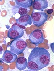
NK-cell therapy in resistant MDS, AML

Results of a phase 1/2 trial suggest treatment with haploidentical natural killer (NK) cells can be effective against relapsed/refractory myelodysplastic syndromes (MDS) and acute myeloid leukemia (AML).
NK-cell therapy elicited responses in 6 of the 16 patients studied and provided a bridge to transplant for 5 patients.
Three responders were still alive at more than 3 years of follow-up.
There were 4 grade 3 adverse events (AEs) and 2 grade 5 AEs considered possibly or probably related to NK-cell therapy.
Investigators reported these results in Clinical Cancer Research.
The trial enrolled 16 patients. Eight had MDS/AML, 3 had de novo AML, and 5 had high-risk MDS, including refractory anemia with excess blasts (RAEB) type 1 progressing toward type 2, RAEB-2, and chronic myelomonocytic leukemia type 2.
The patients’ median age was 64 (range, 40-70), and they had received a median of 3 prior therapies (range, 1-6). Six patients had received an allogeneic hematopoietic stem cell transplant (HSCT).
For this study, all patients received fludarabine, cyclophosphamide, and total lymphoid irradiation prior to receiving haploidentical NK cells.
The median follow-up was 8 months for all patients and 28 months for responders.
Efficacy
Six patients responded to treatment. One patient with de novo AML had a complete response (CR). Two high-risk MDS patients had a marrow CR (mCR), as did 2 MDS/AML patients. One MDS/AML patient had a partial response (PR).
Two patients had stable disease (SD)—1 with MDS and 1 with MDS/AML. One patient with de novo AML had a morphologic leukemia-free state after NK-cell therapy.
Five patients proceeded to HSCT—3 in mCR, 1 in PR, and 1 with SD.
Three patients were still alive at last follow-up—1 with MDS who achieved an mCR and went on to HSCT, 1 with MDS/AML who achieved an mCR and went on to HSCT, and 1 with MDS/AML who achieved an mCR and went on to receive chemotherapy and donor lymphocyte infusion.
One survivor has more than 5 years of follow-up (the MDS patient), and the other 2 have more than 3 years of follow-up.
“Our study shows that patients with MDS, AML, and MDS/AML can be treated with NK cell-based immunotherapy and that the therapy can be highly efficacious,” said study author Hans-Gustaf Ljunggren, MD, PhD, of Karolinska Institutet in Stockholm, Sweden.
Safety
The most common AEs of any grade considered possibly or probably related to NK-cell therapy were chills (n=13) and nausea (n=4).
Two patients had cytokine release syndrome (CRS) likely associated with hemophagocytic lymphohistiocytosis (HLH).
Each of the following potentially related AEs were reported once: headache, vomiting, encephalitis infection, sinus tachycardia, bone pain, pain in extremity, and maculopapular rash.
There were 4 grade 3 AEs—CRS/HLH (n=1), chills (n=1), and nausea (n=2)—but no grade 4 AEs.
There were 2 grade 5 AEs—CRS/HLH and encephalitis infection. These occurred in a single patient who died with HLH, human herpes virus-6 encephalitis, and AML relapse.
Two investigators involved in this study serve on the scientific advisory board of Fate Therapeutics. Dr Ljunggren serves on the scientific advisory board of CellProtect, Nordic Pharmaceuticals, and HOPE Bio-Sciences. He is also on the board of directors of Vycellix and is a collaborator with Fate Therapeutics.
Team creates device to study hemostasis

Researchers have engineered a miniature model system for studying hemostasis.
They believe the device could serve as a drug discovery platform and potential diagnostic tool.
The team has already used the device to assess the effects of an antiplatelet agent and analyze blood from hemophilia A patients.
The researchers described this work in Nature Communications.
The team noted that hemostasis encompasses the interactions of platelets, coagulation factors, blood cells, endothelium, and hemodynamic forces.
“Current methods to study blood clotting require isolation of each of these components, which prevents us from seeing the big picture of what’s going with the patient’s blood clotting system,” said study author Wilbur Lam, MD, PhD, of Georgia Institute of Technology and Emory University in Atlanta, Georgia.
With this in mind, Dr Lam and his colleagues developed their device.
They believe it is the first system to reproduce all aspects of blood vessel injury seen in the microvasculature—blood loss due to trauma, clot formation by whole blood, and repair of the blood vessel lining. However, it does not reproduce aspects of larger blood vessels.
The researchers’ device has 3 layers. The top “vascular” layer consists of human endothelial cells cultured in a microchannel. The middle valve layer consists of a polydimethylsiloxane membrane, and the bottom is a “valve actuator” layer.
To assess hemostatic response, the researchers create a “wound” in this system. They exert both positive and negative pressure to create an opening about 130 micrometers across. Donated human blood can flow through this opening for testing.
The researchers said they used this system to demonstrate the importance of von Willebrand factor and endothelial phosphatidylserine in hemostasis.
The team used the device to test the antiplatelet agent eptifibatide as well. They found the drug leads to decreased clot contraction and a lower density of platelets within the hemostatic plug.
Finally, the researchers analyzed blood from hemophilia A patients and found that it “confers unstable hemostatic plug formation and altered fibrin architecture.”
Drug receives orphan designation for MM
The US Food and Drug Administration (FDA) has granted orphan designation to PT-112 as a treatment for multiple myeloma (MM).
PT-112 is a small-molecule conjugate of pyrophosphate and platinum that promotes apoptosis with damage-associated molecular patterns, leading to downstream T-cell recruitment in the tumor microenvironment.
PT-112 is currently under investigation in a phase 1/2 study of patients with relapsed or refractory MM (NCT03288480).
Phosplatin Therapeutics LLC, the company developing PT-112, has enrolled the first cohort of patients in this trial.
In preclinical experiments, PT-112 demonstrated synergy with lenalidomide and bortezomib in RPMI-8226 cells and dexamethasone-resistant MM1R cells.
Single-agent PT-112 produced responses in mice with established MM. Researchers said PT-112 had “pronounced” activity against bortezomib-refractory Vk12598 tumors, which significantly improved overall survival in the mice.
This research was presented at the 2017 ASH Annual Meeting (abstract 1797).
About orphan designation
The FDA grants orphan designation to products intended to treat, diagnose, or prevent diseases/disorders that affect fewer than 200,000 people in the US.
The designation provides incentives for sponsors to develop products for rare diseases. This may include tax credits toward the cost of clinical trials, prescription drug user fee waivers, and 7 years of market exclusivity if the product is approved.
Abstract: Glyburide Versus Metformin and Their Combination for the Treatment of Gestational Diabetes Mellitus: A Randomized Controlled Study
The video associated with this article is no longer available on this site. Please view all of our videos on the MDedge YouTube channel
Nachum, Z., et al, Diab Care 40(3):332, March 2017
METHODS: These Israeli authors performed an open-label trial of the efficacy and safety of glyburide, metformin and their combination for gestational diabetes mellitus (GDM). The study included 104 women aged 18-45 (mean age, 33) with elevated blood glucose due to GDM diagnosed at 13-33 weeks’ gestation who were randomized to begin treatment with glyburide or metformin. Treatment was changed as follows: the patient was switched to the other drug in case of adverse events, changed to combination therapy in the event of treatment failure (preprandial glucose above 95mg/dL, postprandial glucose above 130mg/dL, or daily glucose above 100mg/dL), changed to insulin if both drugs failed. The primary outcome was treatment failure (adverse events or poor glycemic control) after the first study drug based on patients’ daily glucose charts.
RESULTS: Treatment failed in 18 glyburide patients (34%) and 15 metformin patients (29%), a nonsignificant difference (p=0.6). Failure with glyburide was due to hypoglycemia in 11% and poor glycemic control in 23%; metformin failure was due to gastrointestinal events in 2% and poor glycemic control in 28%. Second-line treatment with metformin was more effective than second-line glyburide (87% versus 50%; p=0.03), and glyburide patients were more likely to require insulin (17% versus 4%; p=0.03). Combining the drugs reduced the need for insulin from 32% to 11% (p=0.0002). Safety parameters and obstetric outcomes were similar between groups.
CONCLUSIONS: In this study, glyburide and metformin were generally comparable in efficacy and safety for treating GDM. There may be a slight advantage to starting with metformin. Combining the two agents reduced the risk of treatment failure and the need for insulin. 22 references ([email protected] – no reprints)
Learn more about the Primary Care Medical Abstracts and podcasts, for which you can earn up to 9 CME credits per month.
Copyright © The Center for Medical Education
FDA approves new HPV assay
Manufacturer Becton Dickinson announced on Feb. 13 that it had received premarket approval from the U.S. Food and Drug Administration for a human papillomavirus (HPV) assay.
The Onclarity assay detects 14 types of high-risk HPV from specimens collected from cervical cancer screening via a SurePath liquid-based Pap test. It has previously been approved in Europe, Canada, and Japan. The bench-top molecular testing platform used for the assay has received prior FDA approval for chlamydia and gonorrhea infection testing.
The assay in particular identifies the HPV genotypes 16, 18, and 45, which are associated with more than two-thirds of cervical cancers and precancerous cervical lesions and as many as 94% of glandular cervical cancer cases.
Cutaneous multiple myeloma is a deadly indicator
Cutaneous involvement was present in less than 1.2% of patients with multiple myeloma (MM) and was associated with reduced overall survival, according to the results of a small, retrospective analysis.
Cutaneous manifestations of MM can be divided into nonspecific and specific lesions, according to according to Yu Ri Woo, MD, and colleagues at Yeouido St. Mary’s Hospital, the Catholic University of Korea, Seoul, South Korea.
Nonspecific cutaneous manifestations include amyloidosis, cryoglobulinemia, Raynaud’s phenomenon, xanthomas, pyoderma gangrenosum, and purpura, and were excluded from the study, while the specific cutaneous manifestation in MM was exemplified by secondary extramedullary plasmacytomas, seen as cutaneous waxy dome-shaped nodules with variable sizes in various locations. These were assessed in the study published in the Journal of the American Academy of Dermatology.
The medical records of 1,228 patients with MM seen at two institutions from Jan.1, 1996, to Dec. 31, 2016, were examined. Among these patients, 14 (1.14%) had specific cutaneous involvement of MM indicated, and their charts were evaluated further for their clinical and histopathologic findings.
There were no significant differences seen among patients in terms of age, sex, the presence of heavy or light chain disease, International Staging System stage, or albumin level.
Patients with cutaneous involvement showed significantly reduced overall survival, compared with patients without cutaneous involvement (median, 28 months vs. 57 months; hazard ratio, 1.9; 95% confidence interval, 1.0-3.6).
In a subgroup analyses of those patients who had MM with cutaneous involvement, the presence of erythematous nodules (P = .004), multiple cutaneous lesions (P = .002), and the absence of a grenz zone (P = .004) were associated with reduced overall survival.
Although the investigators found a relatively low incidence of cutaneous involvement in their database study, they pointed out that the exact incidence of cutaneous involvement in MM might be higher than expected.
“Many clinicians are less interested in the cutaneous lesions specifically,” they wrote, indicating that the original physicians may not have been looking for such lesions closely or reporting them if found. “Additional large sample prospective research is needed to determine the exact incidence of cutaneous involvement in MM.”
The investigators reported that they had no funding sources or conflicts of interest.
SOURCE: Woo YR et al. J Am Acad Dermatol 2018;78:471-8.
FROM JOURNAL OF THE AMERICAN ACADEMY OF DERMATOLOGY
Key clinical point:
Major finding: Patients with cutaneous involvement showed significantly reduced overall survival, compared with those without cutaneous involvement (median, 28 months vs. 57 months; HR, 1.9).
Study details: A retrospective study of 1,228 patients with multiple myeloma, of whom 14 patients had cutaneous involvement (1.14%).
Disclosures: The investigators reported that they had no funding sources or conflicts of interest.
Source: Woo YR et al. J Am Acad Dermatol. 2018;78:471-8.
Postcesarean SSI rate declines with care bundle*
DALLAS – A surgical site infection care bundle reduced the rate of surgical site infections (SSIs) after cesarean delivery by more than half, according to a case-control study examining data from more than 2,000 patients.
At the health center where the SSI bundle was implemented, rates per 1,000 women undergoing cesarean delivery fell from 2.44 to 1.10 (P = .013).
The study showed the effectiveness of implementing evidence-based and -supported recommendations, and of having standardized protocols with little variation, said Christina Davidson, MD, presenting the pre-post findings during a plenary session at the meeting sponsored by the Society for Maternal-Fetal Medicine.
The bundle of interventions was developed over the course of 3 months in late 2013 and early 2014 by a multidisciplinary task force, drawing from colorectal surgery literature about SSI prevention. Both nurses and physicians were on the task force, and representatives came from the departments of obstetrics and gynecology, anesthesia, and infection prevention, said Dr. Davidson of the Baylor College of Medicine, Houston. All inpatient and outpatient clinical care sites had representation.
After the bundle elements were identified, a full month was devoted to education and team training, with full bundle implementation occurring in April 2014. “Visual aids were placed in close proximity to the operating rooms,” said Dr. Davidson. For example, antimicrobial prophylaxis cards were placed on all anesthetic carts.
“A surgical checklist was placed in the chart for each patient undergoing cesarean delivery and compliance was tracked for the first 12 months of implementation,” said Dr. Davidson. Additionally, members of the care team received feedback in the form of quarterly reports on SSI rates and statistics about bundle compliance.
Care bundle elements included a set of instructions for pre- and postoperative antiseptic skin cleaning, wound care, and glycemic control in patients. Women were given chlorhexidine cleanser and asked to use it when showering the day before and the morning of surgery for planned deliveries. Forced warm-air blankets maintained patient normothermia in the preoperative holding area.
A group of intraoperative interventions included use of antiseptic skin and vaginal preparations, double-gloving, and having all scrubbed members of the surgical team change their outer gloves for fascial closure. A new instrument tray also was used for fascial closure. Prophylactic antibiotics were administered within 1 hour of skin incision, and doses were readministered based on the length of the procedure.
Postoperatively, said Dr. Davidson, “a set of insulin orders within the electronic medical record [was] used to maintain euglycemia in all diabetic patients.”
After the surgical dressing was removed on the 2nd postoperative day, patients were given a handout and education about wound control and infection prevention.
Finally, all patients received postdischarge follow-up calls from nurses within 72 hours after discharge.
Patient characteristics generally were similar before (n = 1,085) and after (n = 1,261) SSI bundle implementation. Body mass index was slightly higher in the postbundle group, and women in this group also were less likely to have had a prior cesarean delivery. There were no significant differences in age, gravidity, ethnicity, or race.
The study showed that with continued tracking, data-sharing, and reeducation efforts, “The SSI rate was sustained after bundle implementation,” said Dr. Davidson. The implementation team, working with hospital departments, was able to achieve a high compliance rate. And, she said, the effect size of the intervention was large enough to show significant reduction from an already low SSI rate.
However, Dr. Davidson also noted some limitations: All of the bundle elements were implemented simultaneously, so it wasn’t possible to tell which components had the greatest effect. Also, not all demographic data were available, and the type of SSI was sometimes unavailable from the deidentified data repository used for analysis, she said. “We weren’t able to tease out individual patient-level characteristics” about the timing and type of SSI in a patient-by-patient fashion, she said during discussion following her presentation.
All in all, she said, the bundle’s effectiveness “supports the synergistic effects of multiple strategies and the impact of a multidisciplinary team approach.”
The study authors reported no conflicts of interest.
SOURCE: Davidson C et al. Am J Obstet Gynecol. 2018 Jan;218:S46.
Correction, 3/5/18: An earlier version of this article omitted the word "rate" from the headline and Vitals section.
REPORTING FROM THE PREGNANCY MEETING
Key clinical point: The postcesarean surgical site infection rate dropped by more than half after a multicomponent care bundle was put in place.*
Major finding: SSIs patients went from 2.44 to 1.10/1,000 after the bundle was implemented (P = .013).
Study details: Case-control study of 1,085 women pre– and 1,261 women post–care bundle implementation.
Disclosures: The authors reported no conflicts of interest.
Source: Davidson C et al. Am J Obstet Gynecol. 2018 Jan;218:S46.