User login
Severe OA sparks depression, surgery “ameliorates” depression in RA
AMSTERDAM – Structural severity in OA is related to the onset of depressive symptoms while surgery “ameliorates” depression in RA, according to the results of two separate studies presented at the European Congress of Rheumatology.
Using data on more than 1,600 individuals with knee OA from the Osteoarthritis Initiative, Alan Rathbun, PhD, and his associates looked at the components of disease severity and how they might individually contribute to the development of depression. They found that the odds of having depression more than doubled as joint space width increased (odds ratio, 2.25) and gait speed decreased (OR, 2.08), and rose 60% as pain became more severe (OR, 1.60).

Worsening knee OA could set off depression
“Studies have consistently shown that depressive symptoms are associated with worse osteoarthritis disease severity, however, there is a lack of research focused on identifying the specific components that contribute to the onset of depressive symptoms in nondepressed OA patients,” said Dr. Rathbun in an interview ahead of his presentation.
Dr. Rathbun, a research associate in the departments of epidemiology and of public health and medicine at the University of Maryland, Baltimore, also said that while OA guidelines do advise on treating depression, there is no standardized way to manage comorbid depression in routine clinical practice.
“If OA disease severity contributes to the development and worsening of depressive symptoms, it may be necessary to intervene on both conditions simultaneously in order to successfully manage them,” he suggested.
“Depression is a frequently occurring comorbidity in persons with OA,” Dr. Rathbun later observed during a press conference. Around one in five people with knee OA have depression, he said. This is an important fact if you consider how common symptomatic knee OA is – affecting 10% of men and 13% of women aged over 60 years – and the impact that it has on people’s quality of life, healthcare utilization, and mortality.
Dr. Rathbun and colleagues examined data on 1,652 men and women who were part of the Osteoarthritis Initiative, a multicenter, prospective, longitudinal study of knee health sponsored by the National Institutes of Health. For inclusion in the study, participants had to have radiographic knee OA, no depressive symptoms, and complete data on the disease severity components of interest: minimum joint space width, 20-meter gait speed, and the pain subscale of the Western Ontario and McMaster Universities Osteoarthritis Index. Depression was assessed using the Center for Epidemiological Studies Depression Scale, with no depression ascribed a score of 16 or under. Data from three additional annual follow-up visits was also required.
“All three components of OA disease progression were associated with an increased risk for the onset of depressive symptoms in those with radiographic knee OA,” Dr. Rathbun said in the interview. While pain severity has previously been linked to depressive symptoms in OA, the finding of worsening structural disease is new.
“The clinical implications of our findings are that the onset of depressive symptoms in OA patients is related to worsening pain, physical function, and structural disease severity,” he added. They also mean that these components and depression need to be targeted at the same time.
“Future studies need to ascertain whether depressive symptoms modify clinical response to analgesic medications in OA patients,” Dr. Rathbun suggested. “Considering that analgesics are often the first-line treatment for OA patients and the high prevalence of depressive symptoms in this population, comorbid depression may be an important contributor to ineffective medical management in the many OA patients who undergo total joint replacement.”
Depression “ameliorated” by orthopedic surgery for RA
While the effects of analgesic medications might be something to look at in relation to depression in OA, research presented elsewhere at the congress suggested that appropriate surgical intervention might also be key to dealing with depressive symptoms, at least in patients with RA.
After a 1-year follow-up in patients with RA who underwent orthopedic surgery, the mean BDI-II score improved from 13.0 to 11.5 (P less than .01), and the percentage of patients with a BDI-II score of 14 or more fell from 43% to 35%. Improvements in other health assessments – the Japanese version of the Health Assessment Questionnaire and the EuroQol 5 dimensions instrument – were also seen.
The prospective, observational cohort study included 276 patients with structural damage caused by RA. The most common site of joint damage requiring elective surgery was the wrist (n = 74), followed by the hand (n = 63), knee (n = 50), forefoot (n = 50), elbow (n = 26), hand and wrist (n = 18), hip (n = 13), ankle (n = 12), and shoulder (n = 6).
Looking at the improvement in depression scores by surgical site revealed a significant difference from baseline for the elbow (P less than .001), wrist (P less than .001), and forefoot (P less than .05). The magnitude of decrease in the BDI-II scores was independently related to Steinbrocker stage and pain measured on a visual analog scale.
“Depression was ameliorated by surgical intervention in patients with RA,” Hajime Ishikawa, MD, PhD, of the department of rheumatology at Niigata Rheumatic Center in Shibata, Japan, and associates concluded in their poster presentation. They added that the psychological changes observed were “related to the preoperative severity of joint damage and pain in the affected joint.”
Dr. Rathbun’s work was supported by a Rheumatology Research Foundation Scientist Development Award. Dr. Ishikawa and associates stated they had no disclosures of interest.
SOURCES: Rathbun AM et al. Ann Rheum Dis. 2018;77(Suppl 2):50-1. Abstract OP0003; Ishikawa H et al. Ann Rheum Dis. 2018;77(Suppl 2):297-8. Abstract THU0156.
AMSTERDAM – Structural severity in OA is related to the onset of depressive symptoms while surgery “ameliorates” depression in RA, according to the results of two separate studies presented at the European Congress of Rheumatology.
Using data on more than 1,600 individuals with knee OA from the Osteoarthritis Initiative, Alan Rathbun, PhD, and his associates looked at the components of disease severity and how they might individually contribute to the development of depression. They found that the odds of having depression more than doubled as joint space width increased (odds ratio, 2.25) and gait speed decreased (OR, 2.08), and rose 60% as pain became more severe (OR, 1.60).
Worsening knee OA could set off depression
“Studies have consistently shown that depressive symptoms are associated with worse osteoarthritis disease severity, however, there is a lack of research focused on identifying the specific components that contribute to the onset of depressive symptoms in nondepressed OA patients,” said Dr. Rathbun in an interview ahead of his presentation.
Dr. Rathbun, a research associate in the departments of epidemiology and of public health and medicine at the University of Maryland, Baltimore, also said that while OA guidelines do advise on treating depression, there is no standardized way to manage comorbid depression in routine clinical practice.
“If OA disease severity contributes to the development and worsening of depressive symptoms, it may be necessary to intervene on both conditions simultaneously in order to successfully manage them,” he suggested.
“Depression is a frequently occurring comorbidity in persons with OA,” Dr. Rathbun later observed during a press conference. Around one in five people with knee OA have depression, he said. This is an important fact if you consider how common symptomatic knee OA is – affecting 10% of men and 13% of women aged over 60 years – and the impact that it has on people’s quality of life, healthcare utilization, and mortality.
Dr. Rathbun and colleagues examined data on 1,652 men and women who were part of the Osteoarthritis Initiative, a multicenter, prospective, longitudinal study of knee health sponsored by the National Institutes of Health. For inclusion in the study, participants had to have radiographic knee OA, no depressive symptoms, and complete data on the disease severity components of interest: minimum joint space width, 20-meter gait speed, and the pain subscale of the Western Ontario and McMaster Universities Osteoarthritis Index. Depression was assessed using the Center for Epidemiological Studies Depression Scale, with no depression ascribed a score of 16 or under. Data from three additional annual follow-up visits was also required.
“All three components of OA disease progression were associated with an increased risk for the onset of depressive symptoms in those with radiographic knee OA,” Dr. Rathbun said in the interview. While pain severity has previously been linked to depressive symptoms in OA, the finding of worsening structural disease is new.
“The clinical implications of our findings are that the onset of depressive symptoms in OA patients is related to worsening pain, physical function, and structural disease severity,” he added. They also mean that these components and depression need to be targeted at the same time.
“Future studies need to ascertain whether depressive symptoms modify clinical response to analgesic medications in OA patients,” Dr. Rathbun suggested. “Considering that analgesics are often the first-line treatment for OA patients and the high prevalence of depressive symptoms in this population, comorbid depression may be an important contributor to ineffective medical management in the many OA patients who undergo total joint replacement.”
Depression “ameliorated” by orthopedic surgery for RA
While the effects of analgesic medications might be something to look at in relation to depression in OA, research presented elsewhere at the congress suggested that appropriate surgical intervention might also be key to dealing with depressive symptoms, at least in patients with RA.
After a 1-year follow-up in patients with RA who underwent orthopedic surgery, the mean BDI-II score improved from 13.0 to 11.5 (P less than .01), and the percentage of patients with a BDI-II score of 14 or more fell from 43% to 35%. Improvements in other health assessments – the Japanese version of the Health Assessment Questionnaire and the EuroQol 5 dimensions instrument – were also seen.
The prospective, observational cohort study included 276 patients with structural damage caused by RA. The most common site of joint damage requiring elective surgery was the wrist (n = 74), followed by the hand (n = 63), knee (n = 50), forefoot (n = 50), elbow (n = 26), hand and wrist (n = 18), hip (n = 13), ankle (n = 12), and shoulder (n = 6).
Looking at the improvement in depression scores by surgical site revealed a significant difference from baseline for the elbow (P less than .001), wrist (P less than .001), and forefoot (P less than .05). The magnitude of decrease in the BDI-II scores was independently related to Steinbrocker stage and pain measured on a visual analog scale.
“Depression was ameliorated by surgical intervention in patients with RA,” Hajime Ishikawa, MD, PhD, of the department of rheumatology at Niigata Rheumatic Center in Shibata, Japan, and associates concluded in their poster presentation. They added that the psychological changes observed were “related to the preoperative severity of joint damage and pain in the affected joint.”
Dr. Rathbun’s work was supported by a Rheumatology Research Foundation Scientist Development Award. Dr. Ishikawa and associates stated they had no disclosures of interest.
SOURCES: Rathbun AM et al. Ann Rheum Dis. 2018;77(Suppl 2):50-1. Abstract OP0003; Ishikawa H et al. Ann Rheum Dis. 2018;77(Suppl 2):297-8. Abstract THU0156.
REPORTING FROM THE EULAR 2018 CONGRESS
Key clinical point: Structural severity is related to the onset of depressive symptoms in OA while surgery “ameliorates” depression in RA.
Major findings: Depression increased the odds of having worse OA (2.25 for greater joint space width, 2.08 for slower gait, and 1.60 for pain severity). The mean Beck Depression Inventory–II score of RA patients who underwent orthopedic surgery was 11.5 postsurgery, an improvement of 1.5 points versus presurgery scores (P less than .01).
Study details: Data on more than 1,600 individuals with knee OA from the Osteoarthritis Initiative and a separate prospective, observational cohort study of 276 patients with structural joint damage caused by RA who underwent elective surgery.
Disclosures: Dr. Rathbun’s work was supported by a Rheumatology Research Foundation Scientist Development Award. Dr. Ishikawa and associates reported no disclosures of interest.
Sources: Rathbun AM et al. Ann Rheum Dis. 2018;77(Suppl 2):50-1. Abstract OP0003; Ishikawa H et al. Ann Rheum Dis. 2018;77(Suppl 2):297-8. Abstract THU0156.
Extreme heat and mental health: Protecting patients
Now that the summer is in full swing, it is incumbent upon the psychiatric and mental health community to learn about the specific effects on behavior, psychiatric risks, and outcomes – and to plan for ways to protect our patients and communities.
Extreme heat has significant effects on mental health and behavior. Research shows1 that the number of people exposed to extreme heat is expected to rise in many American cities, particularly across the southern United States. Records were set in May 2018 across the United States and around the world. In the United States, those May temperatures were the warmest ever recorded, representing the hottest spring ever.2 Around the world, the warmer-than-average conditions that engulfed much of the land and sea surfaces made May the fourth-warmest since records started being kept in 1880.2
In short, these trends are not remitting. Extreme heat and climate disruption are the new normal.
Extreme heat makes many people cranky, agitated, or listless. However, heat waves are not benign, uncomfortable periods; they have profound health risks tied to increasing rates of anxiety, depression, posttraumatic stress disorder, and even death. In fact, extreme heat is now considered to be the single largest weather-related cause of death, exceeding hurricanes, lightning, tornadoes, floods, and earthquakes combined. The Centers for Disease Control and Prevention reports 7,800 deaths attributable to extreme heat between 1999 and 2009 – and predicts more frequent and extreme heat.3
In addition, extreme heat has been linked to increases in aggression and violence. One standard deviation of temperature increase and rainfall is associated with a 4% increase in interpersonal violence and 14% increase in intergroup violence.4 Anecdotal stories underscore the well known lore among prison staff of increased inmate violence during heat waves.5
Complex cognitive tasks such as working memory (spatial span test, pattern recognition) have been observed to be significantly impaired through heat stress.6 Increased heat also contributes to insomnia and worsens with increased humidity.7 A study in England and Wales showed a link between a possible association between hot weather and an increased risk of suicide.8 People with mental illness and those who abuse substances are considered an especially vulnerable population to the impacts of extreme heat and other climate change–related events. Co-occurring variables such as poverty, substandard housing, and lack of access to cool environments all contribute to this increased vulnerability. Homeless mentally ill have little control over their environments and have very limited ability to protect themselves from heat exposures and therefore are at extreme risk.9 . Regretfully, these kinds of cooling systems are out of reach for many people who live on the margins of society.
Furthermore, patients with severe psychotic or mood disorders, substance abuse disorders, and cognitive impairments who are able to compensate with marginal executive functioning during periods of normal weather are challenged during intense heat, and can lose their fragile ability to make plans, have good judgment, and care effectively for themselves. These patients are more likely to experience heat stroke and other heat-related morbidity.
Here is evidence that supports the greater impact of extreme heat on psychiatric patients:
- Increased emergency department and hospitalization for patients with preexisting psychiatric illness during heat waves.10,11
- Preexisting mental illness alone increases the risk of mortality during extreme heat events by 2 to 3 times.12,13
- Patients with schizophrenia might have underlying impairments in thermoregulation that are intrinsic to the disease. Such impairments would explain the perplexing sight of psychotic patients bundled up in layers on hot days.14
- Psychiatric medications (antipsychotics, anticholinergics, and antidepressants) have the potential to impair the body’s heat regulatory functioning; lithium affects fluid homeostasis.15

The negative effects of climate change are not equally distributed, and people with mental illness are among the most vulnerable. Given the predictable future of extreme heat waves (potentially increasing the population exposure by four- to sixfold by midcentury),1 we must do everything we can to educate our patients so that they take preventive measures to protect themselves from the adverse effects of extreme heat.
References
1. Nature Climate Change. 2015 May 18;5:652-5.
2. National Oceanic and Atmospheric Administration Global Climate Report. May 2018.
3. “Climate Change and Extreme Heat Events.” Centers for Disease Control and Prevention.
4. Science. 2013 Sep 13;341(6151).
5. Personal communication.
6. Int J Hyperthermia. 2003 May-Jun;19(3):355-72.
7. J Physiol Anthropol. 2012 May 31;31(14).
8. Br J Psychiatry. 2007 Aug;191:106-12.
9. U.S. Global Change Research Program, 2016. “The Impacts of Climate Change on Human Health in the United States: A Scientific Assessment.” Chapters 8 and 9.
10. J Affect Disord. 2014 Feb;155:154-61.
11. Environ Health Perspect. 2008 Oct;116(10):1369-75.
12. Psychiatr Serv. 1998 Aug;49(8):1088-90.
13. Arch Intern Med. 2007 Nov. 12;167(20):2170-6.
14. Schizophr Res. 2004 Aug 1;69(2-3):149-57.
15. Eur Psychiatry. 2007 Sep;22(6):335-8.
Strategies for patients, communities
Part of the job of mental health professionals is psychoeducation, or teaching patients and families about the health risks tied to the psychological and physical impacts of heat exposure. Also, we should provide advice about effective management of psychiatric medications – such as monitoring lithium levels and considering medication dose adjustments – to reduce risks. Another key step is engaging caregivers, case managers, visiting nurses, and family members so that they closely monitor vulnerable populations. Providing information about the availability of respite care and cooling centers is another concrete step clinicians can take to help minimize the impact of extreme heat on patients.
Information that can be shared with patients about the threat include:
- A brochure from the Climate Psychiatry Alliance that offers specific ways to keep cool in extreme heat.
- A guidebook produced by the Environmental Protection Agency and the Centers for Disease Control and Prevention that explains what people can do to prepare for extreme heat.
- More extensive interventions can be found at https://www.climatepsychiatry.org/what-to-do/.
Dr. Cooper is in private practice and is affiliated with the department of psychiatry at the University of California, San Francisco. She is a Distinguished Life Fellow of the American Psychiatric Association.
Now that the summer is in full swing, it is incumbent upon the psychiatric and mental health community to learn about the specific effects on behavior, psychiatric risks, and outcomes – and to plan for ways to protect our patients and communities.
Extreme heat has significant effects on mental health and behavior. Research shows1 that the number of people exposed to extreme heat is expected to rise in many American cities, particularly across the southern United States. Records were set in May 2018 across the United States and around the world. In the United States, those May temperatures were the warmest ever recorded, representing the hottest spring ever.2 Around the world, the warmer-than-average conditions that engulfed much of the land and sea surfaces made May the fourth-warmest since records started being kept in 1880.2
In short, these trends are not remitting. Extreme heat and climate disruption are the new normal.
Extreme heat makes many people cranky, agitated, or listless. However, heat waves are not benign, uncomfortable periods; they have profound health risks tied to increasing rates of anxiety, depression, posttraumatic stress disorder, and even death. In fact, extreme heat is now considered to be the single largest weather-related cause of death, exceeding hurricanes, lightning, tornadoes, floods, and earthquakes combined. The Centers for Disease Control and Prevention reports 7,800 deaths attributable to extreme heat between 1999 and 2009 – and predicts more frequent and extreme heat.3
In addition, extreme heat has been linked to increases in aggression and violence. One standard deviation of temperature increase and rainfall is associated with a 4% increase in interpersonal violence and 14% increase in intergroup violence.4 Anecdotal stories underscore the well known lore among prison staff of increased inmate violence during heat waves.5
Complex cognitive tasks such as working memory (spatial span test, pattern recognition) have been observed to be significantly impaired through heat stress.6 Increased heat also contributes to insomnia and worsens with increased humidity.7 A study in England and Wales showed a link between a possible association between hot weather and an increased risk of suicide.8 People with mental illness and those who abuse substances are considered an especially vulnerable population to the impacts of extreme heat and other climate change–related events. Co-occurring variables such as poverty, substandard housing, and lack of access to cool environments all contribute to this increased vulnerability. Homeless mentally ill have little control over their environments and have very limited ability to protect themselves from heat exposures and therefore are at extreme risk.9 . Regretfully, these kinds of cooling systems are out of reach for many people who live on the margins of society.
Furthermore, patients with severe psychotic or mood disorders, substance abuse disorders, and cognitive impairments who are able to compensate with marginal executive functioning during periods of normal weather are challenged during intense heat, and can lose their fragile ability to make plans, have good judgment, and care effectively for themselves. These patients are more likely to experience heat stroke and other heat-related morbidity.
Here is evidence that supports the greater impact of extreme heat on psychiatric patients:
- Increased emergency department and hospitalization for patients with preexisting psychiatric illness during heat waves.10,11
- Preexisting mental illness alone increases the risk of mortality during extreme heat events by 2 to 3 times.12,13
- Patients with schizophrenia might have underlying impairments in thermoregulation that are intrinsic to the disease. Such impairments would explain the perplexing sight of psychotic patients bundled up in layers on hot days.14
- Psychiatric medications (antipsychotics, anticholinergics, and antidepressants) have the potential to impair the body’s heat regulatory functioning; lithium affects fluid homeostasis.15
The negative effects of climate change are not equally distributed, and people with mental illness are among the most vulnerable. Given the predictable future of extreme heat waves (potentially increasing the population exposure by four- to sixfold by midcentury),1 we must do everything we can to educate our patients so that they take preventive measures to protect themselves from the adverse effects of extreme heat.
References
1. Nature Climate Change. 2015 May 18;5:652-5.
2. National Oceanic and Atmospheric Administration Global Climate Report. May 2018.
3. “Climate Change and Extreme Heat Events.” Centers for Disease Control and Prevention.
4. Science. 2013 Sep 13;341(6151).
5. Personal communication.
6. Int J Hyperthermia. 2003 May-Jun;19(3):355-72.
7. J Physiol Anthropol. 2012 May 31;31(14).
8. Br J Psychiatry. 2007 Aug;191:106-12.
9. U.S. Global Change Research Program, 2016. “The Impacts of Climate Change on Human Health in the United States: A Scientific Assessment.” Chapters 8 and 9.
10. J Affect Disord. 2014 Feb;155:154-61.
11. Environ Health Perspect. 2008 Oct;116(10):1369-75.
12. Psychiatr Serv. 1998 Aug;49(8):1088-90.
13. Arch Intern Med. 2007 Nov. 12;167(20):2170-6.
14. Schizophr Res. 2004 Aug 1;69(2-3):149-57.
15. Eur Psychiatry. 2007 Sep;22(6):335-8.
Strategies for patients, communities
Part of the job of mental health professionals is psychoeducation, or teaching patients and families about the health risks tied to the psychological and physical impacts of heat exposure. Also, we should provide advice about effective management of psychiatric medications – such as monitoring lithium levels and considering medication dose adjustments – to reduce risks. Another key step is engaging caregivers, case managers, visiting nurses, and family members so that they closely monitor vulnerable populations. Providing information about the availability of respite care and cooling centers is another concrete step clinicians can take to help minimize the impact of extreme heat on patients.
Information that can be shared with patients about the threat include:
- A brochure from the Climate Psychiatry Alliance that offers specific ways to keep cool in extreme heat.
- A guidebook produced by the Environmental Protection Agency and the Centers for Disease Control and Prevention that explains what people can do to prepare for extreme heat.
- More extensive interventions can be found at https://www.climatepsychiatry.org/what-to-do/.
Dr. Cooper is in private practice and is affiliated with the department of psychiatry at the University of California, San Francisco. She is a Distinguished Life Fellow of the American Psychiatric Association.
FDA Approves a Cannabinoid Medicine for Two Forms of Epilepsy
Epidiolex (cannabidiol) oral solution may treat seizures in patients with Lennox-Gastaut syndrome and Dravet syndrome.
The FDA has approved Epidiolex (cannabidiol [CBD]) oral solution for the treatment of seizures associated with Lennox-Gastaut syndrome and Dravet syndrome in patients age 2 and older. Epidiolex is the first FDA-approved drug that contains a derivative of marijuana. It also is the first drug approved by the FDA for the treatment of Dravet syndrome.
The approval was based on three randomized, double-blind, placebo-controlled clinical trials that included 516 patients with Lennox-Gastaut syndrome or Dravet syndrome. Epidiolex taken with other epilepsy medications reduced the frequency of seizures, compared with placebo. The most common side effects included lethargy, elevated liver enzymes, decreased appetite, diarrhea, rash, weakness, sleep disorder, and infection.
“Because of the adequate and well-controlled clinical studies that supported this approval, prescribers can have confidence in the drug’s uniform strength and consistent delivery that support appropriate dosing needed for treating patients with these complex and serious epilepsy syndromes,” said FDA Commissioner Scott Gottlieb, MD. “We will continue to support rigorous scientific research on the potential medical uses of marijuana-derived products…. But at the same time, we are prepared to take action when we see the illegal marketing of CBD-containing products with serious, unproven medical claims.”

CBD, a component of Cannabis sativa, does not cause intoxication or euphoria, unlike tetrahydrocannabinol (THC), the plant’s primary psychoactive component. CBD currently is a Schedule I substance because it is a chemical component of the cannabis plant. The Drug Enforcement Administration (DEA) is expected reschedule CBD within 90 days.
Epidiolex will be marketed in the US by Carlsbad, California-based Greenwich Biosciences, the US subsidiary of GW Pharmaceuticals, which is headquartered in London. Access to Epidiolex is expected to be similar to that for other branded antiepileptic drugs, and the treatment is expected to be available by Fall 2018, the company said.
Epidiolex (cannabidiol) oral solution may treat seizures in patients with Lennox-Gastaut syndrome and Dravet syndrome.
Epidiolex (cannabidiol) oral solution may treat seizures in patients with Lennox-Gastaut syndrome and Dravet syndrome.
The FDA has approved Epidiolex (cannabidiol [CBD]) oral solution for the treatment of seizures associated with Lennox-Gastaut syndrome and Dravet syndrome in patients age 2 and older. Epidiolex is the first FDA-approved drug that contains a derivative of marijuana. It also is the first drug approved by the FDA for the treatment of Dravet syndrome.
The approval was based on three randomized, double-blind, placebo-controlled clinical trials that included 516 patients with Lennox-Gastaut syndrome or Dravet syndrome. Epidiolex taken with other epilepsy medications reduced the frequency of seizures, compared with placebo. The most common side effects included lethargy, elevated liver enzymes, decreased appetite, diarrhea, rash, weakness, sleep disorder, and infection.
“Because of the adequate and well-controlled clinical studies that supported this approval, prescribers can have confidence in the drug’s uniform strength and consistent delivery that support appropriate dosing needed for treating patients with these complex and serious epilepsy syndromes,” said FDA Commissioner Scott Gottlieb, MD. “We will continue to support rigorous scientific research on the potential medical uses of marijuana-derived products…. But at the same time, we are prepared to take action when we see the illegal marketing of CBD-containing products with serious, unproven medical claims.”
CBD, a component of Cannabis sativa, does not cause intoxication or euphoria, unlike tetrahydrocannabinol (THC), the plant’s primary psychoactive component. CBD currently is a Schedule I substance because it is a chemical component of the cannabis plant. The Drug Enforcement Administration (DEA) is expected reschedule CBD within 90 days.
Epidiolex will be marketed in the US by Carlsbad, California-based Greenwich Biosciences, the US subsidiary of GW Pharmaceuticals, which is headquartered in London. Access to Epidiolex is expected to be similar to that for other branded antiepileptic drugs, and the treatment is expected to be available by Fall 2018, the company said.
Abortion not safer at an ambulatory surgical center
Abortion performed in an ambulatory surgery center (ASC) was not associated with a significant difference in abortion-related complications, compared with procedures performed in an office-based setting, according to results of a retrospective cohort study.
These findings might help inform decisions about the type of facility where induced abortions are performed, according to Sarah C. M. Roberts, DrPH, of the University of California, San Francisco, and her coauthors.
The U.S. Supreme Court ruled in 2016 that a Texas law requiring abortion facilities to meet ASC standards was unconstitutional, Dr. Roberts and her coauthors wrote in JAMA.
“Despite this ruling, 13 states currently have laws that require abortions to be provided in ASCs,” the authors wrote, noting that supporters of the laws argue that these requirements make abortions safer.
The laws have requirements such as separate procedure and recovery rooms, and specified hall and door widths. “Many of these apply only at a specific gestational week or gestational duration, typically in the second trimester,” they noted, adding that over 95% of induced abortions are performed in outpatient settings such as clinics or physician offices.
Their retrospective cohort study included a total of 50,311 induced abortions, of which 89% took place in office based settings and 11% in ASCs. Nearly half (47%) were first-trimester aspiration procedures, while 27% were first-trimester medication and 26% were second trimester or later.
Abortion-related morbidity or adverse events were reported for 3.33% of procedures overall. The adjusted incidence rate was 3.25% for ASC-based procedures, and similarly, 3.33% for office-based procedures.
The overall complication rate was higher than previous estimates based on insurance claims data, they said, but the estimate of major events was similar at 0.32%, breaking down to 0.26% for ASCs and 0.33% for office-based settings. The rate of infections was 0.58% for ASCs and 0.77% for office-based settings.
This is not the first study looking at the association between abortion-related events and the procedure setting, though the literature is limited, according to Dr. Roberts and her coauthors. One previous study showed fewer abortion-related events in clinics than in hospitals, while a recent review found similar abortion-related events following first-trimester abortions in hospitals, ASCs, and office-based settings.
One limitation of the current study is that the database included only abortions that were paid for by private insurance, which represents about 15% of the nearly 1 million procedures done each year in the United States.
“Thus, findings may not be generalizable to all abortions in the United States,” Dr. Roberts and her coauthors wrote.
The study was supported by a grant from the Society of Family Planning Research Fund. Study authors reported no conflicts of interest.
SOURCE: Roberts SCM et al. JAMA. 2018 Jun 26;319(24):2497-2506.
This new analysis provides further support that office settings are appropriate for abortion care and that office-based abortion care is appropriately safe, effective, and patient centered.
Results of this study support the safety of office-based abortions, including a low risk of infection, they added.
This comparison study of office-based abortion to abortion provided in an ambulatory surgery center (ASC) is important because 16 states impose restrictions that require abortion facilities adhere to ASC or ASC-equivalent standards.
Converting an office to an ASC is slow, complex, and although the cost of retrofitting a facility is moderately less, building an ASC costs an estimated $5 million, according to industry experts.
Requiring an office to meet an ASC-equivalent standard with no medical justification is too high a hurdle in many areas and serves to restrict women’s access to abortion.
Carolyn L. Westhoff, MD, and Anne R. Davis, MD, are with the department of obstetrics and gynecology at Columbia University, New York. These comments are based on their editorial in JAMA (2018 Jun 26;319[24]:2481-2483). Dr. Westhoff is the editor of Contraception and a senior medical advisor at Planned Parenthood Federation of America. Dr. Davis is consulting medical director for Physicians for Reproductive Health, a consultant for the New York City Department of Health, and an expert for the American Civil Liberties Union.
Abortion performed in an ambulatory surgery center (ASC) was not associated with a significant difference in abortion-related complications, compared with procedures performed in an office-based setting, according to results of a retrospective cohort study.
These findings might help inform decisions about the type of facility where induced abortions are performed, according to Sarah C. M. Roberts, DrPH, of the University of California, San Francisco, and her coauthors.
The U.S. Supreme Court ruled in 2016 that a Texas law requiring abortion facilities to meet ASC standards was unconstitutional, Dr. Roberts and her coauthors wrote in JAMA.
“Despite this ruling, 13 states currently have laws that require abortions to be provided in ASCs,” the authors wrote, noting that supporters of the laws argue that these requirements make abortions safer.
The laws have requirements such as separate procedure and recovery rooms, and specified hall and door widths. “Many of these apply only at a specific gestational week or gestational duration, typically in the second trimester,” they noted, adding that over 95% of induced abortions are performed in outpatient settings such as clinics or physician offices.
Their retrospective cohort study included a total of 50,311 induced abortions, of which 89% took place in office based settings and 11% in ASCs. Nearly half (47%) were first-trimester aspiration procedures, while 27% were first-trimester medication and 26% were second trimester or later.
Abortion-related morbidity or adverse events were reported for 3.33% of procedures overall. The adjusted incidence rate was 3.25% for ASC-based procedures, and similarly, 3.33% for office-based procedures.
The overall complication rate was higher than previous estimates based on insurance claims data, they said, but the estimate of major events was similar at 0.32%, breaking down to 0.26% for ASCs and 0.33% for office-based settings. The rate of infections was 0.58% for ASCs and 0.77% for office-based settings.
This is not the first study looking at the association between abortion-related events and the procedure setting, though the literature is limited, according to Dr. Roberts and her coauthors. One previous study showed fewer abortion-related events in clinics than in hospitals, while a recent review found similar abortion-related events following first-trimester abortions in hospitals, ASCs, and office-based settings.
One limitation of the current study is that the database included only abortions that were paid for by private insurance, which represents about 15% of the nearly 1 million procedures done each year in the United States.
“Thus, findings may not be generalizable to all abortions in the United States,” Dr. Roberts and her coauthors wrote.
The study was supported by a grant from the Society of Family Planning Research Fund. Study authors reported no conflicts of interest.
SOURCE: Roberts SCM et al. JAMA. 2018 Jun 26;319(24):2497-2506.
FROM JAMA
Key clinical point: Abortions in an ambulatory surgical center were not associated with a significant difference in abortion-related complications versus abortions in an office-based setting.
Major finding: The adjusted incidence rate of complications was 3.25% for ambulatory surgery centers and 3.33% for office-based settings.
Study details: A retrospective cohort study including 49,287 women with U.S. private health insurance who had 50,311 induced abortions.
Disclosures: The study was supported by a grant from the Society of Family Planning Research Fund. Study authors reported no conflicts of interest.
Source: Roberts SCM et al. JAMA 2018 Jun 26;319(24):2497-2506.
Boston Children’s Hospital named nation’s best
For the fourth consecutive year, Boston Children’s Hospital has been named the top children’s hospital by U.S. News & World Report.
The hospital finished among the top five in all 10 pediatric specialties included in the rankings: cancer (third), cardiology and heart surgery (second), diabetes and endocrinology (second), gastroenterology and GI surgery (second), neonatology (third), nephrology (first), neurology and neurosurgery (first), orthopedics (first), pulmonology (fourth), and urology (third), according to the 2018-2019 Best Children’s Hospitals rankings.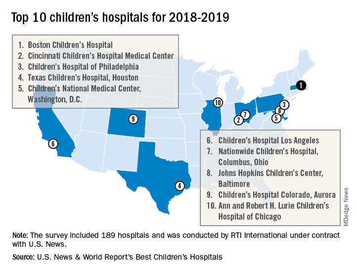
Of the 189 facilities that qualified for inclusion this year, 118 submitted sufficient data to be considered in at least 1 of the 10 specialties and 86 were ranked among the top 50 in at least 1 specialty. In addition, a survey of individuals conducted to establish the hospitals’ reputations – generally worth about 15% of a hospital’s score in each specialty – was completed by 4,165 physicians.
RTI International, a research and consulting firm, conducted the physician survey and produced the methodology and national rankings under contract with U.S. News.
Significant figures: The honesty in being precise
Physicists have strict rules about significant figures. Medical journals lack this professional discipline and it produces distortions that mislead readers.
Whenever you measure and report something in physics, the precision of the measurement is reflected in how the value is written. Writing a result with more digits implies that a higher precision was achieved. If that wasn’t the case, you are falsely claiming skill and accomplishment. You’ve entered the zone of post-truth.
This point was taught by my high school physics teacher, Mr. Gunnar Overgaard, may he rest in peace. Suppose we measured the length of the lab table with the meter stick. We repeated the action three times. We computed an average. Our table was 243.7 cm long. If we wrote 243.73 or 243.73333 we got a lower grade. Meter sticks only have markings of 0.1 cm. So the precision of the reported measurement should properly reflect that limitation.
Researchers in medicine seem to have skipped that lesson in physics lab. In medical journals, the default seems to be to report measurements with two decimal points, such as 16.67%, which is a gross distortion of the precision when I know that that really means 2 out of 12 patients had the finding.
This issue of precision came up recently in two papers published about the number of deaths caused by Hurricane Maria in Puerto Rico. The official death toll was 64. This number became a political hot potato when President Trump cited it as if it was evidence that he and the current local government had managed the emergency response better than George W. Bush did for Katrina.
On May 29, 2018, some researchers at the Harvard School of Public Health, a prestigious institution, published an article in The New England Journal of Medicine, a prestigious journal. You would presume that pair could report properly. The abstract said “This rate yielded a total of 4,645 excess deaths during this period (95% CI, 793 to 8,498).”1 Many newspapers published the number 4,645 in a headline. Most newspapers didn’t include all of the scientific mumbo jumbo about bias and confidence intervals.

However, the number 4,645 did not pass the sniff test at many newspapers, including the Washington Post. Their headline began “Harvard study estimates thousands died”2 and that story went on to clarify that “The Harvard study’s statistical analysis found that deaths related to the hurricane fell within a range of about 800 to more than 8,000.” That is one significant digit. Then the fact checkers went to work on it. They didn’t issue a Pinocchio score, but under a headline of “Did exactly 4,645 people die in Hurricane Maria? Nope”3 the fact checkers concluded that “it’s an egregious example of false precision to cite the ‘4,645’ number without explaining how fuzzy the number really is.”
The situation was compounded 3 days later when another news report had the Puerto Rico Department of Public Health putting the death toll at 1,397. Many assumptions go into determining what an excess death is. If the false precision makes it appear the scientists have a political agenda, it casts shade on whether the assumptions they made are objective and unbiased.
The result on social media was predictable. Outrage was expressed, as always. Lawsuits have been filed. The reputations of all scientists have been impugned. The implication is that, depending on your political polarization, you can choose the number 64, 1,000, 1,400, or 4,645 and any number is just as true as another. Worse, instead of focusing on the severity of the catastrophe and how we might have responded better then and better now and with better planning for the future, the debate has focused on alternative facts and fake scientific news. Thanks, Harvard.

So in the spirit of thinking globally but acting locally, what can I do? I love my editor. I have hinted before about how much easier it is to read, as well as more accurate scientifically, to round the numbers that we report. We've done it a few times recently, but now that the Washington Post has done it on a major news story, should this practice become the norm for journalism? If medical journal editors won't handle precision honestly, other journalists must step up. I'm distressed when I review an article that says 14.6% agreed and 79.2% strongly agreed and I know those percentages with 3 digits really mean 7/48 and 38/48, so they should be rounded to two significant figures. And isn’t it easier to read and comprehend if reporting that three treatment groups had positive findings of 4.25%, 12.08%, and 9.84% when rounded to 4%, 12%, and 10%?
Scientists using this false precision (and peer reviewers who allow it) need to be corrected. They are trying to sell their research as a Louis Vuitton handbag when we all know it is only a cheap knockoff.
Dr. Powell is a pediatric hospitalist and clinical ethics consultant living in St. Louis. Email him at [email protected]
References
1. N Eng J Med. 2018 May 29. doi: 10.1056/NEJMsa1803972
2. “Harvard study estimates thousands died in Puerto Rico because of Hurricane Maria,” by Arelis R. Hernández and Laurie McGinley, The Washington Post, May 29, 2018.
3. “Did exactly 4,645 people die in Hurricane Maria? Nope.” by Glenn Kessler, The Washington Post, June 1, 2018.
Physicists have strict rules about significant figures. Medical journals lack this professional discipline and it produces distortions that mislead readers.
Whenever you measure and report something in physics, the precision of the measurement is reflected in how the value is written. Writing a result with more digits implies that a higher precision was achieved. If that wasn’t the case, you are falsely claiming skill and accomplishment. You’ve entered the zone of post-truth.
This point was taught by my high school physics teacher, Mr. Gunnar Overgaard, may he rest in peace. Suppose we measured the length of the lab table with the meter stick. We repeated the action three times. We computed an average. Our table was 243.7 cm long. If we wrote 243.73 or 243.73333 we got a lower grade. Meter sticks only have markings of 0.1 cm. So the precision of the reported measurement should properly reflect that limitation.
Researchers in medicine seem to have skipped that lesson in physics lab. In medical journals, the default seems to be to report measurements with two decimal points, such as 16.67%, which is a gross distortion of the precision when I know that that really means 2 out of 12 patients had the finding.
This issue of precision came up recently in two papers published about the number of deaths caused by Hurricane Maria in Puerto Rico. The official death toll was 64. This number became a political hot potato when President Trump cited it as if it was evidence that he and the current local government had managed the emergency response better than George W. Bush did for Katrina.
On May 29, 2018, some researchers at the Harvard School of Public Health, a prestigious institution, published an article in The New England Journal of Medicine, a prestigious journal. You would presume that pair could report properly. The abstract said “This rate yielded a total of 4,645 excess deaths during this period (95% CI, 793 to 8,498).”1 Many newspapers published the number 4,645 in a headline. Most newspapers didn’t include all of the scientific mumbo jumbo about bias and confidence intervals.
However, the number 4,645 did not pass the sniff test at many newspapers, including the Washington Post. Their headline began “Harvard study estimates thousands died”2 and that story went on to clarify that “The Harvard study’s statistical analysis found that deaths related to the hurricane fell within a range of about 800 to more than 8,000.” That is one significant digit. Then the fact checkers went to work on it. They didn’t issue a Pinocchio score, but under a headline of “Did exactly 4,645 people die in Hurricane Maria? Nope”3 the fact checkers concluded that “it’s an egregious example of false precision to cite the ‘4,645’ number without explaining how fuzzy the number really is.”
The situation was compounded 3 days later when another news report had the Puerto Rico Department of Public Health putting the death toll at 1,397. Many assumptions go into determining what an excess death is. If the false precision makes it appear the scientists have a political agenda, it casts shade on whether the assumptions they made are objective and unbiased.
The result on social media was predictable. Outrage was expressed, as always. Lawsuits have been filed. The reputations of all scientists have been impugned. The implication is that, depending on your political polarization, you can choose the number 64, 1,000, 1,400, or 4,645 and any number is just as true as another. Worse, instead of focusing on the severity of the catastrophe and how we might have responded better then and better now and with better planning for the future, the debate has focused on alternative facts and fake scientific news. Thanks, Harvard.
So in the spirit of thinking globally but acting locally, what can I do? I love my editor. I have hinted before about how much easier it is to read, as well as more accurate scientifically, to round the numbers that we report. We've done it a few times recently, but now that the Washington Post has done it on a major news story, should this practice become the norm for journalism? If medical journal editors won't handle precision honestly, other journalists must step up. I'm distressed when I review an article that says 14.6% agreed and 79.2% strongly agreed and I know those percentages with 3 digits really mean 7/48 and 38/48, so they should be rounded to two significant figures. And isn’t it easier to read and comprehend if reporting that three treatment groups had positive findings of 4.25%, 12.08%, and 9.84% when rounded to 4%, 12%, and 10%?
Scientists using this false precision (and peer reviewers who allow it) need to be corrected. They are trying to sell their research as a Louis Vuitton handbag when we all know it is only a cheap knockoff.
Dr. Powell is a pediatric hospitalist and clinical ethics consultant living in St. Louis. Email him at [email protected]
References
1. N Eng J Med. 2018 May 29. doi: 10.1056/NEJMsa1803972
2. “Harvard study estimates thousands died in Puerto Rico because of Hurricane Maria,” by Arelis R. Hernández and Laurie McGinley, The Washington Post, May 29, 2018.
3. “Did exactly 4,645 people die in Hurricane Maria? Nope.” by Glenn Kessler, The Washington Post, June 1, 2018.
How to Manage Diabetes While Keeping Costs Down
A cost-effective community program at Pennsylvania State University helped most participants change their behavior and significantly improve their HbA1c and blood pressure, according to a report in Preventing Chronic Disease.
The researchers for the extension program, Dining with Diabetes, collected data on 2,738 adults with type 2 diabetes or prediabetes and adult family members without diabetes. The program consisted of 4 weekly 2-hour classes and a follow-up class conducted 3 months later. The classes included hands-on food preparation, food tastings, and physical activity.
At the follow-up class, participants who completed the program had significant improvements in diabetes-related biomarkers. A greater percentage said they were confident they could keep their diabetes under control, compared with the number at baseline (67% v 58%). At baseline, most participants were adhering to medications; the researchers found no significant change in adherence.
Participants also increased the number of days per week on which they exercised for ≥ 20 minutes (from 2.9 to 3.4 days), and slightly increased the number of days on which they ate a variety of fruits and vegetables.
Nearly half of participants with baseline and follow-up measurements had a drop in HbA1c. At follow-up, 21% had a reduction large enough to lower their diabetes status. The changes translated to a 5.9% decrease in HbA1c for 27% of those who had uncontrolled diabetes at baseline. More than half (59%) had a drop in blood pressure, including 60% of those with uncontrolled diabetes.
The program, which was free to participants, cost Penn State Extension $407 per person. The researchers estimate that extending the program to half of the 1.3 million people with diabetes in Pennsylvania would save the state approximately $195 million at 1 year.
Acute Hepatitis E Superinfection Reactivates Chronic HBV
Many things can reactivate chronic hepatitis B virus (HBV) infection—withdrawal of antiviral therapy, pregnancy, and chemotherapy, to name a few. So when a patient with stable chronic HBV virus presented with significant hepatitis flare, clinicians from Beth Israel Deaconess Medical Center in Boston, Massachusetts, had a long list to check.
They first ruled out drug-associated hepatotoxicity and screened the patient for common causes of acute hepatitis. Beyond the HBV, the patient did not have other significant medical conditions, had not had close contact with anyone ill, and was not pregnant. Tests were negative for cytomegalovirus, Epstein-Barr syndrome, HIV, hepatitis A, C, and D. The patient tested negative for both antihepatitis E virus (HEV) IgM and IgG in a visit about 9 months before.
However, she reported regularly consuming pork, including a recent barbecue meal. Thus, the clinicians focused on HEV serology, which confirmed that she had an acute HEV infection.
Pigs act as a “natural reservoir” for HEV; contaminated meats and direct contact with animals are the most common causes of HEV human infection in industrialized countries. Recent data reveal the prevalence of HEV antibodies in the US is about 6%, illustrating that it is not as uncommon as it was thought to be. Although there was no direct evidence to confirm the source of her infection, it seemed likely due to the pork consumption.
The patient was started on tenofovir but stopped it 4 months later because she felt well. After a subsequent flare, “repeated counseling” persuaded the patient to start on entecavir, with successful viral suppression.
Hepatitis E superinfection on chronic HBV can contribute to significant morbidity and mortality, the clinicians say, particularly in patients with cirrhosis. Concurrent infection with another viral hepatitis should be considered in both immunodeficient and immunocompetent patients with chronic HBV reactivation.
Source:
Aslam A, Susheela A, Iriana S, Chan SS, Lau D. BMJ Case Rep. 2018;2018. pii: bcr-2017-223616.
doi: 10.1136/bcr-2017-223616.
Voxelotor benefits adolescents with SCD

STOCKHOLM—An ongoing phase 2 study suggests voxelotor (GBT440) can benefit adolescents with sickle cell disease (SCD).
In the HOPE-KIDS 1 study, voxelotor produced sustained improvements in hemoglobin levels and a reduction in clinical measures of hemolysis in a cohort of adolescents with SCD, most of whom were also receiving hydroxyurea (HU).
The most common adverse events (AEs) related to voxelotor were nausea, vomiting, headache, and rash.
These results were presented in a poster (abstract PF709) at the 23rd Congress of the European Hematology Association (EHA).
HOPE-KIDS 1 is sponsored by Global Blood Therapeutics, Inc.
In this study, researchers are evaluating voxelotor in SCD patients ages 6 to 17. In part A, researchers evaluated a 600 mg daily dose of voxelotor. In part B, they are testing voxelotor at daily doses of 900 mg and 1500 mg in patients ages 12 to 17.
At EHA, the researchers presented data on 25 patients who received voxelotor at 900 mg/day for 24 weeks in part B. Eighty-eight percent of the patients (n=22) were also taking HU.
The patients’ median age was 14 (range, 12-17), and 56% were male. Ninety-six percent (n=24) had the HbSS genotype.
Forty-eight percent of patients had 1 to 4 vaso-occlusive crises (VOCs) in the past year, 8% had more than 4 VOCs, and 44% had 0 VOCs.
At baseline, the median hemoglobin was 8.9 g/dL, the median fetal hemoglobin was 10.8 g/dL, and the median time-averaged mean of maximum velocity was 110 cm/s.
All 25 patients were dosed with voxelotor, and 22 completed 24 weeks of dosing. One patient withdrew consent, 1 was lost to follow-up, and 1 patient discontinued due to noncompliance.
Of the 22 patients who completed 24 weeks of voxelotor treatment, all but 3 were receiving concurrent HU.
Results
Voxelotor-related AEs occurring in at least 2 patients included nausea (12%, n=3), vomiting (8%, n=2), headache (8%, n=2), and rash (8%, n=2).
There was 1 case of grade 3 urticaria, which resolved and did not recur with continued dosing. There were no discontinuations of voxelotor due to AEs.
Patients experienced increased hemoglobin levels and improved clinical measures of hemolysis at 24 weeks, as evaluated by changes from baseline in hemoglobin, percent of reticulocytes, and percent of unconjugated bilirubin.
In all, 43% of patients (9/21) achieved a hemoglobin response (>1 g/dL) at 24 weeks. The median hemoglobin change from baseline was 0.7 g/dL, the median reduction in reticulocytes was 22.9%, and the median reduction in unconjugated bilirubin was 38.6%.
Sixty-two percent of patients (13/21) had a reduction in daily symptoms at 24 weeks, as assessed by total symptom scores (TSS). There was a 39% median reduction in TSS from baseline.
Fifty-five percent of patients (11/20) had a numerical decrease in transcranial doppler (TCD) flow at 24 weeks. Among hemoglobin responders (>1 g/dL), 88% (7/8) had a numerical decrease in TCD at 24 weeks.
“We continue to be encouraged by the results of the ongoing HOPE-KIDS 1 study, which are consistent with inhibition of HbS polymerization by voxelotor and support its ongoing clinical evaluation as a potential disease-modifying therapy for both adults and adolescents with SCD,” said Ted W. Love, MD, president and chief executive officer of Global Blood Therapeutics.
“Results to date support our ongoing development of voxelotor in a broad range of patients, including in our phase 3 HOPE study, which is also evaluating voxelotor at doses of 900 mg and 1500 mg per day in adolescents and adults. We continue to expect to announce top-line clinical data from part A of the HOPE study by the end of this quarter.”
Ibrutinib sNDA receives priority review

The US Food and Drug Administration (FDA) has accepted for priority review a supplemental new drug application (sNDA) for ibrutinib (Imbruvica®) to be used in combination with rituximab in patients with Waldenström’s macroglobulinemia (WM).
The FDA intends to take action on a priority review application within 6 months of receiving it, rather than the standard 10 months.
The FDA grants priority review to applications for products that may provide significant improvements in the treatment, diagnosis, or prevention of serious conditions.
Ibrutinib is a first-in-class Bruton’s tyrosine kinase inhibitor jointly developed and commercialized by Pharmacyclics LLC, an AbbVie company, and Janssen Biotech, Inc.
Ibrutinib is already FDA-approved to treat chronic lymphocytic leukemia/small lymphocytic lymphoma, previously treated mantle cell lymphoma, previously treated marginal zone lymphoma, previously treated chronic graft-versus-host disease, and WM.
If the new sNDA is approved, the use of ibrutinib in WM will extend beyond its current approved use as a single agent.
Phase 3 trial
The sNDA for ibrutinib and rituximab in WM is supported by data from the phase 3 iNNOVATE study. Results from this trial were presented at the 2018 ASCO Annual Meeting (abstract 8003) and were simultaneously published in NEJM.
iNNOVATE is a placebo-controlled, double-blind, phase 3 study that enrolled 150 patients with relapsed/refractory and treatment-naïve WM.
All patients received rituximab at 375 mg/m2 with weekly infusions at weeks 1 to 4 and 17 to 20. They also received either ibrutinib (420 mg) or placebo once daily continuously until criteria for permanent discontinuation were met.
Overall response rates were significantly higher in the ibrutinib arm than the placebo arm—92% and 47%, respectively (P<0.0001). Complete response rates were 3% and 1%, respectively.
The median time to next treatment was not reached for the ibrutinib arm and was 18 months for the placebo arm (hazard ratio=0.096; P<0.0001). Of the patients randomized to ibrutinib plus rituximab, 75% continued on treatment at last follow-up.
The 30-month progression-free survival rates were 82% in the ibrutinib arm and 28% in the placebo arm. The median progression-free survival was not reached in the ibrutinib arm and was 20.3 months in the placebo arm (hazard ratio=0.20; P<0.0001).
The 30-month overall survival rates were 94% in the ibrutinib arm and 92% in the placebo arm.
Grade 3 or higher treatment-emergent adverse events (AEs) occurred in 60% of patients in the ibrutinib arm and 61% in the placebo arm.
Serious AEs occurred in 43% and 33% of patients, respectively. There were no fatal AEs in the ibrutinib arm and 3 in the rituximab arm.
Grade 3 or higher AEs that occurred more frequently in the ibrutinib arm than the placebo arm included atrial fibrillation (12% vs 1%) and hypertension (13% vs 4%).
AEs that occurred less frequently in the ibrutinib arm than the placebo arm included grade 3 or higher infusion reactions (1% vs 16%) and any-grade IgM flare (8% vs 47%).