User login
Inflammation in schizophrenia tied to poor functioning
Targeting inflammation in people with schizophrenia could lead to improvements in daily functioning, a study by Sophia Kogan, MD, PhD, and her associates at the Icahn School of Medicine at Mount Sinai, New York, has suggested.
“Our findings invite speculation about the underlying link between inflammation and neurocognitive dysfunction in schizophrenia,” Dr. Kogan and her associates wrote in Brain, Behavior, and Immunity. “Our results suggest that these associations also extend to poor daily functioning in this population.”
to analyze neurocognition, functions of daily living, and inflammatory markers in 41 adults aged 18-55 years with schizophrenia, schizoaffective disorder, or psychosis. Among the individuals in the trial, 36% were female, the average age was 37 years, and the average body mass index was 31 kg/m2. In addition, all of the individuals were either taking antipsychotics or were on doses of injectable depot antipsychotics. People with active substance use and mild depressive symptoms were excluded.
Dr. Kogan and her associates found that poorer neurocognition was tied to increased levels of peripheral tumor necrosis factor–alpha (TNF-a) and interleukin-12 (IL-12p70).
“For TNF-a, these findings were driven primarily by significant associations with poorer neurocognitive performance in the domains of speed of processing (P = .02), visual learning (P = .02), and reasoning and problem solving (P = .03),” wrote Dr. Kogan and her associates. “Similarly, the association between neurocognition and IL-12p70 was driven primarily by decreased speed of processing (P less than .01) and visual learning (P = .05).”
The small sample size was cited as a limitation, as was the exclusion of people with substance use and mild depressive symptoms.
The National Institute of Mental Health funded the study. Dr. Kogan reported no conflicts of interest. Coauthor David Kimhy, PhD, is a consultant to NeuroCog Trials relating to another project.
SOURCE: Kogan S et al. Brain Behav Immun. 2018 Sep 12. doi: 10.1016/j.bbi.2018.09.016.
FROM BRAIN, BEHAVIOR, AND IMMUNITY
Simplified SYNTAX holds promise, but still has doubters
SAN DIEGO – A simplified version of the SYNTAX score for coronary artery disease complexity strongly correlated with the unmodified SYNTAX score and could make it easier for cardiologists to employ it in everyday practice. The modified version can be easily memorized and has simplified values that a clinician can calculate and use to determine an appropriate treatment without breaking their work flow to consult a computerized system.
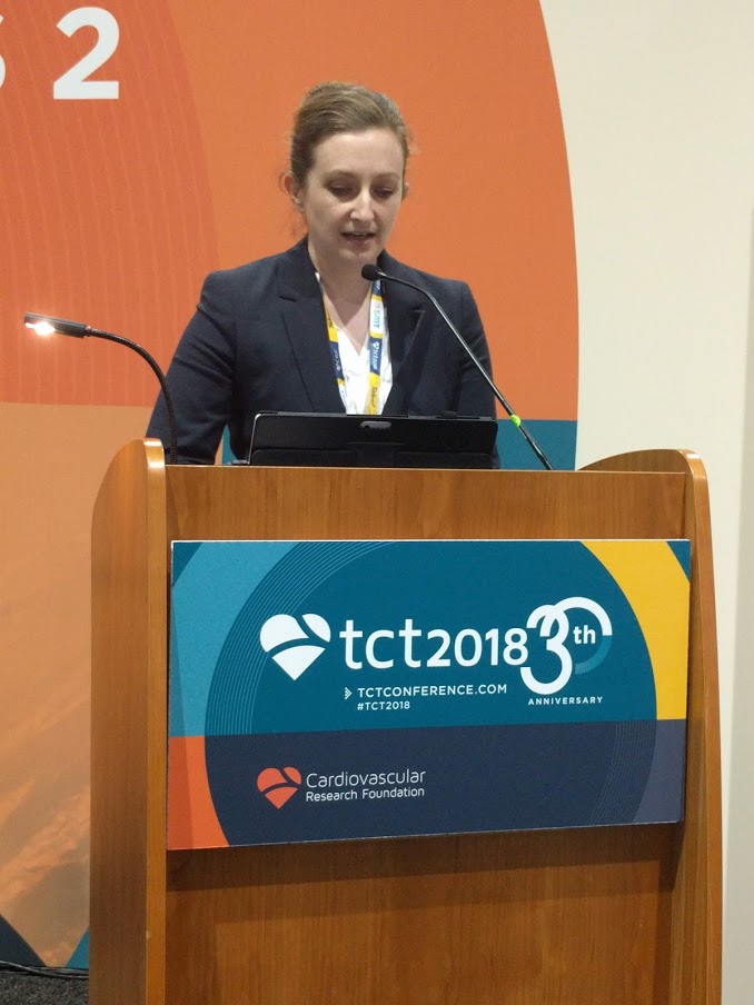
“It’s primarily simplifying the values for the location of the lesions that has allowed it to be more memorizable. That does make it a slightly blunter tool, but only slightly,” said Sonya Burgess, MBCHB, during a presentation at the Transcatheter Cardiovascular Therapeutics annual meeting. Dr. Burgess is an interventional cardiologist at the University of New South Wales, Sydney.
In common practice, SYNTAX scores often go uncalculated because of their complexity, despite guidelines that recommend it. “My guess might be that 1 in every 10 [cardiologists] do it, and that’s not right for our patients,” said Dr. Burgess.
Others at the session agreed that SYNTAX is underutilized, but not all agreed with Dr. Burgess’ solution. Notable among the attendees was Patrick Serruys, MD, of Erasmus University, Rotterdam, the Netherlands, who originally published the SYNTAX system. He doesn’t like the idea of simplifying SYNTAX, based in part on previous experience. “About 10 years ago, Boston Scientific found the score too difficult and they wanted to simplify it, but the score completely lost its prognostic value. I was very emotional about that and I said, ‘no, we have to keep all of the components until we understand [the risk factors] much better,’ ” said Dr. Serruys.
But Dr. Burgess argued that the simplified system, which she and her coinvestigators created, makes concessions to the realities of an interventional cardiology suite. To obtain a standard SYNTAX score, “they have to stop doing something or rebook something, or there’s a lesion there that they just want to treat. [With the simplified score], at least before they go on to treat that lesion, they don’t have to take their gowns and gloves off. You can even have a card hanging on the bar [to refer to].” Dr. Burgess did not provide details of the system.

Dr. Serruys agreed with the source of the problem. “Dr. Burgess is right, it is somewhat demanding,” he allowed, but he also called on clinicians to consider the needs of the patient, even going so far as picturing the patient as a family member. “People complain that doing the SYNTAX is on average 7 minutes, with a standard deviation of 7, so you could work 15 minutes just looking at film. But when you think that the future of your father or brother is depending on the surgeon and the cardiologist looking carefully, I find the argument absolutely obnoxious,” said Dr. Serruys at the meeting sponsored by the Cardiovascular Research Foundation.
Rather than simplification, he stresses teamwork. With at least three people looking at the angiogram, the results are consistent and useful. “You look at the film with the trainees, and that’s how they learn. Never do a SYNTAX score alone,” said Dr. Serruys.
To assess the simplified SYNTAX score, the researchers conducted a retrospective assessment of both the SYNTAX score and the simplified SYNTAX score in 617 patients who had multivessel disease. They performed both assessments in subgroups of patients with 169 patients with ST-segment elevation MI, 78 patients with chronic total occlusion, and 113 patients with left main coronary artery (LMCA) stenosis. They used a 100-patient derivation cohort to determine cutoffs for patients who would not be suitable for percutaneous coronary intervention. They also looked at the accuracy of the simplified version compared with standard SYNTAX in 517 patients from five tertiary centers.
The Spearman’s rho value was 0.93 overall and at least 0.91 for all subgroups (P less than .001 for all). In patients with LMCA stenosis, the simplified version had a sensitivity of 100%, specificity of 85%, negative predictive of 100%, and an area under the curve of 1.0 (P less than .001). For patients with multivessel disease and no LMCA stenosis, the values were 98%, 82%, 99%, and 0.971, respectively (P less than .001).
Dr. Burgess and Dr. Serruys reported no financial conflicts of interest.
SAN DIEGO – A simplified version of the SYNTAX score for coronary artery disease complexity strongly correlated with the unmodified SYNTAX score and could make it easier for cardiologists to employ it in everyday practice. The modified version can be easily memorized and has simplified values that a clinician can calculate and use to determine an appropriate treatment without breaking their work flow to consult a computerized system.
“It’s primarily simplifying the values for the location of the lesions that has allowed it to be more memorizable. That does make it a slightly blunter tool, but only slightly,” said Sonya Burgess, MBCHB, during a presentation at the Transcatheter Cardiovascular Therapeutics annual meeting. Dr. Burgess is an interventional cardiologist at the University of New South Wales, Sydney.
In common practice, SYNTAX scores often go uncalculated because of their complexity, despite guidelines that recommend it. “My guess might be that 1 in every 10 [cardiologists] do it, and that’s not right for our patients,” said Dr. Burgess.
Others at the session agreed that SYNTAX is underutilized, but not all agreed with Dr. Burgess’ solution. Notable among the attendees was Patrick Serruys, MD, of Erasmus University, Rotterdam, the Netherlands, who originally published the SYNTAX system. He doesn’t like the idea of simplifying SYNTAX, based in part on previous experience. “About 10 years ago, Boston Scientific found the score too difficult and they wanted to simplify it, but the score completely lost its prognostic value. I was very emotional about that and I said, ‘no, we have to keep all of the components until we understand [the risk factors] much better,’ ” said Dr. Serruys.
But Dr. Burgess argued that the simplified system, which she and her coinvestigators created, makes concessions to the realities of an interventional cardiology suite. To obtain a standard SYNTAX score, “they have to stop doing something or rebook something, or there’s a lesion there that they just want to treat. [With the simplified score], at least before they go on to treat that lesion, they don’t have to take their gowns and gloves off. You can even have a card hanging on the bar [to refer to].” Dr. Burgess did not provide details of the system.
Dr. Serruys agreed with the source of the problem. “Dr. Burgess is right, it is somewhat demanding,” he allowed, but he also called on clinicians to consider the needs of the patient, even going so far as picturing the patient as a family member. “People complain that doing the SYNTAX is on average 7 minutes, with a standard deviation of 7, so you could work 15 minutes just looking at film. But when you think that the future of your father or brother is depending on the surgeon and the cardiologist looking carefully, I find the argument absolutely obnoxious,” said Dr. Serruys at the meeting sponsored by the Cardiovascular Research Foundation.
Rather than simplification, he stresses teamwork. With at least three people looking at the angiogram, the results are consistent and useful. “You look at the film with the trainees, and that’s how they learn. Never do a SYNTAX score alone,” said Dr. Serruys.
To assess the simplified SYNTAX score, the researchers conducted a retrospective assessment of both the SYNTAX score and the simplified SYNTAX score in 617 patients who had multivessel disease. They performed both assessments in subgroups of patients with 169 patients with ST-segment elevation MI, 78 patients with chronic total occlusion, and 113 patients with left main coronary artery (LMCA) stenosis. They used a 100-patient derivation cohort to determine cutoffs for patients who would not be suitable for percutaneous coronary intervention. They also looked at the accuracy of the simplified version compared with standard SYNTAX in 517 patients from five tertiary centers.
The Spearman’s rho value was 0.93 overall and at least 0.91 for all subgroups (P less than .001 for all). In patients with LMCA stenosis, the simplified version had a sensitivity of 100%, specificity of 85%, negative predictive of 100%, and an area under the curve of 1.0 (P less than .001). For patients with multivessel disease and no LMCA stenosis, the values were 98%, 82%, 99%, and 0.971, respectively (P less than .001).
Dr. Burgess and Dr. Serruys reported no financial conflicts of interest.
REPORTING FROM TCT 2018
Key clinical point: A simplified SYNTAX score could lead to broader implementation of evidence-based decision making for complex coronary artery disease.
Major finding: The simplified score had a Spearman’s rho value of 0.93 overall.
Study details: A retrospective analysis of 617 patients with multivessel disease.
Disclosures: Dr. Burgess and Dr. Serruys reported no financial conflicts of interest.
Recommendations to Improve Asthma Outcomes: Work Group Call to Action
Click here to read the supplement.

What can be done to address the burden of asthma beyond pharmacotherapy? A panel of experts discuss steps for addressing sensitization to allergens that trigger increased asthma burden.
Topics Include:
- Identifying Patients with Allergic Components of Asthma
- Identifying and Addressing Allergen Exposure in Daily Practice
- The Opportunity for Payers and Health Systems for Supporting Trigger Avoidance Education
Click here to read the supplement.
Click here to read the supplement.
What can be done to address the burden of asthma beyond pharmacotherapy? A panel of experts discuss steps for addressing sensitization to allergens that trigger increased asthma burden.
Topics Include:
- Identifying Patients with Allergic Components of Asthma
- Identifying and Addressing Allergen Exposure in Daily Practice
- The Opportunity for Payers and Health Systems for Supporting Trigger Avoidance Education
Click here to read the supplement.
Obesity is linked to some RCC subtypes
The link between obesity and renal cell carcinoma (RCC) appears to be complex and may provide insight into differing etiologies for various histologic subtypes of this cancer, according to a nested case-control study and subsequent meta-analysis.
Investigators led by Catherine L. Callahan, PhD, a postdoctoral fellow with the Division of Cancer Epidemiology and Genetics, National Cancer Institute, Bethesda, Md., first analyzed data from the Kaiser Permanente Northern California health care network. They matched 685 patients with RCC (421 clear cell, 65 papillary, 24 chromophobe, 35 other, and 140 not otherwise specified) with 4,266 unaffected control patients on age, sex, race/ethnicity, duration of network membership, and medical center of diagnosis.
Compared with normal-weight counterparts (body mass index less than 25 kg/m2), obese patients (body mass index of at least 30 kg/m2), had a significantly elevated risk of clear cell RCC (odds ratio, 1.5) and a nonsignificantly elevated risk of chromophobe RCC (2.5), but a similar risk of papillary RCC (1.0), according to results reported in Cancer Epidemiology. Associations weakened when cases were restricted to stage II or higher RCC, suggesting potential bias from incidental diagnoses related to abdominal imaging. Patients who were overweight (body mass index of 25 to 29.9 kg/m2) did not have significantly elevated risks of any subtype of RCC.
The investigators next conducted a meta-analysis, including this new study and three others. Results showed a significant link between obesity and clear cell RCC (summary relative risk, 1.8) and chromophobe RCC (2.2), but not papillary RCC (1.2). Here, however, patients who were overweight also had elevated risks of clear cell RCC (1.3) and chromophobe RCC (1.9).
“Our results provide support for the hypothesis that histologic subtypes of RCC represent distinct etiologic pathways, and that obesity is more strongly associated with risk of clear cell RCC. Additional research to elucidate the underlying biology of specific subtypes of RCC is warranted,” wrote Dr. Callahan and her coinvestigators. “More generally, our findings underscore the importance of accounting for histologic subtype in investigations of RCC etiology.”
The investigators disclosed that they had no conflicts of interest. The research was supported by the Intramural Research Program of the NIH and the National Cancer Institute.
SOURCE: Callahan CL et al. Cancer Epidemiol. 2018 Jul 18, doi: 10.1016/j.canep.2018.07.002.
FROM CANCER EPIDEMIOLOGY
Key clinical point: Obesity is a risk factor for only certain histologic subtypes of RCC.
Major finding: Obese individuals had a significantly elevated risk of clear cell RCC (odds ratio, 1.5) and a nonsignificantly elevated risk of chromophobe RCC (OR, 2.5), but a similar risk of papillary RCC (OR, 1.0).
Study details: A nested case-control study of 685 patients with RCC and 4,266 unaffected matched control patients.
Disclosures: The investigators disclosed that they had no conflicts of interest. The research was supported by the Intramural Research Program of the NIH and the National Cancer Institute.
Source: Callahan CL et al. Cancer Epidemiol. 2018 Jul 18. doi: 10.1016/j.canep.2018.07.002.
Predicting failure of nonoperative management of spinal epidural abscess
Clinical question: Can one predict whether nonoperative management of spinal epidural abscesses will fail?
Background: Even though spinal epidural abscesses have a low incidence and nonspecific presentation, a delay in treatment can lead to significant morbidity. Previously, operative management was the preferred treatment; however, improvements in imaging and timing of diagnosis have led to an increased interest in nonoperative management. Few studies have identified possible predictors of failure for nonoperative management, and no algorithm exists for weighing the different possible predictors with the outcome of nonoperative management failure.
Study design: Retrospective cohort study.
Setting: A Massachusetts hospital system with two tertiary academic medical centers and three regional community hospitals.
Synopsis: The study evaluated 1,053 patients admitted with a spinal epidural abscess during 1993-2016. Of these, 432 patients were managed nonoperatively, and 367 were included in the analysis. Failure of nonoperative management occurred in 99 patients (27%). These patients were compared with 266 patients with successful nonoperative management with more than 60 days of follow-up. Six independent factors were associated with failure of nonoperative management including motor deficit at presentation (odds ratio, 7.85), pathological or compression fractures (OR, 6.12), active malignancy (OR, 3.32), diabetes (OR, 2.92), sensory changes at presentation (3.48), and location of the abscess dorsal to the thecal sac (OR, 0.29). Subsequently, a clinical algorithm was created to predict the likelihood of failure of nonoperative management.
Because of its retrospective design, the study was unable to assess the efficacy of surgery versus nonoperative management.
Bottom line: Specific measures of general health, neurologic status at presentation, and anatomical data of a patient with a spinal epidural abscess have led to the development of a clinical algorithm to determine the risk of failure in nonoperative management of spinal epidural abscesses.
Citation: Shah AA et al. Nonoperative management of spinal epidural abscess: Development of a predictive algorithm for failure. J Bone Joint Surg Am. 2018;100(7):546-55.
Dr. Tsien is a hospitalist in the division of hospital medicine in the department of medicine at Loyola University Chicago, Maywood, Ill.
Research Advances Look Bright for VA
Key leadership in research and oncology addressed attendees of the recent AVAHO annual meeting in Chicago. Carolyn Clancy, MD, deputy undersecretary for health discovery, education and affiliated networks, discussed the important role of cancer in the VA’s research and clinical mission. Neil Spector, MD, director of the National Precision Oncology Program (NPOP) outlined the significant growth in the use of tumor sequencing by NPOP, while Michael Kelley, MD, director of operations for National Oncology discussed the significant strides in opening up access to clinical trials at the VA.
Dr. Clancy addressed AVAHO for the first time and remarked upon the impressive array of research conducted by AVAHO members. “I love the idea of telehealth for genomics,” she remarked about the Genomic Medicine Service Uses Group Telehealth Appointments poster abstract, “it’s brilliant and it’s only the beginning.” The VA’s unique blend of clinical care and research puts it in a unique position to provide cutting edge care to its patients.
Clancy also addressed the larger shift in the VA as it moves from a closed integrated health care system to a high performing network. “We are closer than most systems in this country—public or private—to having a research enterprise that is integral to our mission of providing veterans with great care,” she said. “The magic of bringing [research and clinical] groups together is to enhance the visibility. But frankly it’s also to enhance our capability to take advantage of these assets strategically.” That means providing veterans with “cutting-edge care, and what could be better than that? When we do great things in how we deliver health care that helps your work,” she told attendees.
This shift, as outlined by the VA’s new leadership under Secretary Robert Wilkie and Richard A. Stone, MD, executive in charge of the Veterans Health Administration (VHA), is designed to restore veterans’ trust and confidence in the system, foster an environment of continuous learning to improve quality, and transform the VHA into a “high-reliability organization,” to reduce medical errors. The goal, according to Dr. Clancy, is to develop a culture—like the National Aeronautics and Space Administration or air traffic control systems—where all members of the organization search for and eliminate potential problems. Improving safety, she insisted, has to be a top priority for everyone.
Dr. Clancy also reported that 700,000 veterans enrolled in the VA’s Million Veteran Program (MVP) “We not only have the largest repository of genomic information on people but we also have their clinical data” Later, she told AVAHO members “I am hugely optimistic about the work that is being done in oncology research.” In July, the VA made an arrangement with the National Cancer Institute to allows veterans access to clinical trials. “We need to do more of that, she said, “this is only the beginning of the exciting work we will be doing in cancer research.”
Dr. Spector reported on the progress made by NPOP over the previous year. Currently, NPOP is sequencing solid tumors with a recent biopsy (liquid biopsies are acceptable), but hopes to begin examination of sarcomas and hematologic malignancies. NPOP has grown from about 100 samples analyzed monthly in January 2017 to nearly 350 in June 2018 with a goal of reaching 600 monthly samples across the VA. “You should be sending tumor tissues to be sequenced,” he explained. “It’s free, sequencing tumor tissue is the standard of care, and we need to be sequencing our patients to provide them with an opportunity to get patients onto clinical trials.”
Although the initial analysis can take up to 21 days, the program offers a 72-hour turn-around time for e-consultations. Depending on the quality of trial data, patients may be eligible for treatment even if there is no FDA-approved treatment. According to Spector, the goal of the program is to get patients on the right treatment and avoid costly treatment that will not work for a patient’s cancer type. “We do not want to be giving an expensive drug to someone who will not respond,” he explained.
Multiple efforts are underway to streamline and increase access to oncology care in the VA, according to Dr. Kelley. The development of a national cancer strategy is “long overdue” he admitted, but multiple efforts are underway to including the Fast Track to VA Cancer Care, a single national point of entry for VA cancer care, mechanisms to streamline enrolling patients in non-VA clinical trials, virtual tumor boards, and oncology-specific dashboards. “We have to be transparent and show not only to ourselves, but the whole worlds that we are doing a great job,” he told attendees.
One of the biggest challenges the VA faces will be the roll out of a new electronic health record system. While the new Cerner system has an oncology package, it does not have a cancer registry. According to Kelley, the VA is searching for a commercial system that can interface with Cerner to provide a cancer registry.
Kelley also focused on Annie, a new VA texting platform that allow patients to report on symptoms and get advice The Annie system is automated and allows patients to provide self-care. Already, cancer care providers are experimenting with Annie and Kelley expects the program to develop further.
FDA approves Arikayce for MAC lung diseases
that has been caused by members of the Mycobacterium avium complex and is refractory to other treatments.

In a randomized, controlled trial, patients with refractory M. avium complex infections were assigned to receive either Arikayce plus a multidrug antibacterial regimen or just the antibacterial regimen. By 6 months, sputum cultures for 29% of those treated with the combination had shown no mycobacterial growth for 3 consecutive months, whereas this was only true for the cultures for 9% of patients on the multidrug antibacterial regimen alone.
The Arikayce prescribing information includes a boxed warning regarding the increased risk of respiratory conditions, including hypersensitivity pneumonitis, bronchospasm, exacerbation of underlying lung disease, and hemoptysis, some of which have proven serious enough to lead to hospitalization. Other side effects include dysphonia, cough, musculoskeletal pain, nausea, and fatigue.
According to the press announcement from the FDA, this is the first approval under the Limited Population Pathway for Antibacterial and Antifungal Drugs, which was set up by Congress “to advance development and approval of antibacterial and antifungal drugs to treat serious or life-threatening infections in a limited population of patients with unmet need.” It does so by allowing a more streamlined clinical development program that may involve smaller, shorter, or fewer clinical trials.
More information can be found in the full press announcement.
that has been caused by members of the Mycobacterium avium complex and is refractory to other treatments.
In a randomized, controlled trial, patients with refractory M. avium complex infections were assigned to receive either Arikayce plus a multidrug antibacterial regimen or just the antibacterial regimen. By 6 months, sputum cultures for 29% of those treated with the combination had shown no mycobacterial growth for 3 consecutive months, whereas this was only true for the cultures for 9% of patients on the multidrug antibacterial regimen alone.
The Arikayce prescribing information includes a boxed warning regarding the increased risk of respiratory conditions, including hypersensitivity pneumonitis, bronchospasm, exacerbation of underlying lung disease, and hemoptysis, some of which have proven serious enough to lead to hospitalization. Other side effects include dysphonia, cough, musculoskeletal pain, nausea, and fatigue.
According to the press announcement from the FDA, this is the first approval under the Limited Population Pathway for Antibacterial and Antifungal Drugs, which was set up by Congress “to advance development and approval of antibacterial and antifungal drugs to treat serious or life-threatening infections in a limited population of patients with unmet need.” It does so by allowing a more streamlined clinical development program that may involve smaller, shorter, or fewer clinical trials.
More information can be found in the full press announcement.
Adalimumab safety update finds no new signals
not included in the previous 2009 analysis; their evaluation of data from these 18 trials found no new safety signals, they reported in the British Journal of Dermatology.
Adverse event incidence rates were expressed as events per 100 patient-years of exposure to adalimumab and, among the 3,727 patients who were aged 18 years or older and had moderate to severe plaque psoriasis for at least 6 months, there were 5,430 patient-years of cumulative exposure at the December 2015 cutoff date.
There were 3,798 treatment-related events altogether (70 events/100 patient-years); 269 events (5 events/100 patient-years ) led to discontinuation of treatment. The rates for serious adverse events and serious infections were 8.4 and 1.8 events per 100 patient-years, respectively; the most common types of serious infections were pneumonia and cellulitis.
The rates of the most frequently reported adverse events were comparable with those in the 2009 data set, with the most common being nasopharyngitis, upper respiratory tract infection, and headache. Furthermore, the rates of serious adverse events, serious infections, and malignancies were also stable, even with the increasing adalimumab exposure, and these were mostly consistent with what has been seen in large real-world registries.
The researchers did note that the rates of melanoma and nonmelanoma skin cancer were higher than would be expected in the general population, but they suspected this was at least partly because these psoriasis patients were receiving more frequent skin examinations and more skin cancers were being detected. (Incidence rates for these two cancers were stable during 2009-2015).
The analysis had certain limitations, such as a lack of a long-term comparator group. Also, while some patients continue to receive adalimumab for more than 10 years, the maximum duration of treatment in this analysis was only 5.5 years. Finally, the population in these clinical trials may differ from that seen in general practice settings because of the inclusion/exclusion criteria.
Six authors of the study reported multiple disclosures with pharmaceutical companies, including serving as a consultant, speaker, and/or adviser for, receiving honoraria from, and/or receiving grant/research support from AbbVie, which developed adalimumab and funded/advised this study; two authors are AbbVie employees, one is a former employee.
SOURCE: Leonardi C et al. Br J Dermatol. 2018 Aug 31. doi: 10.1111/bjd.17084.
FROM THE BRITISH JOURNAL OF DERMATOLOGY
Marcia Morris: College drinking
Dr. Morris is associate professor of psychiatry and associate program director for Student Health Psychiatry at the University of Florida.
Dr. Morris is associate professor of psychiatry and associate program director for Student Health Psychiatry at the University of Florida.
Dr. Morris is associate professor of psychiatry and associate program director for Student Health Psychiatry at the University of Florida.

Neurological disease and aging
Also today, trivalent adjuvant influenza vaccine aIIV3 is safe in elderly adults, pill burden affects the ability to reach systolic BP control, and breast cancer risk in type 2 diabetes related to adiposity.
Amazon Alexa
Apple Podcasts
Spotify
Also today, trivalent adjuvant influenza vaccine aIIV3 is safe in elderly adults, pill burden affects the ability to reach systolic BP control, and breast cancer risk in type 2 diabetes related to adiposity.
Amazon Alexa
Apple Podcasts
Spotify
Also today, trivalent adjuvant influenza vaccine aIIV3 is safe in elderly adults, pill burden affects the ability to reach systolic BP control, and breast cancer risk in type 2 diabetes related to adiposity.
Amazon Alexa
Apple Podcasts
Spotify