User login
Abnormal EEG in Patients with Autism May Signal Developmental Problems
Children with autism who also have an abnormal EEG or epilepsy are more likely to experience problems with developmental and adaptive functioning, according to an analysis of 443 patients with autism spectrum disorder (ASD).
- The medical records of children with autism were reviewed by researchers at Cincinnati Children’s Hospital Medical Center.
- The children were divided into 3 categories: those with ASD, no epilepsy, and abnormal EEG results; those with ASD, no epilepsy, and normal EEG; and those with ASD and epilepsy.
- Among 372 patients with ASD without epilepsy, 25.5% had an abnormal EEG; these patients were more likely to have more impaired adaptive functioning when compared to patients with normal EEG readings.
- Children with abnormal EEG readings presented with similar abnormalities to the group with epilepsy.
- Patients with epilepsy had lower scores on all the tests that measure developmental and adaptive functioning, when compared to those with normal EEG readings.
Capal JK, Carosella C, Corbin E, et al. EEG endophenotypes in autism spectrum disorder [published online ahead of print Oct 17, 2018]. Epilepsy Behav.
Secukinumab shows promise in hidradenitis suppurativa
PARIS – David Rosmarin, MD, reported at the annual congress of the European Academy of Dermatology and Venereology.
“It was especially notable that secukinumab was effective in five of the six patients who had previously failed anti-TNF [tumor necrosis factor] therapy,” said Dr. Rosmarin, a dermatologist at Tufts University, Boston.
At present, the sole medication approved for treatment of hidradenitis suppurativa (HS) is the TNF inhibitor adalimumab (Humira). The rationale for investigating secukinumab (Cosentyx), a biologic that blocks the interleukin-17A receptor and is approved for treatment of psoriasis, psoriatic arthritis, and ankylosing spondylitis, lies in the observation that HS lesions exhibit an increased ratio of Th17- to T-regulatory cells, compared with normal skin. Lesional skin also features elevated IL-17 levels. These abnormalities can be reversed by anti-TNF therapy, he explained.
Dr. Rosmarin presented a 28-week, open-label study in which 18 patients with moderate to severe HS received an induction regimen consisting of 300 mg secukinumab given subcutaneously once weekly for 5 weeks and were then randomized to the same dose of the biologic given either every 2 or 4 weeks until week 24.
The primary endpoint was achievement of a Hidradenitis Suppurativa Clinical Response (HiSCR) at 24 weeks. This requires no increase in the number of draining fistulae or abscesses, compared with baseline, plus at least a 50% reduction in total inflammatory nodules. Secondary endpoints were the mean change from baseline in the Sartorius Scale as well as on the Dermatology Life Quality Index (DLQI).
Participants were an average of 33 years and had a disease duration of 12 years; 14 patients were Hurley Stage II, the rest Stage III. Their mean baseline DLQI was 12.
“Patients with hidradenitis suppurativa have a horrible quality of life,” Dr. Rosmarin commented. “When we compare it to patients who have atopic dermatitis, psoriasis, or chronic idiopathic urticaria, oftentimes patients with hidradenitis suppurativa suffer the worst and have the most quality of life impairment.”
Of the 18 patients, 14 – 7 of 9 in each group – achieved HiSCR. This included five of the six patients who were previous nonresponders to adalimumab or another anti-TNF biologic.
“The other thing we noticed was the rapidity of response, which is important to a lot of our patients. It took an average of 7 weeks to achieve HiSCR, and eight patients achieved HiSCR during the induction phase of treatment,” the dermatologist said.
Mean scores on the Sartorius Scale dropped by 28%. Similarly, scores on the DLQI improved by a mean of 3.6 points, or 26%. Nine patients experienced a reduction of 5 points or more on the DLQI. “This happened largely in the first 1-2 months of therapy,” Dr. Rosmarin continued.
Secukinumab was well tolerated. There were no treatment discontinuations because of adverse events. Four patients, all in the biweekly dosing arm, developed Candida infections, all easily cured using topical ketoconazole.
The next step will be to conduct a large, placebo-controlled, randomized trial to firmly establish the efficacy of secukinumab for HS. Also, the optimal dosing of the biologic for induction and long-term maintenance therapy have yet to be determined. Over the long term, it will be important to see whether marked improvement in HS is accompanied by a reduction in the elevated cardiovascular risk associated with this inflammatory disease, he added.
In 2019, a trial will get underway to compare two doses of secukinumab for patients with HS. Based on a search of clinical trials at ClinicalTrials.gov, a wide range of monoclonal antibody therapies are being investigated for the treatment of HS.
The results of this preliminary study of secukinumab emphasize the importance of the Th17 pathway in HS and open the door to alternative strategies targeting this pathway. Dr. Rosmarin noted that he and his coinvestigators have collected a case series of positive responses to guselkumab (Tremfya), which targets the IL-23 p19 subunit, which also lies along the Th17 pathway.
The secukinumab study was sponsored by Novartis. Dr. Rosmarin reported serving as a consultant to or on speakers’ bureaus for that company and more than half a dozen other pharmaceutical companies.
REPORTING FROM THE EADV CONGRESS
Key clinical point: Secukinumab shows considerable promise for treatment of hidradenitis suppurativa.
Major finding: Hidradenitis suppurativa improved markedly in response to secukinumab in 14 of 18 patients.
Study details: This prospective, open-label, 28-week study included 18 patients with hidradenitis suppurativa who were randomized to one of two secukinumab dosing regimens.
Disclosures: The study was sponsored by Novartis. The presenter reported serving as a consultant to or on speakers’ bureaus for that company and more than half a dozen other pharmaceutical companies.
Young adults with hypertension may be at higher CVD risk
a pair of recent studies published in JAMA suggest.

In one study, investigators applied the 2017 American College of Cardiology/American Heart Association blood pressure criteria to nearly 5,000 U.S. young adults followed for approximately 20 years and who had up to a 3.5-fold risk associated with hypertension versus normal blood pressure.
The second study of almost 2.5 million Korean young adults, followed for 10 years, similarly found increased risks of cardiovascular disease later in life for those who had stage 1 or 2 hypertension between the ages of 20 and 39 years.
“These findings from a second country on the opposite side of the globe are consistent with those of the U.S. study, providing further support for the ACC/AHA guideline definitions of hypertension,” Naomi D.L. Fisher, MD, deputy editor, JAMA, and Gregory Curfman, MD, Brigham and Women’s Hospital, Boston, said in an editorial also appearing in JAMA.
Disagreement over the ACC/AHA Guideline for the Prevention, Detection, Evaluation, and Management of High Blood Pressure in Adults threatens to distract from their potential benefits, Dr. Fisher and Dr. Curfman wrote in that editorial.
By redefining stage 1 hypertension as 130/80 mm Hg or higher, down from 140/90 mm Hg or higher, the 2017 ACC/AHA increased the prevalence of hypertension in the United States from 31.9% to 45.6%, they noted.
“Given the magnitude and reach of the global problem of hypertension, it is imperative that dedicated control efforts at the population level intensify,” they said.
U.S. study
The U.S. study, described in JAMA by Yuichiro Yano, MD, PhD, department of community and family medicine, Duke University, Durham, N.C., and his colleagues, was based on analysis of a prospective cohort study, CARDIA (Coronary Artery Risk Development in Young Adults Study), which started in 1985 and enrolled 5,115 black and white adults aged 18-30 years.
They applied the ACC/AHA blood pressure criteria based on each participants’ highest measurement before the age of 40 years, and correlated that with incident cardiovascular disease events that occurred over a median follow-up of 18.8 years.
Patients with normal blood pressure had a cardiovascular disease incidence rate of 1.37/1,000 person-years, compared with 2.74/1,000 person-years for those with elevated blood pressure, 3.15 for stage 1 hypertension, and 8.04 for stage 2 hypertension, investigators found.
That translated into increased risks of cardiovascular disease for those with elevated blood pressure versus those with normal blood pressure. After multivariable adjustment, the hazard ratio for cardiovascular disease was 1.67 (95% confidence interval, 1.01-2.77) for elevated blood pressure, 1.75 (95% CI, 1.22-2.53) for stage 1 hypertension, and 3.49 (95% CI, 2.42-5.05) for stage 2, Dr. Yano and his colleagues reported.
“The ACC/AHA blood pressure classification system may help identify young adults at higher risk for CVD events,” they concluded.
South Korean study
Similar findings were shown in a population-based cohort study, also published in JAMA, that included 2,488,101 adults aged 20-39 years in Korean National Health Insurance Service records.
The investigators looked at mean blood pressure levels from an initial health examination that took place during 2002-2003 and a second examination during 2004-2005.
Follow-up was shorter than the U.S. study, with a median duration of 10 years, reported Joung Sik Son, MD, department of family medicine and biomedical sciences, Seoul (South Korea) National University, and coauthors.
Even so, investigators detected an elevated risk of cardiovascular events for individuals with stage 1 or 2 hypertension versus those with normal blood pressure.
For men with baseline stage 1 hypertension based on the mean values and using the latest ACC/AHA blood pressure criteria, the incidence of cardiovascular disease was 215/100,000 person-years, versus 164 for those with normal blood pressure, with an adjusted hazard ratio of 1.25 (95% CI, 1.21-1.28), the authors said. Likewise, women with stage 1 hypertension had an incidence of 131/100,000 person-years versus 40 for women with normal blood pressure, with a hazard ratio of 1.27 (95% CI, 1.21-1.34).
Men with stage 2 hypertension likewise had a higher cardiovascular disease incidence than did those with normal blood pressure (336 vs. 164 per 100,000 person-years; adjusted HR 1.76), with similar findings seen in women, the report shows.
“Despite the relatively low absolute risk, the difference in absolute risk and the fact that sustained hypertension during longer durations is associated with higher risk of CVD [cardiovascular disease] indicate that early blood pressure management among young adults may lead to significant public health benefits by reducing CVD risk later in life,” Dr. Son and colleagues wrote in a discussion of the results.
Authors of the U.S. study reported disclosures related to Amarin, Amgen, and Novartis outside of the submitted work, as well as grants from the National Heart, Lung, and Blood Institute and National Institutes of Health during the conduct of the study.
Authors of the South Korean study reported no conflict of interest disclosures. That study was supported by the Ministry of Health and Welfare and the Ministry of Education of Korea, along with grants from the National Research Foundation of Korea.
SOURCES: Yano Y et al. JAMA. 2018;302(17):1774-82; Son JS et al. JAMA. 2018;302(17):1783-92.
These two studies suggest that a higher blood pressure level in young adulthood is associated with a greater hazard of premature cardiovascular disease, according to Ramachandran S. Vasan, MD.
However, observing an elevated risk of premature cardiovascular disease does necessarily prove causality, or establish that intervening to lower blood pressure in this age group would lessen that risk, he said in an editorial.
The studies are notable for showing that half to nearly 60% of younger adults had levels of blood pressure considered not normal, he added in the editorial, which appears in JAMA.
It is not clear why so many young adults would manifest higher blood pressure levels in these studies, he said, noting that the umbrella of young adults with hypertension likely includes patients with a variety of subtypes. Those including white-coat hypertension, peripheral amplification with normal central blood pressure, hyperadrenergic state, isolated systolic hypertension, and a smaller subset with secondary hypertension.
“These distinct pathophenotypes may have varying natural histories and their management approaches may be distinctive, suggesting the importance and potential role of subphenotyping of elevated blood pressure in young adults to facilitate treatment decisions,” he wrote in his editorial.
The two studies raise key questions, such as whether there are modifiable social, behavioral, or cultural factors that could prevent elevated blood pressure in younger people, he said.
To date, a substantial body of evidence does suggest that blood pressure levels evolve over the course of life, driven by environmental factors superimposed on genetic risks, and modified by sex and race.
“Overall, these data emphasize that primary prevention of higher blood pressure levels must begin in childhood,” he said.
Ramachandran S. Vasan, MD, is with the section of preventive medicine and epidemiology at Boston University. He reported no conflict of interest disclosures related to his editorial, which was supported by the National Heart, Lung, and Blood Institute’s Framingham Heart Study and a grant from the National Institutes of Health. JAMA. 2018;320(17):1760-3. doi:10.1001/jama.2018.16068.
a pair of recent studies published in JAMA suggest.
In one study, investigators applied the 2017 American College of Cardiology/American Heart Association blood pressure criteria to nearly 5,000 U.S. young adults followed for approximately 20 years and who had up to a 3.5-fold risk associated with hypertension versus normal blood pressure.
The second study of almost 2.5 million Korean young adults, followed for 10 years, similarly found increased risks of cardiovascular disease later in life for those who had stage 1 or 2 hypertension between the ages of 20 and 39 years.
“These findings from a second country on the opposite side of the globe are consistent with those of the U.S. study, providing further support for the ACC/AHA guideline definitions of hypertension,” Naomi D.L. Fisher, MD, deputy editor, JAMA, and Gregory Curfman, MD, Brigham and Women’s Hospital, Boston, said in an editorial also appearing in JAMA.
Disagreement over the ACC/AHA Guideline for the Prevention, Detection, Evaluation, and Management of High Blood Pressure in Adults threatens to distract from their potential benefits, Dr. Fisher and Dr. Curfman wrote in that editorial.
By redefining stage 1 hypertension as 130/80 mm Hg or higher, down from 140/90 mm Hg or higher, the 2017 ACC/AHA increased the prevalence of hypertension in the United States from 31.9% to 45.6%, they noted.
“Given the magnitude and reach of the global problem of hypertension, it is imperative that dedicated control efforts at the population level intensify,” they said.
U.S. study
The U.S. study, described in JAMA by Yuichiro Yano, MD, PhD, department of community and family medicine, Duke University, Durham, N.C., and his colleagues, was based on analysis of a prospective cohort study, CARDIA (Coronary Artery Risk Development in Young Adults Study), which started in 1985 and enrolled 5,115 black and white adults aged 18-30 years.
They applied the ACC/AHA blood pressure criteria based on each participants’ highest measurement before the age of 40 years, and correlated that with incident cardiovascular disease events that occurred over a median follow-up of 18.8 years.
Patients with normal blood pressure had a cardiovascular disease incidence rate of 1.37/1,000 person-years, compared with 2.74/1,000 person-years for those with elevated blood pressure, 3.15 for stage 1 hypertension, and 8.04 for stage 2 hypertension, investigators found.
That translated into increased risks of cardiovascular disease for those with elevated blood pressure versus those with normal blood pressure. After multivariable adjustment, the hazard ratio for cardiovascular disease was 1.67 (95% confidence interval, 1.01-2.77) for elevated blood pressure, 1.75 (95% CI, 1.22-2.53) for stage 1 hypertension, and 3.49 (95% CI, 2.42-5.05) for stage 2, Dr. Yano and his colleagues reported.
“The ACC/AHA blood pressure classification system may help identify young adults at higher risk for CVD events,” they concluded.
South Korean study
Similar findings were shown in a population-based cohort study, also published in JAMA, that included 2,488,101 adults aged 20-39 years in Korean National Health Insurance Service records.
The investigators looked at mean blood pressure levels from an initial health examination that took place during 2002-2003 and a second examination during 2004-2005.
Follow-up was shorter than the U.S. study, with a median duration of 10 years, reported Joung Sik Son, MD, department of family medicine and biomedical sciences, Seoul (South Korea) National University, and coauthors.
Even so, investigators detected an elevated risk of cardiovascular events for individuals with stage 1 or 2 hypertension versus those with normal blood pressure.
For men with baseline stage 1 hypertension based on the mean values and using the latest ACC/AHA blood pressure criteria, the incidence of cardiovascular disease was 215/100,000 person-years, versus 164 for those with normal blood pressure, with an adjusted hazard ratio of 1.25 (95% CI, 1.21-1.28), the authors said. Likewise, women with stage 1 hypertension had an incidence of 131/100,000 person-years versus 40 for women with normal blood pressure, with a hazard ratio of 1.27 (95% CI, 1.21-1.34).
Men with stage 2 hypertension likewise had a higher cardiovascular disease incidence than did those with normal blood pressure (336 vs. 164 per 100,000 person-years; adjusted HR 1.76), with similar findings seen in women, the report shows.
“Despite the relatively low absolute risk, the difference in absolute risk and the fact that sustained hypertension during longer durations is associated with higher risk of CVD [cardiovascular disease] indicate that early blood pressure management among young adults may lead to significant public health benefits by reducing CVD risk later in life,” Dr. Son and colleagues wrote in a discussion of the results.
Authors of the U.S. study reported disclosures related to Amarin, Amgen, and Novartis outside of the submitted work, as well as grants from the National Heart, Lung, and Blood Institute and National Institutes of Health during the conduct of the study.
Authors of the South Korean study reported no conflict of interest disclosures. That study was supported by the Ministry of Health and Welfare and the Ministry of Education of Korea, along with grants from the National Research Foundation of Korea.
SOURCES: Yano Y et al. JAMA. 2018;302(17):1774-82; Son JS et al. JAMA. 2018;302(17):1783-92.
FROM JAMA
Transcatheter repair for tricuspid regurgitation holds up at 1 year
SAN DIEGO – Findings were reported in a session and press conference at the Transcatheter Cardiovascular Therapeutics annual meeting.

Research on the tricuspid valve, “the so-called forgotten valve,” is limited, commented lead investigator Jörg Hausleiter, MD, of the Medizinische Klinik und Poliklinik I at the Klinikum der Universität München and the Munich Heart Alliance. But there is unmet need for transcatheter treatment of high-risk patients having symptomatic tricuspid regurgitation (TR).
“The MitraClip has been used in several sites in off-label and compassionate-use programs to treat these patients,” he noted. “But the data which are available so far are really just looking at the early outcome, like 30 days.”
Dr. Hausleiter and his colleagues undertook a retrospective cohort study of a subgroup of 249 patients undergoing edge-to-edge valve repair for symptomatic TR from the international, multidevice TriValve Registry. All received conventional MitraClips (Abbott Vascular) through off-label or compassionate-use programs.
The procedure was successful, reducing regurgitation to mild or moderate levels in nearly four-fifths of patients by discharge. And procedural success was associated with lower risk of rehospitalization and death.
At 1 year, more than two-thirds of all patients had achieved a New York Heart Association (NYHA) functional class of I or II. In addition, prevalence of peripheral edema had fallen dramatically.
“We were able to demonstrate that the TR reduction is durable and that this also improves the clinical outcome at 1 year,” Dr. Hausleiter concluded.
Uptake and applicability
This procedure will likely be increasingly used in Europe and will find its way into U.S. practice in the not-so-distant future, Dr. Hausleiter predicted. “The MitraClip actually is being used now in a modified version in trials, so that this edge-to-edge therapy is applied for TR. And the TRILUMINATE trial has just finished its enrollment in Europe. I guess that we are going to see EU Mark approval for this therapy also next year. At the same time, a U.S. study is currently being planned and will start very soon with this device, so you are going to see this type of therapy at least being investigated within the next few months.”
The procedure is applicable to a large proportion of patients with TR, including the sizable share having comorbid mitral regurgitation (MR), according to Dr. Hausleiter. In fact, more than half of the study patients had treatment of MR during the same procedure for their tricuspid valve.
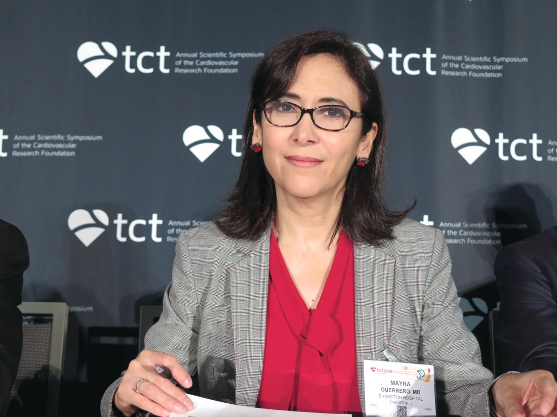
“How were outcomes compared, mitral clip plus tricuspid clip, versus tricuspid clip alone? Could some of this benefit be attributed to the mitral clip procedure?” asked press conference panelist Mayra Guerrero, MD, a senior associate consultant in interventional cardiology in the department of cardiovascular medicine at the Mayo Clinic Hospital, Rochester, Minn.
The two groups had essentially the same mortality rates and improvements in NYHA class, Dr. Hausleiter said. “So we did not observe any difference between those patients who were just treated on the tricuspid side and those patients who had combined treatment. Of course the patients differed a bit in their baseline characteristics, but the outcome was very much the same.”
“With the new data we have, operators and teams may be encouraged to start treating functional MR. I personally think that, if we do that, we should probably evaluate the response and reevaluate the severity of TR after all therapies to the mitral valve have been provided, before we intervene on the tricuspid valve, until we have more data,” Dr. Guerrero further commented. “Do you agree?”
“The tricuspid regurgitation can also improve after treatment of the mitral side. However, when we look at least at the published data, in at least 50% of patients who have severe TR, this TR is not improving,” Dr. Hausleiter replied. In addition, registry data suggest that these patients with severe TR have higher in-hospital, 30-day, and 1-year mortality, compared with patients whose TR is not severe. “Since these are frail patients and you don’t want to bring them too often back to the hospital, if the procedure can be performed very easily, I think there might be a good rationale to combine this.”
Study details
The patients Dr. Hausleiter and his coinvestigators studied had symptomatic TR, predominantly of grade 3+ or 4+, despite receiving adequate medical therapy, as well as a high operative risk, with an average EuroSCORE II of 11.2%. On average, two MitraClips were placed in their tricuspid valve during the procedure.
“We were able to demonstrate that this procedure can be performed very safely. There was a mortality of only 2% in the first 30 days, and one conversion to surgery,” Dr. Hausleiter reported at the meeting, which is sponsored by the Cardiovascular Research Foundation. “We were able to reduce the TR by at least one grade in 89% of patients, and concomitant treatment for mitral regurgitation was also performed in the same procedure in 52%.”
The rate of procedural success, defined as achievement of TR grade of 1+ or 2+ at discharge, was 77%. Independent predictors of procedural failure were noncentral/nonanteroseptal TR jet location and larger TR effective regurgitant orifice area, tenting area, and leaflet gap.
With a mean follow-up of about 10 months, compared with peers in whom the procedure failed, patients in whom it was successful had a higher 1-year rate of freedom from unplanned rehospitalization or death (70.1% vs. 49.7%; P less than .0001).
The 77% rate of procedural success at discharge was largely maintained at 1 year, when it was 72%. There was also a significant improvement in NYHA class distribution in the entire cohort (P less than .001), with 69% of patients attaining class I or II at this time point, compared with virtually none at baseline. Prevalence of peripheral edema fell from 84% to 26% (P less than .001).
Dr. Hausleiter reported that he receives research support and speaker honoraria from Abbott Vascular and Edwards Lifesciences. The registry is sponsored by the University of Zürich.
SAN DIEGO – Findings were reported in a session and press conference at the Transcatheter Cardiovascular Therapeutics annual meeting.
Research on the tricuspid valve, “the so-called forgotten valve,” is limited, commented lead investigator Jörg Hausleiter, MD, of the Medizinische Klinik und Poliklinik I at the Klinikum der Universität München and the Munich Heart Alliance. But there is unmet need for transcatheter treatment of high-risk patients having symptomatic tricuspid regurgitation (TR).
“The MitraClip has been used in several sites in off-label and compassionate-use programs to treat these patients,” he noted. “But the data which are available so far are really just looking at the early outcome, like 30 days.”
Dr. Hausleiter and his colleagues undertook a retrospective cohort study of a subgroup of 249 patients undergoing edge-to-edge valve repair for symptomatic TR from the international, multidevice TriValve Registry. All received conventional MitraClips (Abbott Vascular) through off-label or compassionate-use programs.
The procedure was successful, reducing regurgitation to mild or moderate levels in nearly four-fifths of patients by discharge. And procedural success was associated with lower risk of rehospitalization and death.
At 1 year, more than two-thirds of all patients had achieved a New York Heart Association (NYHA) functional class of I or II. In addition, prevalence of peripheral edema had fallen dramatically.
“We were able to demonstrate that the TR reduction is durable and that this also improves the clinical outcome at 1 year,” Dr. Hausleiter concluded.
Uptake and applicability
This procedure will likely be increasingly used in Europe and will find its way into U.S. practice in the not-so-distant future, Dr. Hausleiter predicted. “The MitraClip actually is being used now in a modified version in trials, so that this edge-to-edge therapy is applied for TR. And the TRILUMINATE trial has just finished its enrollment in Europe. I guess that we are going to see EU Mark approval for this therapy also next year. At the same time, a U.S. study is currently being planned and will start very soon with this device, so you are going to see this type of therapy at least being investigated within the next few months.”
The procedure is applicable to a large proportion of patients with TR, including the sizable share having comorbid mitral regurgitation (MR), according to Dr. Hausleiter. In fact, more than half of the study patients had treatment of MR during the same procedure for their tricuspid valve.
“How were outcomes compared, mitral clip plus tricuspid clip, versus tricuspid clip alone? Could some of this benefit be attributed to the mitral clip procedure?” asked press conference panelist Mayra Guerrero, MD, a senior associate consultant in interventional cardiology in the department of cardiovascular medicine at the Mayo Clinic Hospital, Rochester, Minn.
The two groups had essentially the same mortality rates and improvements in NYHA class, Dr. Hausleiter said. “So we did not observe any difference between those patients who were just treated on the tricuspid side and those patients who had combined treatment. Of course the patients differed a bit in their baseline characteristics, but the outcome was very much the same.”
“With the new data we have, operators and teams may be encouraged to start treating functional MR. I personally think that, if we do that, we should probably evaluate the response and reevaluate the severity of TR after all therapies to the mitral valve have been provided, before we intervene on the tricuspid valve, until we have more data,” Dr. Guerrero further commented. “Do you agree?”
“The tricuspid regurgitation can also improve after treatment of the mitral side. However, when we look at least at the published data, in at least 50% of patients who have severe TR, this TR is not improving,” Dr. Hausleiter replied. In addition, registry data suggest that these patients with severe TR have higher in-hospital, 30-day, and 1-year mortality, compared with patients whose TR is not severe. “Since these are frail patients and you don’t want to bring them too often back to the hospital, if the procedure can be performed very easily, I think there might be a good rationale to combine this.”
Study details
The patients Dr. Hausleiter and his coinvestigators studied had symptomatic TR, predominantly of grade 3+ or 4+, despite receiving adequate medical therapy, as well as a high operative risk, with an average EuroSCORE II of 11.2%. On average, two MitraClips were placed in their tricuspid valve during the procedure.
“We were able to demonstrate that this procedure can be performed very safely. There was a mortality of only 2% in the first 30 days, and one conversion to surgery,” Dr. Hausleiter reported at the meeting, which is sponsored by the Cardiovascular Research Foundation. “We were able to reduce the TR by at least one grade in 89% of patients, and concomitant treatment for mitral regurgitation was also performed in the same procedure in 52%.”
The rate of procedural success, defined as achievement of TR grade of 1+ or 2+ at discharge, was 77%. Independent predictors of procedural failure were noncentral/nonanteroseptal TR jet location and larger TR effective regurgitant orifice area, tenting area, and leaflet gap.
With a mean follow-up of about 10 months, compared with peers in whom the procedure failed, patients in whom it was successful had a higher 1-year rate of freedom from unplanned rehospitalization or death (70.1% vs. 49.7%; P less than .0001).
The 77% rate of procedural success at discharge was largely maintained at 1 year, when it was 72%. There was also a significant improvement in NYHA class distribution in the entire cohort (P less than .001), with 69% of patients attaining class I or II at this time point, compared with virtually none at baseline. Prevalence of peripheral edema fell from 84% to 26% (P less than .001).
Dr. Hausleiter reported that he receives research support and speaker honoraria from Abbott Vascular and Edwards Lifesciences. The registry is sponsored by the University of Zürich.
REPORTING FROM TCT 2018
Key clinical point: Transcatheter edge-to-edge valve repair for tricuspid regurgitation is safe and effective at 1 year.
Major finding: The procedure had a success rate of 77% and netted a 1-year improvement in New York Heart Association class (P less than .001), with 69% of patients achieving class I or II.
Study details: A retrospective cohort study of a subgroup of 249 patients undergoing edge-to-edge valve repair for symptomatic tricuspid regurgitation from the TriValve Registry.
Disclosures: Dr. Hausleiter reported that he receives research support and speaker honoraria from Abbott Vascular and Edwards Lifesciences. The registry is sponsored by the University of Zürich.
Osteoporosis: Breaking Down the Treatment Options
Ms. B, a 72-year-old woman, presents with new-onset low back pain. A comprehensive workup is performed, and a radiograph reveals compression fractures of the L1 and L2 vertebral bodies. The patient recalls no trauma to account for her fractures. Dual-energy x-ray absorptiometry (DXA) is ordered; the results show evidence of osteoporosis. Ms. B asks about initiating longterm treatment.
Osteoporosis is a disease of significant public health concern.1 According to the NIH Osteoporosis and Related Bone Diseases National Resource Center, more than 53 million people in the United States either have osteoporosis or are at high risk for it.2 The total cost of osteoporosis-related fractures is expected to reach $25.3 billion by 2025.3 It is estimated that one in three women (and one in five men) older than 50 will sustain osteoporotic fractures.4 The morbidity and mortality associated with these fractures must be recognized by health care providers in all medical specialties. Appropriate preventive and treatment modalities should be employed when providing care to persons with or at risk for osteoporosis. Advances in medical science have yielded multiple options for the prevention and treatment of osteoporosis.
CASE CONTINUED Ms. B’s medical history includes hypertension and GERD, for which she uses twice-daily dosing of a proton pump inhibitor (PPI). At age 53, she was diagnosed with left breast cancer, which required surgical excision and radiation therapy. She took tamoxifen for a total of five years, and the cancer did not recur. She takes no OTC products, including vitamins. She has no history of systemic inflammatory conditions, kidney stones, or extended treatment with corticosteroids. No history of gastrointestinal surgeries is reported. Ms. B has never smoked cigarettes and has never consumed two or more alcoholic beverages a day. She has no family history of osteoporosis in first-degree relatives. She is otherwise healthy but is physically inactive, with no regular weight-bearing exercise routine. It is also notable that she experienced an uneventful early menopause at age 41 and did not take estrogen replacement therapy.
NONPHARMACOLOGIC OPTIONS
Regular weight-bearing exercise, adequate calcium and vitamin D intake, smoking cessation, avoidance of heavy alcohol use, and education in fall prevention are vital. Recommended calcium intake varies by age, ranging from 1,000 mg/d to 1,200 mg/d in divided doses.2 Vitamin D intake is recommended at 600 IU/d until age 70; 800 IU/d after age 70;and additional units if deficiency is noted.2 Avoidance of medications that contribute to bone loss (eg, corticosteroids) is also encouraged, if possible. Patient education should include balance training and a home safety assessment.
CASE POINT Nonpharmacologic strategies should be encouraged for every patient to promote optimal bone health and to prevent or treat osteoporosis.
PHARMACOLOGIC OPTIONS
Oral bisphosphonates are considered firstline treatment for osteoporosis; currently available options include alendronate, risedronate, and ibandronate. Bisphosphonates work by inhibiting osteoclast function, thereby reducing bone resorption.5
Oral bisphosphonates have been clinically available since the 1990s and have demonstrated their efficacy, safety, and cost-effectiveness.6-8 However, a thoughtful approach should be taken to their use in specific patient populations: those with esophageal disorders, chronic kidney disease, and/or a history of bariatric gastrointestinal procedures. Bisphosphonates of any form should be avoided in a patient with chronic kidney disease with a glomerular filtration rate ≤ 30 mL/min or ≤ 35 mL/min (based on the package insert for the specific product).7 Patients with a recent or upcoming tooth extraction should also avoid using bisphosphonates until they have healed, due to concerns for osteonecrosis of the jaw.
Continue to: Administration of oral bisphosphonates requires...
Administration of oral bisphosphonates requires special attention. Oral bisphosphonates must be taken first thing in the morning with water; for the next 30 to 60 minutes, the patient must stay upright and not have any food, drink, or additional medications by mouth. These specifications may affect patient adherence to treatment.
Intravenous bisphosphonates. Depending on the IV bisphosphonate chosen—ibandronate and zoledronic acid are the currently available options—administration is recommended either every three or 12 months. A common adverse effect of IV bisphosphonates is flulike symptoms, which are generally brief in duration. Hypocalcemia has also been associated with IV administration, more so than with oral bisphosphonate use. Osteonecrosis of the jaw, while rare, must also be considered.
CASE POINT Because of Ms. B’s GERD requiring PPI use, oral bisphosphonates are not the most ideal treatment for her osteoporosis; they could exacerbate her gastrointestinal symptoms. IV bisphosphonates are a potential option for her, as this method of administration would eliminate the gastrointestinal risk associated with oral bisphosphonates.
Selective estrogen receptor modulators (SERMs), which are administered orally, are another option for osteoporosis treatment for vertebral fractures. One medication in this class, raloxifene, selectively acts on estrogen receptors—it works as an agonist in bone estrogen receptors (preventing bone loss) and an estrogen antagonist in other tissue (eg, breast, uterine). SERMs are not considered firstline treatment for osteoporosis because they appear to be less potent than other currently available agents. However, a postmenopausal patient with a high risk for invasive breast cancer without a history of fragility fracture might consider this option, as raloxifene can reduce the risk for invasive breast cancer.9 SERMs have been associated with an increase in thromboembolic events and hot flashes.
Calcitonin nasal spray is used much less commonly now because its effect on bone mineral density is weaker than other currently available options. Calcitonin nasal spray is administered as one spray in one nostril each day. There has been some concern regarding calcitonin use and its association with malignancy.10
Continue to: CASE POINT
CASE POINT Ms. B’s history of compression fractures suggests the need for potent pharmacologic options to treat her osteoporosis. SERMS and calcitonin nasal spray are felt to be less potent and therefore are not the preferred treatment recommendations for her
Parathyroid hormone analogs. The availability of the parathyroid hormone analogs teriparatide and abaloparatide gives patients and health care providers another treatment option for osteoporosis.11 These potent stimulators of bone remodeling help reduce future fracture risk. Teriparatide and abaloparatide are considered anabolic bone agents, rather than antiresorptive medications. These medications are administered subcutaneously daily for no more than two years. Many health care providers use parathyroid hormone analogs for patients with severe osteoporosis (T score, ≤ –3.5 without fragility fracture history or ≤ –2.5 with fragility fracture history).12 The cost of these agents must be considered when recommending them to eligible patients.8
Parathyroid hormone analogs do carry a black box warning because of an increased risk for osteosarcoma observed in rat studies.13,14 These products should therefore be avoided in patients with increased risk for osteosarcoma: those who have Paget disease of the bone or unexplained elevations of alkaline phosphatase; pediatric and young adult patients with open epiphyses; or those who have had external beam or implant radiation therapy involving the skeleton.13,14
CASE POINT Because of Ms. B’s prior history of breast cancer requiring radiation treatment, parathyroid hormone analogs are not recommended.
Denosumab is a human monoclonal antibody, a RANKL inhibitor, that works by preventing the development of osteoclasts. This medication is administered subcutaneously every six months. There are no dosing adjustments recommended for hepatic impairment.11 The denosumab package insert does not specify a dosage adjustment for patients with renal impairment; however, clinical studies have indicated that patients who have a creatine clearance < 30 mL/min or who are on dialysis are more likely to experience hypocalcemia with denosumab use.15 As with other newer osteoporosis treatments, cost considerations should be discussed with patients.
Continue to: One unique consideration...
One unique consideration is that clinical trials have shown an increased fracture risk and the return of bone mineral density to predenosumab treatment levels within 18 months of discontinuing the medication.15 Health care providers should be prepared to recommend alternative treatment options if denosumab is discontinued.
CASE CONCLUDED After a discussion of the risks, benefits, and expectations associated with each of the available treatment options, Ms. B and her health care provider narrow down her options to use of an IV bisphosphonate or denosumab for her osteoporosis. She ultimately chooses denosumab, based on her preference for an injectable medication.
CONCLUSION
The morbidity and mortality associated with osteoporosis can be improved with an appropriate balance of nonpharmacologic and pharmacologic approaches. The varying mechanisms of action, administration methods, and documented efficacy of the available medications provide an opportunity for patient education and informed decision-making when choosing treatment. For additional guidance, the American College of Physicians, the American Association of Clinical Endocrinologists, and American College of Endocrinology have published guidelines that can help in the decision-making process.16,17
1. Cauley JA. Public health impact of osteoporosis. J Gerontol A Biol Sci Med Sci. 2013;68(10):1243-1251.
2. NIH Osteoporosis and Related Bone Diseases National Resource Center. Osteoporosis overview. February 2017. www.bones.nih.gov/health-info/bone/osteoporosis/overview. Accessed October 1, 2018.
3. Dempster DW. Osteoporosis and the burden of osteoporosis-related fractures. Am J Manag Care. 2011;17: S164-S169.
4. International Osteoporosis Foundation. Osteoporosis facts and statistics. www.iofbonehealth.org/facts-and-statistics/calcium-studies-map. Accessed October 1, 2018.
5. Weinstein RS, Roberson PK, Manolagas SC. Giant osteoclast formation and long-term oral bisphosphonate therapy. N Engl J Med. 2009;360(1):53-62.
6. Bilezikian JP. Efficacy of bisphosphonates in reducing fracture risk in postmenopausal osteoporosis. Am J Med. 2009;122(2):S14-S21.
7. Miller PD. Long-term extension trials to prove the efficacy of and safety of bisphosphonates. Clin Invest. 2014;4(1):35-43.
8. Hiligsmann M, Evers SM, Sedrine B, et al. A systematic review of cost-effectiveness analyses of drugs for postmenopausal osteoporosis. Pharmacoeconomics. 2015;33(3):205-224.
9. Raloxifene [package insert]. Indianapolis, IN: Lilly USA, LLC; 2018.
10. Wells G, Chernoff J, Gilligan JP, Krause DS. Does salmon calcitonin cause cancer? A review and meta-analysis. Osteoporos Int. 2016;27(1):13-19.
11. Leder BZ. Parathyroid hormone and parathyroid hormone-related protein analogs in osteoporosis therapy. Curr Osteoporos Rep. 2017;15:110-119.
12. Kendler DL, Marin F, Zerbini CAF, et al. Effects of teriparatide and risedronate on new fractures in post-menopausal women with severe osteoporosis (VERO): a multicentre, double-blind, double-dummy, randomised controlled trial. Lancet. 2018;391:230-240.
13. Teriparatide [package insert]. Indianapolis, IN: Lilly USA, LLC; 2018.
14. Abaloparatide [package insert]. Waltham, MA: Radius Health, Inc; 2017.
15. Denosumab [package insert]. Thousand Oaks, CA: Amgen Inc; 2018.
16. Camacho PM, Petak SM, Binkley N, et al. American Association of Clinical Endocrinologists and American College of Endocrinology clinical practice guidelines for the diagnosis and treatment of postmenopausal osteoporosis—2016. Endocr Pract. 2016;22(4):1-42.
17. Qaseem A, Forciea MA, McLean RM, et al; Clinical Guidelines Committee of the American College of Physicians. Treatment of low bone density or osteoporosis to prevent fractures in men and women: a clinical practice guideline update from the American College of Physicians. Ann Intern Med. 2017;166(11):818-839.
Clinician Reviews in partnership with
Ben Smith is the Director of Didactic Education in the School of Physician Assistant Practice, College of Medicine, at Florida State University in Tallahassee.
Clinician Reviews in partnership with
Ben Smith is the Director of Didactic Education in the School of Physician Assistant Practice, College of Medicine, at Florida State University in Tallahassee.
Clinician Reviews in partnership with
Ben Smith is the Director of Didactic Education in the School of Physician Assistant Practice, College of Medicine, at Florida State University in Tallahassee.
Ms. B, a 72-year-old woman, presents with new-onset low back pain. A comprehensive workup is performed, and a radiograph reveals compression fractures of the L1 and L2 vertebral bodies. The patient recalls no trauma to account for her fractures. Dual-energy x-ray absorptiometry (DXA) is ordered; the results show evidence of osteoporosis. Ms. B asks about initiating longterm treatment.
Osteoporosis is a disease of significant public health concern.1 According to the NIH Osteoporosis and Related Bone Diseases National Resource Center, more than 53 million people in the United States either have osteoporosis or are at high risk for it.2 The total cost of osteoporosis-related fractures is expected to reach $25.3 billion by 2025.3 It is estimated that one in three women (and one in five men) older than 50 will sustain osteoporotic fractures.4 The morbidity and mortality associated with these fractures must be recognized by health care providers in all medical specialties. Appropriate preventive and treatment modalities should be employed when providing care to persons with or at risk for osteoporosis. Advances in medical science have yielded multiple options for the prevention and treatment of osteoporosis.
CASE CONTINUED Ms. B’s medical history includes hypertension and GERD, for which she uses twice-daily dosing of a proton pump inhibitor (PPI). At age 53, she was diagnosed with left breast cancer, which required surgical excision and radiation therapy. She took tamoxifen for a total of five years, and the cancer did not recur. She takes no OTC products, including vitamins. She has no history of systemic inflammatory conditions, kidney stones, or extended treatment with corticosteroids. No history of gastrointestinal surgeries is reported. Ms. B has never smoked cigarettes and has never consumed two or more alcoholic beverages a day. She has no family history of osteoporosis in first-degree relatives. She is otherwise healthy but is physically inactive, with no regular weight-bearing exercise routine. It is also notable that she experienced an uneventful early menopause at age 41 and did not take estrogen replacement therapy.
NONPHARMACOLOGIC OPTIONS
Regular weight-bearing exercise, adequate calcium and vitamin D intake, smoking cessation, avoidance of heavy alcohol use, and education in fall prevention are vital. Recommended calcium intake varies by age, ranging from 1,000 mg/d to 1,200 mg/d in divided doses.2 Vitamin D intake is recommended at 600 IU/d until age 70; 800 IU/d after age 70;and additional units if deficiency is noted.2 Avoidance of medications that contribute to bone loss (eg, corticosteroids) is also encouraged, if possible. Patient education should include balance training and a home safety assessment.
CASE POINT Nonpharmacologic strategies should be encouraged for every patient to promote optimal bone health and to prevent or treat osteoporosis.
PHARMACOLOGIC OPTIONS
Oral bisphosphonates are considered firstline treatment for osteoporosis; currently available options include alendronate, risedronate, and ibandronate. Bisphosphonates work by inhibiting osteoclast function, thereby reducing bone resorption.5
Oral bisphosphonates have been clinically available since the 1990s and have demonstrated their efficacy, safety, and cost-effectiveness.6-8 However, a thoughtful approach should be taken to their use in specific patient populations: those with esophageal disorders, chronic kidney disease, and/or a history of bariatric gastrointestinal procedures. Bisphosphonates of any form should be avoided in a patient with chronic kidney disease with a glomerular filtration rate ≤ 30 mL/min or ≤ 35 mL/min (based on the package insert for the specific product).7 Patients with a recent or upcoming tooth extraction should also avoid using bisphosphonates until they have healed, due to concerns for osteonecrosis of the jaw.
Continue to: Administration of oral bisphosphonates requires...
Administration of oral bisphosphonates requires special attention. Oral bisphosphonates must be taken first thing in the morning with water; for the next 30 to 60 minutes, the patient must stay upright and not have any food, drink, or additional medications by mouth. These specifications may affect patient adherence to treatment.
Intravenous bisphosphonates. Depending on the IV bisphosphonate chosen—ibandronate and zoledronic acid are the currently available options—administration is recommended either every three or 12 months. A common adverse effect of IV bisphosphonates is flulike symptoms, which are generally brief in duration. Hypocalcemia has also been associated with IV administration, more so than with oral bisphosphonate use. Osteonecrosis of the jaw, while rare, must also be considered.
CASE POINT Because of Ms. B’s GERD requiring PPI use, oral bisphosphonates are not the most ideal treatment for her osteoporosis; they could exacerbate her gastrointestinal symptoms. IV bisphosphonates are a potential option for her, as this method of administration would eliminate the gastrointestinal risk associated with oral bisphosphonates.
Selective estrogen receptor modulators (SERMs), which are administered orally, are another option for osteoporosis treatment for vertebral fractures. One medication in this class, raloxifene, selectively acts on estrogen receptors—it works as an agonist in bone estrogen receptors (preventing bone loss) and an estrogen antagonist in other tissue (eg, breast, uterine). SERMs are not considered firstline treatment for osteoporosis because they appear to be less potent than other currently available agents. However, a postmenopausal patient with a high risk for invasive breast cancer without a history of fragility fracture might consider this option, as raloxifene can reduce the risk for invasive breast cancer.9 SERMs have been associated with an increase in thromboembolic events and hot flashes.
Calcitonin nasal spray is used much less commonly now because its effect on bone mineral density is weaker than other currently available options. Calcitonin nasal spray is administered as one spray in one nostril each day. There has been some concern regarding calcitonin use and its association with malignancy.10
Continue to: CASE POINT
CASE POINT Ms. B’s history of compression fractures suggests the need for potent pharmacologic options to treat her osteoporosis. SERMS and calcitonin nasal spray are felt to be less potent and therefore are not the preferred treatment recommendations for her
Parathyroid hormone analogs. The availability of the parathyroid hormone analogs teriparatide and abaloparatide gives patients and health care providers another treatment option for osteoporosis.11 These potent stimulators of bone remodeling help reduce future fracture risk. Teriparatide and abaloparatide are considered anabolic bone agents, rather than antiresorptive medications. These medications are administered subcutaneously daily for no more than two years. Many health care providers use parathyroid hormone analogs for patients with severe osteoporosis (T score, ≤ –3.5 without fragility fracture history or ≤ –2.5 with fragility fracture history).12 The cost of these agents must be considered when recommending them to eligible patients.8
Parathyroid hormone analogs do carry a black box warning because of an increased risk for osteosarcoma observed in rat studies.13,14 These products should therefore be avoided in patients with increased risk for osteosarcoma: those who have Paget disease of the bone or unexplained elevations of alkaline phosphatase; pediatric and young adult patients with open epiphyses; or those who have had external beam or implant radiation therapy involving the skeleton.13,14
CASE POINT Because of Ms. B’s prior history of breast cancer requiring radiation treatment, parathyroid hormone analogs are not recommended.
Denosumab is a human monoclonal antibody, a RANKL inhibitor, that works by preventing the development of osteoclasts. This medication is administered subcutaneously every six months. There are no dosing adjustments recommended for hepatic impairment.11 The denosumab package insert does not specify a dosage adjustment for patients with renal impairment; however, clinical studies have indicated that patients who have a creatine clearance < 30 mL/min or who are on dialysis are more likely to experience hypocalcemia with denosumab use.15 As with other newer osteoporosis treatments, cost considerations should be discussed with patients.
Continue to: One unique consideration...
One unique consideration is that clinical trials have shown an increased fracture risk and the return of bone mineral density to predenosumab treatment levels within 18 months of discontinuing the medication.15 Health care providers should be prepared to recommend alternative treatment options if denosumab is discontinued.
CASE CONCLUDED After a discussion of the risks, benefits, and expectations associated with each of the available treatment options, Ms. B and her health care provider narrow down her options to use of an IV bisphosphonate or denosumab for her osteoporosis. She ultimately chooses denosumab, based on her preference for an injectable medication.
CONCLUSION
The morbidity and mortality associated with osteoporosis can be improved with an appropriate balance of nonpharmacologic and pharmacologic approaches. The varying mechanisms of action, administration methods, and documented efficacy of the available medications provide an opportunity for patient education and informed decision-making when choosing treatment. For additional guidance, the American College of Physicians, the American Association of Clinical Endocrinologists, and American College of Endocrinology have published guidelines that can help in the decision-making process.16,17
1. Cauley JA. Public health impact of osteoporosis. J Gerontol A Biol Sci Med Sci. 2013;68(10):1243-1251.
2. NIH Osteoporosis and Related Bone Diseases National Resource Center. Osteoporosis overview. February 2017. www.bones.nih.gov/health-info/bone/osteoporosis/overview. Accessed October 1, 2018.
3. Dempster DW. Osteoporosis and the burden of osteoporosis-related fractures. Am J Manag Care. 2011;17: S164-S169.
4. International Osteoporosis Foundation. Osteoporosis facts and statistics. www.iofbonehealth.org/facts-and-statistics/calcium-studies-map. Accessed October 1, 2018.
5. Weinstein RS, Roberson PK, Manolagas SC. Giant osteoclast formation and long-term oral bisphosphonate therapy. N Engl J Med. 2009;360(1):53-62.
6. Bilezikian JP. Efficacy of bisphosphonates in reducing fracture risk in postmenopausal osteoporosis. Am J Med. 2009;122(2):S14-S21.
7. Miller PD. Long-term extension trials to prove the efficacy of and safety of bisphosphonates. Clin Invest. 2014;4(1):35-43.
8. Hiligsmann M, Evers SM, Sedrine B, et al. A systematic review of cost-effectiveness analyses of drugs for postmenopausal osteoporosis. Pharmacoeconomics. 2015;33(3):205-224.
9. Raloxifene [package insert]. Indianapolis, IN: Lilly USA, LLC; 2018.
10. Wells G, Chernoff J, Gilligan JP, Krause DS. Does salmon calcitonin cause cancer? A review and meta-analysis. Osteoporos Int. 2016;27(1):13-19.
11. Leder BZ. Parathyroid hormone and parathyroid hormone-related protein analogs in osteoporosis therapy. Curr Osteoporos Rep. 2017;15:110-119.
12. Kendler DL, Marin F, Zerbini CAF, et al. Effects of teriparatide and risedronate on new fractures in post-menopausal women with severe osteoporosis (VERO): a multicentre, double-blind, double-dummy, randomised controlled trial. Lancet. 2018;391:230-240.
13. Teriparatide [package insert]. Indianapolis, IN: Lilly USA, LLC; 2018.
14. Abaloparatide [package insert]. Waltham, MA: Radius Health, Inc; 2017.
15. Denosumab [package insert]. Thousand Oaks, CA: Amgen Inc; 2018.
16. Camacho PM, Petak SM, Binkley N, et al. American Association of Clinical Endocrinologists and American College of Endocrinology clinical practice guidelines for the diagnosis and treatment of postmenopausal osteoporosis—2016. Endocr Pract. 2016;22(4):1-42.
17. Qaseem A, Forciea MA, McLean RM, et al; Clinical Guidelines Committee of the American College of Physicians. Treatment of low bone density or osteoporosis to prevent fractures in men and women: a clinical practice guideline update from the American College of Physicians. Ann Intern Med. 2017;166(11):818-839.

AAP: Pediatricians can mitigate conflict effects on children
Pediatricians have a central role in addressing the impact to children caused by armed conflict, including diagnosing and managing health conditions resulting from exposure to associated violence, according to a new American Academy of Pediatrics policy statement.

The recommendation, published in Pediatrics, emphasizes the need for child health professionals to understand the prevalence of armed conflict and its pervasive effects on children and to respond both domestically and globally to the problem. The AAP defines armed conflict as any organized dispute that involves the use of weapons, violence, or force, such as international wars, civil wars, ethnic conflicts, and violence associated with gangs and drug trafficking. Children effected by armed conflict may include refugees forcibly displaced from their countries, unaccompanied immigrant children, and former child combatants, known as child soldiers.
The policy details the various health effects triggered by armed conflict, including physical, mental, developmental, and behavioral health conditions in children as well as indirect effects through deprivation and toxic stress. Children affected by armed conflict, for example, are at increased risk for PTSD, depression, anxiety, and behavioral and psychosomatic problems, which can continue well into adulthood, according to the policy statement.
To address these effects, pediatricians should receive training on trauma-informed care that enables them to recognize the dynamics of traumatic life experiences among children exposed to armed conflict and provide optimal responses, the policy advises.
and can work to mitigate the effects of trauma, according to the policy. The AAP recommends that child health professionals be prepared to diagnose and offer initial management for conditions associated with exposure to armed conflict as well as partner with community mental and behavioral health providers to establish collaborative care networks, when possible. Other AAP recommendations in the policy statement include that child health professionals:
- Be trained to provide “culturally effective care,” defined as “the delivery of care within the context of appropriate physician knowledge, understanding, and appreciation of all cultural distinctions, leading to optimal health outcomes.”
- Collaborate with local refugee resettlement groups and other public and private sector organizations, such as schools, health systems, and social services, to “facilitate the integration of children and families into their communities and to help families meet unmet needs.”
- Receive special preparation before working with refugees in camps or in conflict settings to become familiar with recognized standards for child protection and to better manage the physical, sexual, and psychological injuries caused by armed conflict.
The policy statement goes on to offer recommendations for systems that serve children exposed to armed conflict, advising that such systems should protect exposed children from abuse and exploitation, and offer access to physical, mental, behavioral, developmental, oral, and rehabilitation health services.
Finally, the AAP stressed its support of policies that advance the health, development, well-being, and rights of children affected by armed conflict and displacement. The AAP states that children should not be separated from their families during displacement and resettlement, emphasizing that “an intact family is the optimal environment for children’s health and well-being” and that “in the event of separation, family reunification should be prioritized.”
In addition, the AAP strongly advocates that children affected by armed conflict have access to educational opportunities, noting that pediatricians can act as a voice toward this end.
“There is strong evidence to suggest that education for boys and girls at all levels reduces most forms of political violence,” the authors wrote. “However, education currently receives less than 2% of all humanitarian funding, and girls are more likely than boys to be excluded from education. Because education is a priority for many children and essential for their well-being, child health providers may advocate for their educational rights and access, especially during humanitarian emergencies.”
SOURCE: Shenoda S et al. Pediatrics. 2018 Nov 5. doi: 10.1542/peds.2018.2586.
The new policy by the American Academy of Pediatrics regarding the effects of armed conflict on children is an important and interesting statement. At first look, someone reading statements like “children should be protected from land mines” might easily conclude that what is contained here is simply common sense guidelines.

On an individual level, the statement also urges individual child health professionals to consider getting out of their comfort zone and become more involved in community organizations, advocacy, and public policy. It also directly states that child health professionals should be prepared to diagnose and treat several mental health conditions that many might not consider in their current scope of practice. This is an important point, especially considering that stigma surrounding mental health treatment in many developing countries is even worse than it is in the United States – which means that primary care plays a crucial role in identification and treatment of these conditions.
David C. Rettew, MD , is an associate professor of psychiatry and pediatrics and director of child and adolescent psychiatry at the University of Vermont’s Center for Children, Youth, and Families in Burlington, Vt. These comments were taken from an interview with Dr. Rettew for this article.
The new policy by the American Academy of Pediatrics regarding the effects of armed conflict on children is an important and interesting statement. At first look, someone reading statements like “children should be protected from land mines” might easily conclude that what is contained here is simply common sense guidelines.
On an individual level, the statement also urges individual child health professionals to consider getting out of their comfort zone and become more involved in community organizations, advocacy, and public policy. It also directly states that child health professionals should be prepared to diagnose and treat several mental health conditions that many might not consider in their current scope of practice. This is an important point, especially considering that stigma surrounding mental health treatment in many developing countries is even worse than it is in the United States – which means that primary care plays a crucial role in identification and treatment of these conditions.
David C. Rettew, MD , is an associate professor of psychiatry and pediatrics and director of child and adolescent psychiatry at the University of Vermont’s Center for Children, Youth, and Families in Burlington, Vt. These comments were taken from an interview with Dr. Rettew for this article.
Pediatricians have a central role in addressing the impact to children caused by armed conflict, including diagnosing and managing health conditions resulting from exposure to associated violence, according to a new American Academy of Pediatrics policy statement.
The recommendation, published in Pediatrics, emphasizes the need for child health professionals to understand the prevalence of armed conflict and its pervasive effects on children and to respond both domestically and globally to the problem. The AAP defines armed conflict as any organized dispute that involves the use of weapons, violence, or force, such as international wars, civil wars, ethnic conflicts, and violence associated with gangs and drug trafficking. Children effected by armed conflict may include refugees forcibly displaced from their countries, unaccompanied immigrant children, and former child combatants, known as child soldiers.
The policy details the various health effects triggered by armed conflict, including physical, mental, developmental, and behavioral health conditions in children as well as indirect effects through deprivation and toxic stress. Children affected by armed conflict, for example, are at increased risk for PTSD, depression, anxiety, and behavioral and psychosomatic problems, which can continue well into adulthood, according to the policy statement.
To address these effects, pediatricians should receive training on trauma-informed care that enables them to recognize the dynamics of traumatic life experiences among children exposed to armed conflict and provide optimal responses, the policy advises.
and can work to mitigate the effects of trauma, according to the policy. The AAP recommends that child health professionals be prepared to diagnose and offer initial management for conditions associated with exposure to armed conflict as well as partner with community mental and behavioral health providers to establish collaborative care networks, when possible. Other AAP recommendations in the policy statement include that child health professionals:
- Be trained to provide “culturally effective care,” defined as “the delivery of care within the context of appropriate physician knowledge, understanding, and appreciation of all cultural distinctions, leading to optimal health outcomes.”
- Collaborate with local refugee resettlement groups and other public and private sector organizations, such as schools, health systems, and social services, to “facilitate the integration of children and families into their communities and to help families meet unmet needs.”
- Receive special preparation before working with refugees in camps or in conflict settings to become familiar with recognized standards for child protection and to better manage the physical, sexual, and psychological injuries caused by armed conflict.
The policy statement goes on to offer recommendations for systems that serve children exposed to armed conflict, advising that such systems should protect exposed children from abuse and exploitation, and offer access to physical, mental, behavioral, developmental, oral, and rehabilitation health services.
Finally, the AAP stressed its support of policies that advance the health, development, well-being, and rights of children affected by armed conflict and displacement. The AAP states that children should not be separated from their families during displacement and resettlement, emphasizing that “an intact family is the optimal environment for children’s health and well-being” and that “in the event of separation, family reunification should be prioritized.”
In addition, the AAP strongly advocates that children affected by armed conflict have access to educational opportunities, noting that pediatricians can act as a voice toward this end.
“There is strong evidence to suggest that education for boys and girls at all levels reduces most forms of political violence,” the authors wrote. “However, education currently receives less than 2% of all humanitarian funding, and girls are more likely than boys to be excluded from education. Because education is a priority for many children and essential for their well-being, child health providers may advocate for their educational rights and access, especially during humanitarian emergencies.”
SOURCE: Shenoda S et al. Pediatrics. 2018 Nov 5. doi: 10.1542/peds.2018.2586.
FROM PEDIATRICS
AMA Insights
While the American Medical Association (AMA) is the oldest and largest national medical association, many physicians, both CHEST members and nonmembers, have limited understanding of the policies, processes, and strategic foci of the AMA. It is our goal to inform our membership about the workings of the AMA and how those interact with the goals of CHEST and our members. We hope to do this by publishing periodic articles in CHEST Physician. One of the authors (DRM) was the CHEST delegate to the AMA for more than 20 years, and the other (NRD) is CHEST’s new AMA delegate.
The American Medical Association (AMA) had its Annual Meeting of the House of Delegates (HOD) from May 9-13. This meeting was attended by over 1,000 physicians who are delegates from all geographic societies, ie, state societies and specialties and subspecialties, as well as the uniformed services.
Policy and resolutions
Process:
Policies originate via resolutions submitted by individuals or societies. These resolutions then go to one of several Reference Committees for open discussion. These committees then report their recommendations back to the HOD, which then discusses and votes on the recommendations. In some instances, the question is referred for further studies by one of several Councils, which reports go to the Board of Trustees or back to the House. The diagram below explains the flow of resolutions to policies.
Chest/Allergy Section Council (which is composed of CHEST, ATS, SCCM, AASM, and several allergy specialty organizations) meets prior to the voting in the House to discuss the pending business. The Specialty and Service Society (SSS) is the largest caucus in the AMA’s House of Delegates and is composed of all the delegates from the specialty societies, as well as the uniformed services.
There are two categories of groups in the SSS: those societies that have seats in the HOD and those seeking admission to the house.
SSS groups in the HOD include:
• 119 national medical specialties
• 2 professional interest medical associations
• 5 military or uniformed service groups (the Public Health Service is “uniformed” but not “military” or “armed”)
The compendium of policies covers the entire range of topics impacting the practice of medicine – ethics, legislation, regulation, public health, individual health, and medical education, among them. The full range of policies can be found in the AMA’s Policy Manual available on the web site, AMA-assn.org
Some of the issues discussed at the HOD are as follows:
• Health care as a “right.” In response to a resolution asking the AMA to support health care as a “right,” the Board of Trustees reaffirmed current policy supporting expanded access to health care for all but stopped short of calling health care a “right.”
• POLST forms. The Board of Trustees will work with state organizations and others to recognize Physician Orders for Life Sustaining Treatment (POLST) forms and allow for reciprocity between states.
• Influenza vaccination. The HOD enacted as AMA policy that no health-care worker should be terminated from employment due solely to their refusal to be vaccinated for influenza.
• e-Cigarettes and tobacco.
A. The AMA was instructed to urge federal officials, including but not limited to the US Food and Drug Administration (FDA), to prohibit the sale of any e-cigarette cartridge that does not include a complete list of ingredients on its packaging, in the order of prevalence (similar to food labeling). We will also urge federal officials, including but not limited to the FDA, to require that accurate nicotine content of e-cigarettes be prominently displayed on the product alongside a warning of the addictive quality of nicotine (new HOD policy).
B. Develop a report on the individual health and public health implications of a low nicotine standard for cigarettes. Such a report should consider and make recommendations on scientific criteria for selection of a nicotine standard that is nonaddictive, regulatory strategies to ensure compliance with an established standard, and how a low-nicotine standard should work with other nicotine products in a well-regulated nicotine market. American Medical Association consider joining other medical organizations in an amicus brief supporting the American Academy of Pediatrics legal action to compel the US Food and Drug Administration to take timely action to establish effective regulation of e-cigarettes, cigars, and other nicotine tobacco products (Directive to Take Action).
• Prior authorization for durable medical equipment. The AMA will advocate that denials of prior authorization for durable medical equipment must be based on true medical necessity not arbitrary time limits or other paperwork issues and will continue to work to improve the prior authorization process for Medicare Managed Care Plans (Directive to Take Action).
• Medical training (including IMG, Medical students, Residents, and Fellows).
A. EHR and Business training during Med Ed and residency
B. Fellowship Start Date. The AMA will survey physicians who have experienced a fellowship start date of August 1 to further evaluate the benefits and drawbacks from this transition (Directive to Take Action).
• Opioid abuse. Surgeon General Jerome Adams addressed the HOD about several topics but focused on opioid crisis.
• Physician Burnout and Organizational efficiency. Physician burnout is a health-care crisis in all specialties, and critical care has a very high rate of physician burnout. The AMA has several tools available that can help with physician burnout both in the ICU and outpatient medicine. (https://www.stepsforward.org)
This is just a small sampling of the activities at the HOD. More information, including reports from the various Councils, are available on the AMA website, http://ama-assn.org.
Testing the Limits of Low-Dose Aspirin
Unless someone has already had a heart attack or other cardiovascular event, daily low-dose aspirin does not prolong healthy independent living, according to the ASPirin in Reducing Events in the Elderly (ASPREE) trial.
The study began in 2010, enrolling 19,114 adults aged ≥ 65 years. The participants were treated with 100 mg/d of aspirin or placebo and followed for an average of 4.7 years. Of participants randomly assigned aspirin, 90.3% were still alive at the end of the treatment without persistent physical disability or dementia, as were 90.5% of those on placebo. Rates of dementia were almost identical in both groups. Major cardiovascular events were similar: 448 in the aspirin group and 474 in the placebo group.
The aspirin group had a slightly higher risk of death (5.9% vs 5.2%). The researchers advise interpreting this cautiously: Most of the deaths were due to cancer. A small increase in new cases of cancer was reported for the aspirin group but may have been due to chance. Heart disease accounted for 19% of deaths and major bleeding for 5%. People taking aspirin were more likely to have significant bleeding (3.8% vs 2.7%).
The researchers emphasize that, study findings notwithstanding, older adults should follow their physician’s advice about daily aspirin use. The new findings do not apply to people with an indication for aspirin, including stroke, heart attack, or other cardiovascular disease.
Combo appears safe, active in rel/ref NHL

The combination of Hu5F9-G4 (5F9) and rituximab was considered safe and produced durable complete responses (CRs) in patients with relapsed or refractory non-Hodgkin lymphoma (NHL) in a phase 1b trial.
Mainly low-grade adverse events (AEs) were observed with rituximab and 5F9, a macrophage-activating immune checkpoint inhibitor blocking CD47.
In addition, the combination produced an objective response rate (ORR) of 50% and a CR rate of 36%.
Most of the responses were ongoing at the time of data cutoff.
“It was very gratifying to see how the treatment was well-tolerated and showed a clinically meaningful response,” said Ranjana Advani, MD, of Stanford University in California.
She and her colleagues reported these results in The New England Journal of Medicine.
The study included 22 patients with relapsed or refractory NHL. Fifteen had diffuse large B-cell lymphoma (DLBCL), and seven had follicular lymphoma (FL).
The patients had received a median of four prior therapies (range, 2-10). Twenty-one patients had disease that was refractory to rituximab (all FL and 14 DLBCL patients).
All patients received 5F9 starting with a priming dose of 1 mg/kg followed by weekly maintenance doses of 10 to 30 mg/kg in three dose-escalation cohorts. The treatment was given until disease progression or lack of clinical benefit.
Patients received rituximab at 375 mg/m2 weekly starting on the second week of the first cycle and then monthly for cycles two through six.
Results
The most common treatment-related AEs were chills (41%), headache (41%), anemia (41%), and infusion-related reactions (36%).
Serious AEs included infections (18%), anemia (4.5%), dyspnea (4.5%), pyrexia (4.5%), lactic acidosis (4.5%), retroperitoneal mass (4.5%), pulmonary embolism (4.5%), and infusion-related reaction (4.5%).
For the entire cohort, the ORR was 50% (n=11), and the CR rate was 36% (n=8).
Among DLBCL patients, the ORR was 40% (n=6), and the CR rate was 33% (n=5). In FL patients, the ORR was 71% (n=5), and the CR rate was 43% (n=3).
The median duration of response was not reached in either disease cohort. The median follow-up was 6.2 months for DLBCL and 8.1 months for FL.
Ten of 11 responders (91%) were still in response at the time of data cutoff.
The researchers said a phase 2 trial of 5F9 plus rituximab in relapsed or refractory B-cell NHL is ongoing.
The phase 1b study was supported by Forty Seven, Inc., and the Leukemia and Lymphoma Society. Dr. Advani reported disclosures related to Forty Seven, Inc., Bristol-Myers Squibb, Pharmacyclics, Seattle Genetics, and Roche/Genentech, among others.
Changes related to AML relapse may be reversible

New research suggests relapse of acute myeloid leukemia (AML) after allogeneic hematopoietic stem cell transplant (HSCT) is related to changes in immune-related gene expression that may be reversible.
Researchers observed downregulation of major histocompatibility complex (MHC) class II genes in samples from patients who relapsed after HSCT.
However, interferon-gamma “rapidly reversed this phenotype” in vitro, according to the researchers.
Matthew J. Christopher, MD, PhD, of Washington University School of Medicine in St. Louis, Missouri, and his colleagues reported these findings in The New England Journal of Medicine.
The researchers set out to determine how genetic and epigenetic changes after HSCT may allow leukemic cells to avoid the graft-vs-leukemia effect and to see whether immune-related genes are affected by HSCT.
The team analyzed paired samples obtained at diagnosis and relapse from 15 AML patients who relapsed after HSCT and 20 AML patients who relapsed after chemotherapy. The team also analyzed additional samples from patients who relapsed after HSCT to validate initial findings.
Methods of analysis included enhanced exome sequencing, RNA sequencing, flow cytometry, and immunohistochemical analysis.
Findings
The researchers first looked for relapse-specific mutations but found no driver mutations associated with relapse after HSCT.
The mutations seen post-HSCT relapse were generally similar to those seen both before treatment and after relapse in patients who had undergone chemotherapy, and the researchers could not identify any patterns of mutations related to relapse.
They then looked for, but did not find, relapse-specific mutations in genes involved in modulation of immune checkpoints, antigen presentation, or cytokine signaling.
The researchers did, however, find evidence of epigenetic changes that were more common in the samples from patients with post-transplant relapses.
RNA sequencing showed that MHC class II genes (HLA-DPA1, HLA-DPB1, HLA-DQB1, and HLA-DRB1) were downregulated three- to 12-fold after transplant.
Flow cytometry and immunohistochemical analysis confirmed that MHC class II expression was decreased at relapse after HSCT in 17 of 34 samples evaluated.
The researchers said there was no association between the downregulation of MHC class II and donor type or use of immunosuppression.
To see whether the downregulation of MHC class II genes was reversible, the researchers treated three post-HSCT relapse samples with interferon-gamma, which is known to upregulate MHC class II on certain cells.
Culturing patient cells with interferon-gamma “rapidly induced MHC class II protein expression on leukemic blasts,” the researchers said. They observed “essentially full restoration of MHC class II protein expression in nearly all AML blasts after 72 hours.”
This study was supported by the National Institutes of Health, Leukemia and Lymphoma Society, and the Barnes-Jewish Hospital Foundation.
Several study authors reported personal fees and/or research support from industry outside the submitted work.