User login
Experts review recent winners and losers in the RA pipeline
MAUI, HAWAII – Filgotinib, the oral Janus kinase (JAK) inhibitor now under Food and Drug Administration review for the treatment of RA, has a better safety profile than some of the approved oral JAK inhibitors, but that’s unlikely to save it from being saddled with a black-box safety warning label, experts agreed at the 2020 Rheumatology Winter Clinical Symposium.

“There’s probably a class label out there,” according to Mark C. Genovese, MD, professor of medicine and clinical chief of the division of immunology and rheumatology at Stanford (Calif.) University.
He cited the example of upadacitinib (Rinvoq), approved last year as the third oral JAK inhibitor for RA. Even though venous thromboembolic (VTE) events weren’t seen with any significantly increased frequency, compared with placebo, in the upadacitinib development program – unlike for the earlier-approved tofacitinib (Xeljanz) and baricitinib (Olumiant) –the FDA nevertheless required that upadacitinib’s product labeling include this black-box warning: “Thrombosis, including deep vein thrombosis, pulmonary embolism, and arterial thrombosis, have occurred in patients treated with Janus kinase inhibitors used to treat inflammatory conditions.”
“I would fully expect that there’ll be a similar label in the filgotinib package insert saying that VTEs have been seen in other members of the class,” he predicted.
His copanelist Roy Fleischmann, MD, noted that filgotinib displayed a clean long-term safety profile in a pooled analysis of the 24-week results in the 2,088 filgotinib-treated participants in all phase 3 clinical trials for RA. For example, the incidence of herpes zoster in that large treated population was a mere 0.1%.
“Herpes zoster is almost nonexistent across the program,” observed Dr. Fleischmann, a rheumatologist at the University of Texas and medical director of the Metroplex Clinical Research Center, both in Dallas.
That’s consistent with what he’s heard from Japanese investigators about their experience. They tell him that in their studies the incidence of herpes zoster with filgotinib is five times less than with other JAK inhibitors.
The long-term pooled phase 3 filgotinib safety data also show less than a 0.1% incidence of adjudicated VTE/pulmonary embolism through 24 weeks. That’s a substantially lower rate than with tofacitinib or baricitinib, he noted.
The two rheumatologists, long-time observers of the FDA regulatory scene, stressed that they have no inside information regarding what the agency will do about filgotinib. It seems beyond doubt that the JAK inhibitor will be approved. But an open question of practical importance to clinicians is whether the agency will approve only the 100-mg dose or the 200-mg dose as well. The panelists agreed that having access to both would be advantageous since the clinical trials data indicate the higher dose is more effective and this greater efficacy doesn’t come at a cost of additional safety issues.
“If the 100 mg is sufficient, that’s great, but the reality is if you want to push to low disease activity or remission, the 200 mg seems to work better, particularly in patients who’ve already failed TNF [tumor necrosis factor] inhibitors or other biologics,” Dr. Genovese said. “If you’re not having additional safety concerns and you can get additional efficacy, I love the idea of having flexibility.”
Dr. Fleischmann is skeptical that the regulators will see things that way.
“There is a real risk that the FDA will do what it’s done before and say: ‘Well, the 200 works and the 100 works, so we’re going to approve the lower dose.’ But there doesn’t appear to be a big safety difference between 100 and 200. So I can see why they would approve the two doses, but I think that’d be unusual,” according to the rheumatologist.
The RA pipeline
The two speakers also highlighted several agents with novel mechanisms of action moving through the RA developmental pipeline, including olokizumab, otilimab, fenebrutinib, and a promising oral selective interleukin-1 receptor–associated kinase 4 inhibitor (IRAK4).
Olokizumab: This humanized monoclonal antibody targets IL-6. It has a different mechanism of action than the two commercially available IL-6 inhibitors approved for RA, tocilizumab (Actemra) and sarilumab (Kevzara), in that olokizumab uniquely targets the IL-6 ligand.
At the 2019 annual meeting of the American College of Rheumatology, Dr. Genovese presented the positive findings of a double-blind, placebo-controlled, randomized, phase 3 clinical trial of olokizumab in 428 RA patients with an inadequate response to methotrexate. The primary outcome, an ACR 20 response at 12 weeks, occurred in 25.9% of patients on placebo, 63.6% with 64 mg of olokizumab given subcutaneously every 2 weeks, and 70.4% with 64 mg every 4 weeks, with all participants on background methotrexate. An ACR 50 response at week 24 occurred in 7.7%, 42.7%, and 48.6%, respectively, with an acceptable side effect profile.
This was the first phase 3 trial to be presented from a large, ongoing phase 3 olokizumab developmental program for a variety of diseases.
“The results certainly support the idea that a 4-week regimen would probably be quite useful with this medication, although we’ll have to see what happens with the remaining phase 3 trials,” Dr. Genovese said.
Dr. Fleischmann posed a question: Do we really need a third IL-6 inhibitor?
That would make for a crowded field, Dr. Genovese agreed, adding that grabbing a reasonable market share for olokizumab may come down to cost, formulary access, and the convenience factor of once-monthly dosing. Whether the biologic’s unique mechanism of action in blocking the IL-6 ligand makes any practical difference in outcomes is unknown.
IRAK4 inhibitor: PF-06650833 is an oral selective reversible inhibitor of IRAK4, a key signaling kinase for IL-1 and toll-like receptors.
“This should be a really good drug for IL-1-mediated diseases,” according to Dr. Fleischmann.
In a phase 2b, double-blind, randomized, placebo-controlled, 12-week study featuring tofacitinib at 5 mg twice daily as an active comparator, the IRAK4 inhibitor exhibited dose-dependent efficacy for the primary endpoint of improvement from baseline in Simple Disease Activity Index score, compared with placebo. The same was true for the secondary endpoint, change over time in 28-joint Disease Activity Score using C-reactive protein (DAS28-CRP). The trial results were also presented at the 2019 ACR annual meeting.
“This is a drug they should probably take forward and see how far it goes in RA,” he said.
Dr. Genovese concurred.
“We’re still trying to figure out how we can put together rational combinations in RA, and this might be something that could be considered as a combination play. In fact, Pfizer has already teed up a study looking at a JAK inhibitor/IRAK4 combination. The question will be whether this is a standalone or has an opportunity to be part of a combination approach,” the rheumatologist said.
Otilimab: This monoclonal antibody is a granulocyte-macrophage colony-stimulating factor inhibitor. In a secondary analysis of the BAROQUE trial, a phase 2b, 50-week study in RA inadequately responsive to methotrexate, otilimab demonstrated an impressive effect in terms of pain reduction. This new analysis, which was first presented at the 2019 ACR annual meeting, showed that, at week 12, a minimum clinically important difference in pain was achieved in 29% of placebo-treated controls, compared with 65%-75% of patients on low to higher doses of otilimab.
“The question is: Is this pain effect unique to this molecule, this pathway, or is it a simple reflection of the treated patient population?” Dr. Genovese commented. “It’s an interesting molecule. It’s being developed in RA, and it might have unique benefits on the pain side.”
A tale of two BTK inhibitors: Bruton tyrosine kinase (BTK) is an intracellular kinase viewed as a promising target in autoimmune disease. Fenebrutinib is an oral, noncovalent, reversible, and highly selective BTK inhibitor that performed well in a phase 2, randomized, double-blind, placebo-controlled clinical trial with adalimumab as an active comparator in 480 patients with an inadequate response to methotrexate in one branch, and in 98 patients with an inadequate response to TNF inhibitor therapy in the other. All subjects were on background methotrexate. (The study results were published April 9 in Arthritis & Rheumatology.)
In the group with a prior inadequate response to TNF inhibitors, the ACR 50 response rate at 12 weeks was 25% in the group on fenebrutinib at 200 mg twice daily, significantly better than the 12% rate in placebo. And there were favorable effects on biomarkers: The reduction in erythrocyte sedimentation rate from baseline was nearly twice as great with fenebrutinib, the drop in CRP was nearly three times greater than with placebo, and there was also a significantly greater decrease over time in DAS28-CRP.
In the methotrexate-inadequate responders, the ACR50 rates were 28% and 35% with the BTK inhibitor at 150 and 200 mg once daily, respectively, compared with 15% in controls.
The safety picture was encouraging, with similarly low adverse event rates across all treatment arms.
In contrast to the fenebrutinib experience, Dr. Genovese was lead investigator in a 250-patient, phase 2 study of another oral BTK inhibitor, poseltinib, which differs from fenebrutinib in that it is an irreversible covalent inhibitor. It was a failed study, with no significant difference between poseltinib and placebo in ACR 20 response at 12 weeks. It’s unclear whether the problem was insufficient dosing or that poseltinib is a failed molecule, perhaps because of its irreversible covalent binding to BTK, he said.
Other notable failures
The spleen tyrosine kinase (Syk) inhibitor known as GS-9876 showed no clinical efficacy in a phase 2, double-blind, randomized trial in 83 RA patients with an inadequate response to methotrexate or a biologic DMARD.
“This is like the fourth Syk inhibitor that’s failed. Syk inhibition is not sick, Syk is dead,” Dr. Fleischmann declared.
Cadherin-11 is an inflammatory cytokine expressed on fibroblasts in RA joints. In a phase 2, double-blind, randomized trial in 109 patients with RA inadequately responsive to TNF inhibitors, RG6125, a humanized monoclonal antibody directed against cadherin-11, failed to outperform placebo.
“It should have worked. It didn’t. So the question is whether this pathway is not an appropriate pathway, or the molecule was not quite the right molecule. I have a feeling it was possibly not the right molecule and the pathway may be viable,” according to Dr. Fleischmann.
He reported receiving clinical trial research grants from and serving as a consultant to more than a dozen pharmaceutical companies. Dr. Genovese also reported financial relationships with more than a dozen pharmaceutical companies.
MAUI, HAWAII – Filgotinib, the oral Janus kinase (JAK) inhibitor now under Food and Drug Administration review for the treatment of RA, has a better safety profile than some of the approved oral JAK inhibitors, but that’s unlikely to save it from being saddled with a black-box safety warning label, experts agreed at the 2020 Rheumatology Winter Clinical Symposium.
“There’s probably a class label out there,” according to Mark C. Genovese, MD, professor of medicine and clinical chief of the division of immunology and rheumatology at Stanford (Calif.) University.
He cited the example of upadacitinib (Rinvoq), approved last year as the third oral JAK inhibitor for RA. Even though venous thromboembolic (VTE) events weren’t seen with any significantly increased frequency, compared with placebo, in the upadacitinib development program – unlike for the earlier-approved tofacitinib (Xeljanz) and baricitinib (Olumiant) –the FDA nevertheless required that upadacitinib’s product labeling include this black-box warning: “Thrombosis, including deep vein thrombosis, pulmonary embolism, and arterial thrombosis, have occurred in patients treated with Janus kinase inhibitors used to treat inflammatory conditions.”
“I would fully expect that there’ll be a similar label in the filgotinib package insert saying that VTEs have been seen in other members of the class,” he predicted.
His copanelist Roy Fleischmann, MD, noted that filgotinib displayed a clean long-term safety profile in a pooled analysis of the 24-week results in the 2,088 filgotinib-treated participants in all phase 3 clinical trials for RA. For example, the incidence of herpes zoster in that large treated population was a mere 0.1%.
“Herpes zoster is almost nonexistent across the program,” observed Dr. Fleischmann, a rheumatologist at the University of Texas and medical director of the Metroplex Clinical Research Center, both in Dallas.
That’s consistent with what he’s heard from Japanese investigators about their experience. They tell him that in their studies the incidence of herpes zoster with filgotinib is five times less than with other JAK inhibitors.
The long-term pooled phase 3 filgotinib safety data also show less than a 0.1% incidence of adjudicated VTE/pulmonary embolism through 24 weeks. That’s a substantially lower rate than with tofacitinib or baricitinib, he noted.
The two rheumatologists, long-time observers of the FDA regulatory scene, stressed that they have no inside information regarding what the agency will do about filgotinib. It seems beyond doubt that the JAK inhibitor will be approved. But an open question of practical importance to clinicians is whether the agency will approve only the 100-mg dose or the 200-mg dose as well. The panelists agreed that having access to both would be advantageous since the clinical trials data indicate the higher dose is more effective and this greater efficacy doesn’t come at a cost of additional safety issues.
“If the 100 mg is sufficient, that’s great, but the reality is if you want to push to low disease activity or remission, the 200 mg seems to work better, particularly in patients who’ve already failed TNF [tumor necrosis factor] inhibitors or other biologics,” Dr. Genovese said. “If you’re not having additional safety concerns and you can get additional efficacy, I love the idea of having flexibility.”
Dr. Fleischmann is skeptical that the regulators will see things that way.
“There is a real risk that the FDA will do what it’s done before and say: ‘Well, the 200 works and the 100 works, so we’re going to approve the lower dose.’ But there doesn’t appear to be a big safety difference between 100 and 200. So I can see why they would approve the two doses, but I think that’d be unusual,” according to the rheumatologist.
The RA pipeline
The two speakers also highlighted several agents with novel mechanisms of action moving through the RA developmental pipeline, including olokizumab, otilimab, fenebrutinib, and a promising oral selective interleukin-1 receptor–associated kinase 4 inhibitor (IRAK4).
Olokizumab: This humanized monoclonal antibody targets IL-6. It has a different mechanism of action than the two commercially available IL-6 inhibitors approved for RA, tocilizumab (Actemra) and sarilumab (Kevzara), in that olokizumab uniquely targets the IL-6 ligand.
At the 2019 annual meeting of the American College of Rheumatology, Dr. Genovese presented the positive findings of a double-blind, placebo-controlled, randomized, phase 3 clinical trial of olokizumab in 428 RA patients with an inadequate response to methotrexate. The primary outcome, an ACR 20 response at 12 weeks, occurred in 25.9% of patients on placebo, 63.6% with 64 mg of olokizumab given subcutaneously every 2 weeks, and 70.4% with 64 mg every 4 weeks, with all participants on background methotrexate. An ACR 50 response at week 24 occurred in 7.7%, 42.7%, and 48.6%, respectively, with an acceptable side effect profile.
This was the first phase 3 trial to be presented from a large, ongoing phase 3 olokizumab developmental program for a variety of diseases.
“The results certainly support the idea that a 4-week regimen would probably be quite useful with this medication, although we’ll have to see what happens with the remaining phase 3 trials,” Dr. Genovese said.
Dr. Fleischmann posed a question: Do we really need a third IL-6 inhibitor?
That would make for a crowded field, Dr. Genovese agreed, adding that grabbing a reasonable market share for olokizumab may come down to cost, formulary access, and the convenience factor of once-monthly dosing. Whether the biologic’s unique mechanism of action in blocking the IL-6 ligand makes any practical difference in outcomes is unknown.
IRAK4 inhibitor: PF-06650833 is an oral selective reversible inhibitor of IRAK4, a key signaling kinase for IL-1 and toll-like receptors.
“This should be a really good drug for IL-1-mediated diseases,” according to Dr. Fleischmann.
In a phase 2b, double-blind, randomized, placebo-controlled, 12-week study featuring tofacitinib at 5 mg twice daily as an active comparator, the IRAK4 inhibitor exhibited dose-dependent efficacy for the primary endpoint of improvement from baseline in Simple Disease Activity Index score, compared with placebo. The same was true for the secondary endpoint, change over time in 28-joint Disease Activity Score using C-reactive protein (DAS28-CRP). The trial results were also presented at the 2019 ACR annual meeting.
“This is a drug they should probably take forward and see how far it goes in RA,” he said.
Dr. Genovese concurred.
“We’re still trying to figure out how we can put together rational combinations in RA, and this might be something that could be considered as a combination play. In fact, Pfizer has already teed up a study looking at a JAK inhibitor/IRAK4 combination. The question will be whether this is a standalone or has an opportunity to be part of a combination approach,” the rheumatologist said.
Otilimab: This monoclonal antibody is a granulocyte-macrophage colony-stimulating factor inhibitor. In a secondary analysis of the BAROQUE trial, a phase 2b, 50-week study in RA inadequately responsive to methotrexate, otilimab demonstrated an impressive effect in terms of pain reduction. This new analysis, which was first presented at the 2019 ACR annual meeting, showed that, at week 12, a minimum clinically important difference in pain was achieved in 29% of placebo-treated controls, compared with 65%-75% of patients on low to higher doses of otilimab.
“The question is: Is this pain effect unique to this molecule, this pathway, or is it a simple reflection of the treated patient population?” Dr. Genovese commented. “It’s an interesting molecule. It’s being developed in RA, and it might have unique benefits on the pain side.”
A tale of two BTK inhibitors: Bruton tyrosine kinase (BTK) is an intracellular kinase viewed as a promising target in autoimmune disease. Fenebrutinib is an oral, noncovalent, reversible, and highly selective BTK inhibitor that performed well in a phase 2, randomized, double-blind, placebo-controlled clinical trial with adalimumab as an active comparator in 480 patients with an inadequate response to methotrexate in one branch, and in 98 patients with an inadequate response to TNF inhibitor therapy in the other. All subjects were on background methotrexate. (The study results were published April 9 in Arthritis & Rheumatology.)
In the group with a prior inadequate response to TNF inhibitors, the ACR 50 response rate at 12 weeks was 25% in the group on fenebrutinib at 200 mg twice daily, significantly better than the 12% rate in placebo. And there were favorable effects on biomarkers: The reduction in erythrocyte sedimentation rate from baseline was nearly twice as great with fenebrutinib, the drop in CRP was nearly three times greater than with placebo, and there was also a significantly greater decrease over time in DAS28-CRP.
In the methotrexate-inadequate responders, the ACR50 rates were 28% and 35% with the BTK inhibitor at 150 and 200 mg once daily, respectively, compared with 15% in controls.
The safety picture was encouraging, with similarly low adverse event rates across all treatment arms.
In contrast to the fenebrutinib experience, Dr. Genovese was lead investigator in a 250-patient, phase 2 study of another oral BTK inhibitor, poseltinib, which differs from fenebrutinib in that it is an irreversible covalent inhibitor. It was a failed study, with no significant difference between poseltinib and placebo in ACR 20 response at 12 weeks. It’s unclear whether the problem was insufficient dosing or that poseltinib is a failed molecule, perhaps because of its irreversible covalent binding to BTK, he said.
Other notable failures
The spleen tyrosine kinase (Syk) inhibitor known as GS-9876 showed no clinical efficacy in a phase 2, double-blind, randomized trial in 83 RA patients with an inadequate response to methotrexate or a biologic DMARD.
“This is like the fourth Syk inhibitor that’s failed. Syk inhibition is not sick, Syk is dead,” Dr. Fleischmann declared.
Cadherin-11 is an inflammatory cytokine expressed on fibroblasts in RA joints. In a phase 2, double-blind, randomized trial in 109 patients with RA inadequately responsive to TNF inhibitors, RG6125, a humanized monoclonal antibody directed against cadherin-11, failed to outperform placebo.
“It should have worked. It didn’t. So the question is whether this pathway is not an appropriate pathway, or the molecule was not quite the right molecule. I have a feeling it was possibly not the right molecule and the pathway may be viable,” according to Dr. Fleischmann.
He reported receiving clinical trial research grants from and serving as a consultant to more than a dozen pharmaceutical companies. Dr. Genovese also reported financial relationships with more than a dozen pharmaceutical companies.
REPORTING FROM RWCS 2020
AMA asks HHS for ‘immediate’ aid to ease clinicians’ COVID-19 ‘financial peril’
The American Medical Association (AMA) along with scores of specialty and state medical societies are asking the Trump administration to help the nation’s clinicians out with an immediate cash infusion that they say they need to sustain their practices, many of which have been crippled by the COVID-19 crisis.
In an April 7 letter to Secretary of US Department of Health and Human Services (HHS) Alex Azar, the AMA, backed by 137 medical groups, made the case for “immediate financial assistance” from the government for all US physicians and nurse practitioners and physician assistants enrolled in Medicare or Medicaid. These payments would be equal to roughly 1 month’s worth of prepandemic revenue from all payers.
Under the methodology laid out in the letter, HHS would use an individual clinician’s average monthly Medicare payment from October to December 2019 to determine their precrisis monthly revenue.
Because Medicare business generates an average of 35% of practice revenue in most specialties, the letter suggests that HHS triple the monthly Medicare payment to calculate the amount of emergency funding it should provide to each clinician.
The letter acknowledges that this approach wouldn’t work for certain specialties, such as psychiatry, allergy/immunology, obstetrics/gynecology, and pediatrics, which derive far less revenue from Medicare than other specialties do. These physicians’ payouts “should be adjusted upward accordingly,” the letter states.
“Physicians are continuing to put their patients’ needs first to combat this unprecedented public health emergency,” the AMA writes. “We urge you to support them against financial peril while they put their lives and businesses at risk.”
Other Emergency Funding Programs
These disbursements would be separate from the $30 billion in direct provider payments announced on April 7 by Seema Verma, the administrator of the Centers for Medicare and Medicaid Services (CMS). Because these payments are based on Medicare volume, the vast majority of this money is expected to go to hospitals.
The government is also providing financial support to hospitals, physicians, and other clinicians affected by the pandemic through CMS’s accelerated/advance payment program, as reported by Medscape Medical News. Physician practices can apply to receive upfront payments equal to 3 months’ worth of their historical Medicare payments, but they must pay back these loans, starting at 120 days after receiving them.
In addition, providers with less than 500 employees can apply for Small Business Administration (SBA) loans that were authorized by the CURES Act. If they use at least 75% of this money to cover payroll costs, the loans will be forgiven.
Medical leaders defended their request for direct physician relief in excess of what these three government programs are offering.
“From the very beginning, the AMA has been advocating for [financial] support for physician practices,” AMA President Patrice Harris, MD, told Medscape Medical News. “It’s not an either/or, it’s not a choice between hospitals or physician practices, it’s both.”
She made it clear that this applied not only to the direct payments that the CURES Act allocated to healthcare providers, but also to the SBA loans.
“We’ve been pleased to see support through the Small Business Administration, and we know that many practices have applied for loans,” Harris said. “We’ll review this, because physician practices have to be included.”
Thus far, she added, “I haven’t heard of anyone [in a medical practice] who has actually received a loan. We’ll be monitoring that, because that will be key.”
Likewise, Robert Doherty, senior vice president of governmental affairs and public policy for the American College of Physicians (ACP), said he hadn’t heard of any practices receiving SBA loans, although many have applied.
What he has heard is that “people couldn’t even get through the SBA process and the website was freezing up. They also have to find a lender, submit documentation and get approved by the lender. And they’re competing with all the other small businesses” for a finite amount of money.
Doherty said it was unclear how many practices have received advance payments from CMS so far. CMS said it disbursed $34 billion in these payments in the first week of the program. These went to over 17,000 of the more than 25,000 applicants, CMS noted.
The ACP – which joined the AMA in its request to HHS – supports the advanced-payment program, Doherty added, but “a loan is a loan. You have to repay it. It brings in cash now, but it means you don’t have cash a few months from now. That’s different from what we’re recommending, which is an infusion of cash to practices that wouldn’t have to be repaid.”
Another advantage of the AMA-led proposal, he said, is its simplicity. It’s based on data that CMS already has, and it doesn’t require physicians to fill out forms or provide documents.
In contrast, he said, “We don’t think HHS would have the ability to process applications from thousands and thousands of physicians [for direct payments]. To create a situation where they’d have to review applications from physicians for funding out of that [CARES Act] emergency fund is probably almost impossible for HHS to administer effectively.”
Most Practices Need Help
While the medical societies’ letter makes a strong pitch for supporting physicians who are combating COVID-19, Harris and Doherty noted that physicians in all kinds of practice situations desperately need this help.
“We’ve heard from many physician practices that they have trouble making payroll,” Doherty said. “Many of them are not seeking any money out of the practices for themselves right now. They’re just trying to keep their staff employed. And some will shut their doors, unless there’s a significant and immediate infusion of money to them. From a healthcare capacity viewpoint, it’s not going to be to anyone’s benefit to see a substantial number of practices laying off staff or closing up entirely because they don’t have the money coming in to keep the doors open,” he said.
Harris agreed. “We’re hearing from practices large and small all over the country, including solo practices. Even the larger practices are losing revenue,” she pointed out. “They appropriately shut down their offices or reduced their hours. They didn’t want to contribute to the further spread of COVID-19.”
Rural practices and those launched by young physicians are facing especially difficult challenges, Harris added, and some may not make it.
It’s also important for policy makers to look ahead to what lies after the pandemic, she said. “We will come out of this, but when we come out of it there will be a lot of pent-up or unmet need where folks delayed necessary visits. Physicians and practices will have to be ready to go. If practices have to furlough some staff, it’s going to take time to ramp that up. So we’re glad to see support of physician practices so the infrastructure is strong when we start again.”
What happens if HHS turns down the medical societies’ request? “We’re hopeful that the [HHS] secretary will agree to what we’re asking,” Doherty said. While it’s always possible to ask Congress to intervene in the next stimulus bill, he said, that wouldn’t happen fast enough to get the money to physicians when they really need it.
This article first appeared on Medscape.com.
SARS-CoV-2 may confound seasons, persist in warmer months, report shows
Although conflicting, the available data indicate that SARS-CoV-2 could continue to spread in warmer spring and summer months in the US, according to a new report from the National Academies of Science, Engineering, and Medicine (NAS).
Current data suggest that the novel coronavirus may be transmitted less efficiently in higher temperatures and humidity, but the studies are not conclusive because of poor data quality, confounding factors, and the relatively short existence of the pandemic, which makes it difficult to determine its true course, writes David A. Relman, MD, a member of the NAS’ Standing Committee on Emerging Infectious Diseases and 21st Century Health Threats, in a rapid expert consultation letter to the White House Office of Science and Technology Policy on April 7.
A number of factors could influence whether SARS-CoV-2 follows the same seasonal pattern as the influenza virus and other seasonal coronaviruses, which wane during warmer months, writes Relman, a professor of microbiology and immunology at Stanford University in California.
But he pointed out that previous coronavirus strains that have caused serious illness – SARS-CoV and MERS-CoV – “have not demonstrated any evidence of seasonality following their emergence.”
Relman cites an example from the current outbreak: “Given that countries currently in ‘summer’ climates, such as Australia and Iran, are experiencing rapid virus spread, a decrease in cases with increases in humidity and temperature elsewhere should not be assumed…Additional studies as the pandemic unfolds could shed more light on the effects of climate on transmission,” he writes.
And even if SARS-CoV-2 turns out to be less infectious in warmer months, “given the lack of host immunity globally, this reduction in transmission efficiency may not lead to a significant reduction in disease spread without the concomitant adoption of major public health interventions,” writes Relman.
Conflicting Data
Relman cites a handful of studies indicating that, on the one hand, SARS-CoV-2 has declined with increasing humidity and temperatures, but that conversely, infectivity has increased in warmer, more humid climates.
A recent study in China, published on the repository and international journal site SSRN, found that while increased temperatures and humidity decreased the infectivity, “the average R0 (R naught) was still close to 2 at maximum temperatures and humidity in their data set, suggesting that the virus will still spread exponentially at higher temperatures and humidity,” said Relman.
Several other studies found higher growth rates in temperate regions. One study, still in preprint on MedRxiv, looked at 310 geographic regions across 116 countries, and shows an inverse relationship between temperature and humidity and the incidence of COVID-19.
All the available studies so far have significant limitations, including limitation in time and location, confounding factors having to do with geography, access to and the quality of public health and health care systems, human behavior, and the availability of testing, said Relman.
However, he said, “it is useful to note that pandemic influenza strains have not exhibited the typical seasonal pattern of endemic/epidemic strains,” and, regardless of whether they started in a warmer or a cooler month, “all had a peak second wave approximately six months after the emergence in the human population.”
Worrisome Persistence on Masks
Seasonality can also be potentially gauged in the laboratory. Most of the studies on environmental persistence of SARS-CoV-2 have been conducted using virus grown in tissue culture. But that, too, is an imperfect method.
Virus disseminated into the environment from naturally infected humans likely has different survival properties than virus grown in culture, said Relman.
In addition, many labs cannot, or fail to, control and vary relative humidity, the committee letter noted. The aerosol studies so far have used humidity levels of 50% to 65%, which is more favorable to decay, while respiratory fluid is more likely to protect against infectivity, and the 20%-to-40% wintertime indoor humidity in temperate regions is more favorable for virus survival.
Even with these caveats, the committee cited worrisome studies on SARS-CoV-2 survival.
In a study published April 2 online in The Lancet, Hong Kong researchers reported significant reductions in virus in culture starting with temperatures at 37°C (98.6°F) or above.
On surfaces at a room temperature of 22°C (71.6°F) with a relative humidity of 65%, there was no infectious virus on printing paper or tissue papers after just 3 hours. It took 4 days for an infectious level to break down on glass and money, and 7 days for stainless steel and plastic. But after 7 days, investigators found 0.1% of the original inoculum on the outside of a surgical mask.
“The persistence of infectious virus on PPE is concerning,” writes Relman, noting that more studies are needed to guide healthcare workers, especially on what might be used to disinfect personal protective equipment “when they cannot be discarded after single use.”
Chad Roy, PhD, a researcher from Tulane University National Primate Research Center in New Orleans, Louisiana, told Relman by phone that in experiments where the virus was suspended as an aerosol at a temperature of 23°C (73.4° F) and about 50% humidity, SARS-CoV-2 had a longer half-life than the influenza virus, SARS-CoV-1, monkeypox virus, and Mycobacterium tuberculosis.
“This result is also concerning, but quite preliminary,” writes Relman.
This article first appeared on Medscape.com.
CMS loosens clinician scope-of-practice, telehealth rules for COVID-19 crisis
To boost the capacity of frontline clinicians and facilities to fight COVID-19, the Centers for Medicare & Medicaid Services (CMS) on Thursday announced it is temporarily suspending rules to allow physicians to provide telehealth services across state lines, and will permit midlevel practitioners to provide as much care as their state licenses allow.
Physicians can now care for patients at rural hospitals across state lines via phone, radio, or online communications without having to be physically present.
“Remotely located physicians, coordinating with nurse practitioners at rural hospitals, will provide staffs at such facilities additional flexibility to meet the needs of their patients,” a CMS news release said.
At skilled nursing facilities, nurse practitioners will now be able to perform some medical exams that doctors normally conduct on Medicare patients, whether they are COVID-19-related or not, CMS said.
Occupational therapists from home health agencies can now perform initial assessments on certain homebound patients, allowing home health services to start sooner and freeing home health nurses to do more direct patient care.
In addition, hospice nurses will be relieved of hospice aide in-service training tasks so they can spend more time with patients.
“It’s all hands on deck during this crisis,” said CMS Administrator Seema Verma in the press release. “All frontline medical professionals need to be able to work at the highest level they were trained for. CMS is making sure there are no regulatory obstacles to increasing the medical workforce to handle the patient surge during the COVID-19 pandemic.”
The announcement did not directly address the question of whether CMS’ new telemedicine and scope-of-practice policies override state laws. The agency said, “CMS sets and enforces essential quality and safety standards that supplement state scope-of-practice and licensure laws for healthcare workers. CMS has continuously examined its regulations to identify areas where federal requirements may be more stringent than state laws and requirements.”
On March 20, Vice President Pence announced that physicians would be allowed to practice across state lines during the COVID-19 crisis, as reported by Medscape Medical News. Until now, however, CMS had not changed its regulations to allow doctors to conduct telehealth consultations in states other than the ones in which they are licensed.
Other Changes
As part of other rule changes to support the healthcare workforce, CMS said on March 30 that it will pay for more than 80 additional services when furnished via telehealth.
These include emergency department visits, initial skilled nursing facility and discharge visits, and home visits. In addition, the agency said it would cover phone visits with Medicare beneficiaries.
Moreover, while virtual “check-in” visits had previously been limited to established patients, CMS said that doctors would be able to provide these services to both new and established patients.
Among its other regulatory changes in recent weeks, CMS has also temporarily:
- Permitted physicians whose privileges will expire to continue practicing at a hospital, and allowed new physicians to begin working prior to full hospital medical staff/governing body review and approval
- Lifted regulatory requirements regarding hospital personnel qualified to perform specific respiratory care procedures, allowing these professionals to operate to the fullest extent of their licensure
- Waived federal minimum personnel qualifications for clinical nurse specialists, nurse practitioners, and physician assistants so they can work at rural hospitals as long as they meet state licensure requirements
- Allowed physicians and nonphysician practitioners to use telehealth to care for patients at long-term care facilities, rather than having to treat patients at those facilities in person
This article first appeared on Medscape.com.
What do early remdesivir data suggest?
New data on the investigational antiviral drug remdesivir (Gilead) suggest clinical improvement in 36 of 53 patients (68%) hospitalized for severe COVID-19, according to a new study published online April 10 in the New England Journal of Medicine.

But experts are warning that these data come from compassionate use in a wide variety of patients, with no randomization and no control group.
“It is impossible to know the outcome for this relatively small group of patients had they not received remdesivir,” commented Stephen Griffin, PhD, associate professor at the University of Leeds School of Medicine, United Kingdom, who was not involved with the study.
“As the authors point out, a randomized clinical trial is necessary to determine the true effectiveness of this drug,” Griffin added in comments he provided to the Science Media Centre in London. Such trials are underway.
“The data from this paper are almost uninterpretable,” said Stephen Evans, MSc, FRCP, professor of pharmacoepidemiology, London School of Hygiene & Tropical Medicine, who provided comments to the Science Media Centre.
Evans notes that the authors describe multiple caveats that limit interpretation of the results, including the small sample size, the relatively short follow-up, missing data, no follow-up on eight patients, and lack of a randomized control group.
Meanwhile, Josh Farkas, MD, who writes the PulmCrit blog, details his criticisms in a piece entitled, “Eleven reasons the NEJM paper on remdesivir reveals nothing.” Beyond the issues the authors list, he points out several more, including cherry picking of patients. “Remdesivir was aggressively sought-after by thousands of patients with COVID-19,” he writes. “Of these patients, 61 ended up receiving the drug. Why did these patients receive medication, out of scores of patients applying to receive it?”
Also, there are no follow-up data for 8 of the 61 patients who received an initial dose of the drug, leaving 53 for the published analysis, continues Farkas, who is an assistant professor of pulmonary and critical care medicine at the University of Vermont in Burlington.
“What happened to these patients? Did they die from anaphylaxis? Did they get well, sign out against medical advice, and go party? This is unknown — but I’m worried that these patients actually didn’t fare so well,” Farkas writes.
Farkas, like Evans and Griffin, concludes that the data are largely unusable. “Until [a randomized controlled trial] is performed, further compassionate use of remdesivir probably isn’t justified,” he writes.
Data from Compassionate Use Program
The data in the NEJM article come from a compassionate use program set up by Gilead. The company says it has provided emergency access to remdesivir for several hundred patients in the United States, Europe, and Japan.
The authors, led by Jonathan Grein, MD, from Cedars–Sinai Medical Center, Los Angeles, California, report on 61 patients who received remdesivir as part of this program.
The authors, several of whom are employees of Gilead, note that data on 8 patients could not be analyzed (including 7 patients with no posttreatment data and 1 with a dosing error).
Of the 53 patients whose data were included, 22 were in the United States, 22 in Europe or Canada, and 9 in Japan.
These were patients hospitalized with COVID-19 who had confirmed SARS-CoV-2 infection and had an oxygen saturation of 94% or less while they were breathing ambient air, or who were receiving oxygen support.
Patients received a 10-day course of remdesivir, consisting of 200 mg administered intravenously on day 1, followed by 100 mg daily for the remaining 9 days of treatment.
At baseline, 30 patients (57%) were receiving mechanical ventilation and 4 (8%) were receiving extracorporeal membrane oxygenation.
During a median follow-up of 18 days, 36 patients (68%) had an improvement in oxygen-support class, including 17 (57%) of 30 patients receiving mechanical ventilation who were extubated.
A total of 25 patients (47%) were discharged, and 7 patients (13%) died; mortality was 18% (6 of 34) among patients receiving invasive ventilation and 5% (1 of 19) among those not receiving invasive ventilation.
While the authors acknowledge limitations of the data they collected, they nevertheless comment that “comparisons with contemporaneous cohorts from the literature, in whom general care is expected to be consistent with that of our cohort, suggest that remdesivir may have clinical benefit in patients with severe COVID-19.”
“Currently there is no proven treatment for COVID-19. We cannot draw definitive conclusions from these data, but the observations from this group of hospitalized patients who received remdesivir are hopeful,” said Grein in a Cedars–Sinai press release. “We look forward to the results of controlled clinical trials to potentially validate these findings.”
Experts are not convinced, however.
“The drug was being used in patients who were severely ill, but reporting on 61 out of several hundred makes it clear that generalizations about the efficacy and safety must be treated with great caution,” said Evans. “There is some evidence suggesting efficacy, but we simply do not know what would have happened to these patients had they not been given the drug.”
“I would say it’s impossible to discern whether there is a treatment effect or not,” said Duncan Richards, MA, DM, FRCP, clinical pharmacologist and professor of clinical therapeutics, University of Oxford, UK. “This is in part due to the mixed patient population, ranging from those needing low dose oxygen, who are more likely to survive anyway, to much more severe cases ... [who] show a much more mixed picture.”
“There are ongoing large international randomized controlled trials with remdesivir — we really need to see those data, “ he said in comments to Science Media Centre. “Safe and effective treatments for COVID-19 are critically needed and should be expedited wherever possible, but it’s important not to compromise on the quality of the research.”
Multiple coauthors are employees of Gilead, the company developing remdesivir. Griffin, Evans, and Farkas have disclosed no relevant financial relationships. Richards consults for GlaxoSmithKline in the field of drug safety. GSK does not manufacture any of the products mentioned.
N Engl J Med. 2020 Apr 10. Full text.
This article first appeared on Medscape.com.
New data on the investigational antiviral drug remdesivir (Gilead) suggest clinical improvement in 36 of 53 patients (68%) hospitalized for severe COVID-19, according to a new study published online April 10 in the New England Journal of Medicine.
But experts are warning that these data come from compassionate use in a wide variety of patients, with no randomization and no control group.
“It is impossible to know the outcome for this relatively small group of patients had they not received remdesivir,” commented Stephen Griffin, PhD, associate professor at the University of Leeds School of Medicine, United Kingdom, who was not involved with the study.
“As the authors point out, a randomized clinical trial is necessary to determine the true effectiveness of this drug,” Griffin added in comments he provided to the Science Media Centre in London. Such trials are underway.
“The data from this paper are almost uninterpretable,” said Stephen Evans, MSc, FRCP, professor of pharmacoepidemiology, London School of Hygiene & Tropical Medicine, who provided comments to the Science Media Centre.
Evans notes that the authors describe multiple caveats that limit interpretation of the results, including the small sample size, the relatively short follow-up, missing data, no follow-up on eight patients, and lack of a randomized control group.
Meanwhile, Josh Farkas, MD, who writes the PulmCrit blog, details his criticisms in a piece entitled, “Eleven reasons the NEJM paper on remdesivir reveals nothing.” Beyond the issues the authors list, he points out several more, including cherry picking of patients. “Remdesivir was aggressively sought-after by thousands of patients with COVID-19,” he writes. “Of these patients, 61 ended up receiving the drug. Why did these patients receive medication, out of scores of patients applying to receive it?”
Also, there are no follow-up data for 8 of the 61 patients who received an initial dose of the drug, leaving 53 for the published analysis, continues Farkas, who is an assistant professor of pulmonary and critical care medicine at the University of Vermont in Burlington.
“What happened to these patients? Did they die from anaphylaxis? Did they get well, sign out against medical advice, and go party? This is unknown — but I’m worried that these patients actually didn’t fare so well,” Farkas writes.
Farkas, like Evans and Griffin, concludes that the data are largely unusable. “Until [a randomized controlled trial] is performed, further compassionate use of remdesivir probably isn’t justified,” he writes.
Data from Compassionate Use Program
The data in the NEJM article come from a compassionate use program set up by Gilead. The company says it has provided emergency access to remdesivir for several hundred patients in the United States, Europe, and Japan.
The authors, led by Jonathan Grein, MD, from Cedars–Sinai Medical Center, Los Angeles, California, report on 61 patients who received remdesivir as part of this program.
The authors, several of whom are employees of Gilead, note that data on 8 patients could not be analyzed (including 7 patients with no posttreatment data and 1 with a dosing error).
Of the 53 patients whose data were included, 22 were in the United States, 22 in Europe or Canada, and 9 in Japan.
These were patients hospitalized with COVID-19 who had confirmed SARS-CoV-2 infection and had an oxygen saturation of 94% or less while they were breathing ambient air, or who were receiving oxygen support.
Patients received a 10-day course of remdesivir, consisting of 200 mg administered intravenously on day 1, followed by 100 mg daily for the remaining 9 days of treatment.
At baseline, 30 patients (57%) were receiving mechanical ventilation and 4 (8%) were receiving extracorporeal membrane oxygenation.
During a median follow-up of 18 days, 36 patients (68%) had an improvement in oxygen-support class, including 17 (57%) of 30 patients receiving mechanical ventilation who were extubated.
A total of 25 patients (47%) were discharged, and 7 patients (13%) died; mortality was 18% (6 of 34) among patients receiving invasive ventilation and 5% (1 of 19) among those not receiving invasive ventilation.
While the authors acknowledge limitations of the data they collected, they nevertheless comment that “comparisons with contemporaneous cohorts from the literature, in whom general care is expected to be consistent with that of our cohort, suggest that remdesivir may have clinical benefit in patients with severe COVID-19.”
“Currently there is no proven treatment for COVID-19. We cannot draw definitive conclusions from these data, but the observations from this group of hospitalized patients who received remdesivir are hopeful,” said Grein in a Cedars–Sinai press release. “We look forward to the results of controlled clinical trials to potentially validate these findings.”
Experts are not convinced, however.
“The drug was being used in patients who were severely ill, but reporting on 61 out of several hundred makes it clear that generalizations about the efficacy and safety must be treated with great caution,” said Evans. “There is some evidence suggesting efficacy, but we simply do not know what would have happened to these patients had they not been given the drug.”
“I would say it’s impossible to discern whether there is a treatment effect or not,” said Duncan Richards, MA, DM, FRCP, clinical pharmacologist and professor of clinical therapeutics, University of Oxford, UK. “This is in part due to the mixed patient population, ranging from those needing low dose oxygen, who are more likely to survive anyway, to much more severe cases ... [who] show a much more mixed picture.”
“There are ongoing large international randomized controlled trials with remdesivir — we really need to see those data, “ he said in comments to Science Media Centre. “Safe and effective treatments for COVID-19 are critically needed and should be expedited wherever possible, but it’s important not to compromise on the quality of the research.”
Multiple coauthors are employees of Gilead, the company developing remdesivir. Griffin, Evans, and Farkas have disclosed no relevant financial relationships. Richards consults for GlaxoSmithKline in the field of drug safety. GSK does not manufacture any of the products mentioned.
N Engl J Med. 2020 Apr 10. Full text.
This article first appeared on Medscape.com.
Remdesivir tops list of promising COVID-19 treatments in review of nearly 300 trials
, according to authors of a recent review covering nearly 300 active clinical treatment trials underway for the disease.
Remdesivir, which has potent in vitro activity against the severe acute respiratory syndrome coronavirus 2 (SARS-CoV-2), is not approved by the Food and Drug Administration and is currently being tested in randomized trials, according to the review authors, led by James M. Sanders, PhD, of the department of pharmacy at University of Texas Southwestern Medical Center in Dallas.
By contrast, oseltamivir has not demonstrated efficacy against the virus, corticosteroids are not recommended, and promising data from a small French hydroxychloroquine study are balanced by “several major limitations” including small sample size and exclusion of early dropouts from the analysis, among others, Dr. Sanders and coauthors said in their report.
“These limitations coupled with concerns of additive cardiotoxicity with combination therapy [i.e., hydroxychloroquine with azithromycin] do not support adoption of this regimen without additional studies,” the researchers wrote. Their report is in JAMA.
Dr. Sanders and colleagues identified 291 COVID-19–specific studies listed in ClinicalTrials.gov through April 2, including 29 placebo-controlled trials.
This might represent just a sliver of the treatments that could combat COVID-19, according to the researchers, who said more than 3,000 small-molecule drug candidates with potential activity against human coronaviruses have been identified.
“This large amount of potential agents will hopefully yield more candidate therapeutics in the race to find effective treatments or preventive strategies against COVID-19,” said Dr. Sanders and coauthors.
Remdesivir for COVID-19
Remdesivir, an investigational nucleotide analog, is one promising agent because of its broad-spectrum and potent activity against SARS-CoV-2 and other novel coronaviruses, they said, adding that phase 1 trials demonstrated the drug was well tolerated without observed liver or kidney toxicity.
There have been “successful” case reports of remdesivir use in COVID-19, and at least five ongoing clinical trials are evaluating the drug’s safety and antiviral activity in this disease. Among those studies is a National Institutes of Health–sponsored adaptive, randomized, placebo-controlled trial that will provide data on the use of remdesivir versus supportive care.
“As the results from randomized controlled trials are anticipated, inclusion of this agent for treatment of COVID-19 may be considered,” Dr. Sanders and colleagues wrote in their report. To date, remdesivir remains investigational and needs to be obtained via compassionate use, through expanded access, or by participating in a clinical trial, they added.
Hydroxychloroquine and chloroquine
Among the published hydroxychloroquine studies is a “promising” 36-patient open-label nonrandomized French study, in which the antimalarial agent given every 8 hours improved virologic clearance by day 6 versus controls (70% vs. 12.5%, respectively), the review authors said. Moreover, viral clearance was 100% for 6 patients who received hydroxychloroquine plus azithromycin, compared to 57% (8 of 14) for patients treated with hydroxychloroquine alone. However, that study had several important limitations, including the small sample size, variable viral loads at baseline between groups, and a lack of safety and clinical outcomes reporting, according to the investigators. Moreover, six patients in the hydroxychloroquine group were taken out of the analysis because of early treatment stoppage due to medical intolerance or critical illness, the authors noted.
One prospective study including 30 patients in China demonstrated no difference in virologic outcomes for patients randomized to hydroxychloroquine plus standard of care versus standard of care alone, they added. There is also a case series of more than 100 patients with COVID-19 that reportedly improved viral clearance and reduced disease progression, though they said results haven’t been published or presented beyond a news briefing in China.
Randomized, controlled trials of chloroquine and hydroxychloroquine for COVID-19 treatment are underway, and studies are planned or enrolling to look at chloroquine prophylaxis in health care personnel and hydroxychloroquine for postexposure prophylaxis, authors said.
In results from one of those randomized trials, just reported, a higher dose of chloroquine was associated with a cardiac adverse event and an increased mortality risk, leading to the closure of that study arm. In the parallel, double-blinded, phase IIb clinical trial, patients in Brazil with SARS-CoV-2 infection received low or high doses of chloroquine plus ceftriaxone and azithromycin. According to the preprint publication, a higher rate of heart rate–corrected QT interval (QTc) prolongation and a “trend toward higher lethality” was observed in the high-dose group, leading investigators to “strongly recommend” the higher dose be abandoned.
“No apparent benefit of chloroquine was seen regarding lethality in our patients so far, but we will still enroll patients in the low chloroquine dose group to complete the originally planned sample size,” said investigators of the study, which at the time of the report had enrolled 81 out of an anticipated 440 patients.
Other COVID-19 pharmacologic therapies under study
Treatments of note in the review included the following:
- Tocilizumab. This monoclonal antibody IL-6 receptor antagonist, approved by the FDA for treatment of rheumatoid arthritis and for cytokine release syndrome related to chimeric antigen receptor (CAR) T-cell therapy, has yielded success in small series of patients with severe cases of COVID-19, according to authors. In one 21-patient report, 91% had clinical improvement, usually after a single dose. In China, tocilizumab is included in COVID-19 treatment guidelines, and several randomized clinical trials are underway in China including patients with COVID-19 with severe pneumonia.
- Immunoglobulin therapy. Antibodies from recovered COVID-19 patients could help with free virus and infected cell immune clearance, the authors said, adding that further studies are warranted beyond a few small published case series that suggest promise. Furthermore, on March 24 the FDA released guidance for screening donors for COVID-19 convalescent plasma and on emergency investigational new drug applications based on this modality.
- Lopinavir/ritonavir. Despite demonstrated in vitro activity against other novel coronaviruses, there is no published in vitro data for lopinavir/ritonavir in SARS-CoV-2, and likely a “limited role” for this combination anticipated in treating COVID-19, according to the review authors. In an open-label randomized clinical trial published in the New England Journal of Medicine (2020 Mar 18. doi: 10.1056/NEJMoa2001282), there were no differences in clinical improvement, viral clearance, or mortality for antiviral treatment versus standard care. Delayed treatment initiation may explain the ineffectiveness, though a subgroup analysis didn’t show a shorter time to clinical improvement for those who got the treatment earlier.
- Ribavirin. Likewise, this antiviral medication has efficacy and safety data suggesting “limited value” for treatment of COVID-19. Treatment of SARS yielded “inconclusive results” for ribavirin, which was also associated with substantial toxicity that included hemolytic anemia in 60% of SARS patients.
- Oseltamivir. While it may treat influenza, it has no documented activity against SARS-CoV-2 in vitro: “This agent has no role in the management of COVID-19 once influenza has been excluded,” said Dr. Sanders and coauthors.
- Corticosteroids. They could decrease inflammatory responses in the lung, but they could also lead to delays in viral clearance and increases in secondary infection risk. Guidelines for COVID-19 say to avoid corticosteroids, and the authors of the review concur, saying that potential harms and lack of proven benefit mean they usually should not be used outside of a randomized clinical trial setting.
- Vaccines. Clearly, vaccines represent the “most effective long-term strategy” to prevent future COVID-19 outbreaks, though at least 12-18 months would be required until vaccines can be widely deployed, authors said.
Dr. Sanders reported no potential conflicts. Senior author James B. Cutrell, MD, also of the University of Texas Southwestern Medical Center, reported nonfinancial support from Gilead and Regeneron outside of the study. No other authors reported disclosures.
FROM JAMA
Troponins touted as ‘ally’ in COVID-19 triage, but message is nuanced
, cardiologists in the United Kingdom advise in a recently published viewpoint.
The tests can be used to “inform the triage of patients to critical care, guide the use of supportive treatments, and facilitate targeted cardiac investigations in those most likely to benefit,” Nicholas Mills, MD, PhD, University of Edinburgh, United Kingdom, told theheart.org | Medscape Cardiology. He is senior author on the viewpoint published online April 6 in the journal Circulation.
Older adults and those with a history of underlying cardiovascular disease appear to be at greatest risk of dying from COVID-19. “From early reports it is clear that elevated cardiac troponin concentrations predict in-hospital mortality,” said Mills.
In a recent report on hospitalized patients with COVID-19 in Wuhan, China, for example, cardiac injury (hs-cTn above the 99th-percentile upper reference limit) was seen in 1 in 5 patients and was an independent predictor of dying in the hospital. Mortality was 10-fold higher in those with cardiac injury on presentation.
Elevated cardiac troponin in the setting of COVID-19, Mills said, “may reflect illness severity with myocardial injury arising due to myocardial oxygen supply–demand imbalance. Or it may be due to direct cardiac involvement through viral myocarditis or stress cardiomyopathy, or where the prothrombotic and proinflammatory state is precipitating acute coronary syndromes.”
In their viewpoint, the authors note that circulating cTn is a marker of myocardial injury, “including but not limited to myocardial infarction or myocarditis, and the clinical relevance of this distinction has never been so clear.”
Therefore, the consequence of not measuring cardiac troponin may be to “ignore the plethora of ischemic and nonischemic causes” of myocardial injury related to COVID-19. “Clinicians who have used troponin measurement as a binary test for myocardial infarction independent of clinical context and those who consider an elevated cardiac troponin concentration to be a mandate for invasive coronary angiography must recalibrate,” they write.
“Rather than encouraging avoidance of troponin testing, we must harness the unheralded engagement from the cardiovascular community due to COVID-19 to better understand the utility of this essential biomarker and to educate clinicians on its interpretation and implications for prognosis and clinical decision making.”
Based on “same logic” as recent ACC guidance
The viewpoint was to some extent a response to a recent informal guidance from the American College of Cardiology (ACC) that advised caution in use of troponin and natriuretic peptide tests in patients with COVID-19.
Even so, that ACC guidance and the new viewpoint in Circulation are based on the “same logic,” James Januzzi Jr, MD, Massachusetts General Hospital, Boston, told theheart.org | Medscape Cardiology. Both documents:
- Point out that troponins are frequently abnormal in patients with severe cases of COVID-19
- Caution that clinicians should not equate an abnormal hs-cTn with acute myocardial infarction
- Note that, in most cases, hs-cTn elevations are a result of noncoronary mechanisms
- Recognize the potential risk to caregivers and the continued unchecked spread of SARS-CoV-2 related to downstream testing that might not be needed
“The Circulation opinion piece states that clinicians often use troponin as a binary test for myocardial infarction and a mandate for downstream testing, suggesting clinicians will need to recalibrate that approach, something I agree with and which is the central message of the ACC position,” Januzzi said.
Probably the biggest difference between the two documents, he said, is in the Circulation authors’ apparent enthusiasm to use hs-cTn as a tool to judge disease severity in patients with COVID-19.
It’s been known for more than a decade that myocardial injury is “an important risk predictor” in critical illness, Januzzi explained. “So the link between cardiac injury and outcomes in critical illness is nothing new. The difference is the fact we are seeing so many patients with COVID-19 all at once, and the authors suggest that using troponin might help in triage decision making.”
“There may be [such] a role here, but the data have not been systematically collected, and whether troponin truly adds something beyond information already available at the bedside — for example, does it add anything not already obvious at the bedside? — has not yet been conclusively proven,” Januzzi cautioned.
“As well, there are no prospective data supporting troponin as a trigger for ICU triage or for deciding on specific treatments.”
Positive cTn status “common” in COVID-19 patients
In his experience, Barry Cohen, MD, Morristown Medical Center, New Jersey, told theheart.org | Medscape Cardiology, that positive cTn status is “common in COVID-19 patients and appears to have prognostic value, not only in type 1 MI due to atherothrombotic disease (related to a proinflammatory and prothrombotic state), but more frequently type 2 MI (supply–demand mismatch), viral myocarditis, coronary microvascular ischemia, stress cardiomyopathy or tachyarrhythmias.”
Moreover, Cohen said, hs-cTn “has identified patients at increased risk for ventilation support (invasive and noninvasive), acute respiratory distress syndrome, acute kidney injury, and mortality.”
Echoing both the ACC document and the Circulation report, Cohen also said hs-cTn measurements “appear to help risk stratify COVID-19 patients, but clearly do not mean that a troponin-positive patient needs to go to the cath lab and be treated as having acute coronary syndrome. Only a minority of these patients require this intervention.”
Mills discloses receiving honoraria from Abbott Diagnostics, Roche Diagnostics, Siemens Healthineers, and LumiraDx. Januzzi has previously disclosed receiving personal fees from the American College of Cardiology, Pfizer, Merck, AbbVie, Amgen, Boehringer Ingelheim, and Takeda; grants and personal fees from Novartis, Roche, Abbott, and Janssen; and grants from Singulex and Prevencio. Cohen has disclosed no relevant financial relationships.
This article first appeared on Medscape.com.
COVID-19 pandemic brings unexpected pediatric consequences
As physicians and advanced practitioners, we have been preparing to face COVID-19 – anticipating increasing volumes of patients with fevers, cough, and shortness of breath, and potential surges in emergency departments (EDs) and primary care offices. Fortunately, while COVID-19 has demonstrated more mild symptoms in pediatric patients, the heightened public health fears and mandated social isolation have created some unforeseen consequences for pediatric patients. This article presents cases encountered over the course of 2 weeks in our ED that shed light on the unexpected ramifications of living in the time of a pandemic. These encounters should remind us as providers to be diligent and thorough in giving guidance to families during a time when face-to-face medicine has become increasingly difficult and limited.
These stories have been modified to protect patient confidentiality.
Case 1
A 2-week-old full-term infant arrived in the ED after having a fever for 48 hours. The patient’s mother reported that she had called the pediatrician yesterday to ask for advice on treating the fever and was instructed to give acetaminophen and bring the infant into the ED for testing.
When we asked mom why she did not bring the infant in yesterday, she stated that the fever went down with acetaminophen, and the baby was drinking well and urinating normally. Mostly, she was afraid to bring the child into the ED given concern for COVID-19; however, when the fever persisted today, she came in. During the work-up, the infant was noted to have focal seizures and was ultimately diagnosed with bacterial meningitis.
Takeaway: Families may be hesitant to follow pediatrician’s advice to seek medical attention at an ED or doctor’s office because of the fear of being exposed to COVID-19.
- If something is urgent or emergent, be sure to stress the importance to families that the advice is non-negotiable for their child’s health.
- Attempt to call ahead for patients who might be more vulnerable in waiting rooms or overcrowded hospitals.
Case 2
A 5-month-old baby presented to the ED with new-onset seizures. Immediate bedside blood work performed demonstrated a normal blood glucose, but the baby was profoundly hyponatremic. Upon asking the mother if the baby has had any vomiting, diarrhea, or difficulty tolerating feeds, she says that she has been diluting formula because all the stores were out of formula. Today, she gave the baby plain water because they were completely out of formula.
Takeaway: With economists estimating unemployment rates in the United States at 13% at press time (the worst since the Great Depression), many families may lack resources to purchase necessities.
- Even if families have the ability to purchase necessities, they may be difficult to find or unavailable (e.g., formula, medications, diapers).
- Consider reaching out to patients in your practice to ask about their ability to find essentials and with advice on what to do if they run out of formula or diapers, or who they should contact if they cannot refill a medication.
- Are you in a position to speak with your mayor or local council to ensure there are regulations on the hoarding of essential items?
- In a time when breast milk or formula is not available for children younger than 1 year of age, what will you recommend for families? There are no current American Academy of Pediatrics’ guidelines.
Case 3
A school-aged girl was helping her mother sanitize the home during the COVID-19 pandemic. She had her gloves on, her commercial antiseptic cleaner ready to go, but it was not spraying. She turned the bottle around to check the nozzle and sprayed herself in the eyes. The family presented to the ED for alkaline burn to her eyes, which required copious irrigation.
Takeaway: Children are spending more time in the house with access to button batteries, choking hazards, and cleaning supplies.
- Cleaning products can cause chemical burns. These products should not be used by young children.

Case 4
A school-aged boy arrived via emergency medical services (EMS) for altered mental status. He told his father he was feeling dizzy and then lost consciousness. EMS noticed that he had some tonic movements of his lower extremities, and when he arrived in the ED, he had eye deviation and was unresponsive.
Work-up ultimately demonstrated that this patient had a seizure and a dangerously elevated ethanol level from drinking an entire bottle of hand sanitizer. Hand sanitizer may contain high concentrations of ethyl alcohol or isopropyl alcohol, which when ingested can cause intoxication or poisoning.
Takeaway: Many products that we may view as harmless can be toxic if ingested in large amounts.
- Consider making a list of products that families may have acquired and have around the home during this COVID-19 pandemic and instruct families to make sure dangerous items (e.g., acetaminophen, aspirin, hand sanitizer, lighters, firearms, batteries) are locked up and/or out of reach of children.
- Make sure families know the Poison Control phone number (800-222-1222).
Case 5
An adolescent female currently being treated with immunosuppressants arrived from home with fever. Her medical history revealed that the patient’s guardian recently passed away from suspected COVID-19. The patient was tested and is herself found to be positive for COVID-19. The patient is currently being cared for by relatives who also live in the same home. They require extensive education and teaching regarding the patient’s medication regimen, while also dealing with the loss of their loved one and the fear of personal exposure.
Takeaway: Communicate with families – especially those with special health care needs – about issues of guardianship in case a child’s primary caretaker falls ill.
- Discuss with families about having easily accessible lists of medications and medical conditions.
- Involve social work and child life specialists to help children and their families deal with life-altering changes and losses suffered during this time, as well as fears related to mortality and exposure.
Case 6
A 3-year-old boy arrived covered in bruises and complaining of stomachache. While the mother denies any known abuse, she states that her significant other has been getting more and more “worked up having to deal with the child’s behavior all day every day.” The preschool the child previously attended has closed due to the pandemic.
Takeaway: Abuse is more common when the parents perceive that there is little community support and when families feel a lack of connection to the community.1 Huang et al. examined the relationship between the economy and nonaccidental trauma, showing a doubling in the rate of nonaccidental head trauma during economic recession.2
- Allow families to know that they are not alone and that child care is difficult
- Offer advice on what caretakers can do if they feel alone or at their mental or physical limit.
- Provide strategies on your practice’s website if a situation at home becomes tense and strained.

Case 7
An adolescent female arrived to the ED with increased suicidality. She normally follows with her psychiatrist once a month and her therapist once a week. Since the beginning of COVID-19 restrictions, she has been using telemedicine for her therapy visits. While previously doing well, she reports that her suicidal ideations have worsened because of feeling isolated from her friends now that school is out and she is not allowed to see them. Although compliant with her medications, her thoughts have increased to the point where she has to be admitted to inpatient psychiatry.
Takeaway: Anxiety, depression, and suicide may increase in a down economy. After the 2008 global economic crisis, rates of suicide drastically increased.3
- Recognize the limitations of telemedicine (technology limitations, patient cooperation, etc.)
- Social isolation may contribute to worsening mental health
- Know when to advise patients to seek in-person evaluation and care for medical and mental health concerns.
Pediatricians are at the forefront of preventative medicine. Families rely on pediatricians for trustworthy and accurate anticipatory guidance, a need that is only heightened during times of local and national stress. The social isolation, fear, and lack of resources accompanying this pandemic have serious consequences for our families. What can you and your practice do to keep children safe in the time of COVID-19?
Dr. Angelica DesPain is a pediatric emergency medicine fellow at Children’s National Hospital in Washington. Dr. Rachel Hatcliffe is an attending physician at the hospital. Neither physician had any relevant financial disclosures. Email Dr. DesPain and/or Dr. Hatcliffe at [email protected].
References
1. Child Dev. 1978;49:604-16.
2. J Neurosurg Pediatr 2011 Aug;8(2):171-6.
3. BMJ 2013;347:f5239.
As physicians and advanced practitioners, we have been preparing to face COVID-19 – anticipating increasing volumes of patients with fevers, cough, and shortness of breath, and potential surges in emergency departments (EDs) and primary care offices. Fortunately, while COVID-19 has demonstrated more mild symptoms in pediatric patients, the heightened public health fears and mandated social isolation have created some unforeseen consequences for pediatric patients. This article presents cases encountered over the course of 2 weeks in our ED that shed light on the unexpected ramifications of living in the time of a pandemic. These encounters should remind us as providers to be diligent and thorough in giving guidance to families during a time when face-to-face medicine has become increasingly difficult and limited.
These stories have been modified to protect patient confidentiality.
Case 1
A 2-week-old full-term infant arrived in the ED after having a fever for 48 hours. The patient’s mother reported that she had called the pediatrician yesterday to ask for advice on treating the fever and was instructed to give acetaminophen and bring the infant into the ED for testing.
When we asked mom why she did not bring the infant in yesterday, she stated that the fever went down with acetaminophen, and the baby was drinking well and urinating normally. Mostly, she was afraid to bring the child into the ED given concern for COVID-19; however, when the fever persisted today, she came in. During the work-up, the infant was noted to have focal seizures and was ultimately diagnosed with bacterial meningitis.
Takeaway: Families may be hesitant to follow pediatrician’s advice to seek medical attention at an ED or doctor’s office because of the fear of being exposed to COVID-19.
- If something is urgent or emergent, be sure to stress the importance to families that the advice is non-negotiable for their child’s health.
- Attempt to call ahead for patients who might be more vulnerable in waiting rooms or overcrowded hospitals.
Case 2
A 5-month-old baby presented to the ED with new-onset seizures. Immediate bedside blood work performed demonstrated a normal blood glucose, but the baby was profoundly hyponatremic. Upon asking the mother if the baby has had any vomiting, diarrhea, or difficulty tolerating feeds, she says that she has been diluting formula because all the stores were out of formula. Today, she gave the baby plain water because they were completely out of formula.
Takeaway: With economists estimating unemployment rates in the United States at 13% at press time (the worst since the Great Depression), many families may lack resources to purchase necessities.
- Even if families have the ability to purchase necessities, they may be difficult to find or unavailable (e.g., formula, medications, diapers).
- Consider reaching out to patients in your practice to ask about their ability to find essentials and with advice on what to do if they run out of formula or diapers, or who they should contact if they cannot refill a medication.
- Are you in a position to speak with your mayor or local council to ensure there are regulations on the hoarding of essential items?
- In a time when breast milk or formula is not available for children younger than 1 year of age, what will you recommend for families? There are no current American Academy of Pediatrics’ guidelines.
Case 3
A school-aged girl was helping her mother sanitize the home during the COVID-19 pandemic. She had her gloves on, her commercial antiseptic cleaner ready to go, but it was not spraying. She turned the bottle around to check the nozzle and sprayed herself in the eyes. The family presented to the ED for alkaline burn to her eyes, which required copious irrigation.
Takeaway: Children are spending more time in the house with access to button batteries, choking hazards, and cleaning supplies.
- Cleaning products can cause chemical burns. These products should not be used by young children.
Case 4
A school-aged boy arrived via emergency medical services (EMS) for altered mental status. He told his father he was feeling dizzy and then lost consciousness. EMS noticed that he had some tonic movements of his lower extremities, and when he arrived in the ED, he had eye deviation and was unresponsive.
Work-up ultimately demonstrated that this patient had a seizure and a dangerously elevated ethanol level from drinking an entire bottle of hand sanitizer. Hand sanitizer may contain high concentrations of ethyl alcohol or isopropyl alcohol, which when ingested can cause intoxication or poisoning.
Takeaway: Many products that we may view as harmless can be toxic if ingested in large amounts.
- Consider making a list of products that families may have acquired and have around the home during this COVID-19 pandemic and instruct families to make sure dangerous items (e.g., acetaminophen, aspirin, hand sanitizer, lighters, firearms, batteries) are locked up and/or out of reach of children.
- Make sure families know the Poison Control phone number (800-222-1222).
Case 5
An adolescent female currently being treated with immunosuppressants arrived from home with fever. Her medical history revealed that the patient’s guardian recently passed away from suspected COVID-19. The patient was tested and is herself found to be positive for COVID-19. The patient is currently being cared for by relatives who also live in the same home. They require extensive education and teaching regarding the patient’s medication regimen, while also dealing with the loss of their loved one and the fear of personal exposure.
Takeaway: Communicate with families – especially those with special health care needs – about issues of guardianship in case a child’s primary caretaker falls ill.
- Discuss with families about having easily accessible lists of medications and medical conditions.
- Involve social work and child life specialists to help children and their families deal with life-altering changes and losses suffered during this time, as well as fears related to mortality and exposure.
Case 6
A 3-year-old boy arrived covered in bruises and complaining of stomachache. While the mother denies any known abuse, she states that her significant other has been getting more and more “worked up having to deal with the child’s behavior all day every day.” The preschool the child previously attended has closed due to the pandemic.
Takeaway: Abuse is more common when the parents perceive that there is little community support and when families feel a lack of connection to the community.1 Huang et al. examined the relationship between the economy and nonaccidental trauma, showing a doubling in the rate of nonaccidental head trauma during economic recession.2
- Allow families to know that they are not alone and that child care is difficult
- Offer advice on what caretakers can do if they feel alone or at their mental or physical limit.
- Provide strategies on your practice’s website if a situation at home becomes tense and strained.
Case 7
An adolescent female arrived to the ED with increased suicidality. She normally follows with her psychiatrist once a month and her therapist once a week. Since the beginning of COVID-19 restrictions, she has been using telemedicine for her therapy visits. While previously doing well, she reports that her suicidal ideations have worsened because of feeling isolated from her friends now that school is out and she is not allowed to see them. Although compliant with her medications, her thoughts have increased to the point where she has to be admitted to inpatient psychiatry.
Takeaway: Anxiety, depression, and suicide may increase in a down economy. After the 2008 global economic crisis, rates of suicide drastically increased.3
- Recognize the limitations of telemedicine (technology limitations, patient cooperation, etc.)
- Social isolation may contribute to worsening mental health
- Know when to advise patients to seek in-person evaluation and care for medical and mental health concerns.
Pediatricians are at the forefront of preventative medicine. Families rely on pediatricians for trustworthy and accurate anticipatory guidance, a need that is only heightened during times of local and national stress. The social isolation, fear, and lack of resources accompanying this pandemic have serious consequences for our families. What can you and your practice do to keep children safe in the time of COVID-19?
Dr. Angelica DesPain is a pediatric emergency medicine fellow at Children’s National Hospital in Washington. Dr. Rachel Hatcliffe is an attending physician at the hospital. Neither physician had any relevant financial disclosures. Email Dr. DesPain and/or Dr. Hatcliffe at [email protected].
References
1. Child Dev. 1978;49:604-16.
2. J Neurosurg Pediatr 2011 Aug;8(2):171-6.
3. BMJ 2013;347:f5239.
ASH tackles COVID-19 with hematology-related FAQ, promotes new registries
The American Society of Hematology has committed a portion of its website to providing continually updated information addressing specific hematologic disorders in relation to COVID-19.
“As the world grapples with the novel coronavirus, ASH believes that we can help each other be as knowledgeable and prepared as possible,” wrote the society’s president, Stephanie J. Lee, MD, MPH.
On its website, ASH provides relevant COVID-19 information in a series of FAQ divided into malignant and nonmalignant hematologic diseases and disorders. In the malignant category, the various lymphomas and leukemias are individually addressed, as well as other conditions such as myelodysplastic syndromes, myeloproliferative neoplasms, and multiple myeloma. In the nonmalignant category, ASH has provided FAQ on aplastic anemia, thalassemia, sickle cell disease, pulmonary embolism, venous thromboembolism/anticoagulation, coagulopathy, and immune as well as thrombotic thrombocytopenic purpura.
In addition to the continually updated series of relevant FAQ, as part of its response to the pandemic ASH is promoting two unique COVID-19 registries for physicians: the ASH Research Collaborative’s (ASH RC) Data Hub COVID-19 Registry and the Surveillance Epidemiology of Coronavirus (COVID-19) Under Research Exclusion Sickle Cell Disease (SECURE-SCD) Registry.
“The ASH Research Collaborative’s (ASH RC) Data Hub launched the COVID-19 Registry and is currently capturing data on people who test positive for COVID-19 and have been or are currently being treated for hematologic malignancy,” according to the website. The intention is to provide “near real-time observational data summaries,” which will hopefully provide useful information to clinicians treating hematologic malignancies in patients in the midst of the COVID-19 pandemic.
The registry allows clinicians to enter their own cases in a specified format to allow data analysis on clinical practice and patient outcomes that will be aggregated to provide rapid insights for clinicians to help them care for their patients, according to ASH.
The second registry specifically deals with COVID-19 cases in patients with sickle cell disease. It also allows clinicians to add cases with a similar intention of aggregating data to provide near real-time insights into patient care. “We are asking providers caring for these patients to report all of their cases of COVID-19 to this registry,” according to the registry website. The registry is for reporting COVID-19 cases in sickle cell disease patients “after sufficient time has passed to observe the disease course through resolution of acute illness and/or death.”
ASH also provides more generalized information for hematology practitioners dealing with COVID-19 on the topics of conducting their practice and using telemedicine, among others.
Correction, April 15, 2020: This story originally said incorrectly that ASH developed the 2 new registries. The registries are merely being promoted on the ASH website.
Pediatric Hospital Medicine Management, Staffing, and Well-being in the Face of COVID-19
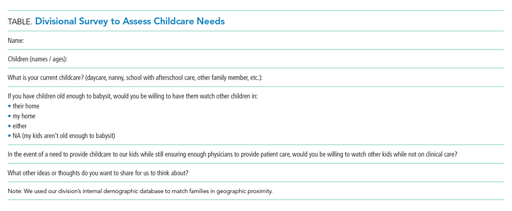
MANAGEMENT AND COMMUNICATION
Establish a Command Team
We benefit from having an existing divisional leadership structure comprising the director, medical directors of our clinical service lines, directors of education and community integration, and associate directors of clinical operations, research, and quality. This established team provides us broad representation of team member expertise and ideas. We maintain our weekly leadership team meeting through video chat and have added daily 30-minute virtual huddles to provide updates from our respective areas and discuss logistical challenges and planning. We use ad hoc phone meetings with relevant team members to address issues of immediate concern.
In the absence of a formal leadership team structure, establish a command team comprising representative leaders of your varied groups (eg, clinical operations, quality improvement, education, research, and business).
Collaborate With Institutional Response
Align divisional command team actions with the institutional response. Our clinical operations leader serves as our primary representative on the institutional emergency preparedness team. This participation allows bidirectional communication, both for institutional updates to be shared with division members and division-specific initiatives to be shared with institutional leadership to facilitate learning across the system.
In conjunction with hospital leadership, our division created a special isolation unit (SIU) to isolate patients positive for COVID-19 and persons under investigation. The institutional emergency preparedness team highlighted the need for such a unit, and our divisional leadership team developed the physician staffing model and medical care delivery system. We collaborated with key stakeholders, including nurses, respiratory therapists, other patient care services members, and subspecialists. The SIU leadership, which includes representatives from hospital medicine, nursing, respiratory therapy, and hospital operations, holds regular phone huddles to provide support and enlist resources based on identified gaps, which allows the frontline SIU physicians to focus on patient care. The calls initially occurred twice daily, but we transitioned to a once-daily schedule after routines were established and resources were procured.
Communicate With Everyone
Frequent communication with the clinical staff is paramount given the rapidly evolving operational changes and medical management recommendations. The divisional leadership team provides frequent email updates to the attending physicians on clinical shifts to communicate clinical updates, send reminders to conserve personal protective equipment (PPE), and share links to COVID-19 resources.
We use our weekly divisional meetings, now held virtually, to provide updates and to allow staff to ask questions and provide input. These meetings routinely include our nonclinical staff, such as administrative assistants and research coordinators, to ensure all team members’ voices are heard and skill sets are utilized. Our divisional infrastructure promotes dialogue and transparency, which is key to our division’s culture. Applying a learning health network approach has allowed us to generate new ideas, accelerate improvement, and encourage everyone to be a part of our community focused on improving outcomes.6 We continue to leverage this approach in our pandemic response.
One idea generated from this approach prompted us to create a centralized communication forum, using Microsoft Teams, to serve as a repository for the most up-to-date information related to COVID-19, the SIU, and general information, including links to divisional and institutional resources.
Maintain Nonclinical Operations
Nonclinical staff are working remotely. The business director and research director hold daily calls with the administrative staff and research coordinators, respectively, to discuss workload and to reallocate responsibilities as needed. This approach allows the business, administrative, and research support teams to function efficiently and redistribute work as the nonclinical priorities shift to meet divisional needs.
STAFFING
Establish a Backup Pool
We anticipate needing a larger pool of backup providers in the event of ill or quarantined staff or in case of increased patient volumes. The latter may be less likely for pediatric patients based on early studies3-5 but could occur if our free-standing children’s hospital expands to include the care of adult patients. We asked physicians to volunteer for backup shifts to augment our existing “jeopardy” backup system with a greater request to those with a lower clinical full-time equivalence. Each day, two backup shift positions are filled by volunteers, with additional positions added on days when medicine-pediatrics providers are scheduled for shifts in case they are needed at the university (adult) hospital.
Minimize Staffing to Reserve Pool
We monitor census closely on all service lines, including our consult service lines and secondary inpatient site, with plans to dissolve unnecessary consult services and combine medical teams, when feasible, to reduce the risk of staff exposure and maintain reserves. For example, after elective procedures were canceled, we reduced physician staffing of our surgical comanagement service to the minimal necessary coverage. We assign nonpatient-facing clinical duties to physicians who are called off their shift, in quarantine, or mildly ill to help off-load the clinical burden. Such duties include accepting direct admission phone calls, triaging patient care calls, entering orders remotely, and assisting with care coordination needs.
Anticipate Adult Care Needs
Our pediatric institution admits select groups of adult patients with congenital or complex healthcare needs who require specialized care. Hospitalists board certified in both pediatrics and internal medicine provide consultative services to many of these patients. Anticipating that these physicians may be needed in adult facilities, we plan to dissolve this consult service and utilize our reserve pool of providers to cover their pediatric shifts if needed. Additionally, if our hospital expands coverage for adult patients, these medicine-pediatrics providers will be instrumental in coordinating that expanded effort and will serve as leaders for teams of physicians and advanced practice providers with limited or no adult medicine training.
Special Isolation Unit
Logistic planning for our SIU evolved over the first few patients with rapid-cycle feedback and learning with each admission. This feedback was facilitated with our twice-daily huddle calls, which involved all key stakeholders, including nursing and respiratory therapy representatives. For division physician staffing, higher-risk team members are excluded from working on this unit. Because the SIU was developed to care for all patients positive for COVID-19 and persons under investigation, subspecialty patients not typically cared for by Hospital Medicine at our institution are being admitted to this unit. Therefore, subspecialty divisions assign attending physicians to provide consultative services to the SIU. These consultants use the unit’s telemedicine capabilities, when feasible, to limit staff exposure and conserve PPE. Our hospital medicine leaders in the SIU proactively worked with subspecialty divisions that are anticipated to have more admissions given their at-risk patient populations, such as pulmonary medicine, cardiology, and oncology. They specifically developed staffing plans for these patients if the SIU census becomes unsustainable under Hospital Medicine alone.
STAFF WELL-BEING
Healthcare workers are experiencing numerous stressors at work and home during this tumultuous time. Our workforce is at risk of developing emotional distress and mental health concerns. A cross-sectional survey of more than 1,200 healthcare workers in China who cared for COVID-19 patients found that many experienced symptoms of psychological distress (71%), as well as depression (51%), anxiety (44%), and insomnia (34%).7 Hospital medicine groups should consider methods to support their staff to mitigate stressors and promote self-care.
Anticipate Childcare Issues
When we were faced with impending school and daycare closures, we surveyed our division to assess childcare needs (Table) and share resources. We created a system of emergency childcare coverage options by connecting parents with similarly aged children and who lived in geographic proximity. This approach to childcare contingency planning was shared with and adopted by other divisions within the institution.
Build Support Measures
To support each other during this particularly stressful time, we divided division members into groups or “support pods,” each facilitated by a leadership team member. Group text messages and weekly phone or video chats have promoted connectivity and peer support.
Promote Self-care
The divisional leadership team provides food and drink for staff on clinical shifts. We also collated self-care resources to share via a central repository. These resources include ideas for meditation, home education for children, parenting, exercise, faith communities, entertainment, methods to support our local community through volunteerism and donations, and mental health resources, as well as online links to these resources.
Adult health systems will be disproportionately affected as this pandemic evolves. Pediatric hospitalists have the unique opportunity to support the response efforts by maintaining teams that are flexible and adaptable to evolving community needs. To do this, team leaders need to promote transparency, share learnings, and leverage the diverse skills of team members to ensure we are ready to meet the challenges of the moment.
1. World Health Organization. Coronavirus disease 2019 (COVID-19) Situation Report - 51. [Situation Report]. 2020. https://www.who.int/docs/default-source/coronaviruse/situation-reports/20200311-sitrep-51-covid-19. Accessed March 26, 2020.
2. Centers for Disease Control and Prevention. Interim Guidance for Healthcare Facilities: Preparing for Community Transmission of COVID-19 in the United States. 2020. https://www.cdc.gov/coronavirus/2019-ncov/healthcare-facilities/guidance-hcf.html. Accessed March 27, 2020.
3. Dong Y, Mo X, Hu Y, et al. Epidemiological characteristics of 2143 pediatric patients with 2019 coronavirus disease in China. Pediatrics. 2020. https://doi.org/10.1542/peds.2020-0702.
4. Cruz A, Zeichner S. COVID-19 in children: initial characterization of pediatric disease. Pediatrics. 2020;e20200834. https://doi.org/10.1542/peds.2020-0834.
5. Wu Z, McGoogan J. Characteristics of and important lessons from the coronavirus disease 2019 (COVID-19) outbreak in China: summary of a report of 72 314 cases from the Chinese Center for Disease Control and Prevention. JAMA. 2020. https://doi.org/10.1001/jama.2020.2648.
6. James M Anderson Center of Health Systems Excellence. The Power of Learning Networks. https://www.cincinnatichildrens.org/research/divisions/j/anderson-center/learning-networks. Accessed April 2, 2020.
7. Lai J, Ma S, Wang Y, et al. Factors Associated With Mental Health Outcomes Among Health Care Workers Exposed to Coronavirus Disease 2019. JAMA Netw Open. 2020;3(3):e203976. https://doi.org/10.1001/jamanetworkopen.2020.3976.
MANAGEMENT AND COMMUNICATION
Establish a Command Team
We benefit from having an existing divisional leadership structure comprising the director, medical directors of our clinical service lines, directors of education and community integration, and associate directors of clinical operations, research, and quality. This established team provides us broad representation of team member expertise and ideas. We maintain our weekly leadership team meeting through video chat and have added daily 30-minute virtual huddles to provide updates from our respective areas and discuss logistical challenges and planning. We use ad hoc phone meetings with relevant team members to address issues of immediate concern.
In the absence of a formal leadership team structure, establish a command team comprising representative leaders of your varied groups (eg, clinical operations, quality improvement, education, research, and business).
Collaborate With Institutional Response
Align divisional command team actions with the institutional response. Our clinical operations leader serves as our primary representative on the institutional emergency preparedness team. This participation allows bidirectional communication, both for institutional updates to be shared with division members and division-specific initiatives to be shared with institutional leadership to facilitate learning across the system.
In conjunction with hospital leadership, our division created a special isolation unit (SIU) to isolate patients positive for COVID-19 and persons under investigation. The institutional emergency preparedness team highlighted the need for such a unit, and our divisional leadership team developed the physician staffing model and medical care delivery system. We collaborated with key stakeholders, including nurses, respiratory therapists, other patient care services members, and subspecialists. The SIU leadership, which includes representatives from hospital medicine, nursing, respiratory therapy, and hospital operations, holds regular phone huddles to provide support and enlist resources based on identified gaps, which allows the frontline SIU physicians to focus on patient care. The calls initially occurred twice daily, but we transitioned to a once-daily schedule after routines were established and resources were procured.
Communicate With Everyone
Frequent communication with the clinical staff is paramount given the rapidly evolving operational changes and medical management recommendations. The divisional leadership team provides frequent email updates to the attending physicians on clinical shifts to communicate clinical updates, send reminders to conserve personal protective equipment (PPE), and share links to COVID-19 resources.
We use our weekly divisional meetings, now held virtually, to provide updates and to allow staff to ask questions and provide input. These meetings routinely include our nonclinical staff, such as administrative assistants and research coordinators, to ensure all team members’ voices are heard and skill sets are utilized. Our divisional infrastructure promotes dialogue and transparency, which is key to our division’s culture. Applying a learning health network approach has allowed us to generate new ideas, accelerate improvement, and encourage everyone to be a part of our community focused on improving outcomes.6 We continue to leverage this approach in our pandemic response.
One idea generated from this approach prompted us to create a centralized communication forum, using Microsoft Teams, to serve as a repository for the most up-to-date information related to COVID-19, the SIU, and general information, including links to divisional and institutional resources.
Maintain Nonclinical Operations
Nonclinical staff are working remotely. The business director and research director hold daily calls with the administrative staff and research coordinators, respectively, to discuss workload and to reallocate responsibilities as needed. This approach allows the business, administrative, and research support teams to function efficiently and redistribute work as the nonclinical priorities shift to meet divisional needs.
STAFFING
Establish a Backup Pool
We anticipate needing a larger pool of backup providers in the event of ill or quarantined staff or in case of increased patient volumes. The latter may be less likely for pediatric patients based on early studies3-5 but could occur if our free-standing children’s hospital expands to include the care of adult patients. We asked physicians to volunteer for backup shifts to augment our existing “jeopardy” backup system with a greater request to those with a lower clinical full-time equivalence. Each day, two backup shift positions are filled by volunteers, with additional positions added on days when medicine-pediatrics providers are scheduled for shifts in case they are needed at the university (adult) hospital.
Minimize Staffing to Reserve Pool
We monitor census closely on all service lines, including our consult service lines and secondary inpatient site, with plans to dissolve unnecessary consult services and combine medical teams, when feasible, to reduce the risk of staff exposure and maintain reserves. For example, after elective procedures were canceled, we reduced physician staffing of our surgical comanagement service to the minimal necessary coverage. We assign nonpatient-facing clinical duties to physicians who are called off their shift, in quarantine, or mildly ill to help off-load the clinical burden. Such duties include accepting direct admission phone calls, triaging patient care calls, entering orders remotely, and assisting with care coordination needs.
Anticipate Adult Care Needs
Our pediatric institution admits select groups of adult patients with congenital or complex healthcare needs who require specialized care. Hospitalists board certified in both pediatrics and internal medicine provide consultative services to many of these patients. Anticipating that these physicians may be needed in adult facilities, we plan to dissolve this consult service and utilize our reserve pool of providers to cover their pediatric shifts if needed. Additionally, if our hospital expands coverage for adult patients, these medicine-pediatrics providers will be instrumental in coordinating that expanded effort and will serve as leaders for teams of physicians and advanced practice providers with limited or no adult medicine training.
Special Isolation Unit
Logistic planning for our SIU evolved over the first few patients with rapid-cycle feedback and learning with each admission. This feedback was facilitated with our twice-daily huddle calls, which involved all key stakeholders, including nursing and respiratory therapy representatives. For division physician staffing, higher-risk team members are excluded from working on this unit. Because the SIU was developed to care for all patients positive for COVID-19 and persons under investigation, subspecialty patients not typically cared for by Hospital Medicine at our institution are being admitted to this unit. Therefore, subspecialty divisions assign attending physicians to provide consultative services to the SIU. These consultants use the unit’s telemedicine capabilities, when feasible, to limit staff exposure and conserve PPE. Our hospital medicine leaders in the SIU proactively worked with subspecialty divisions that are anticipated to have more admissions given their at-risk patient populations, such as pulmonary medicine, cardiology, and oncology. They specifically developed staffing plans for these patients if the SIU census becomes unsustainable under Hospital Medicine alone.
STAFF WELL-BEING
Healthcare workers are experiencing numerous stressors at work and home during this tumultuous time. Our workforce is at risk of developing emotional distress and mental health concerns. A cross-sectional survey of more than 1,200 healthcare workers in China who cared for COVID-19 patients found that many experienced symptoms of psychological distress (71%), as well as depression (51%), anxiety (44%), and insomnia (34%).7 Hospital medicine groups should consider methods to support their staff to mitigate stressors and promote self-care.
Anticipate Childcare Issues
When we were faced with impending school and daycare closures, we surveyed our division to assess childcare needs (Table) and share resources. We created a system of emergency childcare coverage options by connecting parents with similarly aged children and who lived in geographic proximity. This approach to childcare contingency planning was shared with and adopted by other divisions within the institution.
Build Support Measures
To support each other during this particularly stressful time, we divided division members into groups or “support pods,” each facilitated by a leadership team member. Group text messages and weekly phone or video chats have promoted connectivity and peer support.
Promote Self-care
The divisional leadership team provides food and drink for staff on clinical shifts. We also collated self-care resources to share via a central repository. These resources include ideas for meditation, home education for children, parenting, exercise, faith communities, entertainment, methods to support our local community through volunteerism and donations, and mental health resources, as well as online links to these resources.
Adult health systems will be disproportionately affected as this pandemic evolves. Pediatric hospitalists have the unique opportunity to support the response efforts by maintaining teams that are flexible and adaptable to evolving community needs. To do this, team leaders need to promote transparency, share learnings, and leverage the diverse skills of team members to ensure we are ready to meet the challenges of the moment.
1. World Health Organization. Coronavirus disease 2019 (COVID-19) Situation Report - 51. [Situation Report]. 2020. https://www.who.int/docs/default-source/coronaviruse/situation-reports/20200311-sitrep-51-covid-19. Accessed March 26, 2020.
2. Centers for Disease Control and Prevention. Interim Guidance for Healthcare Facilities: Preparing for Community Transmission of COVID-19 in the United States. 2020. https://www.cdc.gov/coronavirus/2019-ncov/healthcare-facilities/guidance-hcf.html. Accessed March 27, 2020.
3. Dong Y, Mo X, Hu Y, et al. Epidemiological characteristics of 2143 pediatric patients with 2019 coronavirus disease in China. Pediatrics. 2020. https://doi.org/10.1542/peds.2020-0702.
4. Cruz A, Zeichner S. COVID-19 in children: initial characterization of pediatric disease. Pediatrics. 2020;e20200834. https://doi.org/10.1542/peds.2020-0834.
5. Wu Z, McGoogan J. Characteristics of and important lessons from the coronavirus disease 2019 (COVID-19) outbreak in China: summary of a report of 72 314 cases from the Chinese Center for Disease Control and Prevention. JAMA. 2020. https://doi.org/10.1001/jama.2020.2648.
6. James M Anderson Center of Health Systems Excellence. The Power of Learning Networks. https://www.cincinnatichildrens.org/research/divisions/j/anderson-center/learning-networks. Accessed April 2, 2020.
7. Lai J, Ma S, Wang Y, et al. Factors Associated With Mental Health Outcomes Among Health Care Workers Exposed to Coronavirus Disease 2019. JAMA Netw Open. 2020;3(3):e203976. https://doi.org/10.1001/jamanetworkopen.2020.3976.
© 2020 Society of Hospital Medicine