User login
New data illustrate pandemic pivot to telehealth by patients, physicians
Telehealth use, although much higher than before the COVID-19 pandemic, accounted for less than 20% of weekly outpatient visits 6 months into the pandemic, according to a new report from the American Medical Association. Ten percent of weekly visits were conducted via videoconferencing, and 8.1% of visits were conducted using the telephone.
Those figures may overstate the true level of telehealth use in fall 2020. A study by the Commonwealth Fund, Harvard University, Boston, and Phreesia found that in December of that year, only 8% of outpatient visits involved the use of telemedicine – and that was up from 6% in October. In contrast to the AMA results, which came from its 2020 benchmark survey of physicians, the Commonwealth Fund study used data from practice management systems and an online patient registration platform, as well as electronic health record data.
A more recent survey of hospital executives found that as of September 2021, hospital telehealth visits had leveled off at 10% to 20% of appointments. Similarly, a McKinsey survey in July showed that telehealth encounters made up 13% to 17% of evaluation and management visits across all specialties.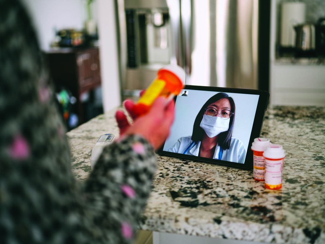
Big jump during pandemic
The AMA report offers a wealth of data on how physicians use telehealth and the differences between specialties in this area.
The report found that 70.3% of physicians worked in practices that used videoconferencing to provide patient visits in September 2020, compared to 14.3% of physicians in September 2018. Sixty-seven percent of physicians worked in practices that used telephone visits (the comparable figure for 2018 was unavailable).
Overall, 79% of physicians worked in a practice that used telehealth, compared to 25% in 2018.
Not every doctor in practices that utilized telehealth conducted virtual visits. In contrast to the 70.3% of doctors who were in practices that had video visits, only 59.1% of the respondents had personally conducted a videoconferencing visit in the previous week. The average numbers of weekly video and telephone visits per physician were 9.9 and 7.6, respectively, including those who did none.
There were big differences in virtual visit use among specialties as well. Eighty-five percent of psychiatrists were in practices that provided online appointments, according to the AMA survey, and three-quarters of primary care physicians said their practices offered telehealth appointments. Pediatricians were much less likely than family practice/general practice physicians (FPs/GPs) or general internists to do so.
The practices of many medical specialists were also highly likely to provide telehealth. Over 75% of practices in cardiology, endocrinology/diabetes, gastroenterology, nephrology, and neurology offered telehealth visits. About 88% of hematologists/oncologists offered video visits. Far fewer surgeons reported that their practice used virtual visits; the exceptions were urologists and dermatologists, 87% of whose practices used telehealth.
How telehealth was used
Across all specialties, 58% of physicians said clinicians in their practices used it to diagnose or treat patients; 59.2%, to manage patients with chronic disease; 50.4%, to provide acute care; and 34.3%, to provide preventive care.
Seventy-two percent of FP/GP and pediatric practices used telehealth to diagnose or treat patients. Just 64.9% of internists said their practices did so, and only 61.9% of them said their practices provided acute care via telehealth, versus 70% of FPs/GPs and pediatricians.
Among medical specialties, endocrinologists/diabetes physicians were those most likely to report the practice-level use of telehealth to diagnose or treat patients (71.9%), manage patients with chronic disease (92.1%), and provide preventive care (52.6%).
Significantly, 33% of medical specialists said their practices used remote patient monitoring. This finding was driven by high rates of use among cardiology practices (63.3%) and endocrinology practices (41.6%). Overall, the practice-level use of remote patient monitoring rose from 10.4% of practices in 2018 to 19.9% in 2020.
Virtual consults with peers
Some practices used telehealth to enable physicians to consult with colleagues. Twelve percent of respondents said their practices used telehealth to seek a second opinion from a health care professional in 2020, compared to 6.9% in 2018. Formal consultations via telehealth were also increasingly common: 17.2% of doctors said their practices did this in 2020, compared to 11.3% in 2018.
Also of note, 22.4% of physicians said their practices used telehealth for after-hours care or night calls in 2020, versus 9.9% in 2018.
The AMA report credited telehealth and expanded coverage and payment rules for enabling physician practices to keep their revenue streams positive and their practices open. However, the Commonwealth Fund study found “a substantial cumulative reduction in visits across all specialties over the course of the pandemic in 2020.” These ranged from a drop of 27% in pediatric visits to a decline of 8% in rheumatology visits during the period from March to December 2020.
A version of this article first appeared on Medscape.com.
Mortality-related risk factors for hospitalized patients with COVID-19
Key clinical point: A meta-analysis has identified 10 key risk factors for mortality in hospitalized patients with COVID-19.
Major finding: The significant mortality-related risk factors in hospitalized patients with COVID-19 included older age, male sex, smoking, obesity, cardiovascular disease, diabetes, hypertension, chronic obstructive pulmonary disease, acute kidney injury, and elevated D-dimer levels.
Study details: The data come from a meta-analysis of 42 studies involving 423,117 patients with COVID-19.
Disclosures: The study did not receive any funding. The authors declared no conflict of interests.
Source: Dessie ZG et al. BMC Infect Dis. 2021 Aug 21. doi: 10.1186/s12879-021-06536-3.
COVID-19: Antibody cocktail effective in preventing household transmission
Key clinical point: A single subcutaneous dose of the antibody cocktail REGEN-COV (casirivimab plus imdevimab) is effective in preventing symptomatic and asymptomatic infection in household contacts of COVID-19-positive individuals.
Major finding: The antibody cocktail group developed fewer symptomatic SARS-CoV-2 infections than the placebo group (relative risk reduction, 81.4%). The antibody cocktail effectively prevented symptomatic and asymptomatic infections overall (relative risk reduction, 66.4%).
Study details: In a randomized, double-blind, placebo-controlled trial, unaffected household members (age, 12 years or older) of individuals testing positive for SARS-CoV-2 received either the antibody cocktail (n=753) or placebo (n=752).
Disclosures: The study was funded by Regeneron Pharmaceuticals, F. Hoffmann-LaRoche, and the National Institutes of Health. Several authors were employees and/or stockholders of Regeneron Pharmaceuticals.
Source: O'Brien MP et al. N Engl J Med. 2021 Aug 4. doi: 10.1056/NEJMoa2109682.
Hospitalized COVID-19 patients with cardiometabolic risk do not benefit from dapagliflozin
Key clinical point: Dapagliflozin does not improve COVID-19 hospitalization outcomes for patients with cardiometabolic risk factors.
Major finding: Dapagliflozin vs placebo failed to meet the primary composite outcome of organ dysfunction or all-cause death (hazard ratio, 0.80; 95% confidence interval, 0.58-1.10). There was no difference in the rates of new/worsened organ dysfunction, deaths, or clinical improvement between the groups.
Study details: In the DARE-19 phase 3 trial, patients were randomly assigned to receive either dapagliflozin (n=625) or placebo (n=625).
Disclosures: The study was funded by AstraZeneca. R Esterline, J Oscarsson, SB Gasparyan, J Buenconsejo, AM Langkilde, and P Ambery are employees and stockholders of AstraZeneca. M Aboudara, E Akin, WKS Barroso, ADM Feitosa, CRH Filho, A Fonseca, K Gosch, RA Gordon, CP Jaeger, LN Maia, DDF Moia, JRL Soto, F Tang, SL Windsor, O Mukhtar, V Chopra, RVP Soares, V Garla, PE Leaes, FS Silveira, and M Pursley declared no conflict of interests. The remaining authors disclosed relationships with pharmaceutical companies and/or research institutions.
Source: Kosiborod MN et al. Lancet Diabetes Endocrinol. 2021 Jul 21. doi: 10.1016/S2213-8587(21)00180-7.
Canakinumab fails to improve survival in hospitalized patients with severe COVID-19
Key clinical point: Canakinumab does not improve survival in hospitalized patients with severe COVID-19.
Major finding: There was no significant difference in survival without invasive mechanical ventilation between days 3 and 29 with canakinumab vs. placebo (88.8% vs. 85.7%; rate difference, 3.1 percentage points; 95% confidence interval [CI], −3.1 to 9.3). COVID-19 mortality also did not differ with canakinumab vs. placebo (4.9% vs 7.2%; rate difference, −2.3 percentage points; 95% CI, −6.7 to 2.2).
Study details: The data come from the randomized, double-blind, placebo-controlled phase 3 CAN-COVID trial (n=454).
Disclosures: The study was sponsored by Novartis Pharma AG, Basel, Switzerland. All authors received funding from Novartis during the conduct of the study. The authors also reported relationships with other pharmaceutical companies.
Source: Caricchio R et al. JAMA. 2021 Jul 20. doi: 10.1001/jama.2021.9508.
COVID-19: Inhaled budesonide may shorten recovery time
Key clinical point: Inhaled budesonide is associated with a shorter time to recovery but fails to reduce the risk for hospitalization or death in high-risk primary care patients with COVID-19.
Major finding: Budesonide vs usual care was associated with a shorter time to recovery (11.8 days vs 14.7 days). Budesonide was associated with a nonsignificant 2.0% reduction in hospitalization or death compared with usual care.
Study details: The data come from the PRINCIPLE trial, where 2,530 patients were randomly assigned to either inhaled budesonide (n=787), usual care alone (n=1,069), or usual care plus other interventions (n=674).
Disclosures: The study was funded by the National Institute of Health Research and United Kingdom Research Innovation. M Bafadhel, D Richards, BR Saville, N Berry, MA Detry, M Fitzgerald, S de Lusignan, MI Andersson, PJ Barnes, REK Russell, S Ramakrishnan, FDR Hobbs, and CC Butler reported relationships with pharmaceutical companies and/or research institutions. The remaining authors declared no conflict of interests.
Source: Yu LM et al. Lancet. 2021 Aug 10. doi: 10.1016/S0140-6736(21)01744-X.
COVID-19: Pulmonary embolism not tied to increased mortality risk
Key clinical point: Presence of pulmonary embolism (PE) is not associated with increased mortality in patients with COVID-19 risk.
Major finding: Risk factors for PE in patients with COVID-19 included male sex, mechanical ventilation, intensive care unit admission, and circulating D-dimer. Patients with PE did not have an increased risk for mortality compared with those without PE (odds ratio, 1.31; P = .25).
Study details: The data come from a meta-analysis of 16 cohort studies involving 5,826 patients with COVID-19.
Disclosures: No funding information was available. The authors declared no conflict of interests.
Source: Gómez CA et al. Sci Rep. 2021 Aug 6. doi: 10.1038/s41598-021-95512-7.
COVID-19: Early administration of plasma fails to prevent disease progression
Key clinical point: Administration of convalescent plasma within 7 days of symptom onset fails to prevent disease progression in acutely ill, high-risk patients with COVID-19.
Major finding: There was no difference between the convalescent plasma group and the placebo group in disease progression (30.0% vs 31.9%; risk difference, 1.9 percentage points; 95% credible interval, −6.0 to 9.8).
Study details: The multicenter, single-blind SIREN-C3PO trial included patients with COVID-19 (n=511) randomly assigned to receive either plasma or placebo in the emergency department.
Disclosures: The study was supported by the National Institutes of Health, the Biomedical Advanced Research and Development Authority, and the Operation Warp Speed interagency program. D Beiser, A Burnett R Davenport, L Dumont, V Durkalski-Mauldin, N El Kassar, L Foster, C Greineder, N Haas, J Hah, A Kaplan, B Kea, F Korley, E Lowell, J McDyer, J Quinn, J Reynolds, R Silbergleit, C Van Huysen, and K Yadav declared no conflict of interests. The remaining authors disclosed relationships with pharmaceutical companies and/or research institutions.
Source: Korley FK et al. N Engl J Med. 2021 Aug 18. doi: 10.1056/NEJMoa2103784.
Baricitinib plus standard of care may reduce mortality risk in hospitalized patients with COVID-19
Key clinical point: Addition of baricitinib to standard of care treatment may reduce 28-day mortality risk in patients hospitalized with COVID-19, but does not significantly reduce the frequency of disease progression.
Major finding: There was a 38.2% relative reduction in 28-day all-cause mortality risk with baricitinib vs placebo. There was no significant difference between the baricitinib and placebo groups in the primary composite endpoint of disease progression outcomes (odds ratio, 0.85; P = .18).
Study details: In a phase 3, double-blind trial, hospitalized patients with COVID-19 receiving standard of care were randomly assigned to either baricitinib (n=764) or placebo group (n=761).
Disclosures: The study was funded by Eli Lilly and Company. S de Bono, CE Kartman, V Krishnan, R Liao, MLB Piruzeli, A Cardose, S Chakladar, B Crowe, P Reis, X Zhang, and DH Adams are employees and shareholders of Eli Lilly and Company. RD Pellegrini declared no conflict of interests. Other authors reported relationships with pharmaceutical companies including Eli Lilly and Company.
Source: Marconi VC et al. Lancet Respir Med. 2021 Sep 1. doi: 10.1016/S2213-2600(21)00331-3.
Lower risk for COVID-19 in patients with asthma
Key clinical point: Individuals with asthma have a 17% lower risk for COVID-19 infection than those without asthma.
Major finding: Individuals with asthma had a lower risk for COVID-19 infection (risk ratio [RR], 0.83; P = .01), but not for COVID-19-related hospitalization (RR, 1.18; P = .08), intensive care unit admission (RR, 1.21; P = .09), and ICU admission (RR, 1.06; P = .65).
Study details: The data come from a meta-analysis of 51 studies involving 965,551 individuals with and without asthma who tested positive for COVID-19.
Disclosures: This study was self-funded. The authors declared no conflict of interests.
Source: Sunjaya AP et al. Eur Respir J. 2021 Aug 24. doi: 10.1183/13993003.01209-2021.