User login
Imaging links insula and frontal cortex to anxiety in Parkinson’s disease
Anxiety occurs in approximately 31% of Parkinson’s disease patients, but the underlying mechanisms are not well understood, wrote Nacim Betrouni, MD, of the University of Lille, France, and colleagues. Previous research has shown associations between anxiety severity and increased activity in brain areas of emotion processing, based on MRI and positron emission tomography, but electroencephalography (EEG) has not been widely used, they said.
In a study published in Neurophysiologie Clinique , the researchers compared EEG spectral patterns and functional resting-state networks in Parkinson’s disease patients with and without anxiety disorders. They identified data from 33 PD patients who met criteria for anxiety, and 75 without anxiety. The average age of the patients was 65 years, and the average disease duration was 9.76 years in anxiety patients and 7.83 years in patients without anxiety.
Overall, findings on spectral analysis showed an association between anxiety and changes in the alpha activity in the right frontal cortex, the researchers said. They also found the relative power in the alpha1 frequency band in the right prefrontal cortex was lower in patients with anxiety than without; this finding was significantly associated with avoidance behavior on a subscale of the Parkinson’s Anxiety Scale (PAS_C, P = .035). A trend toward a significant association with episodic anxiety was noted (PAS_B, P = .06), but no significant associations were noted for persistent anxiety or the total scale score.
The imaging also showed an increased connectivity between the insula and the posterior cingulate cortex in several frequency bands in the anxiety patients, the researchers said. “The increased connectivity observed here may be a marker of the maintenance of avoidance behaviors that characterize anxiety in PD,” they noted.
The study findings were limited by several factors including the small and unbalanced proportion of the study population with anxiety, and the consideration of global anxiety only, without distinguishing anxiety subtypes, the researchers noted. Another limitation was the use only of static EEC patterns, without using dynamic patterns, they said.
The study is the first known to use EEG to explore the mechanisms of PD-related anxiety and “the reported results provide new insights, supporting findings of previous studies using other modalities, mainly rs-fMRI, and show that EEG could be a relevant technique to explore these disorders,” the researchers wrote.
However, more research is needed to confirm the findings in patients with a larger panel of anxiety disorders, they concluded.
The study was supported by the Michael J. Fox Foundation. The researchers had no financial conflicts to disclose.
FROM NEUROPHYSIOLOGIE CLINIQUE
About 73% of U.S. estimated to be immune to Omicron variant
, a university health institute says.
About half of eligible Americans have received booster shots, and about 80 million confirmed COVID-19 infections have been reported. Many more infections have occurred but haven’t been officially recorded, The Associated Press reported.
The high percentage of immunity from vaccination and previous infection tends to prevent or shorten new illnesses and reduce the amount of virus circulating overall. Health experts are now discussing whether the number is high enough to stop new waves or reduce the burden on hospitals.
“I am optimistic even if we have a surge in summer, cases will go up, but hospitalizations and deaths will not,” Ali Mokdad, PhD, a professor of health metrics sciences at the University of Washington in Seattle, told the AP.
Dr. Mokdad works on COVID-19 forecasting for the university’s Institute for Health Metrics and Evaluation, which has been a reliable model during the pandemic. Dr. Mokdad calculated the 73% number for the AP.
“We have changed,” he said. “We have been exposed to this virus and we know how to deal with it.”
The United States is now reporting about 125,000 new cases per day, according to the data tracker from the New York Times, marking a 68% decrease from the past 2 weeks. Hospitalizations are also down 39%, and about 2,300 new deaths are being reported daily, marking a 13% decline.
There will be more outbreaks as new variants emerge, immunity wanes, and some people remain unvaccinated, Dr. Mokdad said. But the coronavirus is no longer new, and the entire population is no longer “immunologically naive.” Scientists are now trying to understand how long booster protection will last against Omicron and how many people have been infected who had mild or no symptoms that were never reported.
By the end of the Omicron surge, about three out of four people in the United States will have been infected, Shaun Truelove, PhD, an epidemiologist and disease modeler at Johns Hopkins University, told the AP.
“We know it’s a huge proportion of the population,” he said. “This varies a lot by location, and in some areas, we expect the number infected to be closer to one in two.”
That means different regions and groups of people have different levels of protection and risk. In Virginia, for instance, disease modelers estimate that about 45% of residents have the highest level of immunity by being vaccinated and boosted or vaccinated with a recent Omicron infection. Another 47% have immunity that has waned somewhat.
“That’s going to be a nice shield of armor for our population as a whole,” Bryan Lewis, PhD, an epidemiologist who leads the University of Virginia’s COVID-19 modeling team, told the outlet. “If we do get to very low case rates, we certainly can ease back on some of these restrictions.”
About 7% of Virginians are considered the most vulnerable because they were never vaccinated or infected, he noted. Nationwide, about 80 million Americans are still vulnerable, the AP reported.
“The 26% who could still get Omicron right now have to be very careful,” Dr. Mokdad said.
The percentages will continue to change as immunity wanes and new variants circulate in the country. For now, the Institute for Health Metrics and Evaluation model estimates that about 63% to 81% of Americans are protected.
“We’ve reached a much better position for the coming months, but with waning immunity, we shouldn’t take it for granted,” Dr. Mokdad said.
A version of this article first appeared on WebMD.com.
Breast cancer trials enrolling now: Could your patient benefit?
• Menopausal women at moderate risk of developing breast cancer. A phase 2 study sponsored by the National Cancer Institute is seeking women aged 45-60 in late menopause or post menopause who are at “moderate” risk of developing breast cancer. Examples of criteria for moderate risk include prior proliferative disease on breast biopsy or having a first- or second-degree relative who developed breast cancer at aged 60 or younger. Researchers are looking for a signal that bazedoxifene plus conjugated estrogens (Duavee), a hot-flash therapy, could prevent breast cancer in at-risk people. Participants in the active-therapy group will receive once-daily oral medication for 6 months. The control patients will have the option of taking the medication after 6 months. The trial aims to enroll 120 participants. It began recruiting on Dec. 2, 2021, at the University of Kansas Medical Center; sites in California, Illinois, and Massachusetts are planned. The primary outcome is the change in fibroglandular volume. Overall survival (OS) and quality of life (QOL) will not be measured. More details at clinicaltrials.gov.
• Early high-risk nonmetastatic HER2+ breast cancer with no prior treatment. Adults with this type of breast cancer are invited to join a phase 3 trial of trastuzumab deruxtecan (T-DXd; Enhertu) as neoadjuvant therapy. T-DXd is currently approved for patients with advanced disease, so this study could lead to a new indication. Participants will receive standard intravenous regimens of either T-DXd monotherapy; T-DXd followed by paclitaxel (Taxol), trastuzumab (Herceptin), and pertuzumab (Perjeta), referred to as the THP regime; or doxorubicin plus cyclophosphamide followed by THP. The primary outcome is rate of pathologic complete response, and a secondary outcome is OS over approximately 5 years. QOL won’t be measured. The study opened on Oct. 25, 2021, and eventually hopes to recruit 624 participants in 19 countries and 15 U.S. states. More details at clinicaltrials.gov.
“[This is an] important early trial to move trastuzumab deruxtecan to early disease. If successful as monotherapy, this would be a big win for patients,” commented Kathy Miller, MD, professor of oncology and medicine at Indiana University, Indianapolis, a contributor to this news organization. She cautioned that monitoring rates of pneumonitis will be important in this curable setting.
• Locally advanced unresectable or metastatic HER2+ breast cancer with no prior tyrosine-kinase inhibitor therapy. Adult patients with these clinical features are eligible for a phase 3 study that is also testing a drug in an earlier setting than its current label – tucatinib (Tukysa) as first-line anti-HER2 therapy in advanced disease. Tucatinib was approved in April 2020 by the U.S. Food and Drug Administration as second-line therapy in such patients, so this study could also lead to a new indication. Participants in the experimental arm will receive tucatinib tablets twice daily and a combination of trastuzumab and pertuzumab intravenously or subcutaneously every 3 weeks for up to approximately 3 years. Patients in the control arm will take a placebo instead of tucatinib. Seven sites across Florida, Kentucky, Maryland, and South Carolina aim to start recruiting 650 participants on Feb. 28, 2022. The primary outcome is progression-free survival (PFS). OS and QoL will be tracked. More details at clinicaltrials.gov.
“Tucatinib has real activity,” commented Dr. Miller, adding that “we haven’t [yet] found the best way to exploit that activity for our patients.”
• Inoperable or metastatic HR+ HER2– breast cancer after one or two lines of systemic chemotherapy. Adults with this type of breast cancer are being recruited for a phase 3 study to compare datopotamab deruxtecan (Dato-DXd), an experimental antibody-drug conjugate (ADC), against a range of standard single-agent chemotherapies. Participants will receive either intravenous Dato-DXd or investigator’s choice of one of four chemotherapies: oral capecitabine (Xeloda), IV gemcitabine (Gemzar), IV eribulin (Halaven), or IV vinorelbine (Navelbine). The trial began recruiting for 700 participants at sites worldwide on Oct. 18, 2021. U.S. sites are in Michigan and California; trial centers in 15 other states are planned. Primary outcomes are OS over approximately 3.5 years and PFS over approximately 2 years. QOL is tracked. More details at clinicaltrials.gov.
Commenting on this trial, Dr. Miller said: “ADCs will play an expanded role in our management. This may be one of the first to move into the ER+ population.”
• Advanced ER+, HER2– breast cancer. Adult patients with this type of cancer can join a phase 3 trial testing oral imlunestrant, an experimental selective estrogen-receptor degrader (SERD), against standard endocrine therapy. For up to 3 years, people in the study will take either daily tablets of imlunestrant or once-daily pills of imlunestrant and another SERD, abemaciclib (Verzenio). A third group of participants will receive their investigator’s choice of either daily tablets of exemestane (Aromasin) or monthly intramuscular injections of fulvestrant (Faslodex). The study opened to 800 participants on Oct. 4, 2021, at sites in 11 U.S. states and worldwide. The primary outcome is PFS over approximately 3 years; 5-year OS is a secondary outcome. QOL is not assessed. More details at clinicaltrials.gov
Dr. Miller predicted that “oral SERDs will replace fulvestrant in the future: We already have positive phase 3 data with elacestrant.”
Dr. Miller has a regular column with this news organization, Miller on Oncology. She has disclosed no relevant financial relationships. A version of this article first appeared on Medscape.com.
When your medical error harmed a patient and you’re wracked with guilt
Peter Schwartz, MD, was chair of the department of obstetrics and gynecology at a hospital in Reading, Pa., in the mid-1990s when a young physician sought him out. The doctor, whom Dr. Schwartz regarded as talented and empathetic, was visibly shaken. The expectant mother they were caring for had just lost her unborn child.
“The doctor came into my office within an hour of the event and asked me to look at the case,” Dr. Schwartz recalled. “I could see that they had failed to recognize ominous changes in the fetal heart rate, and I faced the pain of having to tell them, ‘I think this could have been handled much better.’” Dr. Schwartz delivered the news as compassionately as he could, but a subsequent review confirmed his suspicion: The doctor had made a serious error.
“The doctor was devastated,” he said. “She got counseling and took time off, but in the end, she quit practicing medicine. She said, ‘If I keep practicing, something like that could happen again, and I don’t think I could handle it.’”
To err may be human, but in a health care setting, the harm can be catastrophic. that their feelings of guilt, shame, and self-doubt can lead to depression, anxiety, post-traumatic stress disorder, and even suicidal ideation. The trauma can be so profound that, in a now famous 2000 editorial in the British Medical Journal, Albert Wu, MD, gave the phenomenon a name: “second victim syndrome.”
Today, as quality improvement organizations and health systems work to address medical errors in a just and transparent way, they’re realizing that finding ways to help traumatized clinicians is integral to their efforts.
Are doctors really ‘second victims?’
Although the medical field is moving away from the term “second victim,” which patient advocates argue lacks a ring of accountability, the emotional trauma doctors and other clinicians endure is garnering increased attention. In the 2 decades since Dr. Wu wrote his editorial, research has shown that many types of adverse health care events can evoke traumatic responses. In fact, studies indicate that from 10.4% to 43.3% of health care workers may experience negative symptoms following an adverse event.
But for doctors – who have sworn an oath to do no harm – the emotional toll of having committed a serious medical error can be particularly burdensome and lingering. In a Dutch study involving more than 4,300 doctors and nurses, respondents who were involved in a patient safety incident that resulted in harm were nine times more likely to have negative symptoms lasting longer than 6 months than those who were involved in a near-miss experience.
“There’s a feeling of wanting to erase yourself,” says Danielle Ofri, MD, a New York internist and author of “When We Do Harm: A Doctor Confronts Medical Error.”
That emotional response can have a profound impact on the way medical errors are disclosed, investigated, and ultimately resolved, said Thomas Gallagher, MD, an internist and executive director of the Collaborative for Accountability and Improvement, a patient safety program at the University of Washington.
“When something goes wrong, as physicians, we don’t know what to do,” Dr. Gallagher says. “We feel awful, and often our human reflexes lead us astray. The doctor’s own emotions become barriers to addressing the situation.” For example, guilt and shame may lead doctors to try to hide or diminish their mistakes. Some doctors might try to shift blame, while others may feel so guilty they assume they were responsible for an outcome that was beyond their control.
Recognizing that clinicians’ responses to medical errors are inextricably tangled with how those events are addressed, a growing number of health systems are making clinician support a key element when dealing with medical errors.
Emotional first aid
Although it’s typical for physicians to feel isolated in the wake of errors, these experiences are far from unique. Research conducted by University of Missouri Health Care nurse scientist Susan Scott, RN, PhD, shows that just as most individuals experiencing grief pass through several distinct emotional stages, health care professionals who make errors go through emotional stages that may occur sequentially or concurrently.
An initial period of chaos is often followed by intrusive reflections, haunting re-enactments, and feelings of inadequacy. The doctor’s thinking moves from “How did that happen?” to “What did I miss?” to “What will people think about me?” As the error comes under scrutiny by quality improvement organizations, licensing boards, and/or lawyers, the doctor feels besieged. The doctor may want to reach out but is afraid to. According to Dr. Scott, only 15% of care providers ask for help.
Recognizing that physicians and other care providers rarely ask for support – or may not realize they need it – a growing number of health systems are implementing Communication and Resolution Programs (CRPs). Rather than respond to medical errors with a deny-and-defend mentality, CRPs emphasize transparency and accountability.
This approach, which the Agency for Healthcare Research and Quality has embraced and codified with its Communication and Optimal Resolution (CANDOR) toolkit, focuses on prompt incident reporting; communication with and support for patients, family members, and caregivers affected by the event; event analysis; quality improvement; and just resolution of the event, including apologies and financial compensation where appropriate.
The CANDOR toolkit, which includes a module entitled Care for the Caregiver, directs health systems to identify individuals and establish teams, led by representatives from patient safety and/or risk management, who can respond promptly to an event. After ensuring the patient is clinically stable and safe, the CANDOR process provides for immediate and ongoing emotional support to the patient, the family, and the caregiver.
“A lot of what CRPs are about is creating structures and processes that normalize an open and compassionate response to harm events in medicine,” says Dr. Gallagher, who estimates that between 400 and 500 health systems now have CRPs in place.
Wisdom through adversity
While clinicians experience many difficult and negative emotions in the wake of medical errors, how they move forward after the event varies markedly. Some, unable to come to terms with the trauma, may move to another institution or leave medicine entirely. Others, while occasionally reliving the trauma, learn to cope. For the most fortunate, enduring the trauma of a medical error can lead to growth, insight, and wisdom.
In an article published in the journal Academic Medicine, researchers asked 61 physicians who had made serious medical errors, “What helped you to cope positively?” Some of the most common responses – talking about their feelings with a peer, disclosing and apologizing for a mistake, and developing system changes to prevent additional errors – are baked into some health systems’ CRP programs. Other respondents said they dedicated themselves to learning from the mistake, becoming experts in a given field, or sharing what they learned from the experience through teaching.
Dr. Ofri said that after she made an error decades ago while managing a patient with diabetic ketoacidosis, her senior resident publicly berated her for it. The incident taught her a clinical lesson: Never remove an insulin drip without administering long-acting insulin. More importantly, the resident’s verbal thumping taught her about the corrosive effects of shame. Today, Dr. Ofri, who works in a teaching hospital, says that when meeting a new medical team, she begins by recounting her five biggest medical errors.
“I want them to come to me if they make a mistake,” she says. “I want to first make sure the patient is okay. But then I want to make sure the doctor is okay. I also want to know: What was it about the system that contributed to the error, and what can we do to prevent similar errors in the future?”
Acceptance and compassion
Time, experience, supportive peers, an understanding partner or spouse: all of these can help a doctor recover from the trauma of a mistake. “But they’re not an eraser,” Dr. Schwartz said.
Sometimes, doctors say, the path forward starts with acceptance.
Jan Bonhoeffer, MD, author of “Dare to Care: How to Survive and Thrive in Today’s Medical World,” tells a story about a mistake that transformed his life. In 2004, he was working in a busy London emergency department when an adolescent girl arrived complaining of breathing trouble. Dr. Bonhoeffer diagnosed her with asthma and discharged her with an inhaler. The next day, the girl was back in the hospital – this time in the ICU, intubated, and on a ventilator. Because he had failed to take an x-ray, Dr. Bonhoeffer missed the tumor growing in the girl’s chest.
Dr. Bonhoeffer was shattered by his error. “After that experience, I knew I wanted to make learning from my mistakes part of my daily practice,” he says. Now, at the end of each workday, Dr. Bonhoeffer takes an inventory of the day and reflects on all his actions, large and small, clinical and not. “I take a few minutes and think about everything I did and what I should have done differently,” he said. The daily practice can be humbling because it forces him to confront his errors, but it is also empowering, he said, “because the next day I get to make a different choice.”
Dr. Bonhoeffer added, “Doctors are fallible, and you have to be compassionate with yourself. Compassion isn’t sweet. It’s not motherhood and honey pies. It’s coming to terms with reality. It’s not a cure, but it’s healing.”
A version of this article first appeared on Medscape.com.
Experimental breast cancer immunotherapy treatment passes important hurdle in pilot study
Three of 6 patients treated had a response, including one that lasted more than five years. TILs were expanded ex vivo, and patients treated with lymphodepleting chemotherapy before the infusion, along with the checkpoint inhibitor pembrolizumab (Keytruda, Merck).
The results pave the way for recruitment of more patients as researchers at the National Cancer Institute ramp up the experimental treatment. They also help to overturn the long-held dogma that breast cancer is not immunogenic, according to lead researcher Steven A. Rosenberg, MD, PhD, who is chief of the surgery branch of the National Cancer Institute.
Although common hormone positive breast cancer doesn’t respond to immunotherapy, the study found that two-thirds of patients with metastatic breast cancer have mutations recognized by TILs. “We can identify the antigens [that] T cells recognize,” said Dr. Rosenberg, who is the lead author on a paper describing the pilot study, published online Feb. 1 in the Journal of Clinical Oncology.
There has been wide speculation that some solid tumors, including common hormone-positive breast cancers, are not immunogenic, because they don’t respond to cancer vaccines or checkpoint inhibitors. However, newer research has unearthed an explanation: Patients with these solid tumors produce immunogenic antigens, but they differ from patient to patient. There was not a single shared antigen among the 42 patients in the study. “Every patient reacts with a unique antigen, so the treatments have to be highly personalized,” said Dr. Rosenberg.
In the phase 2 study, the researchers recruited 42 participants, who underwent screening for novel tumor antigens. Sixty percent were hormone-receptor positive and HER2 negative, 26% were triple negative, and 14% were HER2 enriched.
Of 42 patients, 28 (67%) had at least one detectable, immunogenic tumor antigen, including 46% of HR+/Her2– patients, 32% of triple-negative patients, and 21% of HER2-enriched patients. Thirteen patients had a positive TIL screen, making them candidates for treatment.
Six patients underwent the procedure. Researchers selected TIL culture fragments that responded when stimulated with mutant peptides. They expanded those cells externally over 24 days and then administered lymphodepleting chemotherapy 1 week before the infusion of the expanded TILs. Patients received aldesleukin (Proleukin, Prometheus Laboratories) every 8 hours after TIL infusion, as tolerated. Patients also received pembrolizumab 2 days before the TIL infusion and up to three more doses at 3-week intervals.
Three patients experienced objective tumor regression, including a complete response that has lasted for 5.5 years. Two had partial responses that lasted 6 and 10 months. One patient with a partial response had a limited recurrence that could be excised, followed by further regression of other lesions, and was disease free 2 years after treatment.
The National Cancer Institute has just constructed a new building on the National Institutes of Health campus to pursue this research, and Dr. Rosenberg is actively recruiting patients to further study the treatment protocol. “We’re prepared to start treating large numbers of breast cancer patients with this. It’s highly experimental, it needs to be improved, it’s not ready for primetime. But we have now a signal that it can work,” said Dr. Rosenberg.
The study was funded by the National Cancer Institute. Dr. Rosenberg has received research funding from Kite, Iovance Biotherapeutics, and ZIOPHARM Oncology.
FROM JOURNAL OF CLINICAL ONCOLOGY
Breastfeeding disparities further exacerbated by pending legislation
From the American Medical Association to the Centers for Disease Control and Prevention, health equity is the topic de jour. But how do you get health professionals, lawmakers, lactation providers, and the community on the same page, especially when it comes to addressing breastfeeding disparities?
It depends on who you ask.
In Georgia, a 2018 lawsuit challenging a State Legislature Bill directed toward lactation providers sits on the desk of a trial court judge, with a decision due any day now. The bill requires these providers to be licensed in order to continue to practice and receive compensation, a move that not only threatens the health of mothers and infants, but also jeopardizes a key component of Healthy People 2030: improving breastfeeding initiation, duration, and exclusivity among African American women. A similar bill is in Committee in the New York State Legislature.

“If the Act takes effect, it will force an estimated 800 different practitioners out of business and leave only 162 International Board Certified Lactation Counselors (IBCLCs) for the whole state,” Jaimie Cavanaugh, an attorney at the Institute for Justice and plaintiff coattorney said in an interview.
Ms. Cavanaugh also said that geographical data for the 162 IBCLCs demonstrate that they primarily work in urban vs. rural areas, and mostly in formal settings, factors that will further exacerbate disparities and limit access to much needed resources.
Bridging the breastfeeding divide
While overall breastfeeding initiation rates in the United States have steadily increased over the past decade from 72% to roughly 84%, only a quarter of infants are exclusively breastfed through 6 months, a rate well below the Healthy People 2030 goal of 42.4% (and American Academy of Pediatrics recommendations). Comparatively, breastfeeding initiation (75.8%) and exclusivity (17.2%) rates among African-American women are considerably lower.
The effects are great: Breastfed infants have lower risks for asthma, obesity, and type 1 diabetes, while mothers who breastfeed have lower risks for hypertension, type 2 diabetes, and gynecological cancers. Notably, most of these conditions disproportionately affect African Americans, compared with Whites and other ethnicities.
A key to changing these disparities appears to lie with the type of health care provided as well as the ease by which mothers can access it.
For example, findings of a small cross-sectional study published Jan. 31 in the Journal of Racial and Ethnic Health Disparities highlight the importance of a broad umbrella of support for African American mothers’ feeding choices. Not only does this umbrella include medical professionals and IBCLCs, but also certified lactation counselors (CLCs), peer counselors trained under the National Special Supplemental Nutrition Program for Women, Infants, and Children, partners, family, and the community at-large.

“We thought we were doing it right,” Lydia Furman, MD, lead study author and pediatric specialist at University Hospitals Rainbow Babies & Children’s Ahuja Center for Women & Children in Cleveland, told this news organization. “We have a WIC peer helper, an African American IBCLC and an African American CLC, and a breastfeeding support group twice a week but nobody was using these resources.”
One of the most important findings of the study – which aimed to understand factors driving breastfeeding practices and identify supports – was that women want help when they need it. “It doesn’t mean that you can’t have resources that are available during the day, but it means that a patient support group at 11 a.m. on Tuesday doesn’t help at all if you need it Monday at 2 a.m.” Dr. Furman said.
Take TaNeeka Davis, a 34-year-old mother of three residing outside of Atlanta whose personal experience mimics those of the women in Dr. Furman’s study. “I did breastfeed my first child; when I was in the hospital. I saw lactation one time and he latched perfectly but when I left the hospital, I couldn’t get him to latch anymore,” she said.

Ms. Davis explained that she was told that she would have to wait 2-3 weeks before she was able to meet again with a lactation specialist, so she found herself supplementing with formula, and eventually seeking nontraditional help.
“The traditional medical model does not allow for me to be able to reach out and talk to my doctor immediately, does not allow me to be like, ‘Hey, can you call me back in the next 15-20 minutes or an hour because my baby’s very fussy,’ ” Ms. Davis said. “I don’t have that kind of support.”
It takes a village
A 2017 Cochrane review reinforces the value of providing women with predictable, tailored, and multifaceted breastfeeding support offered by professional or lay/peer people or a combination of both.
This model is embodied in ROSE, a nonprofit organization dedicated to eliminating breastfeeding disparities and barriers experienced by mothers of color, including inadequate medical or family support, lack of shared decision-making, recognition of financial or psychological challenges, and historical antecedents. Many of these women’s ancestors were forced to wet-nurse slave masters’ children instead of breastfeeding their own children.
One of several national organizations solely dedicated to this issue, ROSE offers a variety of services and resources ranging from lactation counseling and peer support training programs to training for health professionals (for example, pediatricians, nurses) that serve communities of color. A companion arm (ROBE, Reaching Our Brothers Everywhere) aims to bring men into the fold through breastfeeding education and peer-to-peer connection. All of these services are provided in a judgment-free, culturally sensitive environment.
“We need to look not only into maternal health issues ... but also offer support to people who are working in the birthing community,” cofounder Mary N. Jackson, a CLC, WIC lactation consultant, and former president of the Georgia Breastfeeding Coalition said in an interview.
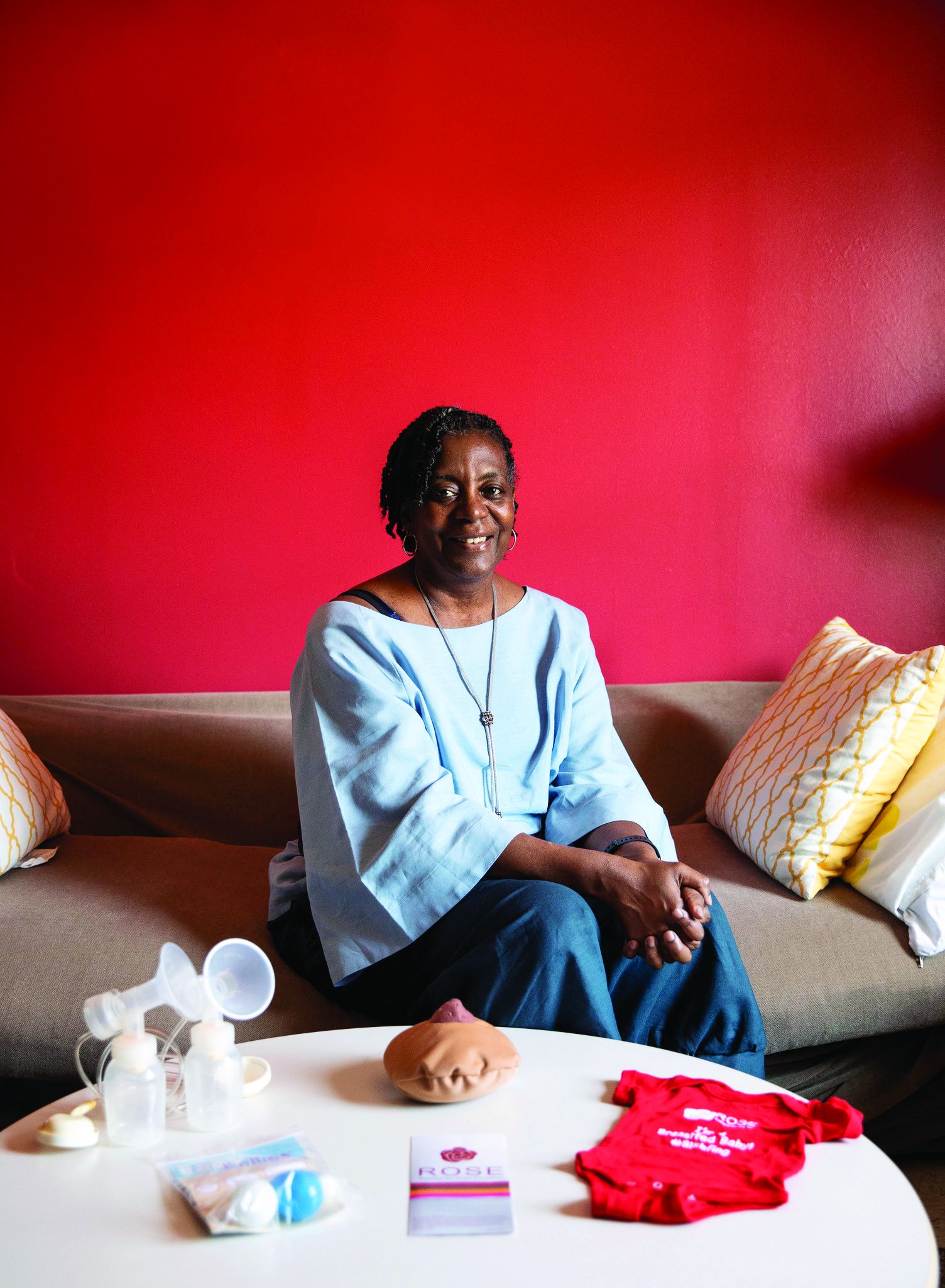
“We have Morehouse pediatricians coming to us just to talk to moms on how they can support them in the community. We have training – Community Transformers – where we talk to moms regardless of their social backgrounds; they’re working in the community helping other moms with breastfeeding, or moms will call them (with) their questions,” Ms. Jackson explained. Ms. Davis is now one of these women.
“Having the women of ROSE support me ... was such a game-changer,” she said. “Sometimes that support that you need, that is helpful, is peer-to-peer,” she noted, adding that ROSE does a lot more than fill in the gaps medically, but also psychologically.”
More pillars, less judgment
TaNeeka Davis pointed out that removing a pillar in the community like ROSE and other grassroots support outside of traditional models will likely have the opposite effect that lawmakers and the lobbyists fighting for certification and licensing aim to achieve, especially if other states adopt the same approach.
“The disparities are going to get even greater, you are going to see bigger gaps, less women even initiating breastfeeding. Why start something that you can’t finish? You can’t tell me that making laws that limit the amount of help we are able to get when it comes to breastfeeding will not have a detrimental effect – health effects – later in life,” she said.
Neither Ms. Jackson nor Ms. Davis believe that medical professionals should be replaced but rather that adjunctive, community-based help is integral for bridging the breastfeeding divide.
As clinicians, “we have to go beyond not judging to trying to figure out where people are, to meet your patients where they are,” said Dr. Furman. “It’s like the difference between cultural competence and cultural humility, which is more of an ongoing process.
Dr. Furman and Ms. Davis report no relevant financial relationships. Ms. Cavanaugh is the coattorney on the lawsuit. Ms. Jackson is employed by ROSE.
*This story was updated on Feb. 11, 2022.
From the American Medical Association to the Centers for Disease Control and Prevention, health equity is the topic de jour. But how do you get health professionals, lawmakers, lactation providers, and the community on the same page, especially when it comes to addressing breastfeeding disparities?
It depends on who you ask.
In Georgia, a 2018 lawsuit challenging a State Legislature Bill directed toward lactation providers sits on the desk of a trial court judge, with a decision due any day now. The bill requires these providers to be licensed in order to continue to practice and receive compensation, a move that not only threatens the health of mothers and infants, but also jeopardizes a key component of Healthy People 2030: improving breastfeeding initiation, duration, and exclusivity among African American women. A similar bill is in Committee in the New York State Legislature.
“If the Act takes effect, it will force an estimated 800 different practitioners out of business and leave only 162 International Board Certified Lactation Counselors (IBCLCs) for the whole state,” Jaimie Cavanaugh, an attorney at the Institute for Justice and plaintiff coattorney said in an interview.
Ms. Cavanaugh also said that geographical data for the 162 IBCLCs demonstrate that they primarily work in urban vs. rural areas, and mostly in formal settings, factors that will further exacerbate disparities and limit access to much needed resources.
Bridging the breastfeeding divide
While overall breastfeeding initiation rates in the United States have steadily increased over the past decade from 72% to roughly 84%, only a quarter of infants are exclusively breastfed through 6 months, a rate well below the Healthy People 2030 goal of 42.4% (and American Academy of Pediatrics recommendations). Comparatively, breastfeeding initiation (75.8%) and exclusivity (17.2%) rates among African-American women are considerably lower.
The effects are great: Breastfed infants have lower risks for asthma, obesity, and type 1 diabetes, while mothers who breastfeed have lower risks for hypertension, type 2 diabetes, and gynecological cancers. Notably, most of these conditions disproportionately affect African Americans, compared with Whites and other ethnicities.
A key to changing these disparities appears to lie with the type of health care provided as well as the ease by which mothers can access it.
For example, findings of a small cross-sectional study published Jan. 31 in the Journal of Racial and Ethnic Health Disparities highlight the importance of a broad umbrella of support for African American mothers’ feeding choices. Not only does this umbrella include medical professionals and IBCLCs, but also certified lactation counselors (CLCs), peer counselors trained under the National Special Supplemental Nutrition Program for Women, Infants, and Children, partners, family, and the community at-large.
“We thought we were doing it right,” Lydia Furman, MD, lead study author and pediatric specialist at University Hospitals Rainbow Babies & Children’s Ahuja Center for Women & Children in Cleveland, told this news organization. “We have a WIC peer helper, an African American IBCLC and an African American CLC, and a breastfeeding support group twice a week but nobody was using these resources.”
One of the most important findings of the study – which aimed to understand factors driving breastfeeding practices and identify supports – was that women want help when they need it. “It doesn’t mean that you can’t have resources that are available during the day, but it means that a patient support group at 11 a.m. on Tuesday doesn’t help at all if you need it Monday at 2 a.m.” Dr. Furman said.
Take TaNeeka Davis, a 34-year-old mother of three residing outside of Atlanta whose personal experience mimics those of the women in Dr. Furman’s study. “I did breastfeed my first child; when I was in the hospital. I saw lactation one time and he latched perfectly but when I left the hospital, I couldn’t get him to latch anymore,” she said.
Ms. Davis explained that she was told that she would have to wait 2-3 weeks before she was able to meet again with a lactation specialist, so she found herself supplementing with formula, and eventually seeking nontraditional help.
“The traditional medical model does not allow for me to be able to reach out and talk to my doctor immediately, does not allow me to be like, ‘Hey, can you call me back in the next 15-20 minutes or an hour because my baby’s very fussy,’ ” Ms. Davis said. “I don’t have that kind of support.”
It takes a village
A 2017 Cochrane review reinforces the value of providing women with predictable, tailored, and multifaceted breastfeeding support offered by professional or lay/peer people or a combination of both.
This model is embodied in ROSE, a nonprofit organization dedicated to eliminating breastfeeding disparities and barriers experienced by mothers of color, including inadequate medical or family support, lack of shared decision-making, recognition of financial or psychological challenges, and historical antecedents. Many of these women’s ancestors were forced to wet-nurse slave masters’ children instead of breastfeeding their own children.
One of several national organizations solely dedicated to this issue, ROSE offers a variety of services and resources ranging from lactation counseling and peer support training programs to training for health professionals (for example, pediatricians, nurses) that serve communities of color. A companion arm (ROBE, Reaching Our Brothers Everywhere) aims to bring men into the fold through breastfeeding education and peer-to-peer connection. All of these services are provided in a judgment-free, culturally sensitive environment.
“We need to look not only into maternal health issues ... but also offer support to people who are working in the birthing community,” cofounder Mary N. Jackson, a CLC, WIC lactation consultant, and former president of the Georgia Breastfeeding Coalition said in an interview.
“We have Morehouse pediatricians coming to us just to talk to moms on how they can support them in the community. We have training – Community Transformers – where we talk to moms regardless of their social backgrounds; they’re working in the community helping other moms with breastfeeding, or moms will call them (with) their questions,” Ms. Jackson explained. Ms. Davis is now one of these women.
“Having the women of ROSE support me ... was such a game-changer,” she said. “Sometimes that support that you need, that is helpful, is peer-to-peer,” she noted, adding that ROSE does a lot more than fill in the gaps medically, but also psychologically.”
More pillars, less judgment
TaNeeka Davis pointed out that removing a pillar in the community like ROSE and other grassroots support outside of traditional models will likely have the opposite effect that lawmakers and the lobbyists fighting for certification and licensing aim to achieve, especially if other states adopt the same approach.
“The disparities are going to get even greater, you are going to see bigger gaps, less women even initiating breastfeeding. Why start something that you can’t finish? You can’t tell me that making laws that limit the amount of help we are able to get when it comes to breastfeeding will not have a detrimental effect – health effects – later in life,” she said.
Neither Ms. Jackson nor Ms. Davis believe that medical professionals should be replaced but rather that adjunctive, community-based help is integral for bridging the breastfeeding divide.
As clinicians, “we have to go beyond not judging to trying to figure out where people are, to meet your patients where they are,” said Dr. Furman. “It’s like the difference between cultural competence and cultural humility, which is more of an ongoing process.
Dr. Furman and Ms. Davis report no relevant financial relationships. Ms. Cavanaugh is the coattorney on the lawsuit. Ms. Jackson is employed by ROSE.
*This story was updated on Feb. 11, 2022.
Post–COVID vaccine AHA cases raise eyebrows in Italy
“The overall number of cases observed does not allow ... any definitive conclusion over a possible causal relationship between SARS-CoV-2 vaccination and AHA, which would need more epidemiological and pharmacovigilance data about suspected vaccine-related adverse events,” Maria Cristina Leone, MD, of Azienda USL-IRCCS di Reggio Emilia (Italy), and colleagues reported online on Jan. 19, 2022, in a letter to the editors of Thrombosis Research.
The cases, observed in Reggio Emilia during the first 8 months of the vaccination campaign, occurred following receipt of mRNA BNT162b2 (Pfizer-BioNTech) vaccine. The AHA patients included two men and two women who ranged in age from 67 to 86 years.
During this time frame, 235,597 people received at least one dose of BNT162b2 vaccine, the authors noted.
In the 5 years prior, from January 2016 to December 2020, only zero to two cases of AHA were observed each year, totaling five cases, or 1.9 cases per million people/year. These numbers are in line with the estimated incidence of the disease, the researchers noted, adding that “it should nonetheless be underlined that vaccination benefits exceed potential side effects and play a central role in individual and public health to effectively protect people from COVID-19 and stop the pandemic.”
However, they also wrote that the “unusual observation of four cases of a rare disease during the first months of the vaccination campaign in our province could be of interest and could sensitize health care personnel toward a possible complication of SARS-CoV-2 immunization.”
AHA is a rare autoimmune disease caused by neutralizing autoantibodies against coagulation factor VIII. It is mainly associated with malignancy, autoimmune diseases, certain medications, and postnatal status.
“Sporadic AHA cases have been reported in association with infectious diseases or vaccinations,” the author noted, adding that associations between the BNT162b2 vaccine immune complications, including AHA, have also been reported by other authors.
Three of the four case patients in Reggio Emilia had “at least one common clinical association of AHA,” they found, suggesting that these associations could “reflect susceptibility to autoimmunity potentially triggered by vaccination.”
“Case four died due to complications from sepsis after being treated with steroid and rituximab, whereas the first three cases underwent clinical and laboratory remission after immunosuppressive therapy, and no relapse has been observed during follow-up, as in the other two cases reported: This could suggest a more favorable prognosis in respect to other non–vaccine-associated cases, but longer-term data are definitely needed,” they concluded.
The authors reported having no disclosures.
FROM THROMBOSIS RESEARCH
AAP approves CDC’s child/adolescent vax schedule for 2022
In a policy statement published online Feb. 17 in Pediatrics, the AAP said the updated recommendations differ little from those released last year by the Advisory Committee on Immunization Practices of the Centers for Disease Control and Prevention.
“The only significant change this year was to add the dengue vaccine to the schedule,” Sean T. O’Leary, MD, MPH, vice chair of the AAP’s 2021-2022 Committee on Infectious Diseases and a coauthor of the statement, told this news organization. “But that is really only relevant for children living in endemic areas, primarily Puerto Rico but some other smaller U.S .territories as well.”
Dengue fever also is endemic in American Samoa and the U.S. Virgin Islands.
Notably, a new section has been added on routine recommendations for use of the Dengvaxia vaccine.
The 2022 policy statement addresses regular immunization of children from birth to 18 years and catch-up vaccination for those aged 4 months to 18 years. In addition to the AAP, multiple complementary physician and nurse organizations have approved the updates. The ACIP schedule is revised annually to reflect current recommendations on vaccines licensed by the U.S. Food and Drug Administration.
Most of the other changes this year involve minor updates to clarify language or improve usability. “CDC and AAP are always working to make the schedule as user-friendly as possible, with improvements made every year,” Dr. O’Leary, professor of pediatric infectious diseases at the University of Colorado at Denver, Aurora, said.
In terms of physician acceptance, he added, “I don’t think any of the changes would be considered controversial.”
Among other updates and clarifications:
- For Haemophilus influenzae type b (Hib) vaccination, the text now includes recommendations for the hexavalent Vaxelis vaccine (diphtheria, tetanus, pertussis, polio, Hib, and hepatitis B) for both routine and catch-up vaccination.
- For hepatitis A, the relevant note has been updated to clarify the age for routine vaccination.
- For human papillomavirus (HPV), the note now clarifies when an HPV series is complete with no additional dose recommended.
- The special situations section has been amended to specify which persons with immunocompromising conditions such as HIV should receive three doses of HPV vaccine regardless of age at initial vaccination.
- For measles, mumps, and rubella, routine vaccination now includes recommendations on the combination measles, mumps, rubella, and varicella vaccine.
- For meningococcal serogroup A, C, W, and Y vaccines, the augmented text explains when these can be simultaneously administered with serogroup B meningococcal vaccines, preferably at different anatomic sites. The language for the dosing schedule for Menveo vaccination in infants also has been clarified.
- In the catch-up immunization schedule for late-starting children aged 4 months to 18 years, the text on Hib has been changed so that the minimum interval between dose two and dose three now refers to Vaxelis, while reference to the discontinued Comvax (Hib-Hep B) vaccine has been removed.
As in other years, graphic changes have been made to table coloration and layout to improve accessibility. And as before, the 2022 childhood and adolescent immunization schedule has been updated to ensure consistency between its format and that of the 2022 adult immunization schedules.
The AAP committee stressed that clinically significant adverse events after immunization should be reported to the Vaccine Adverse Event Reporting System.
The full 2022 schedule can be found on the CDC’s website.
A version of this article first appeared on Medscape.com.
Slow-Growing Pink Nodule in an Active-Duty Service Member
The Diagnosis: Leishmaniasis
Hematoxylin and eosin staining of the tissue specimen revealed a dense histiocytic infiltrate with scattered lymphocytes and neutrophils. There were round to oval basophilic structures within the macrophages consistent with amastigotes. Giemsa staining was not necessary to visualize the organisms. The infiltrate abutted the overlying epidermis, which was acanthotic with pseudoepitheliomatous hyperplasia. There were collections of neutrophils, parakeratosis, and a serum crust overlying the epidermis (Figure). Clinical and histologic findings, as well as travel history, led to a diagnosis of cutaneous leishmaniasis (CL).

Leishmaniases is a group of diseases caused by a parasitic infection with flagellated protozoa of the genus Leishmania. There are more than 20 different Leishmania species that are pathogenic to humans, all presenting with cutaneous findings. The presentation depends on the inoculating species and the host cellular immune response and includes cutaneous, mucosal, and visceral involvement. The disease is transmitted via the bite of an infected bloodsucking female sand fly.1 There are approximately 30 different species of sand flies that are proven to be vectors of the disease, with up to 40 more suspected of involvement in transmission, predominantly from the genera Phlebotomus (Old World) and Lutzomyia (New World).1,2 There are an estimated 1 to 2 million new cases of cutaneous leishmaniasis diagnosed annually in 70 endemic countries of the tropics, subtropics, and southern Europe.1,3,4
The differential diagnosis included cutaneous tuberculosis, which can have a similar progression and clinical appearance. Cutaneous tuberculosis starts as firm, reddish-brown, painless papules that slowly enlarge and ulcerate.5 It may be further differentiated on histopathology by the presence of tuberculoid granulomas, caseating necrosis, and acid-fast bacilli, which are easily detected in early lesions but are less prevalent after the granuloma develops.6 Sporotrichosis presents as a nodule, which may or may not ulcerate, on the extremities. However, the classic morphology is a sporotrichoid pattern, which describes the initial lesion plus subcutaneous nodular spread along the lymphatics.7 On histology, sporotrichosis has a characteristic “sporotrichoid asteroid” comprised of the yeast form surrounded by eosinophilic hyaline material in raylike processes that are found in the center of suppurative granulomas or foci.8
Atypical mycobacteria, principally Mycobacterium marinum (swimming pool granuloma) and Mycobacterium ulcerans (Buruli ulcer), are capable of causing cutaneous infections. They may be differentiated histologically by a neutrophilic infiltrate of poorly formed granulomas without caseation and extensive coagulative necrosis with little cellular infiltrate, respectively.6 Histoplasma capsulatum also infects histiocytes and may appear similar in size and shape; however, histoplasmosis is surrounded by a pseudocapsule and evenly spaced.8
Conversely, the histology of leishmaniasis lacks a pseudocapsule. The amastigotes may form the classic marquee sign by lining the periphery of the macrophage or they can be randomly spaced. Classically, the epidermis shows hyperkeratosis and acanthosis. Sometimes atrophy, ulceration, or intraepidermal abscesses also can be observed. Pseudoepitheliomatous hyperplasia can be seen in some long-standing lesions.1,4 Many of these findings were observed on hematoxylin and eosin staining from a punch biopsy obtained from the center of the lesion in our patient. For further delineation, a speciation kit was obtained from Walter Reed National Military Medical Center (Bethesda, Maryland). A second punch biopsy was obtained from the lesion edge, sectioned into 4 individual pieces, and placed in Schneider tissue culture medium. It was sent for tissue culture, polymerase chain reaction, and histology. Polymerase chain reaction analysis was positive for Leishmania, which was further identified as Leishmania tropica by tissue culture.
Leishmania tropica (Old World CL) commonly causes CL and is endemic to Central Asia, the Middle East, parts of North Africa, and Southeast Asia. Old and New World CL start as a small erythematous papule after a bite from an infected female sand fly. The papule develops into a nodule over weeks to months. The lesion may ulcerate and typically heals leaving an atrophic scar in months to years.1 Speciation of CL is important to guide therapy.
Leishmania mexicana, a New World species that commonly causes CL, classically is found in Central and South America, but there also have been documented cases in Texas. A 2008 case series identified 9 cases in northern Texas in residents without a travel history to endemic locations.9 Similarly, a cross-sectional study identified 41 locally endemic cases of CL over a 10-year period (2007-2017) in Texas; 22 of these cases had speciation by polymerase chain reaction, and all cases were attributed to L mexicana.10
In the United States, CL classically has been associated with travelers and military personnel returning from the Middle East; however, a growing body of literature suggests that it may be endemic to Texas, where it is now a reportable disease. Physicians should have an increased awareness of this entity and a high index of suspicion when treating patients with nonhealing cutaneous lesions.
- Bravo F. Protozoa and worms. In: Bolognia JL, Schaffer JV, Cerroni L, eds. Dermatology. 4th ed. WB Saunders Co; 2018:1470-1502.
- Killick-Kendrick R. The biology and control of phlebotomine sandflies. Clin Dermatol. 1999;17:279-289.
- Reithinger R, Dujardin JC, Louzir H, et al. Cutaneous leishmaniasis. Lancet Infect Dis. 2007;7:581-596.
- Patterson J. Protozoal infections. Weedon’s Skin Pathology. 5th ed. Elsevier; 2021:787-795.
- Ramos-e-Silva M, Ribeiro de Castro MC. Mycobacterial infections. In: Bolognia JL, Schaffer JV, Cerroni L, eds. Dermatology. 4th ed. WB Saunders Co; 2018:1296-1318.
- Patterson J. Bacterial and rickettsial infections. Weedon’s Skin Pathology. 5th ed. Elsevier; 2021:673-709.
- Elewski B, Hughey L, Hunt K, et al. Fungal diseases. In: Bolognia JL, Schaffer JV, Cerroni L, eds. Dermatology. 4th ed. WB Saunders Co; 2018:1329-1363.
- Patterson J. Mycoses and algal infections. Weedon’s Skin Pathology. 5th ed. Elsevier; 2021:721-755.
- Wright NA, Davis LE, Aftergut KS, et al. Cutaneous leishmaniasis in Texas: a northern spread of endemic areas [published online February 4, 2008]. J Am Acad Dermatol. 2008;58:650-652. doi:10.1016/j .jaad.2007.11.008.
- McIlwee BE, Weis SE, Hosler GA. Incidence of endemic human cutaneous leishmaniasis in the United States. JAMA Dermatol. 2018;154:1032-1039. doi:10.1001/jamadermatol.2018.2133
The Diagnosis: Leishmaniasis
Hematoxylin and eosin staining of the tissue specimen revealed a dense histiocytic infiltrate with scattered lymphocytes and neutrophils. There were round to oval basophilic structures within the macrophages consistent with amastigotes. Giemsa staining was not necessary to visualize the organisms. The infiltrate abutted the overlying epidermis, which was acanthotic with pseudoepitheliomatous hyperplasia. There were collections of neutrophils, parakeratosis, and a serum crust overlying the epidermis (Figure). Clinical and histologic findings, as well as travel history, led to a diagnosis of cutaneous leishmaniasis (CL).
Leishmaniases is a group of diseases caused by a parasitic infection with flagellated protozoa of the genus Leishmania. There are more than 20 different Leishmania species that are pathogenic to humans, all presenting with cutaneous findings. The presentation depends on the inoculating species and the host cellular immune response and includes cutaneous, mucosal, and visceral involvement. The disease is transmitted via the bite of an infected bloodsucking female sand fly.1 There are approximately 30 different species of sand flies that are proven to be vectors of the disease, with up to 40 more suspected of involvement in transmission, predominantly from the genera Phlebotomus (Old World) and Lutzomyia (New World).1,2 There are an estimated 1 to 2 million new cases of cutaneous leishmaniasis diagnosed annually in 70 endemic countries of the tropics, subtropics, and southern Europe.1,3,4
The differential diagnosis included cutaneous tuberculosis, which can have a similar progression and clinical appearance. Cutaneous tuberculosis starts as firm, reddish-brown, painless papules that slowly enlarge and ulcerate.5 It may be further differentiated on histopathology by the presence of tuberculoid granulomas, caseating necrosis, and acid-fast bacilli, which are easily detected in early lesions but are less prevalent after the granuloma develops.6 Sporotrichosis presents as a nodule, which may or may not ulcerate, on the extremities. However, the classic morphology is a sporotrichoid pattern, which describes the initial lesion plus subcutaneous nodular spread along the lymphatics.7 On histology, sporotrichosis has a characteristic “sporotrichoid asteroid” comprised of the yeast form surrounded by eosinophilic hyaline material in raylike processes that are found in the center of suppurative granulomas or foci.8
Atypical mycobacteria, principally Mycobacterium marinum (swimming pool granuloma) and Mycobacterium ulcerans (Buruli ulcer), are capable of causing cutaneous infections. They may be differentiated histologically by a neutrophilic infiltrate of poorly formed granulomas without caseation and extensive coagulative necrosis with little cellular infiltrate, respectively.6 Histoplasma capsulatum also infects histiocytes and may appear similar in size and shape; however, histoplasmosis is surrounded by a pseudocapsule and evenly spaced.8
Conversely, the histology of leishmaniasis lacks a pseudocapsule. The amastigotes may form the classic marquee sign by lining the periphery of the macrophage or they can be randomly spaced. Classically, the epidermis shows hyperkeratosis and acanthosis. Sometimes atrophy, ulceration, or intraepidermal abscesses also can be observed. Pseudoepitheliomatous hyperplasia can be seen in some long-standing lesions.1,4 Many of these findings were observed on hematoxylin and eosin staining from a punch biopsy obtained from the center of the lesion in our patient. For further delineation, a speciation kit was obtained from Walter Reed National Military Medical Center (Bethesda, Maryland). A second punch biopsy was obtained from the lesion edge, sectioned into 4 individual pieces, and placed in Schneider tissue culture medium. It was sent for tissue culture, polymerase chain reaction, and histology. Polymerase chain reaction analysis was positive for Leishmania, which was further identified as Leishmania tropica by tissue culture.
Leishmania tropica (Old World CL) commonly causes CL and is endemic to Central Asia, the Middle East, parts of North Africa, and Southeast Asia. Old and New World CL start as a small erythematous papule after a bite from an infected female sand fly. The papule develops into a nodule over weeks to months. The lesion may ulcerate and typically heals leaving an atrophic scar in months to years.1 Speciation of CL is important to guide therapy.
Leishmania mexicana, a New World species that commonly causes CL, classically is found in Central and South America, but there also have been documented cases in Texas. A 2008 case series identified 9 cases in northern Texas in residents without a travel history to endemic locations.9 Similarly, a cross-sectional study identified 41 locally endemic cases of CL over a 10-year period (2007-2017) in Texas; 22 of these cases had speciation by polymerase chain reaction, and all cases were attributed to L mexicana.10
In the United States, CL classically has been associated with travelers and military personnel returning from the Middle East; however, a growing body of literature suggests that it may be endemic to Texas, where it is now a reportable disease. Physicians should have an increased awareness of this entity and a high index of suspicion when treating patients with nonhealing cutaneous lesions.
- Bravo F. Protozoa and worms. In: Bolognia JL, Schaffer JV, Cerroni L, eds. Dermatology. 4th ed. WB Saunders Co; 2018:1470-1502.
- Killick-Kendrick R. The biology and control of phlebotomine sandflies. Clin Dermatol. 1999;17:279-289.
- Reithinger R, Dujardin JC, Louzir H, et al. Cutaneous leishmaniasis. Lancet Infect Dis. 2007;7:581-596.
- Patterson J. Protozoal infections. Weedon’s Skin Pathology. 5th ed. Elsevier; 2021:787-795.
- Ramos-e-Silva M, Ribeiro de Castro MC. Mycobacterial infections. In: Bolognia JL, Schaffer JV, Cerroni L, eds. Dermatology. 4th ed. WB Saunders Co; 2018:1296-1318.
- Patterson J. Bacterial and rickettsial infections. Weedon’s Skin Pathology. 5th ed. Elsevier; 2021:673-709.
- Elewski B, Hughey L, Hunt K, et al. Fungal diseases. In: Bolognia JL, Schaffer JV, Cerroni L, eds. Dermatology. 4th ed. WB Saunders Co; 2018:1329-1363.
- Patterson J. Mycoses and algal infections. Weedon’s Skin Pathology. 5th ed. Elsevier; 2021:721-755.
- Wright NA, Davis LE, Aftergut KS, et al. Cutaneous leishmaniasis in Texas: a northern spread of endemic areas [published online February 4, 2008]. J Am Acad Dermatol. 2008;58:650-652. doi:10.1016/j .jaad.2007.11.008.
- McIlwee BE, Weis SE, Hosler GA. Incidence of endemic human cutaneous leishmaniasis in the United States. JAMA Dermatol. 2018;154:1032-1039. doi:10.1001/jamadermatol.2018.2133
A 36-year-old active-duty male service member with no notable medical history presented to the dermatology clinic with an asymptomatic nodule on the right forearm that he initially noticed approximately 1 year prior while deployed in Syria and thought that it was a mosquito bite; it continued to enlarge slowly since that time. He attempted self-extraction but was only able to express a small amount of clear fluid. No other therapies had been used. He denied any other symptoms on a review of systems and was not taking any medications. Physical examination revealed a 1.5-cm, erythematous, nonulcerated, pink nodule on the right distal volar forearm without other cutaneous findings. A 4-mm punch biopsy was performed.


Gene therapy: A ‘one and done’ hemophilia B treatment?
The report on the gene therapy treatment, known as etranacogene dezaparvovec (EtranaDez), was released at the Feb. 2-4, 2022, annual meeting of the European Association of Hemophilia and Allied Disorders.
In an interview, study lead author Wolfgang Miesbach, MD, PhD, of University Hospital Frankfurt in Germany, touted the decline in ABR. “This statistically significant reduction not only met the primary endpoint for non-inferiority but also demonstrates clear superiority of etranacogene dezaparvovec to prophylaxis in the lead-in period,” he said. “In addition to that, the quality of life improved significantly, [and] there was an overall favorable safety profile.”
Hemophilia B is much rarer than hemophilia A. In a 2020 report, the CDC estimated that type A accounted for less than a quarter of the 29,761-32,985 cases of U.S. males who had hemophilia from 2012-2018. The rest had type B. Most of the males with hemophilia were white (81.2%) and fairly young (just 20.6% were older than 39).
High adherence and high prices
Factor IX (FIX) replacement therapy aims to boost levels of the blood-clotting protein in patients with severe hepatitis B. However, the intravenous prophylactic treatment requires a “high level of adherence” due to the need for self-administration several times a week, Dr. Miesbach said, adding that the treatment does not reliably prevent bleeding and joint destruction.
Also, the price of FIX replacement therapy in the United States is exorbitant, costing an average of $397,491 a year for the conventional treatment and an average of $788,861 a year for an extended half-life treatment, according to a 2019 report.
The gene therapy treatment, formerly known as AMT-061, “consists of a functional FIX gene with higher activity than the wild-type FIX (Padua variant), together with an AAV (AAV5),” Dr. Miesbach said. “AAV 5 is a vector with high liver tropism to transduce the liver cells and lead to the production of the functional FIX gene there.”
For the new open-label, single-dose, single-arm HOPE-B study, researchers treated 54 adult men with severe or moderately severe HB (FIX ≤2%), 31 with and 23 without preexisting AAV5 neutralizing antibodies. The average age was 41.5, 81.5% had severe cases (FIX<1%), and 25.9% had no bleeds at lead-in.
The participants began 12 months of treatment with gene therapy following a 6-month lead-in period of FIX prophylaxis. All but one completed follow-up.
“Mean FIX activity was 39.0 IU/dL (±18.7; 8.2, 97.1) (standard deviation; min, max) at month 6 and 36.9 IU/dL (±21.4; 4.5, 122.9) at month 18,” the researchers reported. ABR dropped by 64% from the lead-in period to the 12-month treatment period (4.19 vs. 1.51, P = .0002), and FIX-treated bleeds fell by 77% (ABR=3.65 vs. 0.83, P < .0001).
Fifty-two of 54 patients stopped full-dose prophylaxis and didn’t return to it. Mean unadjusted annualized FIX use dropped by 97% overall from the lead-in period to months 13-18 (257,338.8 vs. 8,486.6 IU/year/participant).
Thirty-seven participants experienced 92 treatment-related adverse events such as abnormal alanine aminotransferase (16.7%), headache (14.8%), influenza-like illness (13.0%), infusion-related infection (13.0%), and abnormal aspartate aminotransferase (9.3). Researchers determined 74 (80.4%) of the adverse effects were mild.
“Transaminase increases were reported, and corticosteroids were required in nine participants, but the mean duration of corticosteroids, including taper, was only 79 days,” Dr. Miesbach said.
“There was no prophylactic use of steroids in this study. FIX expression was maintained. One death was found to be unrelated to study treatment. One case of hepatocellular carcinoma, which has been reported in detail previously, was reported. But after detailed molecular analysis, this was found to be unrelated to study treatment,” he noted.
Quality of life scores improved by 21.5%-28.78%. The P values, ranging from < .0001 to .0036, were considered to be “nominally significant” due to analysis limitations.
A ‘one and done’ treatment
While the trial is expected to continue until 2025, no further treatment with etranacogene dezaparvovec was given. “Gene therapy is a ‘one and done’ treatment,” Dr. Miesbach said. “According to our current knowledge, it cannot be repeated.”
No information about the expected cost of the treatment is available. CSL Behring, which licensed global rights for the gene therapy from developer uniQure, is expected to seek Food and Drug Administration approval this year.
The trial was funded by CSL Behring. Dr. Miesbach and other study authors report various disclosures including support from CSL Behring and uniQure. Some authors are employees of CSL Behring and uniQure.
REPORTING FROM EAHAD