User login
Many Americans missing an opportunity to prevent dementia
(ADRD), including hypertension, low levels of physical activity, and obesity, new research shows.
Data from the Centers for Disease Control and Prevention reveal that among nearly 162,000 adults aged 45 and older who were surveyed in 2019 as part of the Behavioral Risk Factor Surveillance System (BRFSS), nearly half had high blood pressure and did not achieve aerobic physical activity recommendations. These were the two most common modifiable risk factors for ADRD.
In addition, more than one-third (35%) of adults were obese, 19% had diabetes, 18% had depression, 15% were smokers, 11% had hearing loss, and 10% were binge drinkers.
The findings were published online in the CDC’s Morbidity and Mortality Weekly Report.
A missed prevention opportunity
More than 1 in 10 (11.3%) adults surveyed reported subjective cognitive decline (SCD), an early indicator of possible future ADRD.
The prevalence of SCD increased from about 4% among adults with no modifiable risk factors for ADRD to 25% for those with four or more risk factors.
Adults with SCD were more apt to report having almost all modifiable risk factors and were more likely to report four or more risk factors (34%) than were peers without SCD (13%)
The prevalence of SCD ranged from a high of about 29% in those with depression and 25% in those with hearing loss to 11% in those who reported binge drinking.
In line with previous research, the findings indicate that American Indian or Alaska Native, Black or African American, and Hispanic populations were more likely to have modifiable risk factors for ADRD than other racial groups, the researchers reported.
The CDC’s National Healthy Brain Initiative supports culturally tailored interventions that address ADRD risk factors specifically in these populations.
In 2021, the federal government’s National Plan to Address Alzheimer’s Disease was updated to include a new goal to reduce risk factors for ADRD.
“Given the prevalence of modifiable risk factors for ADRD and anticipated growth of the older adult population and those with ADRD, this new goal has the potential to benefit a large proportion of U.S. adults,” the investigators wrote.
“In addition to helping patients discuss concerns about memory loss, health care professionals should also screen patients for modifiable risk factors, counsel patients with risk factors, and refer them to effective programs and interventions where recommended,” they advised.
A recent report from the Lancet Commission on Dementia Prevention, Intervention, and Care found that modifying 12 risk factors over the life course could delay or prevent 40% of dementia cases.
A version of this article first appeared on Medscape.com.
FROM MMWR
RSV kills 100,000 kids under age 5 a year worldwide
Respiratory syncytial virus (RSV) caused more than 100,000 deaths in children under age 5 years globally in 2019, according to an analysis published online in The Lancet.
Researchers, led by You Li, PhD, of Nanjing (China) Medical University, found that nearly half of those (more than 45,000) occurred in children younger than 6 months old.
They estimated that RSV causes 1 in 50 deaths among children under 5 years old, and 1 in 28 deaths in children under 6 months old.
Additionally, RSV is responsible for an estimated 3.6 million hospital admissions globally each year, according to the report.
This analysis is the first to sift RSV disease burden into narrow age brackets, the authors said.
The numbers highlight that almost all of the deaths (97%) were in low- and middle-income countries.
Messages for prevention
Tina Hartert, MD, MPH, a professor in the division of allergy, pulmonary, and critical care medicine at Vanderbilt University, Nashville, Tenn., who was not part of the study, wrote in an invited commentary that these findings will be important in RSV prevention.
Among the most notable findings, she wrote, is the heavy mortality in the 0- to 6-month age group, which she notes is “the age group targeted by vaccination during pregnancy and birth-dose immunoprophylaxis.”
Dr. Hartert, who coauthored the commentary with Justin R. Ortiz, MD, MS, with the Center for Vaccine Development and Global Health, University of Maryland, Baltimore, told this news organization, “RSV is a respiratory virus that infects nearly every child by the time they are 2-3 years of age, with severe infection and death most common in the youngest infants. Vaccines that prevent the most severe infections in these young infants will likely be one of the best ways to prevent these severe infections and death.”
Though the authors found most deaths occur in low- and middle-income countries, RSV is one of the most common reasons for infant hospitalization in the US and affects 1% to 3% of infants, half of whom are full-term and otherwise healthy, Dr. Hartert said.
It is also one of the most common causes of infant lower respiratory tract infection in young children in the United States, she said, and it causes the most severe disease at the age extremes, with older adults experiencing significant morbidity with RSV.
Dr. Li said in an interview that although the team did not focus on reporting country-specific estimates in this work, their previous work, resulted in estimates of 98,000-155,000 RSV-related hospitalizations in children under 5 years old in the United States in 2019. Between 65,000 and 86,000 were in infants less than 1 year old.
Currently, he said, the only available RSV prophylaxis is palivizumab (Synagis), which is expensive and given only to high-risk infants in high-income countries, including the United States.
“There have been a number of promising RSV prophylactic products including maternal vaccine and monoclonal antibodies that have the potential for targeting the general infant population – not just high-risk infants – in late-phase clinical trials,” he said. “Our estimates of RSV-related disease burden will help anticipate the impact of future RSV immunization programs.”
Pandemic changed patterns
This research was completed before the COVID-19 pandemic, and it is not yet known how that could affect RSV disease burden long term.
However, Dr. Hartert said, RSV circulation has been significantly changed during the pandemic, both in intensity and timing, likely because of a combination of COVID and the public health preventive measures.
“As people return to normal activities and the public health measures put in place to stop the spread of COVID are eased, we are likely to see increases in circulation of RSV and return to its circulation during the winter months – typically similar to circulation of flu – from November through March in temperate climates in the northern hemisphere,” she said.
A coauthor of the paper, Harish Nair, PhD, with the Centre for Global Health, Usher Institute, University of Edinburgh, said in a press release that their findings have particular significance as COVID restrictions ease around the globe.
“The majority of the young children born in the last 2 years have never been exposed to RSV (and therefore have no immunity against this virus),” Nair wrote.
Most deaths occurring outside hospitals
A challenge in reducing the deaths in those 5 years old and younger is that most (76%) of deaths are happening in the community outside hospitals.
The authors wrote: “For every RSV-associated acute lower respiratory infection in-hospital death, we estimate approximately three more deaths attributable to RSV in the community.”
The percentage dying outside hospitals is even larger (81%) in low- to middle-income countries.
This work built on a previous review by the team that analyzed 317 studies. They updated their search with 113 new eligible studies and unpublished data from 51 papers published between Jan. 1, 2017, and Dec. 31, 2020.
The authors acknowledged some limitations, including variations in study settings and in definitions for acute lower respiratory infection, healthcare access, and eligibility for RSV testing.
The study was funded by EU Innovative Medicines Initiative Respiratory Syncytial Virus Consortium in Europe. Dr. Li reported grants from Wellcome Trust and the World Health Organization outside the submitted work. Dr. Hartert, Dr. Ortiz, and Dr. Nair disclosed no relevant financial relationships.
A version of this article first appeared on Medscape.com.
FROM THE LANCET
Persistent dry cough
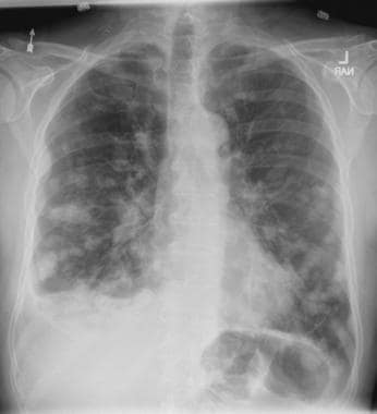
On the basis of the patient's presentation, history, and imaging results, the likely diagnosis is metastatic small cell lung cancer (SCLC). Most patients with SCLC present with hematogenous metastases; only about one third present with limited disease confined to the chest that is amenable to multimodal therapy. Patients with SCLC often present with symptoms of widespread metastases, including weight loss, bone pain, and neurologic compromise. It is uncommon for patients to present with a solitary peripheral nodule. In earlier stages, the differential diagnosis of SCLC spans other neuroendocrine lung tumors and NSCLC, in particular, basaloid carcinoma, extrapulmonary small cell tumors, and lymphoma.
Because the concentration of circulating tumor cells in SCLC is among the highest of any solid tumor, SCLC is characterized by a rapid doubling time, high growth fraction, and early development of widespread metastases. It is likely for this reason that CT screening does not seem effective in detecting early-stage SCLC. Common sites of SCLC metastasis are the contralateral lung, the brain, liver, adrenal glands, and bone. Most cases of SCLC are caused by smoking.
Metastatic spread is often evident on radiologic exam, sometimes showing pleural and pericardial effusions. In general, workup for SCLC includes imaging (contrast-enhanced CT or F-FDG PET–CT of the chest, abdomen, and pelvis and brain MRI with contrast), blood tests (cell count, liver and kidney function, and lactate dehydrogenase), and ECG. Biopsies are generally procured by bronchoscopy with or without endobronchial ultrasonography; if accessible, a biopsy of a distal metastatic site may be obtained. Diagnosis of SCLC is confirmed by histopathologic examination via cytology.
Patients with extensive-stage SCLC are typically treated with systemic chemotherapy with or without immunotherapy. In the early stages, SCLC is very responsive to cytotoxic therapies, with response rates over 60% even in patients with metastatic disease. Until recently, the only second-line therapy for recurrent metastatic SCLC was the topoisomerase I inhibitor topotecan. However, lurbinectedin was granted accelerated approval for second-line therapy after demonstrating a 35% response rate in a single-arm phase 2 study of 105 patients. In addition, the anti–programmed cell death protein 1 monoclonal antibodies nivolumab and pembrolizumab were granted accelerated approval for third-line use. Finally, the National Comprehensive Cancer Network guidelines note that participation in clinical trials should be strongly encouraged for all patients with SCLC.
Karl J. D'Silva, MD, Clinical Assistant Professor, Department of Medicine, Tufts University School of Medicine, Boston; Medical Director, Department of Oncology and Hematology, Lahey Hospital and Medical Center, Peabody, Massachusetts.
Karl J. D'Silva, MD, has disclosed no relevant financial relationships.
Image Quizzes are fictional or fictionalized clinical scenarios intended to provide evidence-based educational takeaways.

A 58-year-old man presents with a persistent dry cough that has developed over the past 8 weeks. He has lost about 8-10 lb in under 3 months. Height is 5 ft 10 in and weight is 172 lb (BMI 24.7). Although he quit smoking about 15 years ago, his wife still smokes. He has been screened twice for non–small cell lung cancer (NSCLC), most recently a year and a half ago. Chest radiograph shows multiple pulmonary nodules of varying sizes and a small right basal effusion.
Distal radial snuffbox technique comes up short in DISCO RADIAL
Distal radial access is not superior to conventional radial access with regard to radial artery occlusion (RAO) but is a valid alternative for use in percutaneous procedures, according to results of the DISCO RADIAL trial.
The primary endpoint of forearm RAO at discharge was not met, occurring in 0.31% of patients whose radial artery was accessed distally (DRA) at the anatomical snuffbox and in 0.91% of patients with conventional transradial access (TRA) in the intention-to-treat analysis (P = .29).
The DRA group was also twice as likely to crossover to another access point (7.5% vs. 3.7%; P = .002) and to experience radial artery spasm (5.4% vs. 2.7%; P < .015).
“The message first is that if you do a good job with transradial access you can end up with a lower [occlusion] rate,” said coprincipal investigator Adel Aminian, MD, Hôpital Civil Marie Curie, Charleroi, Belgium. “On the other hand, it’s a trade-off between a more demanding puncture for distal radial access but also a simpler hemostatic process, which I think is one of the main advantages of distal radial access.”
The results were presented during the annual meeting of the European Association of Percutaneous Cardiovascular Interventions, and published simultaneously in JACC: Cardiovascular Interventions.
DISCO-RADIAL (Distal Versus Conventional RADIAL Access for Coronary Angiography and Intervention) is the largest trial thus far to compare TRA with the distal radial snuffbox technique, which has shown promise for reducing RAO rates in the recent single-center randomized DAPRAO and ANGIE trials.
The trial was conducted at 15 sites across Europe and Japan in 1,309 patients with an indication for percutaneous coronary procedures using the 6Fr Glidesheath Slender (Terumo). The intention-to-treat population included 657 TRA patients and 650 DRA patients.
The two groups were well matched, with most having a chronic coronary syndrome. Operators had to have performed a minimum of 100 procedures by DRA and follow systematic best practices previously reported by the investigators to prevent RAO, Dr. Aminian said.
The use of DRA did not significantly affect the duration of the coronary procedure (27 minutes vs. 24 minutes with TRA; P = .12) or average radiation dose (1298 mGy vs. 1222 mGy; P = .70).
DRA, however, reduced the need for selective compression devices (88% vs. 99.2%) and shortened the median time to hemostasis from 180 minutes to 153 minutes (P for both < .001).
“These results establish compliance to best practice recommendations for RAO avoidance as a mandatory new reference in transradial practice,” Dr. Aminian concluded. “At the same time, distal radial artery arises as a valid alternative associated with higher crossover rates but with a simpler and shorter hemostasis process.”
A show of hands revealed that about 25% of the audience used distal radial access prior to the presentation but that enthusiasm fell off following the results.
Discussant Hany Eteiba, MD, Glasgow Royal Infirmary, said: “I salute your enthusiasm for presenting a negative trial and you tried to persuade the audience to use the distal radial artery results, but nonetheless.”
Dr. Eteiba said he could see a “potential advantage in the shorter hemostasis time,” and asked whether it might be influencing the rapid turnover for day-case angioplasty.
Dr. Aminian responded that “if you do an angioplasty you have to keep the patient for a certain amount of time, but I think for your nurse work and for the health care resources, having a very short hemostasis time is very interesting. We started with a hemostasis time of 2 hours and now we’ve decreased it to 1 hour and it will decrease even more.”
Session moderator Chaim Lotan, MD, Hadassah-Hebrew University Medical Center, Jerusalem, called DISCO-RADIAL an important study and said, “the question now is what’s the indication in your eyes for using distal radial?”
Dr. Aminian said that one message from the trial is that people who are using transradial access “have to do a better job,” and reminded the audience that RAO rates at many centers are too high, at 10% or upward.
At the same time, Dr. Aminian cautioned that operators wanting to use distal radial access “need to master the technique” or they will “end up with a relatively high failure rate.”
Discussant Eliano Navarese, MD, Nicolaus Copernicus University, Toruń, Poland, said, “I still think that it is a very valid approach, we use it for almost 20 years ... but it is very true, it is very demanding. And the learning curve of 100 cases in the trial maybe needed more cases.”
In an accompanying editorial, Grigorios Tsigkas, MD, PhD, University of Patras, Rio Patras, Greece, and colleagues wrote that the incidence of forearm RAO was “surprisingly low” but could be even lower if the authors administered adequate anticoagulation.
Still, they wrote that distal transradial access “for coronary procedures in combination with the systematic implementation of best practices for RAO prevention may be the final solution against RAO.”
The editorialists suggested that exposure to radiation could be the “main limitation of this novel vascular approach” and that forthcoming trials, such as DOSE, could shed light on this issue.
Increased procedure times in the DISCO RADIAL and ANGIE trials are secondary in stable patients, Dr. Tsigkas said, but could be a limitation in patients presenting with ST-segment elevation myocardial infarction (STEMI). Ongoing research, such as the RESERVE trial from China and a Korean trial, will provide insights into the safety and feasibility of distal transradial access in STEMI.
The study was supported by Terumo Europe. Dr. Aminian reported receiving honoraria or consultation fees from Abbott, Boston Scientific, and Terumo Interventional Systems. Dr. Tsigkas reported having no relevant financial relationships.
A version of this article first appeared on Medscape.com.
FROM EUROPCR 2022
CDC signs off on COVID boosters in children ages 5-11
Centers for Disease Control and Prevention Director Rochelle Walensky, MD, signed off May 19 on an advisory panel’s recommendation that children ages 5 to 11 years should receive a Pfizer-BioNTech COVID-19 vaccine booster dose at least 5 months after completion of the primary series.
The CDC’s Advisory Committee on Immunization Practices (ACIP) voted 11:1, with one abstention, on a question about whether it recommended these additional shots in this age group.
The U.S. Food and Drug Administration on May 17 amended the emergency use authorization (EUA) for the Pfizer-BioNTech COVID-19 vaccine to cover a single booster dose for administration to individuals 5 through 11 years of age.
At the request of CDC staff, ACIP members considered whether there should be softer wording for this recommendation, stating that children in this age group “may” receive a booster. This kind of phrasing would better reflect uncertainty about the course of COVID in the months ahead and allow flexibility for a stronger recommendation in the fall.
ACIP panelists and members of key groups argued strongly for a “should” recommendation, despite the uncertainties.
They also called for stronger efforts to make sure eligible children received their initial COVID-19 shots. Data gathered between November and April show only 14.4% of children ages 5 to 11 in rural areas have received at least one dose of COVID-19 vaccination, with top rates of 39.8% in large urban communities and 36% in larger suburban regions, CDC staff said.
CDC staff also said nearly 40% of parents in rural areas reported that their children’s pediatricians did not recommend COVID-19 vaccinations, compared with only 8% of parents in urban communities. These figures concerned ACIP members and liaisons from medical associations who take part in the panel’s deliberations but not in its votes.
“People will hear the word ‘m-a-y’ as ‘m-e-h’,” said Patricia Stinchfield, RN, MS, who served as the liaison for National Association of Pediatric Nurse Practitioners to ACIP. “I think we need to add urgency” to efforts to increase use of COVID vaccinations, she said.
Voting no on Thursday was Helen Keipp Talbot, MD, of Vanderbilt University. She explained after the vote that she is in favor of having young children vaccinated, but she’s concerned about the low rates of initial uptake of the COVID-19 shots.
“Boosters are great once we’ve gotten everyone their first round,” she said. “That needs to be our priority in this.”
Sandra Fryhofer, MD, the American Medical Association’s liaison to ACIP, stressed the add-on benefits from more widespread vaccination of children against COVID. Dr. Fryhofer said she serves adults in her practice as an internal medicine physician, with many of her patients being at high risk for complications from COVID.
Too many people are assuming the spread of infections in the community has lessened the risk of the virus, Dr. Fryhofer said.
“Not everyone’s had COVID yet, and my patients will be likely to get COVID if their grandchildren get it. We’re going through pandemic fatigue in this country,” she said. “Unfortunately, masks are now more off than on. Winter’s coming. They’re more variants” of the virus likely to emerge.
The data emerging so far suggests COVID vaccines will become a three-dose medicine, as is already accepted for other shots like hepatitis B vaccine, Dr. Fryhofer said.
Data gathered to date show the vaccine decreases risk of hospitalization for COVID and for complications such as multisystem inflammatory syndrome in children (MIS-C), she said.
“The bottom line is children in this age group are getting COVID,” Dr. Fryhofer said of the 5- to 11-year-olds. “Some do fine. Some are getting real sick. Some are hospitalized, some have died.”
At the meeting, CDC staff cited data from a paper published in the New England Journal of Medicine in March showing that vaccination had reduced the risk of hospitalization for COVID-19 among children 5 to 11 years of age by two-thirds during the Omicron period; most children with critical COVID-19 were unvaccinated.
COVID-19 led to 66 deaths among children ages 5 to 11 in the October 2020 to October 2021 timeframe, said ACIP member Matthew F. Daley, MD, of Kaiser Permanente Colorado during a presentation to his fellow panel members.
Parents may underestimate children’s risk from COVID and thus hold off on vaccinations, stressed AMA President Gerald E. Harmon, MD, in a statement issued after the meeting.
“It is concerning that only 1 in 3 children between the ages of 5 and 11 in the United States have received two doses of the vaccine, in part because parents believe them to be at lower risk for severe disease than adults,” Dr. Harmon said. “But the Omicron variant brought about change that should alter that calculus.”
Responding to early data
As Dr. Fryhofer put it, the medical community has been learning in “real time” about how COVID vaccines work and how to use them.
The EUA granted on May 17 for booster shots for children ages 5 to 11 was based on an analysis of immune response data in a subset of children from an ongoing randomized placebo-controlled trial, the FDA said.
Antibody responses were evaluated in 67 study participants who received a booster dose 7 to 9 months after completing a two-dose primary series of the Pfizer-BioNTech COVID-19 Vaccine. The EUA for the booster shot was intended to respond to emerging data that suggest that vaccine effectiveness against COVID-19 wanes after the second dose of the vaccine, the FDA said.
CDC seeks help tracking vaccine complications
At the ACIP meeting, a top CDC vaccine-safety official, Tom Shimabukuro, MD, MPH, MBA, asked physicians to make sure their patients know about the agency’s V-Safe program for gathering reports from the public about their experiences with COVID vaccines. This is intended to help the CDC monitor for side effects of these medications.
“We need your help,” he said during a presentation about adverse events reported to date in children ages 5 to 11 who took the Pfizer vaccine.
About 18.1 million doses of Pfizer-BioNTech vaccine have been administered to children ages 5 to 11 years in the United States so far. Most of the reports of adverse events following vaccination were not serious, he said. But there were 20 reports of myocarditis verified to meet CDC case definition among children ages 5 to 11 years.
One case involved a death with histopathologic evidence of myocarditis on autopsy. The CDC continues to assist with case review, he said.
A version of this article first appeared on Medscape.com.
Pancreatic involvement in COVID-19: What do we know?
MADRID – It involves the relationship between COVID-19 and new diagnoses of diabetes and blood glucose disorders, among others, in the post–COVID-19 period. These topics were addressed at the XXXIII National Congress of the Spanish Diabetes Society. They were also the central theme of the inaugural conference, Pancreatic Involvement During COVID-19: From Preclinical Studies to Clinical Relevance, which was led by Alexander Kleger, MD, PhD, head of the department of pancreatology at the Ulm (Germany) University Clinic for Internal Medicine.
The chair of the scientific committee of the congress, Franz Martín, MD, launched the conference by noting that the work of Dr. Kleger and his team has made it possible to ascertain that SARS-CoV-2 can infect pancreatic beta cells that produce insulin. This observation may help in understanding why patients with COVID-19 sometimes experience symptoms related to greater difficulty regulating blood glucose.
“In addition, the German expert and his group have described the abnormalities that occur in beta cells when they are infected by SARS-CoV-2, something especially important, given that knowledge of these abnormalities may be of great importance to understanding the possible appearance of more cases of diabetes in the future,” Dr. Martín added.
“Our data identify the human pancreas as a target of SARS-CoV-2 infection and suggest that pancreatic beta cell involvement could contribute to the metabolic dysregulation seen in COVID-19 patients,” Dr. Kleger pointed out.
In his speech, Dr. Kleger reviewed the evidence on the effects of SARS-CoV-2 that has been garnered since the start of the pandemic, and he presented his research group’s findings on the impact at the pancreatic level.
“Since March 2020, it has been seen that COVID-19 affected the pancreas, and studies published in August of that same year clearly spoke of both a worsening of diabetes and an increase in new cases of this disease diagnosed after SARS-CoV-2 infection. Also, the data showed how hospitalized patients with no previous history of diabetes experienced rapid increases in glucose levels 5 days after admission,” Dr. Kleger said.
Angiotensin-converting enzyme 2
As an example of the pace at which evidence on the pancreatic impact of this virus has been evolving, Dr. Kleger referred to early studies that found no angiotensin-converting enzyme 2 receptor on cells of the endocrine and exocrine pancreas. “To our surprise, in our work, we did observe the obvious presence of angiotensin-converting enzyme 2 specifically expressed in human pancreatic beta cells, something confirmed by other investigations. Another surprising aspect was verifying that the viral infection lasts longer in the pancreas than in the lungs,” said the expert.
These findings caused the researchers to realize that SARS-CoV-2 may be directly or indirectly associated with diabetes. “It is currently the subject of debate whether it may be a direct effect, infecting or directly reaching the pancreatic beta cells, or whether this involvement is a result of the effect of the infection at systemic level, in the context of the cytokine storm and the proinflammatory environment derived from it. Our current challenge is to confirm whether this virus can really replicate in pancreatic beta cells and to assess the possible existence of reinfections, among other aspects,” said Dr. Kleger.
Along with these “developing areas of knowledge,” there are several certainties regarding the link between diabetes and COVID-19. Dr. Kleger summarized the most relevant one. “Preexisting diabetes is known to be a highly prevalent comorbidity seen in 11%-22% of patients and increases the risk of severe disease and mortality.
“SARS-CoV-2 infection has also been shown to affect the exocrine pancreas, manifesting as pancreatitis in 5% of critically ill patients with COVID-19, as well as enlargement of the pancreas and abnormal levels of amylase or lipase in 7.5%-17% of patients.
“Furthermore, it is obvious that SARS-CoV-2 infection produces glycometabolic dysfunction in these patients, with increased hyperglycemia in people with type 2 diabetes and ketoacidosis in 2%-6.4% of patients with and without diabetes.”
After recovery
The most recent research reveals the persistence of this dysregulation long after recovery from COVID-19. “We’ve seen that in a significant proportion of patients, hyperglycemia is maintained for some time; in the specific case of hospitalized patients [without the need for assisted ventilation or other intensive care requirements], for up to more than 2 months after overcoming the illness.
“In the same way, there are studies that have shown that insulin resistance and hyperstimulation of pancreatic beta cells remain at pathological levels in the post–COVID-19 phase. And in line with increased insulin resistance, signs of hyperinflammation have also been detected in these patients.”
Dr. Kleger noted that another research area is the increased incidence of newly diagnosed diabetes after recovery from SARS-CoV-2 infection, “something that seems to be correlated with how severely the disease has been experienced and also depending on whether hospitalization or intensive care was needed. Likewise, retrospective studies have shown that the risk of developing type 2 diabetes is higher in COVID-19 patients, compared with those with other respiratory infections. Regarding the incidence of type 1 diabetes, there is evidence, particularly in the case of children, of a clear correlation between the pandemic waves and the increase in cases.
“Therefore, and in view of this data, we could say that, with regard to the involvement of SARS-CoV-2 in pancreatic beta cells, something is up, but we are not yet able to fully understand what it is. What can be confirmed based on the numerous studies carried out in this regard is that COVID-19 produces a metabolic dysregulation [hyperglycemia, insulin resistance, diabetic ketoacidosis] which in turn favors the development of diabetes in patients with no history of this disease,” said Dr. Kleger.
“Likewise, everything points to the existence of a definitively feasible infection in pancreatic beta cells associated with SARS-CoV-2, but there are still unknown aspects of the physiology that explain this effect that remain the subject of debate and deserve future studies,” he concluded.
Consequences of the pandemic
The experts agreed that, although COVID-19 is no longer at the center of specialist care, it is still a subject of investigation. On the conference’s opening day, an update was made on the approach to diabetes.
Care activity is gradually recovering as the time that professionals devote to COVID-19 care is reduced, “but it will take time to catch up with the care activities not carried out during the pandemic, and, unfortunately, in the coming years, we will see the repercussion of the lack or reduction of care during these years,” stressed the SED chair, Antonio Pérez Pérez, MD, director of endocrinology and nutrition of Hospital de la Santa Creu i Sant Pau, Barcelona.
Dr. Pérez stressed that the pandemic has revealed health system deficiencies in diabetes care. He added that the impact of COVID-19 on diabetes (resulting from the effects of the infection itself or from the inadequacy of prevention, diagnosis, and treatment measures) fostered a deterioration of metabolic control and a delay in the diagnosis of the disease and its complications.
“All this contributes to the fact that we currently continue to see patients with complications, especially in the case of type 2 diabetes, with more serious decompensations and diagnoses in more advanced stages of the disease. This impact has been more significant in older people from disadvantaged areas and with less capacity for self-monitoring and self-adjustment of treatment,” he added.
Describing lessons learned through the experiences accumulated in diabetes care during the pandemic, Dr. Pérez highlighted the push for virtual consultations, accessibility to drugs prescribed in electronic prescriptions, and the use of educational resources online and of telemedicine tools. “The need to invest in the health sector has also been assumed, endowing it with robustness in well-trained health personnel, to promote health education, boost efficient health organization, and invest in innovation aimed at facilitating care.”
Dr. Kleger and Dr. Pérez disclosed no relevant financial relationships.
A version of this article appeared on Medscape.com. This article was translated from the Medscape Spanish edition.
Obesity and lung disease: Much more than BMI
The diverse effects of obesity on lung health and disease are increasingly being teased apart, with researchers honing in on the impact of metabolic dysfunction, circulating inflammatory factors produced by adipose tissue, lipid handling, and other factors – in addition to body mass index – that are associated with the obese state.
“The bird’s eye view is that obesity completely changes lung health. It’s something we’ve only recently begun to appreciate,” said Anne E. Dixon, MA, BM, BCh, director of the Vermont Lung Center at the University of Vermont, Burlington, who is focused on the research field of obesity and lung disease.

Structural, mechanical effects of obesity on lung function are better known and appreciated. Accumulation of fat in the mediastinum and abdominal and thoracic cavities causes reductions in lung volume, in functional residual capacity, and in the compliance of the lungs, chest wall, and entire respiratory system, for instance.
Yet obesity is more than a state of increased BMI, and “what we’ve begun to understand is that [its impact on the lungs and respiratory health] is much more complicated than just a mechanical problem,” said Dr. Dixon, also director of pulmonary and critical care medicine at the University of Vermont Medical Center and professor of medicine at the medical college.
With obesity, adipose tissue changes not only in quantity, but in function, producing proinflammatory cytokines and hormones – such as leptin, tumor necrosis factor-alpha (TNF-alpha), and interleukin-6 – that can have direct effects on the lung. Insulin resistance, which is common with obesity, is also seemingly deleterious. And obesity-associated changes in immune function, lipid handling, diet, and the gut microbiome may also impact lung health and disease, she said.
Dr. Dixon, who wrote about these changes in a 2018 review article in the journal CHEST and another 2019 piece in Expert Review of Respiratory Medicine, has developed a research program focused on obesity and lung disease and has edited a book and organized international conferences on the topic. (CHEST 2018;153[3]:702-9 and Exper Rev Respir Med. 2018;12[9]:755-67.)
“The more I do, the more I realize that there are multiple obesity-associated changes involved, and that [our current high level of] obesity is like a huge population-level natural experiment ... on lung health,” she told this news organization.
Associations between lung disease and the metabolic and other disturbances of obesity are most established in asthma research and have taken hold in the realm of sleep-disordered breathing. But as the prevalence of obesity continues to grow, its role in other lung diseases such as chronic obstructive pulmonary disorder (COPD) and, most recently, pulmonary arterial hypertension (PAH), is getting attention in academia.
And certainly, COVID-19 has highlighted an “urgent need” to better understand how obesity increases susceptibility to severe viral infections, Dr. Dixon added.
Here are some glimpses into current thinking and some examples of research that may have preventive and therapeutic implications in the future:
OSA and OHS
“With sleep apnea we tend to focus on anatomic considerations, but there may be relationships or interactions between obesity and neuromuscular function and neuroventilatory control,” Susheel P. Patil, MD, PhD, director of the sleep medicine program for University Hospitals and assistant professor at Case Western Reserve University, Cleveland, said in an interview.

Some studies suggest, for instance, that TNF-alpha can increase obstructive sleep apnea (OSA) susceptibility and severity through its neuroventilatory modulating properties during sleep. And the potential for additional proinflammatory cytokines produced by adipose tissue to similarly affect upper airway neuroventilatory control is an “intriguing line” of inquiry for researchers in the sleep apnea space, he said.
Leptin is of interest particularly in obesity hypoventilation syndrome (OHS), which is characterized by chronic daytime hypercapnia. Best known as a satiety hormone, leptin is produced by adipose tissue and suppresses appetite at the central nervous system level. But it has long been known that leptin also affects ventilation and the control of breathing.
When transported across the blood-brain barrier, leptin increases the hypercapnic ventilatory response, Babak Mokhlesi, MD, MSc, codirector of the Rush Lung Center and chief of pulmonary, critical care, and sleep medicine at Rush University Medical Center in Chicago, said in an interview.
Research suggests that patients with OHS may have resistance to leptin at the central nervous system level – with leptin not reaching the sites of ventilatory control. This is a “prevailing theory” and could explain why these patients “do not augment their ventilation to maintain homeostasis, normal levels of CO2,” Dr. Mokhlesi said.

“Why some patients with severe obesity develop CO2 retention while others do not is not fully understood,” he said, noting that patients with OHS can normalize their CO2 quickly when instructed to take deep breaths. “What we know is that the centers in the brain responsible for augmenting ventilation when CO2 goes up are somehow blunted.”
In a study of obese mice led by Vsevolod Y. Polotsky, MD, PhD, of Johns Hopkins University, Baltimore – and highlighted by Dr. Mokhlesi as an example of important, recent research – leptin delivered intranasally alleviated hypoventilation (and upper-airway obstruction), while intraperitoneally administered leptin did not, seemingly overcoming “central leptin deficiency.” (Am J Respir Crit Care Med. 2019;199[6]:773-83).
“This proved that there is some level of resistance in this animal model ... and has potential for therapeutics in the future,” Dr. Mokhlesi said.
Understanding the role of insulin resistance in OSA is another research focus. Some data suggest that insulin resistance, which is more common in obesity, is more prevalent in populations with OSA, Dr. Patil said. Researchers have discussed a bidirectional relationship for years, but it’s likely that insulin resistance is a precursor, he said.
In a mechanistic study published in 2016, Dr. Patil and his coinvestigators found that obese individuals with insulin resistance but without frank diabetes or sleep apnea demonstrated preclinical elevations in pharyngeal collapsibility during sleep. The findings suggest that insulin resistance could play a causal role in OSA pathogenesis by “generating requisite elevations in pharyngeal collapsibility,” they wrote (Eur. Respir J. 2016;47[6]:1718-26).
More recently, Dr. Patil noted in the interview, there is increasing appreciation in academia that the type of fat may be important to predicting OSA. “Visceral fat has a completely different cytokine-secretion profile than subcutaneous fat ... it is the more metabolically active fat that may secondarily impact upper airway function though a neuroinflammatory mechanism,” he said. “That is one of the working hypotheses today.”
Asthma
Research has so roundly suggested that metabolic dysfunction contributes to severe, poorly controlled asthma that there’s recent and growing interest in targeting metabolic dysfunction as part of the treatment of obese asthma, said Dr. Dixon, whose own research in obesity and lung disease has focused on asthma.
Data from animal models and some epidemiologic studies have suggested that drugs used to treat type 2 diabetes mellitus, such as glucagon-like peptide receptor-1 (GLPR-1) agonists and metformin, may help control asthma. In one recent study – cited by Dr. Dixon in a 2022 review of obesity and asthma – people with obesity and asthma who were prescribed GLPR-1 agonists for diabetes had fewer asthma exacerbations compared with those who took other medications for diabetes (Semin Respir Crit Care Med. 2022 Feb 17. doi: 10.1055/s-0042-1742384).
There is also research interest in targeting the pro-inflammatory adipokine interleukin 6 (IL-6), since increased circulating levels of IL-6 correlate with asthma severity, and in addressing oxidative stress in asthma through treatment with a mitochondrially targeted antioxidant, she said. Oxidative stress is increased in the airways of people with obesity, and researchers believe it may contribute to the pathophysiology of obese asthma through effects on airway nitric oxide levels.
(Her own research work at the University of Vermont has found associations between poor asthma control and high levels of leptin, and similar associations involving low levels of adiponectin, an anti-inflammatory adipokine that has been shown to downregulate eosinophil recruitment in the airways.)
Weight loss has been shown in mostly small, single-center studies to improve asthma control, but short of weight loss, researchers are also investigating the role of poor dietary quality. Thus far, data suggest that it’s the composition of the diet, and not just its contribution to weight gain, that could be impactful, Dr. Dixon said.
More basic research questions cited by Dr. Dixon include the extent to which adipose tissue inflammation causes inflammation in the lungs. “It’s a little unclear whether all the metabolic dysfunction associated with poor asthma control is causing inflammation in the lungs,” she said, though “we’ve done some work here that shows mediators produced by the adipose tissue could be impacting production of inflammatory mediators by the airway epithelium.”
Overall, she said, “the big questions [in asthma] are, how does adipose tissue affect the airway? Is it through direct effects? Through effects on the immune system? And obesity is affected by diet and the gut microbiome – how can these be [impacting] the airway?”
Obesity “is associated with so many changes – the gut, the immune system, and metabolic dysfunction, in addition to airway mechanics,” she said, “that I no longer think, as I did when I came to this, that it’s just one thing. It’s probably all of these things together.”
In the meantime, questions about potential shared pathways for the development of obesity and asthma remain. “Obesity is a risk factor for developing asthma, but it’s also entirely possible that asthma is a risk factor for developing obesity,” she said. (Some data from pediatric populations, she noted, suggest that nonobese children with asthma are at increased risk of developing obesity.)
Also important, Dr. Dixon said, is “emerging literature in the last 5-10 years” that suggests that people with obesity are more susceptible to the effects of air pollution. Research involving inner-city schoolchildren with asthma, for instance, has shown that those with obesity had worse symptoms with air pollution exposure than did those who were not obese.
Pulmonary arterial hypertension
Some research has looked at adipose tissue–produced substances in PAH, but the most well-established association in obesity and PAH involves insulin resistance.
“I don’t think we’re certain as a community that obesity [in general] is the problem – it’s not itself considered a risk factor for PAH,” Anna R. Hemnes, MD, associate professor of medicine at Vanderbilt University Medical Center in Nashville, Tenn., said in an interview. She noted that it’s “hard to dissect obesity” apart.
Researchers are “more confident,” she said, “that insulin resistance – one feature of obesity [in some people] – is associated with worse outcomes in PAH.” Metabolic disease resembling insulin resistance is common in PAH and is believed to contribute to pulmonary vascular disease and right ventricular (RV) failure – the main cause of mortality in PAH – at least in part because of increased oxidative stress.
Dr. Hemnes led a mechanistic phase II clinical trial of metformin in PAH in which the drug was associated with improved RV fractional area change and reduced RV lipid deposition (J Am Heart Assoc. 2020;9[22]:e018349), and she’s now leading a National Institutes of Health–funded multicenter trial looking at the impact of metformin and an exercise intervention on 6-minute walk distance and World Health Organization functional class in PAH.
At the Rush Lung Center, in the meantime, Dr. Mokhlesi is utilizing animal models of OSA and OHS to explore the effect of hypoxia and nighttime hypercapnia on the development of PAH. “I think the jury is still out as to whether obesity itself is a major risk factor, but if so, by what mechanism?” he said. “Is it worsening [sleep-disordered breathing], which then worsens PAH?”
COPD
The focus in COPD has traditionally been on underweight, but the relationship between obesity and COPD has increasingly been recognized in the last 10-15 years, said Frits M. E. Franssen, MD, PhD, of CIRO, a research institute in Horn, the Netherlands, that treats COPD and other chronic lung diseases, and of the department of respiratory medicine at Maastricht University.
Researchers like Dr. Franssen are trying, for one, to understand obesity’s impact on COPD pathophysiology and to tease apart the impact of both conditions on disease severity and patient-related outcomes such as exercise capacity and exercise-related symptoms.
When Dr. Franssen’s group compared responses to weight-bearing exercise (6-min. walk test) and weight-supported exercise (cycling) in obese and normal weight COPD patients matched for age, gender, and degree of airflow limitation, the researchers found that walking capacity was significantly reduced while cycling capacity was preserved in the obese group (Respirology. 2016;21[3]:483-8).
Exercise-related symptoms (dyspnea and leg fatigue) were largely comparable between the obese and normal-weight COPD patients in both exercise modalities. However, in other studies, dyspnea ratings during cycling – at any given level of ventilation – have been lower in obese patients, indicating that “additional fat mass may have a beneficial effect on lung functioning [in non–weight-bearing exercise],” he said in an interview.
Dr. Franssen’s group also has assessed body composition in overweight and obese patients with COPD and found that a significant number have low muscle mass. These patients had worse lung function, exercise tolerance, and muscle strength compared to patients with comparable BMI and normal muscle mass (Respir Res. 2021 Mar 25. doi: 10.1186/s12931-021-01689-w).
“We’d always thought that obese patients have normal muscle mass ... but now we know it can be dramatically low,” he said. In assessing obesity and formulating any weight loss plans, “we’re now interested not only in weight but in the distribution of fat mass and fat-free mass ... and in maintaining muscle mass in patients who are [prescribed dietary interventions].”
Paradoxically, in patients with severe COPD, obesity is associated with prolonged survival, while in patients with mild to moderate COPD, obesity is associated with increased mortality risk, he noted.
The impact of adipose tissue and the chronic inflammation and metabolic disturbances that characterize obesity are currently largely unexplored, he said. Researchers have not yet studied what optimal weights may be for patients with COPD. “And we’re interested in the questions, are body weight and body composition the result of the disease, or [are they] determining the type of COPD one will get?” Dr. Franssen said.
Patients with COPD who are obese have “more of the phenotype of chronic bronchitis,” he noted, “while typical emphysema patients are normally underweight.”
The diverse effects of obesity on lung health and disease are increasingly being teased apart, with researchers honing in on the impact of metabolic dysfunction, circulating inflammatory factors produced by adipose tissue, lipid handling, and other factors – in addition to body mass index – that are associated with the obese state.
“The bird’s eye view is that obesity completely changes lung health. It’s something we’ve only recently begun to appreciate,” said Anne E. Dixon, MA, BM, BCh, director of the Vermont Lung Center at the University of Vermont, Burlington, who is focused on the research field of obesity and lung disease.
Structural, mechanical effects of obesity on lung function are better known and appreciated. Accumulation of fat in the mediastinum and abdominal and thoracic cavities causes reductions in lung volume, in functional residual capacity, and in the compliance of the lungs, chest wall, and entire respiratory system, for instance.
Yet obesity is more than a state of increased BMI, and “what we’ve begun to understand is that [its impact on the lungs and respiratory health] is much more complicated than just a mechanical problem,” said Dr. Dixon, also director of pulmonary and critical care medicine at the University of Vermont Medical Center and professor of medicine at the medical college.
With obesity, adipose tissue changes not only in quantity, but in function, producing proinflammatory cytokines and hormones – such as leptin, tumor necrosis factor-alpha (TNF-alpha), and interleukin-6 – that can have direct effects on the lung. Insulin resistance, which is common with obesity, is also seemingly deleterious. And obesity-associated changes in immune function, lipid handling, diet, and the gut microbiome may also impact lung health and disease, she said.
Dr. Dixon, who wrote about these changes in a 2018 review article in the journal CHEST and another 2019 piece in Expert Review of Respiratory Medicine, has developed a research program focused on obesity and lung disease and has edited a book and organized international conferences on the topic. (CHEST 2018;153[3]:702-9 and Exper Rev Respir Med. 2018;12[9]:755-67.)
“The more I do, the more I realize that there are multiple obesity-associated changes involved, and that [our current high level of] obesity is like a huge population-level natural experiment ... on lung health,” she told this news organization.
Associations between lung disease and the metabolic and other disturbances of obesity are most established in asthma research and have taken hold in the realm of sleep-disordered breathing. But as the prevalence of obesity continues to grow, its role in other lung diseases such as chronic obstructive pulmonary disorder (COPD) and, most recently, pulmonary arterial hypertension (PAH), is getting attention in academia.
And certainly, COVID-19 has highlighted an “urgent need” to better understand how obesity increases susceptibility to severe viral infections, Dr. Dixon added.
Here are some glimpses into current thinking and some examples of research that may have preventive and therapeutic implications in the future:
OSA and OHS
“With sleep apnea we tend to focus on anatomic considerations, but there may be relationships or interactions between obesity and neuromuscular function and neuroventilatory control,” Susheel P. Patil, MD, PhD, director of the sleep medicine program for University Hospitals and assistant professor at Case Western Reserve University, Cleveland, said in an interview.
Some studies suggest, for instance, that TNF-alpha can increase obstructive sleep apnea (OSA) susceptibility and severity through its neuroventilatory modulating properties during sleep. And the potential for additional proinflammatory cytokines produced by adipose tissue to similarly affect upper airway neuroventilatory control is an “intriguing line” of inquiry for researchers in the sleep apnea space, he said.
Leptin is of interest particularly in obesity hypoventilation syndrome (OHS), which is characterized by chronic daytime hypercapnia. Best known as a satiety hormone, leptin is produced by adipose tissue and suppresses appetite at the central nervous system level. But it has long been known that leptin also affects ventilation and the control of breathing.
When transported across the blood-brain barrier, leptin increases the hypercapnic ventilatory response, Babak Mokhlesi, MD, MSc, codirector of the Rush Lung Center and chief of pulmonary, critical care, and sleep medicine at Rush University Medical Center in Chicago, said in an interview.
Research suggests that patients with OHS may have resistance to leptin at the central nervous system level – with leptin not reaching the sites of ventilatory control. This is a “prevailing theory” and could explain why these patients “do not augment their ventilation to maintain homeostasis, normal levels of CO2,” Dr. Mokhlesi said.
“Why some patients with severe obesity develop CO2 retention while others do not is not fully understood,” he said, noting that patients with OHS can normalize their CO2 quickly when instructed to take deep breaths. “What we know is that the centers in the brain responsible for augmenting ventilation when CO2 goes up are somehow blunted.”
In a study of obese mice led by Vsevolod Y. Polotsky, MD, PhD, of Johns Hopkins University, Baltimore – and highlighted by Dr. Mokhlesi as an example of important, recent research – leptin delivered intranasally alleviated hypoventilation (and upper-airway obstruction), while intraperitoneally administered leptin did not, seemingly overcoming “central leptin deficiency.” (Am J Respir Crit Care Med. 2019;199[6]:773-83).
“This proved that there is some level of resistance in this animal model ... and has potential for therapeutics in the future,” Dr. Mokhlesi said.
Understanding the role of insulin resistance in OSA is another research focus. Some data suggest that insulin resistance, which is more common in obesity, is more prevalent in populations with OSA, Dr. Patil said. Researchers have discussed a bidirectional relationship for years, but it’s likely that insulin resistance is a precursor, he said.
In a mechanistic study published in 2016, Dr. Patil and his coinvestigators found that obese individuals with insulin resistance but without frank diabetes or sleep apnea demonstrated preclinical elevations in pharyngeal collapsibility during sleep. The findings suggest that insulin resistance could play a causal role in OSA pathogenesis by “generating requisite elevations in pharyngeal collapsibility,” they wrote (Eur. Respir J. 2016;47[6]:1718-26).
More recently, Dr. Patil noted in the interview, there is increasing appreciation in academia that the type of fat may be important to predicting OSA. “Visceral fat has a completely different cytokine-secretion profile than subcutaneous fat ... it is the more metabolically active fat that may secondarily impact upper airway function though a neuroinflammatory mechanism,” he said. “That is one of the working hypotheses today.”
Asthma
Research has so roundly suggested that metabolic dysfunction contributes to severe, poorly controlled asthma that there’s recent and growing interest in targeting metabolic dysfunction as part of the treatment of obese asthma, said Dr. Dixon, whose own research in obesity and lung disease has focused on asthma.
Data from animal models and some epidemiologic studies have suggested that drugs used to treat type 2 diabetes mellitus, such as glucagon-like peptide receptor-1 (GLPR-1) agonists and metformin, may help control asthma. In one recent study – cited by Dr. Dixon in a 2022 review of obesity and asthma – people with obesity and asthma who were prescribed GLPR-1 agonists for diabetes had fewer asthma exacerbations compared with those who took other medications for diabetes (Semin Respir Crit Care Med. 2022 Feb 17. doi: 10.1055/s-0042-1742384).
There is also research interest in targeting the pro-inflammatory adipokine interleukin 6 (IL-6), since increased circulating levels of IL-6 correlate with asthma severity, and in addressing oxidative stress in asthma through treatment with a mitochondrially targeted antioxidant, she said. Oxidative stress is increased in the airways of people with obesity, and researchers believe it may contribute to the pathophysiology of obese asthma through effects on airway nitric oxide levels.
(Her own research work at the University of Vermont has found associations between poor asthma control and high levels of leptin, and similar associations involving low levels of adiponectin, an anti-inflammatory adipokine that has been shown to downregulate eosinophil recruitment in the airways.)
Weight loss has been shown in mostly small, single-center studies to improve asthma control, but short of weight loss, researchers are also investigating the role of poor dietary quality. Thus far, data suggest that it’s the composition of the diet, and not just its contribution to weight gain, that could be impactful, Dr. Dixon said.
More basic research questions cited by Dr. Dixon include the extent to which adipose tissue inflammation causes inflammation in the lungs. “It’s a little unclear whether all the metabolic dysfunction associated with poor asthma control is causing inflammation in the lungs,” she said, though “we’ve done some work here that shows mediators produced by the adipose tissue could be impacting production of inflammatory mediators by the airway epithelium.”
Overall, she said, “the big questions [in asthma] are, how does adipose tissue affect the airway? Is it through direct effects? Through effects on the immune system? And obesity is affected by diet and the gut microbiome – how can these be [impacting] the airway?”
Obesity “is associated with so many changes – the gut, the immune system, and metabolic dysfunction, in addition to airway mechanics,” she said, “that I no longer think, as I did when I came to this, that it’s just one thing. It’s probably all of these things together.”
In the meantime, questions about potential shared pathways for the development of obesity and asthma remain. “Obesity is a risk factor for developing asthma, but it’s also entirely possible that asthma is a risk factor for developing obesity,” she said. (Some data from pediatric populations, she noted, suggest that nonobese children with asthma are at increased risk of developing obesity.)
Also important, Dr. Dixon said, is “emerging literature in the last 5-10 years” that suggests that people with obesity are more susceptible to the effects of air pollution. Research involving inner-city schoolchildren with asthma, for instance, has shown that those with obesity had worse symptoms with air pollution exposure than did those who were not obese.
Pulmonary arterial hypertension
Some research has looked at adipose tissue–produced substances in PAH, but the most well-established association in obesity and PAH involves insulin resistance.
“I don’t think we’re certain as a community that obesity [in general] is the problem – it’s not itself considered a risk factor for PAH,” Anna R. Hemnes, MD, associate professor of medicine at Vanderbilt University Medical Center in Nashville, Tenn., said in an interview. She noted that it’s “hard to dissect obesity” apart.
Researchers are “more confident,” she said, “that insulin resistance – one feature of obesity [in some people] – is associated with worse outcomes in PAH.” Metabolic disease resembling insulin resistance is common in PAH and is believed to contribute to pulmonary vascular disease and right ventricular (RV) failure – the main cause of mortality in PAH – at least in part because of increased oxidative stress.
Dr. Hemnes led a mechanistic phase II clinical trial of metformin in PAH in which the drug was associated with improved RV fractional area change and reduced RV lipid deposition (J Am Heart Assoc. 2020;9[22]:e018349), and she’s now leading a National Institutes of Health–funded multicenter trial looking at the impact of metformin and an exercise intervention on 6-minute walk distance and World Health Organization functional class in PAH.
At the Rush Lung Center, in the meantime, Dr. Mokhlesi is utilizing animal models of OSA and OHS to explore the effect of hypoxia and nighttime hypercapnia on the development of PAH. “I think the jury is still out as to whether obesity itself is a major risk factor, but if so, by what mechanism?” he said. “Is it worsening [sleep-disordered breathing], which then worsens PAH?”
COPD
The focus in COPD has traditionally been on underweight, but the relationship between obesity and COPD has increasingly been recognized in the last 10-15 years, said Frits M. E. Franssen, MD, PhD, of CIRO, a research institute in Horn, the Netherlands, that treats COPD and other chronic lung diseases, and of the department of respiratory medicine at Maastricht University.
Researchers like Dr. Franssen are trying, for one, to understand obesity’s impact on COPD pathophysiology and to tease apart the impact of both conditions on disease severity and patient-related outcomes such as exercise capacity and exercise-related symptoms.
When Dr. Franssen’s group compared responses to weight-bearing exercise (6-min. walk test) and weight-supported exercise (cycling) in obese and normal weight COPD patients matched for age, gender, and degree of airflow limitation, the researchers found that walking capacity was significantly reduced while cycling capacity was preserved in the obese group (Respirology. 2016;21[3]:483-8).
Exercise-related symptoms (dyspnea and leg fatigue) were largely comparable between the obese and normal-weight COPD patients in both exercise modalities. However, in other studies, dyspnea ratings during cycling – at any given level of ventilation – have been lower in obese patients, indicating that “additional fat mass may have a beneficial effect on lung functioning [in non–weight-bearing exercise],” he said in an interview.
Dr. Franssen’s group also has assessed body composition in overweight and obese patients with COPD and found that a significant number have low muscle mass. These patients had worse lung function, exercise tolerance, and muscle strength compared to patients with comparable BMI and normal muscle mass (Respir Res. 2021 Mar 25. doi: 10.1186/s12931-021-01689-w).
“We’d always thought that obese patients have normal muscle mass ... but now we know it can be dramatically low,” he said. In assessing obesity and formulating any weight loss plans, “we’re now interested not only in weight but in the distribution of fat mass and fat-free mass ... and in maintaining muscle mass in patients who are [prescribed dietary interventions].”
Paradoxically, in patients with severe COPD, obesity is associated with prolonged survival, while in patients with mild to moderate COPD, obesity is associated with increased mortality risk, he noted.
The impact of adipose tissue and the chronic inflammation and metabolic disturbances that characterize obesity are currently largely unexplored, he said. Researchers have not yet studied what optimal weights may be for patients with COPD. “And we’re interested in the questions, are body weight and body composition the result of the disease, or [are they] determining the type of COPD one will get?” Dr. Franssen said.
Patients with COPD who are obese have “more of the phenotype of chronic bronchitis,” he noted, “while typical emphysema patients are normally underweight.”
How social determinants of health impact disparities in IBD care, outcomes
The incidence of inflammatory bowel disease (IBD) is on the rise among racial and ethnic minority groups in the United States, and social determinants of health (SDOH) contribute to disparities in IBD care and outcome, say the authors of a new paper on the topic.
It’s an “overdue priority to acknowledge the weight and influence of the SDOH on health disparities in IBD care,” write Adjoa Anyane-Yeboa, MD, PhD, with Massachusetts General Hospital, Boston, and co-authors.
“Only after this acknowledgement can we begin to develop alternative systems that work to rectify the deleterious effects of our current policies in a more longitudinal and effective manner,” they say.
Their paper was published online in Clinical Gastroenterology and Hepatology.
Upstream factors propagate downstream outcomes
The authors found multiple examples in the literature of how upstream SDOH (for example, racism, poverty, neighborhood violence, and under-insurance) lead to midstream SDOH (for example, lack of social support, lack of access to specialized IBD care, poor housing conditions, and food insecurity) that result in poor downstream outcomes in IBD (for example, delayed diagnosis, increased disease activity, IBD flares, and suboptimal medical management).
The IBD literature shows that Black/African American adults with IBD often have worse outcomes across the IBD care continuum than White peers, with higher hospitalization rates, longer stays, increased hospitalization costs, higher readmission rates, and more complications after IBD surgery.
Unequal access to specialized IBD care is a factor, with Black/African American patients less likely to undergo annual visits to a gastroenterologist or IBD specialist, twice as likely than White patients to visit the emergency department over a 12-month period, and less likely to receive treatment with infliximab.
As has been shown for other chronic digestive diseases and cancers, disparities in outcomes related to IBD exist across race, ethnicity, differential insurance status and coverage, and socioeconomic status, the authors note.
Yet, they point out that, interestingly, a 2021 study of patients with Medicaid insurance from four states revealed no disparities in the use of IBD-specific medications between Black/African American and White patients, suggesting that when access to care is equal, disparities diminish.
Target multiple stakeholders to achieve IBD health equity
Achieving health equity in IBD will require strategies targeting medical trainees, providers, practices, and health systems, as well as community and industry leaders and policymakers, Dr. Anyane-Yeboa and colleagues say.
At the medical trainee level, racism and bias should be addressed early in medical student, resident, and fellow training and education. Curricula should move away from race-based training, where race is considered an independent risk factor for disease and often used to guide differential diagnoses and treatment, they suggest.
At the provider level, they say self-reflection around one’s own beliefs, biases, perceptions, and interactions with diverse and vulnerable patient groups is “paramount.” Individual self-reflection should be coupled with mandatory and effective implicit bias and anti-racism training.
At the practice or hospital system level, screening for SDOH at the point of care, addressing barriers to needed treatment, and connecting patients to appropriate resources are all important, they write.
The researchers also call for policy-level changes to increase funding for health equity research, which is historically undervalued and underfunded.
“Focusing on SDOH as the root cause of health inequity in IBD is essential to improve outcomes for marginalized patients,” they write.
Given that research describing specific interventions to address SDOH in IBD is currently nonexistent, “our paper serves as a call to action for more work to be done in this area,” they say.
“As medical providers and health care organizations, we all have a responsibility to address the SDOH when caring for our patients in order to provide each patient with IBD the opportunity to achieve the best health possible,” they conclude.
This research had no specific funding. The authors have disclosed no relevant financial relationships.
AGA applauds researchers who are working to raise our awareness of health disparities in digestive diseases. AGA is committed to addressing this important societal issue head on. Learn more about AGA’s commitment through the AGA Equity Project.
A version of this article first appeared on Medscape.com.
FROM CLINICAL GASTROENTEROLOGY AND HEPATOLOGY
Some smokers don’t get lung cancer; genetics might explain it
These people have genes that help limit mutations to DNA that would turn cells malignant and make them grow into tumors, the researchers say.
Scientists have long suspected that smoking leads to lung cancer by triggering DNA mutations in healthy cells. But it was hard for them to identify the mutations in healthy cells that might help predict future cancer risk, Jan Vijg, PhD, a senior author of the study and researcher at the University School of Medicine, Shanghai, China, said in a statement.
His team used a process called single-cell whole genome sequencing to examine cells lining the lungs of 19 smokers and 14 nonsmokers ranging in age from their pre-teens to their mid-80s. The cells came from patients who had tissue samples collected from their lungs during diagnostic testing unrelated to cancer. The scientists reported their findings in Nature Genetics.
The researchers specifically looked at cells lining the lungs because these cells can survive for years and build up mutations over time that are linked to aging and smoking.
“Of all the lung’s cell types, these are among the most likely to become cancerous,” says Simon Spivack, MD, a senior author of the study and professor at the Albert Einstein College of Medicine, New York.
Smokers had far more gene mutations that can cause lung cancer than nonsmokers, the analysis found.
“This experimentally confirms that smoking increases lung cancer risk by increasing the frequency of mutations, as previously hypothesized,” says Dr. Spivack. “This is likely one reason why so few nonsmokers get lung cancer, while 10 to 20 percent of lifelong smokers do.”
Among the smokers, people had smoked a maximum of 116 pack-years. A pack-year is the equivalent of smoking one pack a day for a year. The number of mutations detected in smokers’ lung cells increased in direct proportion to the number of pack-years they smoked.
But after 23 pack-years, the lung cells in smokers didn’t appear to add more mutations, the researchers report, suggesting that some people’s genes might make them more likely to fight mutations.
“The heaviest smokers did not have the highest mutation burden,” says Dr. Spivack. “Our data suggest that these individuals may have survived for so long in spite of their heavy smoking because they managed to suppress further mutation accumulation.”
While it’s possible these findings could one day help doctors come up with better ways to screen for lung cancer and treat the disease, that’s still a long way off. Many more lab tests and larger studies will be needed to better pinpoint which smokers might be more prone to lung cancer and why.
A version of this article first appeared on WebMD.com.
FROM NATURE GENETICS
Meet a miracle: Man with trisomy 13 to celebrate 20th birthday
Lloyd Tyler Rochez, born in 2002 with trisomy 13, a genetic disorder that can involve severe learning problems and health woes that affect nearly every organ.
Lloyd’s diagnosis was confirmed shortly after he was born, when his doctors noticed that his facial features weren’t measuring right for a baby of his size, he had an extra finger on his left hand, and his fingers were joined on the right. His heart was also on the right side of his chest instead of the left. When he had breathing issues, he was quickly rushed to the neonatal ICU (NICU) in the New York City hospital where he was born.
Ms. Nunez wasn’t sure exactly what was wrong with her newborn, but the next morning, a genetics expert came to her room to discuss her medical history and whether anyone in the family had Down syndrome. That same health care provider told her that the next step was to run some tests and do more bloodwork.
Four days later, when Ms. Nunez was told that Lloyd had trisomy 13 and was likely to live for only 2 weeks, she was unable to come to terms with the news.
“There was so much information being told to me at once,” recalls Ms. Nunez, now 42, who is also the mom of two daughters, ages 8 and 10. “I had just turned 22, and this was my first experience giving birth. I can’t even remember everything the doctors told me.”
But she does remember her doctor telling her something about faith.
“After he tried to explain trisomy 13 to me, the downside and the prognosis, at the end he said, ‘I don’t know if you believe in some supernatural being, but if you want to ask that someone for a miracle, I would advise you to do that. Pray for your miracle, and you may get it.’”
Prepared for the worst, Ms. Nunez, who now works from her Martinsburg, WV, home as a case manager for unaccompanied minors coming to the U.S., decided that she would commit to providing the best possible care for her new baby no matter how long he lived.
Thus began an incredible story of Lloyd defying all the odds. While he stayed in the hospital for 2 weeks, his breathing soon began to stabilize, and he could eat by mouth. With that, he was discharged and allowed to go home.
“I was this inexperienced first-time mom who had been told to watch for all sorts of things, like making sure he didn’t turn blue at night,” she says. “I spent so many sleepless nights, but I was dedicated to Lloyd.”
Then, when Lloyd was 6 months old, Ms. Nunez made another important choice.
“I decided that I wasn’t going to live each day as if he was going to die,” she says. “I decided, instead, to enjoy him every day.”
But many health complications still came about, including a serious intestine issue at 8 months, at which point Lloyd’s doctors suggested waiting until he was a year old to have surgery.
Lloyd was able to get through the procedure, but while he was in the recovery room, he stopped breathing.
“I started screaming ‘my son is dying,’” Ms. Nunez recalls. “The nurses put me in a room, and I think I was in there for 10 minutes, but it felt like an eternity of me screaming.”She soon learned that Lloyd had had a seizure. He spent the next 3 weeks in the hospital.
“That was our life,” she says. “He would have respiratory pneumonia, for example, and we would go back to the hospital. We were in and out and in and out.”
But she kept the faith, and since then, Lloyd’s health has mostly stabilized. Ms. Nunez can care for him at home on her own and with family members who help out from time to time.
And, while Lloyd is unable to speak, he smiles and laughs when he’s happy, he’s quiet when he feels ill, and, when he wants to be alone, he groans, Ms. Nunez says. He can stand up, and he crawls from place to place. He also can’t go to the bathroom on his own and is fed by a gastrostomy tube, or G-tube.
In December, when Lloyd was diagnosed with COVID-19, Ms. Nunez started worrying all over again.
“Seeing him in the ICU, all I could think of was ‘please don’t make my son suffer,’” she says. “If he goes, I want him to go in peace, and I don’t want to see him in a machine and suffering.”
But Lloyd once again defied the odds against him and came home again. He has since faced yet another health challenge: He recently had a pelvic fracture.
“When I saw the orthopedist, he told me that Lloyd has a bone deficiency and that his bones don’t have enough room to grow,” she says. “I’m afraid this will be the beginning of a new journey.”
How this mom finds strength
While Ms. Nunez doesn’t go to a support group or speak with a mental health professional about all that she’s juggling, she says she draws strength from Lloyd himself.
“I’m very private, and I come from a culture where you don’t want people feeling sorry for you,” she says. “But I want to give Lloyd everything – he goes to school, we go to church, he had a quinceañera when he was 15, we’ve been to Disney, and we’ve both gotten on a roller coaster. I haven’t limited his life.”
She also draws comfort from her daughters.
“Everyone calls him ‘Baby Lloyd,’” she says. “My girls come right home from school, wash their hands, and throw themselves on his bed and watch TV with him. They also worry about him a lot. When he goes to the hospital, they suffer more than I do.”
In the end, Ms. Nunez hopes her story inspires others to think beyond a prognosis.
“Don’t lose hope,” she says. “I want people to feel hopeful when they read about Lloyd. He’s going to be 20 years old, and no one ever believed he would be here today ... I feel blessed.”
A version of this article first appeared on WebMD.com.