User login
ED troponin testing called big time saver
DENVER – Adopting troponin I point-of-care testing in the emergency department for patients presenting with chest pain significantly cuts door-to-troponin-result times and shortens ED length-of-stay, according to Dr. George Hertner of Memorial Hospital, Colorado Springs, Colo.
Traditionally, troponin testing entails sending a blood sample to a hospital’s central lab. That’s a rate-limiting step that slows patient flow through the ED, he said at the annual meeting of the American College of Emergency Physicians.
Dr. Hertner presented a retrospective before-and-after study involving 5,251 consecutive patients who presented with chest pain to the Memorial Hospital ED and for whom a cardiac troponin test was ordered. A total of 344 patients were from the pre–point-of-care-test era; their samples were sent to the hospital’s central lab for triple-marker testing of cardiac troponin, creatine kinase MB and myoglobin. A CBC, comprehensive metabolic panel, and basic metabolic panel tests were also routinely performed. The subsequent 4,907 patients underwent troponin I point-of-care testing in the ED. During both testing periods, the ED averaged 48-50 visits per day by patients with chest pain.
The mean door-to-troponin-result time dropped by 51% after the introduction of point-of-care testing, from 105 minutes to 51 minutes. The average ED length of stay for chest pain patients decreased from 290 minutes when troponin testing was done in the central lab to 255 minutes with point-of-care testing. And with the hospital’s central lab freed from having to perform troponin assays, the lab’s turnaround time for CBCs and comprehensive metabolic panels dropped by 25 minutes, from an average of 50 minutes in the pre–point-of-care-test era to 25 minutes.
Surveys of ED physicians, nurses, and technicians found a high degree of satisfaction with bedside point-of-care troponin testing using the i-STAT system marketed by Abbott Point of Care. They stated that the test improved staff productivity, patient flow, and quality of care.
In an ACEP conference wrap-up session highlighting the top research presented at the annual meeting, panelist Dr. Judd E. Hollander singled out Dr. Hertner’s study for special attention. He said that while the reported 35-minute reduction in ED length of stay for chest pain patients after introduction of troponin I point-of-care testing is meaningful, what really caught his eye was the 25-minute decrease in turnaround time for other tests that was accomplished by taking troponin measurement away from the hospital’s central lab.
"That’s got the potential to be a really big savings. We have point-of-care troponin testing in our ED, too, but I never really thought about what it does for the turnaround time for other tests. To me, this is a big, clinically relevant finding that to the best of my knowledge no one has ever looked at before with point-of-care biomarker testing," said Dr. Hollander, professor of emergency medicine at the University of Pennsylvania, Philadelphia.
This study was funded by Abbott. Dr. Hertner is on the speakers bureau for the company.
AT THE ANNUAL MEETING OF THE AMERICAN COLLEGE OF EMERGENCY PHYSICIANS
Major Finding: Switching from troponin measurement in the hospital’s central laboratory to point-of-care troponin I testing in the ED for patients presenting with chest pain reduced door-to-troponin-result times by 54 minutes and ED length of stay for chest pain patients by 35 minutes.
Data Source: A retrospective observational study conducted at a single medical center averaging 105,000 ED visits annually.
Disclosures: The study was sponsored by Abbott Point of Care. The presenter is on the company’s speakers bureau.
Beriplex outperforms plasma for rapid warfarin reversal
DENVER – A four-factor prothrombin complex concentrate bettered plasma for urgent reversal of warfarin and other vitamin K antagonists in patients experiencing major bleeding.
Marketed as Beriplex, the product had a higher rate of INR reversal than did plasma at 30 minutes after the start of infusion, based on results from a phase IIIb prospective, multicenter, randomized clinical trial.
The prothrombin complex concentrate (PCC) also proved more successful than plasma at early replacement of depleted coagulation factors and was noninferior to plasma in terms of blinded investigator–rated hemostatic efficacy in the first 24 hours, Dr. Joshua N. Goldstein said at the annual meeting of the American College of Emergency Physicians.
The study included 202 adults on warfarin or another vitamin K antagonist who presented with acute major bleeding. The objectives were to correct their INR as quickly as possible and thereby reduce their blood loss. Participants were randomized to INR- and weight-based dosing of the PCC or to plasma on top of background vitamin K given by slow intravenous infusion in all cases.
Thirty minutes after the start of the infusion, the mean INR was significantly lower in patients on the PCC than in those given plasma. The higher the baseline INR, the greater the benefit of the PCC. For example, in 58 patients with a baseline INR above 6, the mean INR dropped from 10.6 preinfusion to 1.5 at 30 minutes in the PCC group and to 3.7 in the plasma recipients, reported Dr. Goldstein of the University of Rochester (N.Y.).
In 44 patients with a baseline INR of 4-6, the INR fell from a mean of 4.6 preinfusion to 1.4 at 30 minutes in the PCC group and to 3.2 in patients on plasma. And in patients with a baseline INR of 2 to less than 4, mean INR fell from 2.9 preinfusion to 1.6 in the PCC group and to 2.2 in the plasma recipients.
One hour after the start of infusion, roughly 70% of PCC recipients had corrected their INR as defined by an INR of 1.3 or less, compared with less than 5% of plasma recipients.
Median factor levels were below 50% at baseline. Levels increased significantly more within 30 minutes of starting PCC infusion than with plasma. This wasn’t particularly surprising, since the PCC contains factors II, VII, IX, and X, along with proteins C and S, Dr. Goldstein observed.
Blinded investigators rated hemostatic efficacy in the first 24 hours as good or excellent in 72% of the PCC group and in 65% of patients on plasma, a nonsignificant difference.
Thromboembolic event rates through 51 days of follow-up were 7.8% with the PCC and 5.5% with plasma.
Dr. Goldstein reported that he serves as a consultant to and advisory board member for CSL Behring, which markets Beriplex and sponsored the study.
AT THE ANNUAL MEETING OF THE AMERICAN COLLEGE OF EMERGENCY PHYSICIANS
Major Finding: One hour after the start of infusion, roughly 70% of prothrombin complex concentrate recipients and less than 5% of plasma recipients had an INR of 1.3 or less.
Data Source: This was a randomized, multicenter, prospective phase IIIb clinical trial involving 202 patients.
Disclosures: Dr. Goldstein reported that he serves as a consultant to and advisory board member for CSL Behring, which markets Beriplex and sponsored the study.
ED syncope observation protocol safely saves money
DENVER – An emergency department protocol for observing patients who present with unexplained syncope substantially reduced health care services and costs with no evidence of harm, compared with routine hospital admission for observation.
The findings were seen in a multicenter randomized trial conducted in five diverse hospital settings, indicating the syncope observation protocol is applicable in EDs regardless of whether they’re located in academic medical centers, small community hospitals, or big county public hospitals, Dr. Benjamin Sun noted at the annual meeting of the American College of Emergency Physicians.
The observation protocol is designed for syncope patients over age 50 identified in the ED as being at intermediate risk for subsequent serious events such as stroke, MI, or life-threatening arrhythmia.
Patients clearly at high risk, such as those who presented with syncope related to a dangerous arrhythmia or an MI, were routinely admitted straightaway for inpatient care. Those whose syncope was identified in the ED as low-risk – that is, likely due to a benign cause such as vasovagal or orthostatic syncope – were quickly discharged, explained Dr. Sun of Oregon Health and Sciences University, Portland.
The core elements of the observation protocol consisted of serial troponin testing, 12-24 hours of ECG monitoring, and selective use of echocardiography. Head CT and other ancillary testing was done at physician’s discretion and varied widely among the study sites according to local practice. The ED syncope observation units were run by emergency physicians and staffed by mid-level providers.
Dr. Sun reported on 124 intermediate-risk patients at five participating EDs who were randomized to the ED observation protocol or to routine inpatient unstructured care, which is how such patients are typically managed in the United States.
The ED-run protocol resulted in dramatic savings: Only 11% of patients assigned to that strategy were ultimately admitted to the hospital. Roughly 80% of those admissions occurred because an arrhythmia was identified during ED observation, while the other 20% were due to troponin leaks. The mean length of stay for syncope patients assigned to ED observation was 29 hours, compared with 46 hours for controls. Thirty-day hospital costs averaged $2,100 in the ED observation group compared with $3,500 for controls.
Rates of serious adverse events such as stroke, MI, or pulmonary embolism within 30 days and 6 months post discharge were in similarly low single figures in both study arms. Quality of life scores in the two groups were also similar, bolstering the conclusion that the ED syncope observation protocol saved money without causing harm, Dr. Sun continued.
The ED protocol was developed in an effort to come up with a better way to manage the more than 700,000 patients per year who present with syncope to EDs. Such patients are frequently hospitalized, even if they’re at low or intermediate risk. Hospitalizations for syncope account for more than $2.4 billion annually in health care costs with little evidence of benefit. Medicare has identified this line item as a high-priority target for cost reduction, he noted.
Dr. Sun reported having no financial conflicts.
AT THE ANNUAL MEETING OF THE AMERICAN COLLEGE OF EMERGENCY PHYSICIANS
Major Finding: An ED-based observation protocol for patients presenting with unexplained syncope drastically decreased hospital admissions, reduced mean length of stay by one-third, and cut 30-day hospital costs by 40% compared with conventional inpatient observation.
Data Source: A randomized trial of 124 patients who were judged to be at intermediate risk of future adverse events such as MI or arrhythmia.
Disclosures: Dr. Sun reported having no financial conflicts.
Top Recent Articles: One ED Professor's View
DENVER – Results of the first-ever head-to-head comparison of MRI vs. CT for diagnosis of occult hip fractures in the elderly show MRI to be the unequivocal winner.
"The take-home message is clear: MRI is the study of choice in this setting. If you suspect hip fracture in a patient whose plain films are difficult to interpret, get advanced imaging. Push for MRI," Dr. William K. Mallon said in his annual standing-room-only talk on recent highlights in the literature at the annual meeting of the American College of Emergency Physicians.
This study by investigators at the University of California, San Francisco’s Fresno campus earned a spot on Dr. Mallon’s short list of papers published in the last year with which he believes every emergency physician should be familiar. That’s because hip fracture in the elderly is such a common problem in community hospital EDs as well as trauma centers. It’s estimated that 4% of all hip fractures in the elderly are occult, meaning not diagnosable by plain x-ray.
"If you think you’re going to encounter 25 broken hips in your career, you’re going to encounter an occult fracture, so this is an important issue," observed Dr. Mallon of the University of Southern California, Los Angeles.
The study involved 235 patients aged 60 years and over with hip fracture, 211 of which were apparent on plain films. Of the 24 occult fractures, MRI detected 4 that 64-slice CT missed (J. Emerg. Med. 2012;43:303-7).
In his animated and entertaining talk before a capacity audience in the largest hall in the convention center, Dr. Mallon steered clear of articles published in the Annals of Emergency Medicine, reasoning that board-certified emergency physicians have already seen them. Here are some of his top picks from other journals on key topics:
• ALARA: This is an acronym for ‘As Low as Reasonably Achievable’ radiation exposure.
"I believe that in the year 2050, they’ll look back at us as the barbarians of our day, shamelessly and heedlessly irradiating an entire generation and causing cancer," Dr. Mallon declared. "They’re going to say, ‘Did they not think about what happened in Hiroshima? You can’t get away with this radiation crap.’ "
ALARA is not about finding workable alternatives to CT, such as ultrasound, whenever possible. That’s a given. It’s about developing lower-radiation methods of CT when nothing but CT will do. One commercially available low-radiation-exposure device, known as the Lodox Statscan, is on line at L.A. CountyUSC Medical Center, where Dr. Mallon practices.
"It’ll do a whole body AP and lateral in a multisystem trauma patient for less radiation than a chest x-ray," he noted.
Dr. Mallon singled out as one of the past year’s most provocative studies a South Korean trial in which 891 patients with suspected acute appendicitis were randomized single-blind to diagnostic evaluation using either low-dose or standard-dose CT. The low-dose group received 116 mGy/cm, a radiation exposure 80% less than in the standard-dose arm.
"This paper asks, with 80% less zap, can you still make the diagnosis? And the answer is yes," he said.
The negative appendectomy rate was 3.5% in the low-dose group and not statistically different at 3.2% in the standard-dose group. The perforation rate was 26.5% in the low-dose group and 23.3% with regular CT imaging.
Low-dose abdominal CT yields grainier images than physicians are accustomed to. Yet the 3.2% secondary imaging rate in the low-dose group wasn’t statistically different from that in the standard-dose arm (N. Engl. J. Med. 2012;366:1596-605).
This study will have to be replicated in the United States before American radiologists and surgeons will accept low-radiation CT to rule out appendicitis. The academic community must lead the way here, in Dr. Mallon’s view.
"As an emergency physician, I think I can easily live with those numbers, if they are the real numbers. We could start now with lower-radiation protocols. I think this is an important thing, and if we as a specialty aren’t going to advocate and push for it, I just think it’ll stay status quo forever," he added.
• Pulmonary embolism overdiagnosis: Pulmonary embolism, in Dr. Mallon’s view, is the bane of the emergency department, a double-edged sword.
"If you miss the diagnosis, they could die of the next one. And if you diagnose it and treat it, they risk serious anticoagulation-related complications. The fact is, the most dangerous inpatient drug in terms of serious, life-threatening complications is heparin. And the most dangerous outpatient drug in all of medicine in terms of serious, life-threatening complications is Coumadin," he asserted.
An analysis of time trends in pulmonary embolism (PE) in the United States nicely captured his frustration on this score, thereby making his ‘best-of’ list. The investigators compared national rates of PE and treatment outcomes before and after 1988, the year that CT pulmonary angiography became widely available. After 1988, the incidence of PE climbed by 81% through 2006, and the rate of in-hospital anticoagulation-related complications rose by 71%, from 3.1 to 5.3 cases per 100,000. Yet there was no significant reduction in the death rate due to PE (Arch. Intern. Med. 2011;171:831-7).
"Ouch! All we’ve done is harm a lot more people without a lot of evidence that we’ve saved more people," Dr. Mallon commented.
"I remember when I was in medical school I was told this: If a person has a PE of significance, they will have the diagnostic duet of tachypnea and tachycardia. They’ll be sick from their PE. And PE morphed from that life-threatening thing to us finding these small, subsegmental little things we don’t even really know the meaning of in a person with a totally normal ECG who maybe had a twinge of chest pain and couldn’t catch their breath for 5 minutes," he said. "I think there’s compelling evidence that what we used to think a PE was is not what PE is today, and that the need for treatment of these newer PEs isn’t being talked about in a sensible way. There’s room for that discussion."
• Hyperbaric oxygen therapy for carbon monoxide poisoning: The hyperbaric oxygen chamber is well accepted as standard therapy for divers with the bends, but its utility in cases of domestic acute carbon monoxide toxicity has been highly controversial. A pair of recent French prospective randomized trials concluded it is ineffective and possibly harmful.
One study randomized 179 noncomatose patients with transient loss of consciousness to a real or sham hyperbaric oxygen therapy session. There was no difference in outcomes.
The other trial involved 206 comatose carbon monoxide overdose patients randomized to one or two sessions of hyperbaric oxygen therapy. It was halted early because the group that received more hyperbaric oxygen had less complete recovery, worse delayed neurologic symptoms, and more persistent sequelae (Intensive Care Med. 2011;37:486-92).
• Nonsurgical hemorrhage control: Hyperfibrinolysis is present in 10%-15% of trauma patients and is associated with sharply higher 6- and 24-hour mortality. It can now be detected in 15 minutes at the bedside using thromboelastogram measurement. And, in several recent studies, early administration of tranexamic acid to trauma patients in order to block hyperfibrinolysis has demonstrated improved survival.
The most recent of these studies, MATTERS, (Military Application of Tranexamic Acid in Trauma Emergency Resuscitation), was a retrospective observational study that included 896 trauma patients who received packed red blood cells, 293 of whom also received tranexamic acid.
Unadjusted mortality was significantly lower in tranexamic acid recipients by a margin of 17.4%, compared with 23.9%, even though they had higher mean Injury Severity Scores and thus should have done worse.
The survival benefit associated with tranexamic acid was greatest in the subgroup who received massive transfusions. Their mortality rate was 14.4% compared to 28.1% in controls. In a multivariate analysis, administration of tranexamic acid in this subgroup was associated with a 7.2-fold increased likelihood of survival (Arch. Surg. 2012;147:113-9).
Tranexamic acid is a relatively inexpensive, Food and Drug Administration–approved synthetic blocker of the conversion of plasminogen to plasmin. It has a reasonable safety profile. Remaining questions regarding its incorporation into trauma management protocols include optimal dosing and whether thromboelastogram measurement should be used to guide therapy such that only those patients with hyperfibrinolysis would get tranexamic acid.
Look for answers to come from two ongoing major randomized trials: CRASH-3 (Clinical Randomisation of an Antifibrinolytic in Significant Head Injury) and PROPPR (Pragmatic, Randomized Optimal Platelets and Plasma Ratios), Dr. Mallon said.
• ‘ARDS, acronyms, and the Pinocchio effect’: This was the title of an essay by British physicians (Anaesthesia 2010; 65: 976-9) who argued that the medical world has gone acronym-crazy, with vast research money being spent trying to find cures for ARDS (acute respiratory distress syndrome), SIRS (systemic inflammatory response syndrome), ARF (acute respiratory failure), and the like.
"This is the oldest paper in the series, but I thought it was important enough to bring up," Dr. Mallon explained. "The Pinocchio effect is an important new concept in medicine. The authors point out that these aren’t diseases, they’re just acronyms. When you look at ARDS or SIRS, there are dozens of causes of each of them. Why on earth would we think there’s going to be a magic bullet therapy when there’s such tremendous underlying heterogeneity in the cause? Yet, we keep doing research trials trying to find a treatment for SIRS or ARDS."
"These authors point out, ‘Pinocchio, you’re lying. Organ failure is not a specific diagnosis but a constellation of signs and symptoms.’ We’ve got to stop lying to ourselves that there’s going to be a unifying therapy," Dr. Mallon said.
He reported having no financial conflicts.
EXPERT ANALYSIS FROM THE ANNUAL MEETING OF THE AMERICAN COLLEGE OF EMERGENCY PHYSICIANS
'Death Rashes' Are More Than Skin Deep
DENVER – Most rashes and other skin conditions are not worrisome, but a few are signs of potentially fatal infections or disease processes.
Recognizing and knowing how best to treat these "death rashes" literally make the difference between life and death, Dr. Heather Murphy-Lavoie said at the annual meeting of the American College of Emergency Physicians.
"You don’t want to miss it. You want to make the diagnosis early and use specific interventions that reduce morbidity and mortality," said Dr. Murphy-Lavoie of Louisiana State University, New Orleans.
She co-created a free app for iPhones and iPads colloquially called "EM Rashes" to help emergency physicians who might be puzzled by a patient with an undifferentiated rash. The app user picks from a selection of rash types, then answers a series of simple questions, such as, "Is the patient febrile?" The app then uses an algorithm to narrow the differential diagnoses, discuss possible causes and findings, and list treatments.
Dr. Murphy-Lavoie offered the following pearls for physicians who are thinking about rashes.
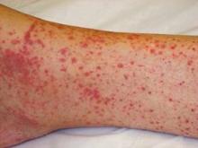
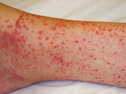
Petechiae and fever? Worry! Only about 50% of people with Rocky Mountain spotted fever will recall a tick bite, so you have to be suspecting it if you’re in an appropriate geographic area. Patients get febrile and toxic. The rash starts out on the wrist and ankle as a maculopapular rash and then spreads and becomes petechial.
"The tip-off is that it spreads to the palms and the soles," she said. "It’s a vasculitis, so it’s a palpable petechiae."
Rocky Mountain spotted fever is a bit of a misnomer, because the disease occurs largely in central and eastern states like North Carolina, Georgia, Tennessee, and Arkansas.
"This is a clinical diagnosis," she said. Don’t wait the 4-5 days it takes to get serology results from a laboratory to decide on treatment. Up to 15% of people with Rocky Mountain spotted fever may develop permanent neurologic deficits. Treatment with doxycycline reduces the risk of death from 30% to 5%. In pregnant women consider chloramphenicol instead of doxycycline, she suggested.
Palpable petechiae, vasculitis could mean infection. A patient with fever, mental status changes, vasculitis, and palpable petechiae should scare you, Dr. Murphy-Lavoie said. This could be meningococcemia, which carries a 3%-50% mortality rate depending on the promptness of treatment. Over the last 10 years in the United States, the mortality has hovered around 13%, she said.
Ceftriaxone is the drug of choice. "Because of diagnostic uncertainty, any time you have a suspected case of bacterial meningitis, you’re going to add on vancomycin to cover resistant Streptococcus," and dexamethasone has been shown to decrease neurologic sequelae, she said. People exposed to a patient with this disease should get prophylactic therapy
Palpable petechiae also are a feature of many types of bacteremia. Petechiae are the most common skin manifestation of bacterial endocarditis, but it’s only present in 20%-40% of cases. When you suspect bacterial endocarditis, get three sets of blood cultures because nailing down the type of bacteria will inform the long-term treatment strategy. Treat initially with broad-spectrum IV antibiotics such as vancomycin and gentamicin to cover methicillin-resistant Staphylococcus aureus (MRSA).
"If you have a S. aureus–related endocarditis, your mortality is almost double that of a streptococcal endocarditis, so you really, really have to make sure that you’re covering for MRSA," she said.
Nonpalpable petechiae = thrombocytopenia. "If petechiae are not palpable, it’s thrombocytopenia unless proven otherwise," Dr. Murphy-Lavoie said.
A febrile, tachycardic woman came to her emergency room complaining of a rash on both legs comprising diffuse, nonpalpable petechiae. She appeared generally ill, had no past medical or medication history of note, and had a very low platelet count. "You should be scared to death if you get this patient in your ER," because she had thrombotic thrombocytopenia purpura (TTP).
More than 90% of patients will die if TTP is not treated with plasmapheresis (also called exchange transfusion), which reduces the mortality risk to 10%. If this treatment is not available at your institution, transfer the patient, she said. Manage the patient in the ICU, and treat the underlying cause of the TTP. Do not give platelets to patients with TTP, which will trigger increased end-organ damage, she warned.
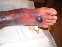
Hemorrhagic bullae? Ominous! "Nothing good causes hemorrhagic bullae," Dr. Murphy-Lavoie said.
Anything that can cause disseminated intravascular coagulation can cause purpura fulminans, which may present with hemorrhagic bullae, rapid hemorrhagic skin necrosis, ecchymosis, purpura, fever, shock, multiorgan failure, and bleeding from multiple sites.
Lab results will show thrombocytopenia, hemolytic anemia, increases in prothrombin time and partial thromboplastin time, and an increase in fibrin degradation products. As the disease progresses, the fibrinogen level falls.
Treatment starts off similar to treating TTP, with an emergent consultation with hematology/oncology, ICU admission, and treatment of the underlying illness. Add supporting vitamin K and folate. "Then it gets tricky," she said. "You’re going to need to balance how much of their problem is from bleeding and not enough coagulation factors and platelets, and how much is from overactivation of the fibrinolytic system and bleeding, and whether or not you need heparin to treat their thrombus. You really don’t want to be doing this in your emergency department."
Hemorrhagic bullae also are a classic presentation of necrotizing fasciitis, along with pain out of proportion for typical cellulitis, systemic toxicity, crepitus, and rapid spread along fascial planes. Not all cases of necrotizing fasciitis will have hemorrhagic bullae, but worry if you see this, she said.
Treat with strategic debridement and broad-spectrum antibiotics. Studies have shown that adding hyperbaric oxygen therapy decreases mortality risk. It’s not worth transferring someone with necrotizing fasciitis to someplace hours away in order to get hyperbaric oxygen, but if this is available, "please use it," she urged. In the literature, mortality rates with necrotizing fasciitis range from 0% to 75%. "Guess which ones had 0% mortality patients who got hyperbaric oxygen."
Consider steroids for bullous rash with mucosal involvement. A woman came to Dr. Murphy-Lavoie’s emergency department complaining of a rash and pain with swallowing. She appeared moderately toxic, was tachycardic, had a bullous rash, and had dry mucous membranes but with oral lesions. Diagnosis: pemphigus vulgaris, which will involve mucosal surfaces 70% of the time.
The bullae may coalesce, and there may be a positive Nikolsky sign (sloughing of full-thickness skin with lateral pressure) and a positive Asboe-Hansen sign (light lateral pressure on the blister edge spreads the blister into adjacent clinically normal skin).
The biggest favor that emergency physicians can do for these patients is to start them on steroids. Before the advent of steroid therapy for pemphigus vulgaris, 50%-90% of patients died, compared with 4%-15% who are treated with steroids. Also provide local wound care to prevent secondary infection. Pemphigus vulgaris is associated with autoimmune diseases, and long-term management with immunosuppressive drugs probably will be handled by a rheumatologist, she said.
Dr. Murphy-Lavoie reported having no financial disclosures. She codeveloped the free app "EM Rashes."
EXPERT ANALYSIS FROM THE ANNUAL MEETING OF THE AMERICAN COLLEGE OF EMERGENCY PHYSICIANS
Acute Dyspnea: Try Physiologic Approach in Differential Diagnosis
DENVER – If you presume that a patient who comes to the emergency department with acute dyspnea primarily has a pulmonary cause, you’ll almost always be right. Those few other cases, though, take a bit of detective work.
In the approximately 5% of cases in which dyspnea is not easily referable to the lungs, the culprit may be a cardiac problem (usually in a very young child) or, rarely, other problems – hemoglobinopathies, diseases that cause metabolic acidosis, or neurologic disorders, Dr. Jeffrey Sankoff said at the annual meeting of the American College of Emergency Physicians.
Take a physiologic approach that can guide you through the differential diagnosis, he suggested. "As somebody who trained in critical care, everything boils down to physiology," said Dr. Sankoff of the University of Colorado, Denver, and director of quality and patient safety at Denver Health Medical Center.
To begin, think of diseases that cause hypoxemia, hypercapnia, or metabolic acidosis, which lead to dyspnea.
Hypoxemia
The most common cause of hypoxemia is ventilation-perfusion (V-Q) mismatch, in which blood flows in the lungs but areas are not getting oxygen. Diffusion abnormalities, in which oxygen gets into alveoli but oxygen transit to the bloodstream is impaired, also cause hypoxemia. These occur in primary pulmonary disease.
But extrapulmonary disease processes create four causes of hypoxemia: a shunt, low mixed venous oxygen saturation (MVO2), decreased fraction of inspired oxygen (FiO2), and alveolar hypoventilation.
A V-Q mismatch at its most extreme is a shunt, in which blood bypasses the lungs altogether, he said. Disease processes that cause blood to go directly from the right to the left side of circulation result in hypoxemia. A shunt is almost always intracardiac, rarely intrapulmonary.
In children, shunts are seen at characteristic times for the development of cyanotic congenital heart disease, most commonly patent ductus arteriosus in an infant.
Look for a shunt by its hallmark – oxygen saturation will not improve when you give the patient oxygen. "This is the test for any young child under the age of 6 weeks who comes to the emergency department hypoxemic," Dr. Sankoff said.
Adults with shunts will have a murmur as well as dyspnea. "Adults don’t develop shunts de novo. This is going to be happening as part of some acute process," he said. The chest x-rays in adults with shunts often are normal.
The second cause of hypoxia – low MVO2 – occurs mainly when blood flows too slowly through the capillary bed, allowing excess oxygen extraction, and blood returns to the heart in a deoxygenated state. When you see this, focus on right ventricular impairment to identify the etiology. Left ventricular impairment will show up on x-ray as pulmonary edema. A right-sided infarction or cor pulmonale from pulmonary embolism will impair right ventricular function. Cardiac tamponade from infectious, inflammatory, or neoplastic processes also can cause low MVO2 and dyspnea, though nobody really understands why, he added.
The third cause of hypoxia– decreased FiO– usually is a problem relegated to people at high altitudes or industrial workers in enclosed spaces, where hypoxemia (low partial pressure of oxygen in blood) causes hypoxia (low oxygen levels in tissues). But Dr. Sankoff uses this category to remind himself to look for diseases that are not associated with lower FiO2 and hypoxemia but still are associated with hypoxia – primarily hemoglobinopathies.
"If a patient has 100% oxygen saturation yet is hypoxic, they have a problem with hemoglobin," Dr. Sankoff said. It may be a severe or acute garden-variety anemia causing the dyspnea, or occasionally a hereditary hemoglobinopathy such as thalassemia or sickle cell disease. To diagnose these, have a high index of suspicion. You’ll see no patient improvement on oxygen therapy, and some diseases create a characteristic appearance of the blood.
Alveolar hypoventilation may be the most insidious cause of hypoxemia, and dyspnea and may be a flag for impending respiratory compromise if the patient has peripheral weakness. The most common acquired causes of peripheral neuromuscular weakness are Guillain-Barre syndrome, amyotrophic lateral sclerosis, and Colorado tick paralysis.
Make the diagnosis in context with other findings, he said. Expect an abnormal motor exam. Check the negative inspiratory force; if it isn’t at least –20 cm H2O, it’s abnormal and the patient likely will need respiratory support.
Hypercapnia
Diseases that cause hypercapnia can cause dyspnea. Three things cause carbon dioxide levels in the blood to rise: increased metabolic rate (which tends to be seen in the ICU, not in the emergency department), decreased minute ventilation, and increased pulmonary dead space. All can be diagnosed by checking arterial blood gases.
Metabolic Acidosis
Acidosis, usually due to high levels of lactate, stimulates respiratory drive to try and balance pH. Sepsis is the most important cause of acidosis. When sepsis is developing, dyspnea frequently is a subtle sign. Have a high index of suspicion for sepsis, and be wary of a normal oxygen saturation level in a patient with dyspnea, he said. Other causes of metabolic acidosis that lead to dyspnea include diabetic or alcoholic ketoacidosis.
Putting this physiologic approach to dyspnea into context, consider three scenarios, Dr. Sankoff suggested. A patient with dyspnea who responds to oxygen therapy and has an abnormal chest x-ray has a primary pulmonary problem. A patient who responds to oxygen but has a normal chest x-ray may have sepsis, another cause of acidosis, or alveolar hypoventilation; their response to oxygen may be transient. They will respond to oxygen but continue to be tachypneic.
The third scenario – normal x-ray, but the patient does not respond to oxygen therapy – raises a broad differential diagnosis including sepsis, other causes of acidosis, hypercapnia, cardiac causes, and hemoglobinopathies. Narrow the differential by recalling the history and physical findings and getting arterial blood gas tests.
Only after everything else has been excluded should you consider anxiety or pain as the cause of dyspnea, he said.
Dr. Sankoff reported having no financial disclosures.
EXPERT ANALYSIS AT THE ANNUAL MEETING OF THE AMERICAN COLLEGE OF EMERGENCY PHYSICIANS
New Strategy Distinguishes Inferior STEMI from Pericarditis
DENVER – The presence of any ST depression in the ECG lead aVL in a patient with significant ST segment elevation in inferior leads is highly sensitive and specific for inferior ST-elevation MI as opposed to pericarditis or early repolarization, Dr. Johanna E. Bischof reported at the annual meeting of the American College of Emergency Physicians.
In her retrospective study of three different patient populations with ST elevation in leads II, III, and/or aVF, the finding of at least 0.25 mm of ST depression in lead aVL had 99% sensitivity and 100% specificity for inferior STEMI.
"It’s more sensitive than traditional ST elevation intervention criteria, and more sensitive and specific than comparing the difference in ST elevation between leads III and II," said Dr. Bischof of Hennepin County Medical Center, Minneapolis.
Numerous non–life-threatening cardiac conditions can provoke ST elevation in the inferior leads in the presence of normal conduction with no left bundle branch block, Wolff-Parkinson-White syndrome, paced rhythm, or left ventricular hypertrophy. She and her coworkers evaluated the significance of any ST depression in aVL in two of these conditions that are often confused with inferior STEMI based upon the ECG: pericarditis and early repolarization.
Her retrospective study included 156 Hennepin County Medical Center patients with confirmed inferior STEMI, 39 patients with pericarditis and 1 mm or more of ST elevation in two or more inferior leads, and 66 Finns with early repolarization and ST elevation in inferior leads. The subjects with early repolarization came from the Finnish Health 2000 Survey, in which ECGs were recorded for nearly 11,000 asymptomatic adults.
Of the 156 patients with confirmed inferior STEMI, 155 had ST depression in aVL. Not one of the patients with ST elevation in inferior leads and pericarditis or early repolarization did.
Only 86% of patients with a true inferior STEMI met traditional intervention criteria for STEMI. That means 14% of them might not have gotten to the cardiac catheterization promptly. Using the novel ECG criterion of ST depression in aVL, everyone with an inferior STEMI would have been sent to the cath lab straight away.
Moreover, based upon the finding of ST elevation in inferior leads, 49% of patients in the pericarditis group would have been sent to the cath lab for what would have turned out to be a negative angiogram. With application of the new finding regarding the clinical significance of any ST depression in aVL, none of the patients with pericarditis would have gone to the cath lab, Dr. Bischof continued.
When the degree of ST elevation in lead III exceeds that in lead II, it has traditionally been thought to be more suggestive of inferior STEMI, whereas ST elevation in lead II that’s greater than in lead III is more often thought of as pericarditis. But in Dr. Bischof’s study, only 87% of patients with confirmed inferior STEMI had greater ST elevation in lead III than II. In the pericarditis group, 98% of patients had more ST elevation in II than III. And in subjects with early repolarization, it was roughly 50/50 as to which lead had greater ST elevation.
Senior emergency medicine physicians in the audience called Dr. Bischof’s study "fabulous," adding that they’d like to see the study expanded to include larger numbers of pericarditis patients just to be sure of those excellent sensitivity and specificity figures.
"Truly, if we could take this finding out and utilize it in the ED it would make things very easy for us," one physician commented.
Dr. Bischof reported having no financial conflicts.
AT THE ANNUAL MEETING OF THE AMERICAN COLLEGE OF EMERGENCY PHYSICIANS
Major Finding: The finding of any ST depression in lead aVL in patients with significant ST segment elevation in inferior leads has 99% sensitivity and 100% specificity for inferior ST-elevation MI, effectively ruling out pericarditis or early repolarization.
Data Source: This was a retrospective study including 156 patients with confirmed inferior STEMI, 39 with pericarditis and at least 1 mm of ST elevation in multiple inferior leads, and 66 with early repolarization and ST elevation in inferior leads.
Disclosures: This study was free of commercial sponsorship. The presenter reported having no financial conflicts.
A Look at Upcoming Surviving Sepsis 2012 Guidelines
DENVER – Look for some big changes ahead in the forthcoming update of the Surviving Sepsis Campaign international guidelines.
In the 4 years since the last version of the guidelines came out, major studies have been released that resolved hot debates regarding sepsis management and in some cases overturned established dogma. At the annual American College of Emergency Physicians (ACEP) meeting, emergency physicians deeply involved in crafting the Surviving Sepsis Campaign 2012 guidelines provided a preview of what’s to come.
Among the areas that will see key new recommendations are antibiotic therapy, fluid resuscitation, vasopressors, and lactate monitoring.
• Antibiotics ASAP. The critical importance of getting empiric antibiotics on board within 6 hours after recognizing that a patient has sepsis has been effectively hammered home over the years. But how fast is fast enough? Several recent small studies have reached conflicting conclusions. Now, however, an 800-lb gorilla of a study has provided a definitive answer.
This study, now in press, involved 14,895 patients enrolled in the Surviving Sepsis Campaign’s worldwide database. All had severe sepsis or septic shock, and all received antibiotics within the first 6 hours, explained Dr. Tiffany M. Osborn, a coauthor of the study and of the forthcoming guidelines.
"This was a big question for us. The results strengthen the importance of getting antibiotics started as soon as possible – within the first hour if possible. We found there’s about a 4% increase in mortality for every hour delay. And it’s cumulative: it’s 4% after the first hour, 8% after the second, 12% after the third, and so on," according to Dr. Osborn of Washington University at St. Louis.
A similar time-dependent increase in mortality was observed in patients with severe sepsis as well as in those in septic shock, she added.
• Fluid resuscitation. The initial fluid of choice for resuscitation in severe sepsis and septic shock remains crystalloids, as before. That’s a grade 1A recommendation. What’s brand new is a recommendation to consider adding albumin in the initial fluid resuscitation (grade 2B). This guidance was heavily influenced by a meta-analysis of 17 studies involving close to 2,000 patients that demonstrated a significant protective effect for the use of albumin as an initial resuscitation fluid (Crit. Care Med. 2011;39:386-91).
At this late date, Dr. Osborn said, the guideline panel is strongly leaning toward a recommendation against the use of low-molecular-weight colloids or starches such as hydroxyethyl starch 130/0.42. That would be ground shaking, as synthetic colloids or starches, particularly those of low molecular weight, are a very popular resuscitation fluid both in the United States and abroad. However, recent studies implicate these fluids in increased risks of 90-day mortality and renal failure, compared with the use of Ringer’s lactate.
"Having said this, there are two other trials currently pending. Any month now the results will come out, and we’ll see where we are at that point. But at this time, we’re thinking stay away from low-molecular-weight starches for resuscitation," Dr. Osborn said.
One of the main reasons the Surviving Sepsis Campaign 2012 guidelines won’t actually be published before January 2013 is the guideline panel’s eagerness to see the results of those two studies, she added.
• Vasopressor therapy. The initial target remains, as before, a mean arterial pressure of at least 65 mm Hg. But while the 2008 guidelines recommended either norepinephrine or dopamine as the first-choice vasopressor to correct hypotension in patients not sufficiently responsive to fluid resuscitation, the new guidelines will state that norepinephrine is the preferred agent (grade 1B). That’s a major change. Dopamine has been essentially kicked to the curb in response to multiple studies in the last 4 years implicating it in an increased incidence of arrhythmias and, in some studies, higher mortality.
The final nail in dopamine’s coffin for use in patients with severe sepsis or septic shock was a recent meta-analysis involving five observational and six interventional studies totaling nearly 2,800 patients (Crit. Care Med. 2012;40:725-30).
"Dopamine has actually fallen by the wayside. By far and away, I’m going to choose norepinephrine as the initial vasopressor agent," declared Dr. Michael E. Winters of the University of Maryland, Baltimore.
The new guidelines will suggest that be reserved for highly selected patients: those at very low arrhythmia risk and with a low cardiac output and/or low heart rate (grade 2C), he noted.
Another change in the new guidelines will be a recommendation that epinephrine be added when an additional agent is needed in order to maintain adequate blood pressure (grade 2B).
• Don’t overdo the fluids. "We want to give patients what they need but not more," Dr. Osborn explained. The 2012 guidelines will recommend that physicians use some sort of fluid challenge test while administering fluid boluses. The goal is to keep giving fluid only so long as hemodynamic improvement is seen in response. This can be achieved in a variety of ways, including monitoring change in pulse pressure, stroke volume variation, heart rate, or arterial pressure.
• Lactate clearance. Serum lactate is recognized as an indicator of global organ hypoperfusion and shock. But incorporating lactate clearance as one of the goals of early sepsis therapy has been "a very hot topic," Dr. Winters observed.
Improved clarity was provided by a prospective 556-patient quality improvement study by investigators in the Asian Network to Regulate Sepsis Care. Patients who got the primary severe sepsis management bundle of care as recommended in the 2008 Surviving Sepsis Campaign guidelines had an unadjusted mortality of 43.6%. This bundle includes early antibiotic administration, hemodynamic monitoring and support, and achievement of a central venous oxygen saturation level greater than 70% by 6 hours. However, patients who got the bundle plus lactate clearance had a 20.5% mortality rate (Crit. Care 2011;15:R229 [doi:10.1186/cc10469]).
The importance of lactate clearance was further underscored by the findings in the GENESIS Project (Generalized Early Sepsis Intervention Strategies). This quality improvement initiative, conducted at five 5 U.S. community hospitals and six tertiary centers, showed a 42.8% mortality in 1,554 historical controls treated for sepsis before implementation of the Surviving Sepsis Campaign resuscitation bundle. In another 4,801 patients who got the bundle, mortality was significantly lower at 28.8%. And, in those who received the bundle plus lactate clearance, mortality further fell to about 22% (J. Intensive Care Med. 2012 [doi:10.1177/0885066612453025]).
Thus, the coming Surviving Sepsis Campaign 2012 guidelines will suggest that in patients with elevated lactate levels as a marker of hypoperfusion, resuscitation should be targeted at normalizing lactate as rapidly as possible (grade 2C). Having said that, however, a normal lactate doesn’t indicate absence of shock. Other factors, such as the patient’s central venous oxygen saturation level, need to be considered as well, the physicians emphasized.
The Surviving Sepsis Campaign guidelines are sponsored by 27 medical organizations. Among them are the Society of Critical Care Medicine, ACEP, the Society of Hospital Medicine, the American College of Chest Physicians, the American Thoracic Society, the Infectious Diseases Society of America, the Surgical Infection Society, the Pediatric Acute Lung Injury and Sepsis Investigators, and a host of international groups.
Dr. Osborn and Dr. Winter reported having no financial conflicts.
AT THE ANNUAL MEETING OF THE AMERICAN COLLEGE OF EMERGENCY PHYSICIANS
Standardize Communication to Reduce Medical Errors
DENVER – Do you speak in SBAR?
If not, communication in your department probably could be better, two speakers said at the annual meeting of the American College of Emergency Physicians, in a session on improving teamwork.
SBAR is the acronym for a standardized communication format that organizes the transfer of information under four themes – situation, background; assessment, and recommendation. Developed by anesthesiologist Dr. Michael Leonard and colleagues at Kaiser Permanente of Colorado, Evergreen, the SBAR tool has been endorsed by many national organizations and medical accreditation bodies, said Dr. Jennifer L. Wiler.
It originally was used for acute-care communications from nurses to physicians but now is being used more widely for communication from nurse to nurse and from physician to physician in a variety of care settings. One study found that it was particularly useful for reporting changes in a patient’s status or a patient’s deterioration between health care services or shifts (Healthcare Quarterly 2008;11:72-9).
Using SBAR won’t necessarily save you time – at least one study suggests it increases time for communications. The limited data from seven published studies so far suggest that SBAR use improves the transfer of important information, the perception of a culture of safety, and patient satisfaction, though SBAR has yet to be validated in an emergency department setting, said Dr. Wiler of the University of Colorado, Aurora.
It has worked in hospital medicine. Hospitalists with Kaiser Permanente found SBAR to be helpful, according to a study conducted from May 2008 to August 2009. SBAR was used for end-of-rotation patient handoffs, the researchers wrote. "The Hospitalist Chief finds SBAR useful when communicating with consultants, especially during the initial telephone conversation when it is important for hospitalists to clearly state information needs." Hospitalists communicate primarily with ED physicians, and "communications with KP ED physicians is superb; [we] get a complete diagnosis" (Perm. J. 2011 Summer;15:51-60).
The Joint Commission and the Agency for Healthcare Research and Quality (AHRQ) list communication problems as the No. 1 cause of medical error, she noted. "Addressing errors related to communication is really critical," Dr. Wiler said.
To communicate in SBAR, use the following structure:
– Situation. Identify yourself, your occupation and where you are calling from, if speaking by phone. Identify the patient – the name, date of birth, age, and sex, and the reason for your report. Describe the current status of the patient or your reason for calling. If it’s urgent, say so.
– Background. Give the patient’s presenting complaint, any relevant past medical history, and a brief summary of background.
– Assessment. Describe abnormal vital signs (heart rate, respiratory rate, blood pressure, temperature, oxygen saturation, pain scale, or level of consciousness). Provide your clinical impression, the severity of the patient’s situation, and any additional concerns.
– Recommendation. Explain what you require, how urgently, and when action needs to be taken. Suggest what action might be taken. Clarify what action you expect to be taken.
One of the challenges to good communication is that physicians and nurses practice in parallel environments and too seldom prioritize teamwork, said Eric Christensen, R.N.
Beyond SBAR, a variety of strategies can improve communication between team members, he said. For example, the AHRQ suggests a structured handoff sign-out protocol dubbed ANTICipate (for Administrative data, New clinical information, Tasks to be performed, Illness severity, and Contingency plans).
Develop structured communication events specific to your hospital or department, suggested Mr. Christensen, a staff nurse at Denver Health Medical Center.
These might include a patient’s physiologic parameters that trigger an alert to the nurse and doctor, or required communication to reconcile abnormal vital signs. A "time out" at discharge will lead to safer discharge if all providers are aware of the treatment plan and pending issues are resolved. Face-to-face debriefings at shift changes are a good idea. He also recommended greater use of physician-nurse huddles to discuss significant patient issues, therapies, oxygen status and last vital signs, and any pending issues in clinical management.
"One of the things we’ve done at our institution is to institute huddles throughout the shift," he said in an interview. Every 2-3 hours the nurses and physicians round together to make sure throughout the shift that everyone is "on the same page."
At Dr. Wiler’s institution, team-based huddles are standard practice at presentation of more acute, sicker patient or trauma patients. "We’re moving to that methodology with all our patients," she said in an interview.
If you have concerns to raise with nurses, resist the temptation to direct them only to the most senior nursing staff, Mr. Christensen said. "It makes a world of difference to address a nurse by name," he added. If you don’t know a nurse’s name, ask directly, so the nurse feels included.
Taking 2 minutes to discuss the plan of care for complicated patients directly with the nurse and seek the nurse’s input will pay off in the long run, he said. With any patient, providing brief targeted bedside education for nurses by not just saying what will be done, but why, gets nurses more engaged in the clinical care team and better prepared to anticipate treatment of similar patients in the future.
Being approachable and complimenting nurses who are doing a good job also pays off, Mr. Christensen said.
"Be a leader, not a commander," he said. "Managers manage process and leaders lead people. If you want to know which you are, just look behind you. If there is no one there, you are just going for a walk."
Dr. Wiler and Mr. Christensen reported having no financial disclosures.
EXPERT ANALYSIS FROM THE ANNUAL MEETING OF THE AMERICAN COLLEGE OF EMERGENCY PHYSICIANS
Apps Proliferate Amid Concerns About Medical Use
DENVER – Do you need a stethoscope, a blood pressure monitor, or a tool to assess cardiac rhythms? There are apps for that. In fact, by recent count there are more than 200,000 applications of technology – or "apps" – available for smartphones or tablet devices, and they’re being used more and more for medical purposes.
Need a convenient way to look up drug interactions, pediatric dosing, or clinical decision rules from guidelines? Or how about a translator, a light to examine a finicky infant’s throat, or a "white board" to draw a picture for your patient? Yup – they’re all in apps, and chances are you already may be using some of these.
Dr. Joshua S. Broder expects an exponential increase in the use of apps in medicine as smartphones and tablets continue to proliferate, but their accuracy needs to be verified and potential problems need to be addressed, he said at the annual meeting of the American College of Emergency Physicians.
Apps will be used increasingly for bedside diagnosis and measurement of hemoglobin or other physiologic parameters. "Some of these tests may be taken over by smartphones in the near future," according to Dr. Broder of Duke University, Durham, N.C.
On the other hand, he cautioned, how do you sterilize a smartphone as you move from one hospital room or patient to another, so that you avoid transmitting infection? There are few independent studies so far testing the accuracy and reliability of medical apps, most of which were designed for lay consumers, not physicians.
The Food and Drug Administration is "very interested" in regulating any apps that might substitute for proven technologies such as stethoscopes or that physicians use as accessories to medical devices that already are regulated, he said. The FDA described its approach to deciding which mobile technology to regulate in a draft report in July 2012.
Even the basic functions of smartphones can be convenient in clinical practice, such as taking photos or videos and transmitting information by text or e-mail, but make sure you protect patient privacy and autonomy in ways that maintain trust and comply with HIPAA, Dr. Broder said.
The Duke University Health System has resolved any issues with HIPAA so that it’s safe for physicians to transmit images and video as long as they’re not sent outside the system. Talk to the HIPAA compliance officer at your medical center to establish the ground rules, he said. You can refresh your memory about which parts of data are considered by HIPAA to be protected information via a University of Miami site.
Dr. Broder reviewed some smartphone functions and apps that may be helpful and others that are not yet ready for medical prime time. Many are available for no cost or for as nominal fee. One study of health and fitness apps suggests that apps costing $0.99 or more tend to be higher quality and more trustworthy than less-expensive ones, he noted (J. Med. Internet Res. 2012;14:e72).
• Sleep: One of his residents swears by "smart alarm clock" apps that claim to use a smartphone’s accelerometer to assess where you are in your sleep cycle (based on your movements in bed) to wake you at a time that will leave you feeling less fatigued. You may set for 6 a.m., but the alarm may wake you at 5:45 a.m. Apps like Sleep Cycle ($0.99) and Sleep as Android have some underlying sleep science behind them, but no independent studies have verified their claims.
• CPR: The accelerometer also is used in the free app PocketCPR to give real-time feedback during CPR on the rate and depth of compression. Its has not been cleared by the FDA for use in humans, however, so the app warns that it’s meant for practice only. One prospective, randomized trial in 1,586 cardiac arrests that happened outside of hospitals found that use by emergency services personnel did not significantly change the likelihood of return of spontaneous circulation or other outcomes (BMJ 2011;342:d512).
• Chest: If you’re trying to teach students and residents about heart and lung sounds, or if you still get confused between mitral regurgitation and aortic stenosis, you might want to have a digital stethoscope app handy. These apps interpret heart and lung sounds heard typically through your smartphone’s microphone, which may not be good enough for clinical use. The Thinklabs Stethoscope app at $70 is pricey, compared with others, but it records sounds directly via the smartphone or through an attached electronic stethoscope.
A case that turns an iPhone into an ECG device has been submitted to the FDA for approval. The AliveCor iPhone ECG is expected to sell for between $100 and $200, compared with the usual price tag of thousands of dollars for conventional ECG machines, according to PC Magazine.
One small prospective study of experimental software that programs an iPhone to detect atrial fibrillation by placing a patient’s finger over the camera lens showed it was 98% sensitive and nearly 100% specific in detecting atrial fibrillation (IEEE Trans. Biomed. Eng. 2012 [doi:10.1109/TBME.2012.2208112]).
• Translation: When your hospital’s interpreter isn’t available, a free app like Google Translate can help. You can write or speak in one language and your device will write and say the message in a wide selection of language. You’ll need a wireless Internet connection for some translation apps.
Pull up an app like the free EyeChart on your smartphone or tablet.
• Light: You want to inspect a patient’s sore throat, but the light in the exam room is broken. Use the flash on your smartphone camera, or use one of many free "flashlight" apps that turn the smartphone screen into a light source. Be sure to turn it off when you’re done, though, or your battery will run down quickly.
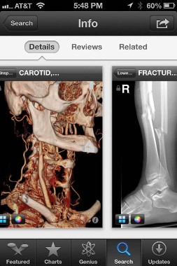
• Ultrasound: The miniaturization of ultrasound devices continues, with systems like the Mobisante MoblUS that attaches a probe to show images on your smartphone screen.
• Skin: For better evaluation of skin lesions, turn your iPhone into a dermatoscope by using the DermScope app ($4.99) and attaching the phone to the DermScope hardware (sold separately).
• Decision support: The PediStat app ($2.99 and up) makes it easy to determine the right pediatric drug dosing, among other features. The free Calculate (Medical Calculator) by QxMD app provides quick intuitive guides to common decision rules and can be customized by medical specialty.
• Drugs: Look up drug dosing, side effects, interactions and other information on free apps from Micromedex and others.
• Photos/videos: These apps are handy for documenting and sharing the appearance of a wound, a patient’s range of motion, or performance on a neurologic exam. Anyone who thinks they see uvula deviation in the throat of a struggling 3-year-old can snap a photo or video for review with other health care providers, medical students, or parents and avoid having to repeat the exam. Images of a wound problem after surgery can be sent to the surgeon when he or she is out of town.
Dr. Broder particularly finds the video useful for children having "pseudoseizures" whose parents demand a neurologic consult, even though the seizure event probably won’t be happening when the neurologist arrives. A video shows the neurologist exactly what Dr. Broder saw. (See the Dos and Don’ts for using photos and videos on the next page.)
Once you’ve got an image or data you want to transmit, avoid texting as first-line means of communication because texts typically are not encrypted. Be careful when e-mailing to make sure it’s going to the correct address and only that address. Use e-mail options such as "confirm delivery" or "request read receipt," and add a sentence to the e-mail saying, "Please delete once no longer necessary for patient care," he advised.
Always document in the patient’s chart that you obtained patient consent and describe what was sent and who received it. Describe any images you send.
Don’t leave images on your portable devices. They’re easily lost, and most have inadequate encryption. Make images part of the medical record by uploading to the patient’s record, printing and scanning, or describing them clearly in the medical record. Then delete them from your device.
Store images and data in "cloud" computing sites with caution, Dr. Broder said. Services such as Google Drive or Dropbox allow sharing of very large files but provide no assurances about the quality of encryption or security. Cloud sites may be best used for giving patients access to instructions, instructional videos, reference papers, anatomic diagrams, etc.
The FDA approved the free Centricity Radiology Mobile Access app, which lets you view CT and MRI images on your iPhone if the images are stored in a GE Centricity PACS (picture archiving and communication system) platform – which may include 20% of U.S. radiology images, according to the company.
The free CloudOn app lets you use MS Office software (including Word, Excel, and Powerpoint) on an iPad.
Various screen replicators that allow you to remotely access your computer desktop from your mobile device (such as ones by Citrix, or Splashtop Remote Desktop) all have the same problem, Dr. Broder said – they’re too clunky and not "touchscreen friendly."
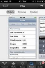
And one final word on an underappreciated perk of medical apps on smartphones: When your medical director stops by, wanting to talk about your productivity, pull out your smartphone to show the data you’ve entered about patient encounters in your free iRVU app, which calculates total RVUs, charges, and average charge per encounter, among other features.
This list only begins to scratch the surface of app use in medicine. Other apps are available for immunization schedules, dictation, infectious disease guides, and teaching aids. Journals provide content to portable devices through apps, and some medical societies offer multifaceted apps such as ACEP Mobile.
Apps on smartphones and tablets will become part of daily medical practice, Dr. Broder predicted, but physicians need to be conscious about their limitations and potential problems as well as their assets.
Dos and Don’ts for Medical Images on Smartphones
Do:
Obtain consent to acquire images or transmit them for the patient’s medical benefit.
Explain to the patient and get consent for any other intended use, such as education or publication.
Tell the patient what you will do with images when their use is completed – delete them or upload them to the medical record.
Confirm receipt if you send to other health care providers.
Specify in your message what that provider should do with the image.
Document in the patient’s chart that consent was obtained, what was sent, who received it, and content of the images.
Don’t:
Obtain images covertly.
Send to any unnecessary recipients.
Show images to anyone for fun.
Post to social media sites.
Blog about "funny" patient encounters.
Dr. Broder owns stock in Apple.
* The photo credits in this story were updated on 10/26/2012.
EXPERT ANALYSIS FROM THE ANNUAL MEETING OF THE AMERICAN COLLEGE OF EMERGENCY PHYSICIANS


