User login
AVAHO
div[contains(@class, 'header__large-screen')]
div[contains(@class, 'read-next-article')]
div[contains(@class, 'nav-primary')]
nav[contains(@class, 'nav-primary')]
section[contains(@class, 'footer-nav-section-wrapper')]
footer[@id='footer']
div[contains(@class, 'main-prefix')]
section[contains(@class, 'nav-hidden')]
div[contains(@class, 'ce-card-content')]
nav[contains(@class, 'nav-ce-stack')]
Low-carb, plant-rich diets tied to breast cancer survival?
TOPLINE:
METHODOLOGY:
- The diets of 9,621 women with stage I-III breast cancer from two ongoing cohort studies – the Nurses’ Health Study and Nurses’ Health Study II – were evaluated.
- Overall low-carb, animal-rich, and plant-rich low-carb diet scores were calculated using food frequency questionnaires after breast cancer diagnosis.
- Cox proportional hazards regression models adjusted for multiple potential confounding factors.
- Follow-up lasted for a median of 12.4 years after breast cancer diagnosis.
TAKEAWAY:
- Overall, 1,269 deaths due to breast cancer and 3,850 all-cause deaths occurred during the follow-up period.
- Researchers found that greater adherence to low-carb (hazard ratio, 0.82 for quintile 5 vs. 1) and plant-rich diet (HR, 0.73 Q5 vs. 1) was associated with a significantly lower risk for overall mortality but not breast cancer–specific mortality.
- Overall, adhering to animal-rich, low-carb diets did not significantly influence all-cause or breast cancer–specific survival rates.
- But replacing 3% of energy intake from available carbohydrates with fish protein was associated with 17% lower risk for breast cancer–specific mortality and 15% lower risk for all-cause mortality.
IN PRACTICE:
“The findings suggest that breast cancer survivors could benefit from limiting intake of carbohydrates, especially from fruit juice, sugar-sweetened beverages, and added sugar, and increasing the amount of protein and fat, in particular from plant sources,” the authors write.
STUDY DETAILS:
The study was led by Maryam Farvid, PhD, with the Data Statistics Group, Mission Viejo, Calif. It was published online in the journal Cancer and supported by National Institutes of Health and the University of Toronto.
LIMITATIONS:
Most women were non-Hispanic White and health professionals, so the results might not generalize to other sociodemographic groups. The authors also noted potential residual confounding, despite controlling for several breast cancer risk factors.
DISCLOSURES:
Dr. Farvid is a founder of the Institute for Cancer Prevention and Healing and the Data Statistics Group.
A version of this article first appeared on Medscape.com.
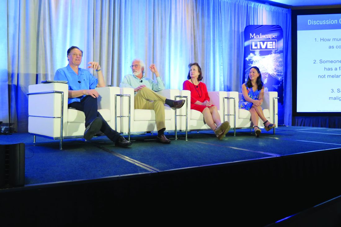
Experts share their sun protection tips for children
“I basically say, ‘sun protection means clothing, shade, [considering the] time of day of exposure, and sunscreen if you are going to be otherwise exposed,’ ” Dr. Eichenfield, chief of pediatric and adolescent dermatology at Rady’s Children’s Hospital, San Diego, said during a panel discussion about sunscreen use at the Hawaii Dermatology Seminar provided by MedscapeLIVE! He recommends photoprotective gear such as rash guards for surfers and other water sport enthusiasts. When patients ask him if they should use sunscreen, he often replies with a question of his own.
“Do you brush your teeth?” he’ll ask.
“Yes, I do.”
“Well, you should put sunscreen on every day.”
Another panelist, Adelaide A. Hebert, MD, professor of dermatology and pediatrics and chief of pediatric dermatology at the University of Texas, Houston, said that she advises new parents to start sun protection efforts early. “Most sunscreens are not approved for use in children under the age of 6 months because testing has not been done in this age group, but I do recommend protective clothing. I also recommend wrap-around sunglasses, which offer 5% more protection from the sun than regular sunglasses.”
In her opinion, stick sunscreens are “a good add-on,” especially for under the eyes and the backs of the hands, but she is not a fan of spray sunscreens, which can leave large areas of skin unprotected if not applied properly.
Fellow panelist Jennifer Huang, MD, a pediatric dermatologist at Boston Children’s Hospital, who has a special interest in taking care of dermatologic conditions of children with cancer, generally recommends mineral-based sunscreens. “There is data to suggest that nonmineral sunscreens are less safe than mineral sunscreens for humans, and mineral sunscreens are considered to be better for the environment,” Dr. Huang said. “Plus, there are more elegant versions of mineral sunscreens that don’t make your skin pasty white.” However, for patients with darker skin tones, “it can be hard to apply a pasty white sunscreen, so I lean on some recommendations for tinted sunscreens, too, so there are options. I specifically recommend sunscreens that have iron oxides in them so that it can block physical rays and help with the cosmetic appearance.”
Moise Levy, MD, professor of internal medicine and pediatrics at the University of Texas at Austin, said that his approach to imparting sunscreen advice to children and their parents involves a mix of spoken information, printed information, and sunscreen samples for children to try in the office, in the presence of a parent. To help patients choose among different samples, be they ointments, gels, or lotions, he will often ask the child: “‘What do you like the feel of better?’ If the child says, ‘I like this one,’ I make sure the parent hears that,” Dr. Levy said.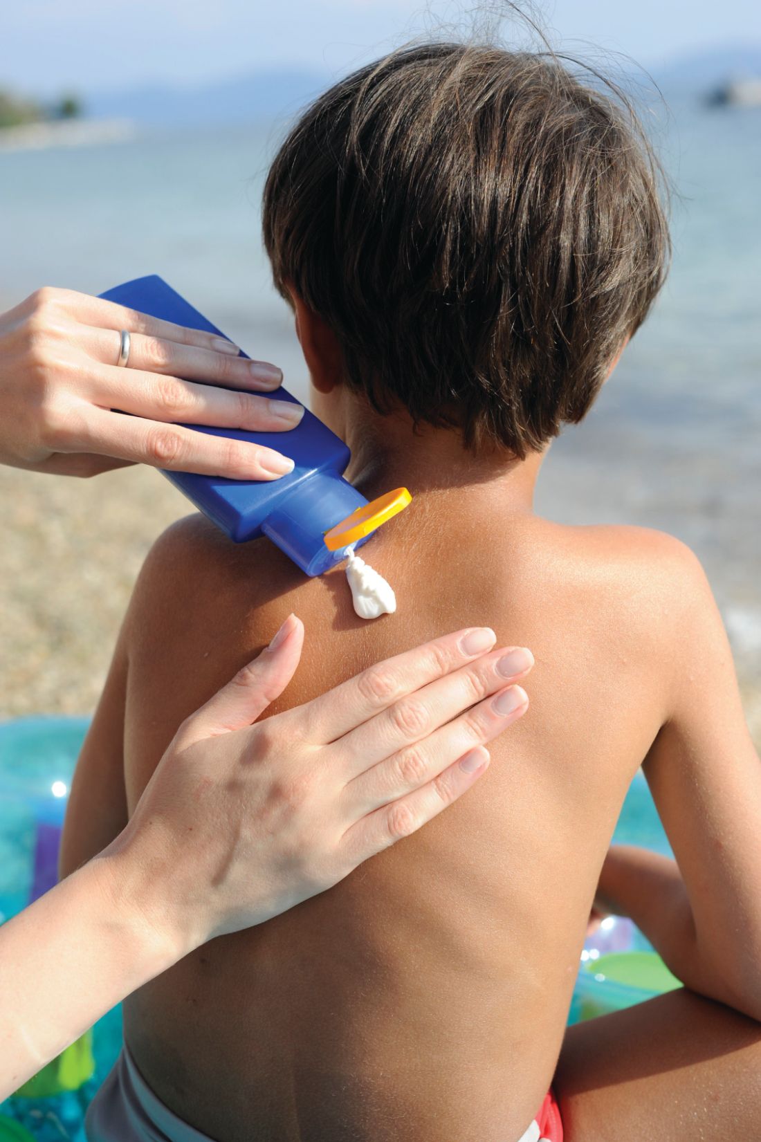
Next, Dr. Eichenfield, who moderated the discussion, asked his fellow panelists how they would counsel someone who comes to their practice for evaluation of moles and has a family history of nonmelanoma skin cancer. “I think this is one of the easier counseling sessions, because there are enough kids who are asked about the moles on their skin when they’re at school,” Dr. Hebert said. “I think they’re very ready to wear sun protective clothing and I certainly don’t want any sun exposure that would pose an increased risk for their child.”
In addition to routine sun protection, Dr. Huang recommends annual mole checks for children who have a first-degree relative with a history of malignant melanoma. Other high-risk groups that should undergo annual skin exams include anyone who has received high doses of radiation, bone marrow transplants, prolonged use of voriconazole, or prolonged systemic immunosuppression. Without a known genetic predisposition syndrome, a family history of nonmelanoma skin cancer would not raise concern for melanoma in an otherwise healthy child.
Dr. Eichenfield added that freckling used to be the secondary risk factor for melanoma, “but it’s flipped over to a primary risk factor. A history of immunosuppression or prior cancer is a major risk factor in childhood and teenage years.”
Dr. Eichenfield disclosed that he is a consultant or adviser for numerous pharmaceutical companies. He has also received research funding from AbbVie, Bausch & Lomb, Galderma Laboratories, and Pfizer. Dr. Hebert disclosed that she is a consultant or adviser for AbbVie, Almirall, Amryt Pharma, Arcutis Biotherapeutics, Beiersdorf, Dermavant Sciences, Galderma Laboratories, L’Oreal, Novan, Ortho Dermatologics, Pfizer, and Verrica. Dr. Levy disclosed that he is consultant or adviser for Abeona, Castle Creek, Dusa Pharma, Krystal Bio, Novan, Regeneron, and Sanofi Genzyme. Dr. Huang disclosed that she is an adviser for EllaOla.
MedscapeLive! and this news organization are owned by the same parent company.
FROM THE MEDSCAPELIVE! HAWAII DERMATOLOGY SEMINAR
Electronic nose may sniff out early lung cancer in COPD
, based on data from a prospective study of approximately 800 individuals.
Lung cancer remains a major cause of death in patients with chronic obstructive pulmonary disease (COPD), but many cases may go undetected in the early stage because of lack of screening and lack of validated predictive biomarkers, wrote Rianne de Vries, PhD, of the University of Amsterdam, and colleagues.
Accurate, noninvasive tests to screen patients with COPD for lung cancer are needed, and molecular profiling of exhaled breath using electronic nose (eNose) technology has shown potential as a method of early detection by identifying patterns of exhaled volatile organic compounds (VOCs), they said.
In a study published in the journal Chest, the researchers reviewed data from 682 adults with COPD and 211 with lung cancer who were enrolled in BreathCloud, a multicenter, observational study of healthy controls and individuals with suspected or confirmed diagnosis of asthma, COPD, or lung cancer.
Patients’ breath profiles were collected at study enrollment, between May 2017 and November 2018, using a metal oxide semiconductor eNose (SpiroNose).
Data from the eNose included the highest sensor peak normalized to the most stable sensor and the ratio between sensor peak and breath hold point. These variables were combined into four principal components (PCs) that captured 78.4% of variance in the dataset, and training and validation sets were constructed for all subjects. The researchers calculated a receiver operating characteristic (ROC) curve, including the area under the curve (AUC).
All patients were treated with standard clinical care and were monitored for development of clinically diagnosed lung cancer for 2 years, confirmed via CT imaging. The mean age of the patients was 64 years, and demographics at baseline were similar for patients with and without lung cancer.
After exclusion of 116 patients with both COPD and lung cancer, the analysis showed an accuracy of 90% and a ROC-AUC of 0.95.
Within 2 years of study enrollment, 37 patients with COPD (5.4%) developed lung cancer. In training sets and validation sets, the principal components one, two, and three were significantly different in patients with COPD who developed lung cancer and those who did not, (P = .002, P < .001, P < .001, respectively). The ROC-AUCs of the testing and validation sets were 0.89 and 0.86, respectively.
“Interestingly, the VOC pattern associated with early development of lung cancer in COPD did not match to the pattern related to lung cancer stages, as the former was mainly captured by PC2 and the latter by PC3,” the researchers wrote in their discussion. “This suggests that early identification of upcoming clinically manifest lung cancer in patients with COPD by eNose is not driven by VOCs that are predominantly associated with a particular stage of the disease,” they said.
The findings were limited by several factors including the lack of CT scanning at baseline because of the real-world design, so the presence of any baseline tumors was unknown, although none of the COPD patients showed symptoms indicative of lung cancer at baseline, the researchers noted.
However, the results suggest that eNose technology can identify lung cancer-specific VOC patterns early in cancer development in COPD patients, which provides a possible opportunity for early intervention, they concluded.
The study received no outside funding. De Vries disclosed personal fees and a substantial interest in the start-up company Breathomix.
A version of this article first appeared on Medscape.com.
FROM CHEST
Cancer drug shortages spur worry, rationing, and tough choices
CHICAGO – Oncologist Denise Yardley, MD, isn’t used to expressing uncertainty when she tells patients about what’s in store for them in terms of drug treatment. But things are dramatically different now amid a severe national shortage of carboplatin and cisplatin, two common and crucial cancer drugs.
“There’s a regimen I’m thinking about,” Dr. Yardley told a new patient recently, “but we’ll have to wait until you finish your staging evaluation to see whether I can deliver this. Another regimen that’s a little more toxic is my second choice.” And, she added, the alternative chemotherapy treatment – anthracycline instead of carboplatin – requires a longer treatment period.
This ambiguity is hardly ideal, said Dr. Yardley, of Tennessee Oncology and Sarah Cannon Research Institute in Nashville. “It’s another factor in being overwhelmed in a first-time visit and wanting to know the details about what your treatment is going to look like. You’re not walking out knowing exactly what you’re going to take or the exact timing so you can start mapping out your calendar and work schedule.”
This kind of scenario is becoming all too familiar this spring, according to oncologists who gathered at the annual meeting of the American Society of Clinical Oncology (ASCO). In interviews, these physicians said the limited supply of multiple cancer drugs – including the chemotherapies carboplatin and cisplatin – is having an unprecedented negative effect since their use is so widespread in cancer care.
“Every patient could get impacted. That’s why we need to address this sooner rather than later,” said oncologist Aditya Baria, MBBS, MPH, director of the Breast Cancer Research Program at Massachusetts General Hospital, Boston.
Shortages of cancer drugs are not unusual. Three-quarters of oncology pharmacists at 68 organizations surveyed from 2019 to 2020 said shortages prompted treatment delays, reduced doses, or alternative regimens. But the current shortages are having a much wider impact.
The National Comprehensive Cancer Network recently reported that 93% of 27 member institutions surveyed in late May are short on carboplatin, and 70% have reported a shortage of cisplatin. Plus, 20% of 19 centers said they weren’t able to continue carboplatin regimens for all patients.
The drugs are mainstays of multiple types of treatment for a long list of cancer types including lung, breast, gynecologic, and many others.
Several scenarios are possible when the drugs are in short supply, said Dr. Yardley, who noted that the shortage is more severe than any she’s seen in her medical career of more than 3 decades. Patients may need to be switched to regimens with more side effects, even when they’re in the middle of a treatment, she said. Or patients might have to go longer between treatments.
In some cases, Dr. Yardley said, the shortage is forcing patients to go without an important component of a larger combination therapy regimen. “The Keynote 522 neoadjuvant regimen for triple-negative breast cancer has carboplatin given with Taxol [paclitaxel] and Keytruda [pembrolizumab]. We are just deleting the carboplatin.”
She added that carboplatin is part of the following so-called TCHP regimen for HER2+ early-stage breast cancer: Taxotere (docetaxel), carboplatin, Herceptin (trastuzumab), and Perjeta (pertuzumab).
“You can delete [carboplatin] or consider substituting cyclophosphamide for carboplatin,” she said. But she cautioned the Keynote 522 and TCHP regimens haven’t been tested without carboplatin in curative-intent trials.
At Duke University in Durham, N.C., doses of carboplatin for many patients are being lowered by a third to the level that’s commonly used for older and frail patients, said oncologist Arif Kamal, MD, MBA, MHS, who works at the academic center and is the chief patient officer at the American Cancer Society.
“We don’t know if [the lower doses will negatively affect cancer patients’ outcomes]. What’s amazing is how many patients [are understanding about having to take smaller amounts of the chemotherapy],” he said.
Medical organizations are offering guidance. The Society of Gynecologic Oncology, for example, in late April recommended that oncologists increase intervals between chemotherapy treatments when appropriate, round down vial sizes to ensure “efficient use,” and eliminate or minimize use of cisplatin and carboplatin in certain platinum-resistant cancers.
In early June, ASCO published guidance regarding alternatives to cisplatin, carboplatin, and 5-fluorouracil, which is also in short supply, in gastrointestinal cancer. As the guidance notes, some alternatives are more untested or more toxic than ideal treatments.
In addition, ASCO has a webpage devoted to news and resources about shortages of cancer drugs. It offers drug availability updates, general guidance, and breast cancer guidance. ASCO also offers ethical guidance about handling drug shortages.
Patients in clinical trials and those who hope to join them are especially vulnerable to the drug shortage, oncologists interviewed for this story said. Cisplatin and carboplatin are the backbones of many clinical trials, Dr. Yardley said. “When you can’t supply a drug in one of the [trial] arms, that puts the whole trial on pause.”
Even clinics that have managed to find adequate supplies of the drugs are planning for when they run out.
“Our institution and other institutions are trying to come up with a rationing protocol, deciding which patients are going to get access, and which ones have reasonable alternatives,” radiation oncologist Corey Speers MD, PhD, of University Hospitals Seidman Cancer Center and Case Western Reserve University, Cleveland, said in an interview. “In some settings, there really isn’t an effective alternative. Or the alternatives are tens of thousands of dollars more expensive.”
Oncologists also noted that cisplatin and carboplatin aren’t the only cancer drugs in short supply.
“Methotrexate is critically low, and 5FU [fluorouracil] is critically low,” Dr. Yardley said, referring to drugs that each treat several types of cancer. According to the May NCNN survey, 67% of respondents reported low supplies of methotrexate, and 26% said they were low on 5FU.
“Viscous lidocaine is a component of many supportive care mouth rinses for the stomatitis caused by our drugs but is not available at all,” Dr. Yardley said.
She added that there are also low supplies of fludarabine, which is used to treat chronic lymphocytic lymphom; clofarabine, which is used to treat acute lymphoblastic leukemia; and rasburicase, which is used to treat high levels of uric acid in patients on chemotherapy.
Dr. Speers said his institution is facing a shortage of capecitabine, which is used to treat several types of cancer.
“Numerous trials have demonstrated the improved, safety, efficacy, and convenience of oral capecitabine. With the shortage we’re having to use infusional 5FU, which not only is less convenient but also ends up being more costly and requires infusion room space or continuous infusion pumps. This impacts our ability to treat cancer patients,” he said. “Our capacity is becoming more limited to accommodate these added patients, and we have to use infusional formulations of a drug that previously was readily available via an oral formulation. Patients and caregivers now have to come to the cancer center for appointments and infusions that previously weren’t needed as they could take an oral pill.”
Dr. Speers added that his institution is rationing methotrexate. “We are now prioritizing patients being treated with curative intent and adjusting protocols to use the lowest allowable doses to conserve supply,” he said.
The roots of the platinum chemotherapy drug shortage link back to the India-based Intas Pharmaceuticals company, a major manufacturer of cisplatin and carboplatin. According to Kellyann Zuzulo, spokeperson for Accord Healthcare, an Instas U.S. subsidiary, a facility inspection in December 2022 prompted a decision to temporarily stop making the drugs. The inspection identified multiple problems.
“Intas and Accord are working with the FDA on a plan to return to manufacturing,” Ms. Zuzulo said in an interview. “This will allow for continued production of products that will be prioritized based on medical necessity. A date has not yet been confirmed in which the facility will return to manufacturing for cisplatin, carboplatin or any other products.”
Ms. Zuzulo said the company is not a health care provider and cannot offer advice to patients about alternatives.
Other companies that make cisplatin and carboplatin have also reported shortages. In interviews, representatives for Fresenius Kabi and Pfizer said the companies have limited supplies because of increased demand – not because of manufacturing problems.
On June 12, the American Society of Health-System Pharmacists (ASHP) reported that carboplatin remains in short supply, with all five companies that sell the drug listed as having limited or back-ordered supplies. Cisplatin is also in short supply, the organization reported in a June 9 update, although some is available.
In a June 12 update on methotrexate, ASHP said manufacturing delays at Accord have caused a shortage, and other companies are running low due to increased demand.
As for the future, Congress and the Biden administration, according to a report by Bloomberg, are trying to figure out what to do regarding shortages of cheap generic drugs such as cisplatin and carboplatin. The FDA is exploring a partnership with a Chinese drugmaker to make cisplatin, NBC News reported.
However, fixes will be challenging, according to former FDA commissioner and Pfizer board member, Scott Gottlieb, MD.
“This generic business, particularly for these complex drugs, these complex formulations, is not a healthy business right now. Yet it’s a vital business from a public standpoint,” he told CBS News.
In an interview, Dr. Kamal said that there is even talk about boosting the prices of cheap generic drugs “to ensure that there’s enough incentive for multiple manufacturers to be involved.”
Dr. Kamal said he is crossing his fingers that cutting chemotherapy doses at his clinic doesn’t result in worse outcomes for his patients.
“Right now, I think dropping someone by 25% or 30% is okay. And for some patients, particularly in a curative setting, we try to keep them at as much as 100% as possible. But there’s just a lot of unknowns,” he said.
AT ASCO 2023
ACS officer provides ASCO highlights: Targeting hidden cancer, AI in oncology
And it didn’t just sparkle because of the sequined Taylor Swift fans clogging the nearby streets during the meeting.
Arif Kamal, MD, MBA, MHS, who is also an oncologist at Duke University, Durham, N.C., said he was impressed by a pair of landmark studies released at the meeting that show hidden cancer can be targeted with “really remarkable outcomes.” He also highlighted sessions that examined the role of artificial intelligence (AI) in oncology, during an interview.
Below are lightly edited excerpts from a conversation with Dr. Kamal:
Question: What are some of most groundbreaking studies released at ASCO?
Answer: One is an interim analysis of the NATALEE trial, which involved patients with early-stage hormone receptor-positive, HER2-negative (HR+/HER2–) breast tumors. This phase 3 randomized trial compared maintenance therapy with the cyclin-dependent kinase 4/6 (CDK4/6) inhibitor ribociclib (Kisqali) plus endocrine therapy with an aromatase inhibitor to endocrine therapy alone in patients with node-positive or node-negative and stage II or III HR+/HER– breast cancer.
For a long time, the standard care in these patients has been to use endocrine therapy alone. This is the first big trial to show that upstream usage of additional therapy in early stages is also beneficial for disease-free survival. The 3-year invasive disease-free survival rate was 90.4% in the rebociclib-endocrine therapy group vs. 87.1% for patients who received only endocrine therapy (P = .0014).
Q: How do these findings add to current knowledge?
A: Typically, we let people get metastatic disease before we use CDK4/6 inhibitors. These findings show that systemic treatment beyond endocrine therapy will be helpful in cases where you’ve got smaller disease that has not spread yet.
Even in patients with node-negative breast cancer, micrometastatic disease is clearly there, because the medication killed the negative lymph nodes.
Q: What else struck you as especially important research?
A: The NATALEE findings match what we saw in another study – the ADAURA trial, which looked at adjuvant osimertinib in non–small-cell lung cancer patients with EGFR-mutated, stage IB to IIIA disease – cancer that has not spread to the lymph nodes.
This is another example where you have a treatment being used in earlier-stage disease that’s showing really remarkable outcomes. The study found that 5-year overall survival was 88% in an osimertinib group vs. 78% in a placebo group (P < .001). This is a disease where, in stage IB, we wouldn’t even necessarily give these patients treatment at all, other than surgical resection of the tumor and maybe give them a little bit of chemotherapy.
Even in these smaller, early tumors, osimertinib makes a difference.
Q: As a whole, what are these studies telling us about cancer cells that can’t be easily detected?
A: To find a disease-free survival benefit with adding ribociclib in a stage II, stage III setting, particularly in node-negative disease, is remarkable because it says that the cells in hiding are bad actors, and they are going to cause trouble. The study shows that medications can find these cells and reverse that risk of bad outcomes.
If you think about the paradigm of cancer, that’s pretty remarkable because the ADAURA trial does the same thing: You do surgery for [early-stage] lung cancers that have not spread to the lymph nodes and you figure, “Well, I’ve got it all, right? The margins are real big, healthy, clean.” And yet, people still have recurrences, and you ask the same question: “Can any medicine find those few cells, the hundreds of cells that are still left somewhere in hiding?” And the answer is again, yes. It’s changing the paradigm of our understanding of minimal residual disease.
That’s why there’s so much interest in liquid biopsies. Let’s say that after treatment we don’t see any cancer radiologically, but there’s a signal from a liquid biopsy [detecting residual cancer]. These two trials demonstrate that there’s something we can do about it.
Q: There were quite a few studies about artificial intelligence released at ASCO. Where do we stand on that front?
A: We’re just at the beginning of people thinking about the use of generative AI for clinical decision support, clinical trial matching, and pathology review. But AI, at least for now, still has the issue of making up things that aren’t true. That’s not something patients are going to be okay with.
Q: How can AI be helpful to medical providers considering its limitations?
A: AI is going to be very good at the data-to-information transition. You’ll start seeing people use AI to start clinical notes for them and to match patients to the best clinical trials for them. But fundamentally, the clinician’s role will continue to be to check facts and offer wisdom.
Q: Will AI threaten the careers of oncologists?
A: The body of knowledge about oncology is growing exponentially, and no one can actually keep up. There’s so much data that’s out there that needs to be turned into usable information amid a shortage of oncologists. At the same time, the prevalence of cancer is going up, even though mortality is going down.
Synthesis of data is what oncologists are waiting for from AI. They’ll welcome it as opposed to being worried. That’s the sentiment I heard from my colleagues.
Dr. Kamal has no disclosures.
AT ASCO 2023
‘New standard of care’ for capecitabine hand-foot syndrome
researchers reported in a study that has been hailed by experts as “practice changing.”
Hand-foot syndrome causes painful, bleeding blisters and ulcers on the palms and soles. It often leads to dose reductions and sometimes even discontinuations, both of which limit the effectiveness of capecitabine, a standard oral chemotherapy drug widely used for colorectal and breast cancers.
In a new study presented at the annual meeting of the American Society of Clinical Oncology, Indian researchers reported that a cheap, safe, and widely available over-the-counter nonsteroidal anti-inflammatory gel containing 1% diclofenac reduced the incidence of hand-foot syndrome by 75% among patients with cancer being treated with capecitabine.
Up until now, the oral anti-inflammatory celecoxib (Celebrex) was the only agent proven to prevent the problem, but it’s rarely used because of the risk for strokes, gastric bleeding, and other issues, none of which are a concern with topical diclofenac, which osteoarthritis patients have used safely for years.
The Indian trial, dubbed D-Torch, establishes “1% topical diclofenac gel as the new standard of care to prevent capecitabine-associated hand-foot syndrome,” said investigator and study presenter Atul Batra, MD, a medical oncologist at the All India Institute of Medical Sciences, New Delhi.
Dr. Batra told ASCO Daily News that there is no need for a second trial. “We don’t feel there’s a need to replicate these results” in a larger study “because this was adequately powered, and the results speak for themselves. There’s no confusion about these results. Diclofenac is clearly effective.”
Dr. Batra also commented that his clinic now uses topical diclofenac routinely during capecitabine treatment and that he hopes oncology practices elsewhere will do the same.
Diclofenac gel is sold under the brand name Voltaren and is also available as a generic; in the United States, a 150-gram tube costs about $18 at Walmart.
‘The most practice-changing study’ at ASCO 2023
Audience members at ASCO’s annual meeting immediately saw the importance of the study.
Tarah Ballinger, MD, a breast cancer specialist at Indiana University, Indianapolis, said on Twitter that “this might be the most practice changing study I heard at ASCO23.” Topical diclofenac is “widely available, affordable, [and] addresses [a] major” quality of life issue.
The study discussant at the meeting, gastrointestinal cancer specialist Pallavi Kumar, MD, of the University of Pennsylvania, Philadelphia, concurred: “For me as a GI oncologist, topical diclofenac for prevention of HFS for patients on capecitabine is practice changing,” she said.
The takeaway is “that topical diclofenac significantly reduces the incidence of grade 2 or higher HFS in patients receiving capecitabine.” The results are “very impressive,” Dr. Kumar said.
Study details
The idea for the new study came after Batra and colleagues realized that celecoxib, a COX-2 enzyme inhibitor, helps prevent capecitabine hand-foot syndrome (HFS) by blocking a key process that leads to it, the up-regulation of COX-2 and subsequent release of proinflammatory prostaglandins.
They turned to diclofenac gel hoping to get the same effect but more safely; diclofenac is also a COX-2 blocker, and its topical formulation has a strong safety record.
To test the approach, the team randomly assigned 130 patients to topical diclofenac and 133 to placebo – the gel vehicle without the medication – while they were being treated with capecitabine for 12 weeks; 56% were being treated for breast cancer and the rest for gastrointestinal cancers.
Subjects rubbed one fingertip’s worth of gel – about half a gram – on each palm and the back of each hand twice a day. The dose was about 4 grams/day, which is well below maximal dosages for osteoarthritis (up to 32 g/day over all affected joints). Adherence to treatment was about 95% in both arms.
By the end of 12 weeks, the incidence of grade 2 or higher HFS was 3.8% in the diclofenac arm (5 patients) versus 15% (n = 20) with placebo (P = .003), a 75% risk reduction.
The incidence of any grade HFS was 6.1% in the treatment group versus 18.1% with placebo (P = .003).
Hand-foot syndrome led to dose reductions of capecitabine in 13.5% of placebo but only 3.8% of those in the diclofenac group (P = .002).
The findings held regardless of whether patients were being treated for breast or GI cancer or if they were men or women.
Other capecitabine-induced adverse events, including diarrhea, mucositis, and myelosuppression, were not significantly different between the groups.
The treatment arms were well balanced, with a median age of 47 years in both groups and women making up about 70% of each. About 40% of subjects in each group were on capecitabine monotherapy with the rest on combination treatments. The mean dose of capecitabine was just over 1,880 mg/m2 in both groups.
At the meeting, Dr. Batra was asked if topical diclofenac would also work for another common problem in oncology: hand-food syndrome occurring as a side-effect with VEGF–tyrosine kinase inhibitors. He didn’t think so because it probably has a different cause than capecitabine HFS, one not strongly related to COX-2 up-regulation.
The study was partly funded by the Indian Supportive Care of Cancer Association. The investigators reported no relevant financial relationships.
A version of this article originally appeared on Medscape.com.
FROM ASCO 2023
It’s okay to say ‘no’: Setting boundaries in oncology
CHICAGO – in order to protect their well-being and reduce their risk of burnout.
This was the message from speakers at a special session on “Setting Boundaries” during the annual meeting of the American Society of Clinical Oncology (ASCO).
Monica Sheila Chatwal, MD, a medical oncologist at Moffitt Cancer Center and Research Institute, Tampa, Fla., suggested that, like a painting in a museum, physicians should have “some level of guardrail” to protect their knowledge and expertise, and also their ability to be able to continue to care for patients.
Having set boundaries “provides more emotional and cognitive flexibility, and less uncertainty, in the relationships that we have with our colleagues, with our patients, with everyone around us,” she argued.
“More importantly, boundaries acknowledge that, as humans, we are multifaceted, multidimensional people,” and that “we have lives outside of medicine, much as we may or may not want to admit that.
“It’s great to be devoted to what we do, but there are so many other aspects of ourselves that make us who we are, and that is wonderful,” she said.
A calling, not a job
However, the idea of demarcating one’s professional and personal life can go against the still-persistent idea that being a doctor is a calling rather than a job.
“I don’t think it matters whether you call it a job or a calling,” commented another speaker at the session, Jonathan M. Marron, MD, MPH, Dana-Farber/Boston Children’s Cancer.
But even if it is a calling, which implies that “you are supposed to devote all of yourself to the work and not to anything else,” there is still a need for setting boundaries, he argued. Saying “no” and allowing “yourself to be yourself” are important measures, Dr. Marron emphasized, as taking time out can make you a better clinician.
Crucial to that is being able to communicate with colleagues and share a degree of “vulnerability,” added Dr. Chatwal. “Showing that you’re vulnerable not only to your trainees, but also to your staff and to your patients really normalizes everything.”
“I have nurses who are feeling like they have to work 24/7 and manage their inbox to answer all of their messages, because they feel like they have to keep up.
“But it’s nice for me to be able to model that and say: ‘Listen, I want you to know it’s not urgent, please take 24 hours and we’ll come back to it.’ ”
Communicating with patients
Dr. Chatwal noted that, while there are clear boundaries related to sexual or physical relationships between doctors and patients and around not treating family members or friends, the boundaries pertaining to communication, and “how frequently [patients] have access to us ... are not so clearly defined.”
The advent of telemedicine has added to that, she believes, as it offers a “patient portal that can allow access 24/7.”
“Does that mean we as physicians or providers also give that level of access? Are we supposed to check messages at all periods of time?”
“More and more people are becoming more cognizant of this,” she commented, noting that the issue has taken on greater import with the rise of social media and the “ability for our patients to request us as friends.”
She pointed out that former president of the American College of Physicians Wayne J. Riley, MD, MPH, MBA, suggested doctors should maintain an air of detachment with their patients, as “it allows us to protect ourselves and continue to provide that great level of care.”
On the other hand, she noted that there has been a sea change in how patients see doctors. Whereas in the past, medicine “was very paternalistic” with doctors seen as the “be all and end all,” now patients tend to be more knowledgeable and Dr. Google “makes them much more engaged in their care.”
But this can also cause problems when patients become “demanding for certain treatments,” she said.
Limits to ethical care?
Dr. Marron posed the question: “Is there a limit to my ethical obligations to ethical care?”
He described a hypothetical scenario where a patient has found their doctor’s email address online and they now sends “frequent emails, despite very clear instructions to use the on-call paging system for something that’s urgent, and the electronic health record messaging system otherwise.”
This patient’s behavior is “causing a huge amount of stress” for the doctor, and this is affecting their care of other patients, as well as their academic work and home life.
Dr. Marron asked the audience: Would it be ethically acceptable to stop seeing such a patient?
Taking a quick straw poll of the audience, Dr. Marron noted that there were “not a lot of hands” raised in favor.
He suggested this is because the notion of nonabandonment comes into play, in which there is an obligation to not let patients go without providing adequate time for them to find an alternative clinician.
In this scenario, for example, the doctor could find “several local oncologists who are willing to accept the patient,” as well as talk the situation through with a trusted colleagues, and only then “compassionately but resolutely” tell the patient that they will be transferred.
Dr. Marron acknowledged that this may seem at odds with the theme of this year’s ASCO annual meeting, which emphasizes “partnering with patients.” But he argued that “it doesn’t have to be.”
When thoughtfully done, setting boundaries “can ethically allow us to give more to, and partner more with, our patients, while supporting our well-being, sense of purpose, and job satisfaction,” he argued.
Goldilocks situation
Speaking more broadly, Dr. Marron said that boundaries might be considered on a spectrum.
Too few boundaries can lead to conflicts of interest, loss of balance in the patient-physician relationship, and overengagement, while too many boundaries may result in insufficient connection with patients, thus reducing the “human element” and increasing a sense of disengagement.
Either way, “we run the risk of having decreased satisfaction what with what we’re doing, and decreasing the quality of patient care.”
“It’s a little bit of a Goldilocks situation: You want to find just the right balance, somewhere in the middle,” he said.
In the past, issues around having too few boundaries related to conflicts of interest. This reduced trust in the medical profession, he commented, which may have affected patient outcomes, and certainly increased the risk of reduced well-being and burnout.
“Today, we probably still lie on the end of the spectrum with too few boundaries,” Dr. Marron said, “but in a very different way, as we worry about limited work-life balance, and always being connected.”
“I don’t think there’s anybody in the room who doesn’t have some kind of electronic device, either in their hand or not too far from their hand,” he continued.
Moreover, “the patients that we’re taking care of have a greater amount of complexity than they’ve ever had before ... [with] greater numbers of needs than ever before,” and as a result, they require “a greater amount of our time as clinicians.”
Just as with the lack of boundaries in the past, this “runs the risk of us having decreased well-being and an increased risk of burnout,” he suggested.
Wearing several hats
The third speaker, Arif Kamal, MD, MBA, MHS, associate professor of medicine and population health, Duke University, Durham, N.C., and chief patient officer at the American Cancer Society, said that every oncologist wears several “hats” in addition to being a clinician.
These may include, in his case, being “a father, a husband, and a brother, and a soccer coach, and a lot of different things.”
Dr. Kamal underlined that recognizing these various roles is “really important,” especially when it comes to the “moment of comparison with others,” as there is a temptation to see one’s own complexity but not that of a colleague.
“The question is: What are all the other competing priorities that a person faces?”
For example, a person’s tally of publications is “just one of many metrics” when it comes to measuring the “success of a career, and, frankly, I’m not sure that’s one of the good ones,” Dr. Kamal said.
He recalled how a mentor of his when he was at the Mayo Clinic had a “remarkable dip” in the number of publications at a certain point in his career, and he explained to Dr. Kamal that this was the time “when my kids needed me the most.”
“That was really important,” Dr. Kamal said, “because it taught me a lesson about having mentors in your life that are not only focused on your career and academic success, but also those who are very interested in the other hats that you wear.”
Fear of saying no
One way of setting boundaries is saying no to certain requests, Dr. Kamal commented.
He gave an example from his own life – when he was at a soccer game and received a call on his cell from a patient who has seen test results before he has had a chance to review them.
Dr. Kamal also painted a hypothetical scenario, where a doctor on junior faculty, staffing a GI oncology clinic 4 days a week, is also volunteering to collect and organize new cases for the tumor board, and is writing several letters of intent for pharmaceutical trials. They are saying “yes” to 90% of the requests for their time, he said, and the result is they go home “most days feeling like their tank is on empty.”
“Then this person gets asked by the division chief to serve on the hospital’s pain committee,” he said, “regardless of the fact that this is not necessarily in their clinical or research interests.”
“So this is really a bit of an [out of] left field request, and how does this person address this?”
Dr. Kamal said that a useful concept to consider is something commonly ascribed to teenagers, that of the fear of missing out, or FOMO.
The problem is that, “due to this concept of FOMO, when opportunities come your way, saying ‘no’ to them gives rise to the question: What if the opportunity never comes back?”
But Dr. Kamal also reminded the audience that “without being able to say no to things ... your capacity will go down.”
“That’s really important to recognize, because for a long time, healing professions have been thought of as [having] people that can continue to expand and expand and expand, without calling out this concept of inflation.”
This is really about “being true to yourself,” and acknowledging that “no one is going to set boundaries for you.”
“That was a tough lesson I learned in my career,” Dr. Kamal commented, and when he looked for guidance, he found that “everyone is struggling with this.”
Setting boundaries, he emphasized, requires “a certain amount of looking inward ... and it requires some bravery.”
“You just have to ask yourself: Is the only reason you’re going to do something because of FOMO?” Dr. Kamal commented. “Maybe that’s okay, but you have to acknowledge that’s the case.”
Dr. Chatwal reported a relationship with Merck. Dr. Marron reported relationships with Genzyme, Partner Therapeutics, ROM Technologies, Arnett, Draper, & Hagood, and Trentalange & Kelley. Dr. Kamal reported relationships with Acclivity Health, Prepped Health, Private Diagnostic Clinic, AstraZeneca, Care4ward, Compassus, HERON, Janssen Oncology, Medtronic, New Century Health, UnitedHealth Group, and Janssen Oncology.
A version of this article first appeared on Medscape.com.
AT ASCO 2023
Less therapy may suit older patients with breast cancer
CHICAGO – By definition, all clinical care is – or should be – patient-centered care, and that is especially true for older women with early stage breast cancer.
“Older women need to be informed of the benefits and risks of their treatment options, including the option of omitting a treatment,” said Mara Schonberg, MD, MPH, of the division of general medicine and primary care at Beth Israel Deaconess Medical Center in Boston.
“High quality shared decision-making considers a woman’s risk of recurrence, her tumor characteristics, her overall prognosis based on her general health, the lag-time to benefit from the treatment – how long will it take for this treatment to likely have an effect or a real chance of having any benefit for her – and her values and preferences,” she explained. Dr. Schonberg was speaking at a session on the management of care for older women with breast cancer held during the recent American Society of Clinical Oncology (ASCO) annual meeting.
, Dr. Schonberg said.
For example, patients may need to choose between mastectomy or breast conserving surgery (BCS), whether to have radiotherapy after BCS, what type of radiotherapy (e.g., whole breast, partial breast, accelerated partial breast irradiation, boost dose) to have, whether to undergo a lymph node biopsy, and whether to opt for primary endocrine therapy instead of surgery or radiation.
“It is really important that we think about all these decisions that older women face in their preference-sensitive decisions and that we include them in the decision-making, probably even starting at the time of mammography,” Dr. Schonberg said.
Decision-making partnership
Doctor–patient shared decision making improves patient care by helping the patients understand the best available evidence on the risks and benefits of specific choices and their alternatives, Dr. Schonberg said. Discussing and considering all the available options allows the doctor and patient to arrive together at an informed decision based on the individual patient’s needs and preferences, she emphasized.
“It’s particularly useful when there are multiple treatment options, when there’s uncertainty regarding the evidence or uncertainty regarding which patients may benefit or on the outcome, when there are both treatment advantages and disadvantages that patients must weigh, and when the decision is high impact, like for breast cancer treatment,” she said.
Shared decision-making can be complicated by barriers of time, how care is organized, lack of clinician training in patient-centered communication, and mistaken assumptions on the part of clinicians about a particular patient’s preferences or willingness to participate in the process.
Dr. Schonberg and colleagues created the website ePrognosis to consolidate prognostic indices designed to aid clinical decision-making for older adults who do not have a dominant terminal diagnosis. The site contains links to prognostic calculators, information about time to benefit for various cancer screening programs based on life expectancy, and helpful information about communicating information about prognosis, risks, and benefits to patients.
De-escalating surgery
Also at the session, Jennifer Tseng, MD, medical director of breast surgery at City of Hope Orange County Cancer Center, Irvine, Calif., discussed de-escalation of locoregional therapy. For some patients, this may mean skipping surgery or radiation.
“How do we de-escalate the extent of surgery, the extent of morbidity that we are imparting on our patients with surgery but still maximizing and preserving oncological outcomes?” she asked.
Currently more than 30% of new breast cancer diagnoses are in women age 70 and older, and estrogen receptor positive, HER2-negative disease is the majority biomarker profile.
At present, more than 70% of women with breast cancer in this older population will receive axillary surgery and/or radiation.
But for many patients with early, node-negative breast cancers with favorable tumor characteristics, less extensive surgery may be an appropriate option, especially for patients who have other significant comorbidities, Dr. Tseng said.
“Just at baseline, we know that mastectomy is a harder operation, it’s a harder recovery. You may be incorporating additional surgery such as reconstructive surgery, so breast-conserving surgery is always considered less invasive, less morbid,” she said.
“Do we absolutely have to do a mastectomy for a patient who has a second episode of cancer in the same breast? The answer is no,” she said, adding that omitting axillary surgery in early-stage disease may also be safe for some older patients.
De-escalating radiotherapy
Options for de-escalating radiation therapy include shortening the course of treatment with hypofractionation or ultra hypofractionation, reduction of treatment volumes with partial breast radiation, reducing radiation dose to normal tissues, or even in appropriate cases eliminating radiation entirely, Dr. Tseng said.
“My radiation oncologist turned to me and said, ‘This patient is now eligible for 3 days [or radiation] based on the latest trial we have open at City of Hope.’ I was like, wow, we went from 6 weeks to 3 days of radiation, but that is in the appropriate patient population with those early stage, really more favorable tumor characteristics,” she said.
Moving forward, the debate in radiation oncology is likely to focus on the option of ultra hypofractionation vs no radiation, she added.
Regarding reducing radiation volume, Dr. Tseng noted that most in-breast tumor recurrences happen within 1 cm of the original tumor bed, and partial breast irradiation targets the tumor bed with a 1- to 2-cm margin and provides excellent clinical outcomes with minimal adverse events, allowing for rapid recovery.
Deep inspiration breath holds and prone-positioning of patients with left-side tumors during beam delivery can also significantly decrease the dose to normal tissues, an especially important consideration for patients with cardiopulmonary comorbidities, she said.
Radiation may also be deferred in many older patients who may benefit from endocrine therapy alone and in those who have a very early stage and less aggressive tumor type.
Systemic therapy in the older patient
Etienne GC Brain, PhD, of the department of medical oncology at the Curie Institute in Paris and Saint-Cloud, France, reviewed evidence regarding systemic therapy in older patients with high-risk breast cancers.
For patients with triple-negative breast cancer pathologic stage T1b or greater he usually advises adjuvant chemotherapy with the option of neoadjuvant chemotherapy if breast-conserving surgery is a goal; for patients with HER2-positive disease, he advises 1 year of therapy with an anti-HER2 agent.
Shorter HER2 regimens may be possible for older patients, and frail older adults may have good outcomes with HER2 therapy alone, as shown recently by Japanese investigators, Dr. Brain noted.
“For lumimal disease, endocrine therapy remains the standard of treatment for me, and chemo, of course can be considered in higher risk, but the problem is we don’t know how to define this high risk, given the poor guidance provided by gene expression profiles,” he said.
For older patients, longer follow-up is needed to assess treatment benefit vs. life expectancy, Dr. Brain said, warning that the standard of care established in younger patients cannot be easily extrapolated to the care of older patients.
Dr. Schonberg disclosed authorship of review pages on preventive health for older adults for UpToDate. Dr. Tseng disclosed that she is a breast surgeon and that her discussion of radiation therapy may reflect personal bias. Dr. Brain disclosed honoraria from Lilly, Pfizer, and Seagen, consulting/advising for Daiichi Sankyo, AstraZeneca, Pfizer, and Sandoz-Novartis, and travel expenses from Pfizer.
A version of this article first appeared on Medscape.com.
AT ASCO 2023
Women hematologists advance MM research, give back
Inspired in childhood to study medicine, Dr. Madduri chose to specialize in oncology after losing a grandparent to cancer. After moving to the United States as a fifth grader, she went back to India every summer. While visiting as a college student, Dr. Madduri found her grandmother pale, with symptoms such as blood in the stool. Diagnosed with stage IV colon cancer, the grandmother died 6 months later.

“I realized I really wanted to be an oncologist because I wanted to see what I could have done to help my grandma,” Dr. Madduri said in an interview.
Today, as a senior medical director at Janssen Oncology, Dr. Madduri joins her colleague Lisa Kallenbach, MD, and others on a team of hematologist oncologists who are working to advance the treatment of multiple myeloma with chimeric antigen receptor (CAR) T-cell therapy. She and Dr. Kallenbach also mentor other blood cancer specialists through a company-sponsored Women in Hematology program.
Dr. Kallenbach, group medical director at the firm, had also long wanted to become a doctor. Unlike Dr. Madduri, however, Dr. Kallenbach took a “long and winding road” and didn’t start med school until age 30.
Put off by college premed requirements, Dr. Kallenbach majored in anthropology and suppressed her desire to study medicine while she got a master’s degree in public administration, worked in public health, and volunteered with the Peace Corps. Ultimately, she decided to do a postbaccalaureate program, entered Brown University in Providence, R.I., and loved it.
“No one in my family was a doctor, so it was all very mystical to me,” she said. “It wasn’t until I worked for a doctor where it was demystified, and I thought, ‘Ah, they’re not any smarter. They just work really hard, and I can work hard. I always do.’”
Time for a change
Hard work brought both Dr. Kallenbach and Dr. Madduri to Janssen at roughly the same time, for similar reasons.
Dr. Madduri had been a junior faculty member at Mount Sinai, where she followed her mentor’s advice and fought hard to become principal investigator of the CARTITUDE-1 trial, which she presented at the annual meetings of the American Society of Hematology in 2019 and 2020. This research led to the Food and Drug Administration’s approval of the CAR T therapy Carvykti for multiple myeloma. Dr. Madduri also launched the CAR T program at Mount Sinai and quickly gained prominence in her field, despite being the hospital’s youngest faculty member for myeloma. But when the pandemic hit, she decided to try something different.
“I was helping one person at a time as a physician, but [Janssen] gave me the opportunity to help people in a much broader sense,” said Dr. Madduri, who joined the firm in April 2021. “I’m now the one designing the trials and looking at what the needs are in myeloma.”
“Janssen’s CAR T product [Carvykti] revolutionized the space because after a one-time treatment, patients are in a deep and durable remission and living much longer,” she said. Furthermore, Janssen offered Dr. Madduri the chance to design the trials toward that long-held goal.
“I want to be part of the team where they’re really dedicated to curing myeloma,” Dr. Madduri said. And she continues to see patients as an adjunct assistant professor at Stanford (Calif.) University, where she did a blood & marrow transplantation fellowship.
Dr. Kallenbach was also drawn to Janssen because of her pandemic experiences – and the promise of broader opportunities, including a better work-life balance. One patient at a time, she was treating a variety of hematologic disorders and malignancies. Although she enjoyed it, she just needed a change.
“It had been 9 months of COVID, and it was just a really busy time and stressful,” Dr. Kallenbach said. When a friend shared the Janssen job posting, she took it as a sign. “I thought, I could really make an impact here. Now I’ve gone from treating one patient at a time to treating tons of patients and helping to get this drug [Carvykti] to patients who can really use it.”
A cancer field with potential
While it was Dr. Madduri’s grandmother’s illness that drew her to study oncology, she chose not to work on the colon cancer that killed her grandmother. It felt too personal, and she didn’t foresee being able to help patients in the ways she wanted. Instead of sending them to hospice when treatment options ran out, Dr. Madduri saw the myeloma landscape advancing rapidly, with more drugs becoming available.
“What really interests me is that this field is going somewhere, and we can potentially find something to cure these patients,” Dr. Madduri said. “There’s great need, but there’s rapid advancement happening as well. I wanted to go into something where I could really make a difference and help these patients that I couldn’t help before.”
She’s currently managing CARTITUDE-6, a head-to-head frontline trial testing CAR T-cell therapy (Carvykti) in patients eligible for transplant. “Right now the standard of care is transplant, so there’s a lot of excitement” with the idea of replacing transplant with CAR T in newly diagnosed patients, something that’s never been done. Dr. Madduri hopes this will move patients into deeper remission and eventually help pave the path to a cure. “We have to change the landscape. We have to push the boundaries, right?”
Similarly, Dr. Kallenbach was drawn to myeloma because of the rush of new therapies.
“From the time I was training to the time I was practicing, the treatments completely changed,” she said. “That’s always exciting when you’re making that much progress on a disease, to see these enormous changes. Now you’re actually seeing people who’ve had tons of prior therapies have responses that I’ve just never seen before.”
Dr. Kallenbach also found fulfillment through patient care. “People really connect with their oncologist, and that relationship is really special,” she said. “The other thing is that you really learn from cancer patients how to live your life, like what’s important. People’s priorities become very clear.”
Importance of mentorship
Both women credit part of their success to finding excellent mentors early on, and both are paying it forward by mentoring other women in their field.
Dr. Madduri met her mentor, Sundar Jagannath, MBBS, when he interviewed her at Icahn School of Medicine’s Tisch Cancer Institute in New York, where he’s director of the multiple myeloma program and the Myeloma Center of Excellence. Noting her enthusiasm and excellent training, Dr. Jagannath recruited Dr. Madduri and quickly discovered her organizational skills. When she expressed interest in running the CAR T program, he let her run with it, while advising her on how to ensure that she got respect and credit for her work.
“You have to do your part, but if you don’t have the right mentor telling you, it’d be really hard for someone who’s just starting out to know what to do,” Dr. Madduri said.
Dr. Jagannath’s guidance paid off. “When she made the ASH presentation, everybody was impressed,” he said. “She captured the attention of my peers who have been in the field for a long time, so she immediately made a national splash.”
Just a few years out of her own fellowship, Dr. Madduri had already begun mentoring other fellows. Through Women in Hematology, she helps gather data about the roles women play in her field and how to further their advancement. “The myeloma field is slowly starting to shift” toward more gender balance, she said – progress she feels happy to support.
Dr. Kallenbach’s mentoring is less formal, yet it makes a deep impact on those she takes under her wing. Her mentees are mostly the students she’s met on the Bryn Mawr College campus where she walks her two Labradors. That’s how she met Louise Breen, who, after a postbaccalaureate there, just graduated from University of Pennsylvania, Philadelphia, and is headed for residency at Mass General Hospital, Boston.
Dr. Breen said her mentor’s greatest gift has been “showing many of us that it’s possible to do it and what life could look like.” While fostering students’ self-confidence as they wrangle with imposter syndrome, Dr. Kallenbach has also demonstrated what a work-life balance in medicine can look like. She learned that from her own mentor, Hedy Smith, MD, PhD, now clinical director of inpatient hematology/oncology at MedStar Washington Hospital Center, and previously an associate professor at Tufts Medical Center.
Dr. Kallenbach quickly made an impression on Dr. Smith by coming to her door in tears one day.
“She was so devastated at the additions I made in her notes,” recalled Dr. Smith. “She felt that she had presented me with this less-than-adequate document. ... I told her, ‘this really says the world about who you are, who you’re going to become in oncology.’ I was struck by her character, a dedication to her work, and her desire to perfect it.”
Three years later, Dr. Smith remembers Dr. Kallenbach coming to her office with a big smile and saying: “Look at this. You didn’t make any changes.” Then Dr. Smith knew that her mentee was ready for the next chapter of her career.
They have kept in touch, with Dr. Kallenbach periodically calling to discuss a difficult case or to plan to meet up at conferences. “It always puts a smile on my face because this person who was once my student has now undergone this metamorphosis, and here we are, now truly equals and colleagues attending the meetings together,” Dr. Smith remarked.
Dr. Kallenbach feels grateful about finding a strong female mentor early in her medical career, especially given some of the everyday sexism she has encountered. A male colleague at a conference once expressed shock that she was practicing medicine full time while also being a mother. Dr. Kallenbach hasn’t encountered such attitudes while working in the pharmaceutical industry.
“I feel more valued as a doctor now than I ever did in practice,” she said. While before, she felt respected, “here, I feel like your expertise is valued, and you can actually help shape programs and inform how doctors practice.”
Dr. Madduri, too, feels like she’s where she’s supposed to be. “I went into the field because I really wanted to help people and make a difference,” she said. “I’m doing everything that I wanted to do.”
Inspired in childhood to study medicine, Dr. Madduri chose to specialize in oncology after losing a grandparent to cancer. After moving to the United States as a fifth grader, she went back to India every summer. While visiting as a college student, Dr. Madduri found her grandmother pale, with symptoms such as blood in the stool. Diagnosed with stage IV colon cancer, the grandmother died 6 months later.
“I realized I really wanted to be an oncologist because I wanted to see what I could have done to help my grandma,” Dr. Madduri said in an interview.
Today, as a senior medical director at Janssen Oncology, Dr. Madduri joins her colleague Lisa Kallenbach, MD, and others on a team of hematologist oncologists who are working to advance the treatment of multiple myeloma with chimeric antigen receptor (CAR) T-cell therapy. She and Dr. Kallenbach also mentor other blood cancer specialists through a company-sponsored Women in Hematology program.
Dr. Kallenbach, group medical director at the firm, had also long wanted to become a doctor. Unlike Dr. Madduri, however, Dr. Kallenbach took a “long and winding road” and didn’t start med school until age 30.
Put off by college premed requirements, Dr. Kallenbach majored in anthropology and suppressed her desire to study medicine while she got a master’s degree in public administration, worked in public health, and volunteered with the Peace Corps. Ultimately, she decided to do a postbaccalaureate program, entered Brown University in Providence, R.I., and loved it.
“No one in my family was a doctor, so it was all very mystical to me,” she said. “It wasn’t until I worked for a doctor where it was demystified, and I thought, ‘Ah, they’re not any smarter. They just work really hard, and I can work hard. I always do.’”
Time for a change
Hard work brought both Dr. Kallenbach and Dr. Madduri to Janssen at roughly the same time, for similar reasons.
Dr. Madduri had been a junior faculty member at Mount Sinai, where she followed her mentor’s advice and fought hard to become principal investigator of the CARTITUDE-1 trial, which she presented at the annual meetings of the American Society of Hematology in 2019 and 2020. This research led to the Food and Drug Administration’s approval of the CAR T therapy Carvykti for multiple myeloma. Dr. Madduri also launched the CAR T program at Mount Sinai and quickly gained prominence in her field, despite being the hospital’s youngest faculty member for myeloma. But when the pandemic hit, she decided to try something different.
“I was helping one person at a time as a physician, but [Janssen] gave me the opportunity to help people in a much broader sense,” said Dr. Madduri, who joined the firm in April 2021. “I’m now the one designing the trials and looking at what the needs are in myeloma.”
“Janssen’s CAR T product [Carvykti] revolutionized the space because after a one-time treatment, patients are in a deep and durable remission and living much longer,” she said. Furthermore, Janssen offered Dr. Madduri the chance to design the trials toward that long-held goal.
“I want to be part of the team where they’re really dedicated to curing myeloma,” Dr. Madduri said. And she continues to see patients as an adjunct assistant professor at Stanford (Calif.) University, where she did a blood & marrow transplantation fellowship.
Dr. Kallenbach was also drawn to Janssen because of her pandemic experiences – and the promise of broader opportunities, including a better work-life balance. One patient at a time, she was treating a variety of hematologic disorders and malignancies. Although she enjoyed it, she just needed a change.
“It had been 9 months of COVID, and it was just a really busy time and stressful,” Dr. Kallenbach said. When a friend shared the Janssen job posting, she took it as a sign. “I thought, I could really make an impact here. Now I’ve gone from treating one patient at a time to treating tons of patients and helping to get this drug [Carvykti] to patients who can really use it.”
A cancer field with potential
While it was Dr. Madduri’s grandmother’s illness that drew her to study oncology, she chose not to work on the colon cancer that killed her grandmother. It felt too personal, and she didn’t foresee being able to help patients in the ways she wanted. Instead of sending them to hospice when treatment options ran out, Dr. Madduri saw the myeloma landscape advancing rapidly, with more drugs becoming available.
“What really interests me is that this field is going somewhere, and we can potentially find something to cure these patients,” Dr. Madduri said. “There’s great need, but there’s rapid advancement happening as well. I wanted to go into something where I could really make a difference and help these patients that I couldn’t help before.”
She’s currently managing CARTITUDE-6, a head-to-head frontline trial testing CAR T-cell therapy (Carvykti) in patients eligible for transplant. “Right now the standard of care is transplant, so there’s a lot of excitement” with the idea of replacing transplant with CAR T in newly diagnosed patients, something that’s never been done. Dr. Madduri hopes this will move patients into deeper remission and eventually help pave the path to a cure. “We have to change the landscape. We have to push the boundaries, right?”
Similarly, Dr. Kallenbach was drawn to myeloma because of the rush of new therapies.
“From the time I was training to the time I was practicing, the treatments completely changed,” she said. “That’s always exciting when you’re making that much progress on a disease, to see these enormous changes. Now you’re actually seeing people who’ve had tons of prior therapies have responses that I’ve just never seen before.”
Dr. Kallenbach also found fulfillment through patient care. “People really connect with their oncologist, and that relationship is really special,” she said. “The other thing is that you really learn from cancer patients how to live your life, like what’s important. People’s priorities become very clear.”
Importance of mentorship
Both women credit part of their success to finding excellent mentors early on, and both are paying it forward by mentoring other women in their field.
Dr. Madduri met her mentor, Sundar Jagannath, MBBS, when he interviewed her at Icahn School of Medicine’s Tisch Cancer Institute in New York, where he’s director of the multiple myeloma program and the Myeloma Center of Excellence. Noting her enthusiasm and excellent training, Dr. Jagannath recruited Dr. Madduri and quickly discovered her organizational skills. When she expressed interest in running the CAR T program, he let her run with it, while advising her on how to ensure that she got respect and credit for her work.
“You have to do your part, but if you don’t have the right mentor telling you, it’d be really hard for someone who’s just starting out to know what to do,” Dr. Madduri said.
Dr. Jagannath’s guidance paid off. “When she made the ASH presentation, everybody was impressed,” he said. “She captured the attention of my peers who have been in the field for a long time, so she immediately made a national splash.”
Just a few years out of her own fellowship, Dr. Madduri had already begun mentoring other fellows. Through Women in Hematology, she helps gather data about the roles women play in her field and how to further their advancement. “The myeloma field is slowly starting to shift” toward more gender balance, she said – progress she feels happy to support.
Dr. Kallenbach’s mentoring is less formal, yet it makes a deep impact on those she takes under her wing. Her mentees are mostly the students she’s met on the Bryn Mawr College campus where she walks her two Labradors. That’s how she met Louise Breen, who, after a postbaccalaureate there, just graduated from University of Pennsylvania, Philadelphia, and is headed for residency at Mass General Hospital, Boston.
Dr. Breen said her mentor’s greatest gift has been “showing many of us that it’s possible to do it and what life could look like.” While fostering students’ self-confidence as they wrangle with imposter syndrome, Dr. Kallenbach has also demonstrated what a work-life balance in medicine can look like. She learned that from her own mentor, Hedy Smith, MD, PhD, now clinical director of inpatient hematology/oncology at MedStar Washington Hospital Center, and previously an associate professor at Tufts Medical Center.
Dr. Kallenbach quickly made an impression on Dr. Smith by coming to her door in tears one day.
“She was so devastated at the additions I made in her notes,” recalled Dr. Smith. “She felt that she had presented me with this less-than-adequate document. ... I told her, ‘this really says the world about who you are, who you’re going to become in oncology.’ I was struck by her character, a dedication to her work, and her desire to perfect it.”
Three years later, Dr. Smith remembers Dr. Kallenbach coming to her office with a big smile and saying: “Look at this. You didn’t make any changes.” Then Dr. Smith knew that her mentee was ready for the next chapter of her career.
They have kept in touch, with Dr. Kallenbach periodically calling to discuss a difficult case or to plan to meet up at conferences. “It always puts a smile on my face because this person who was once my student has now undergone this metamorphosis, and here we are, now truly equals and colleagues attending the meetings together,” Dr. Smith remarked.
Dr. Kallenbach feels grateful about finding a strong female mentor early in her medical career, especially given some of the everyday sexism she has encountered. A male colleague at a conference once expressed shock that she was practicing medicine full time while also being a mother. Dr. Kallenbach hasn’t encountered such attitudes while working in the pharmaceutical industry.
“I feel more valued as a doctor now than I ever did in practice,” she said. While before, she felt respected, “here, I feel like your expertise is valued, and you can actually help shape programs and inform how doctors practice.”
Dr. Madduri, too, feels like she’s where she’s supposed to be. “I went into the field because I really wanted to help people and make a difference,” she said. “I’m doing everything that I wanted to do.”
Ibrutinib + venetoclax: High-risk features don’t lessen CLL response
In the new analysis, published in Clinical Cancer Research, investigators compared outcomes in 66 adults without genetic risk factors to 129 with deletion of 17p, mutated TP53, and/or unmutated immunoglobulin heavy chain, all of which are associated with poor outcomes and poor responses to chemoimmunotherapy.
Over 95% of patients responded regardless of risk factors, with complete response in 61% of patients with and 53% of subjects without high-risk features. Progression free-survival (PFS) lasted at least 3 years in 88% of the high-risk group and 92% of low-risk patients, with over 95% of patients in both groups alive at 3 years
“Since high-risk genetic features inform treatment selection, understanding the efficacy of fixed-duration ibrutinib plus venetoclax in patients with high-risk CLL is important to determine how this regimen fits in the first-line treatment algorithm for the disease,” hematologic oncologist John Allan, MD, a CLL specialist at Weill Cornell Medical Center in New York and the lead investigator, said in a press release from American Association for Cancer Research, publisher of CCR.
Although the analysis was not powered to perform statistical comparisons between the two groups, Dr. Allan said the results “support fixed-duration ibrutinib plus venetoclax as a treatment approach for this patient population.”
The press release also noted that the outcomes “compare favorably” to other upfront targeted therapy approaches for CLL.
Experts respond
Asked for comment, Thomas LeBlanc, MD, a hematologic oncologist at Duke University in Durham, N.C., said “the advent of some fixed duration regimens with novel therapies has been an exciting thing for patients especially, recognizing that at the start of treatment one already knows the completion date, and one can also thus forgo much of the potentially cumulative physical, psychological, and financial toxicity of an indefinite oral therapy.”
As for the new findings, he said they show “that even in this high-risk population ... we can achieve remarkable remission rates and levels of [minimal residual disease] negativity by combining the two best drug classes to date in CLL: BTK inhibitors and venetoclax.”
Another expert, hematologic oncologist John Byrd, MD, a leukemia specialist at the University of Cincinnati, was more cautious.
“These findings confirm the results of many other prior studies of targeted therapies where high complete response rates with absence of detectable disease is observed,” he said.
However, while “such therapeutic combinations for sure enable treatment discontinuation,” Dr. Byrd noted, they “lack long-term follow-up. Given the added toxicities associated with these combinations and lack of long-term follow up, use of treatments such as those brought forth in the CAPTIVATE trial should be considered only in the context of a well-designed clinical trial.”
Study details
The new findings follow previous reports of CAPTIVATE, which found strong first-line response across CLL patients but did not focus as specifically on patients with high-risk genetic features.
Subjects received three 28-day cycles of ibrutinib 420 mg/day followed by twelve 28-day cycles of ibrutinib plus venetoclax, with a 5-week venetoclax ramp-up to 400 mg/day.
Side effects were similar regardless of high-risk features and included, most commonly, diarrhea, neutropenia, nausea, and arthralgia. The most common grade 3/4 treatment-emergent adverse events were neutropenia in 36% of patients in both groups and hypertension in 9% of patients with and 3% of patients without high-risk features.
The study was funded by Pharmacyclics/AbbVie, maker/marketer of both ibrutinib and venetoclax. Investigators had numerous ties to the companies, including Dr. Allan, who reported grants and/or personal fees. Dr. LeBlanc reported speaker/consulting honoraria from AbbVie as well as institutional research funding. Dr. Byrd did not have any connections to the companies.
FROM CLINICAL CANCER RESEARCH