User login
-
div[contains(@class, 'header__large-screen')]
div[contains(@class, 'read-next-article')]
div[contains(@class, 'main-prefix')]
div[contains(@class, 'nav-primary')]
nav[contains(@class, 'nav-primary')]
section[contains(@class, 'footer-nav-section-wrapper')]
footer[@id='footer']
section[contains(@class, 'nav-hidden')]
div[contains(@class, 'ce-card-content')]
nav[contains(@class, 'nav-ce-stack')]
div[contains(@class, 'view-medstat-quiz-listing-panes')]
div[contains(@class, 'pane-article-sidebar-latest-news')]
Space: The final frontier of public health, air pollution data
SAN DIEGO – No matter where on earth you live, there’s likely to be an eye in the sky hovering overhead, and that’s a good thing, at least when it comes to satellite monitoring of air quality, said scientists from the National Aeronautics and Space Administration (NASA).
In a special symposium held at the American Thoracic Society’s international conference, NASA health and air quality specialists described the use of space-based systems and earth science applications to improve understanding of respiratory health risks worldwide, and to help enrich pulmonary research with galaxies of data.
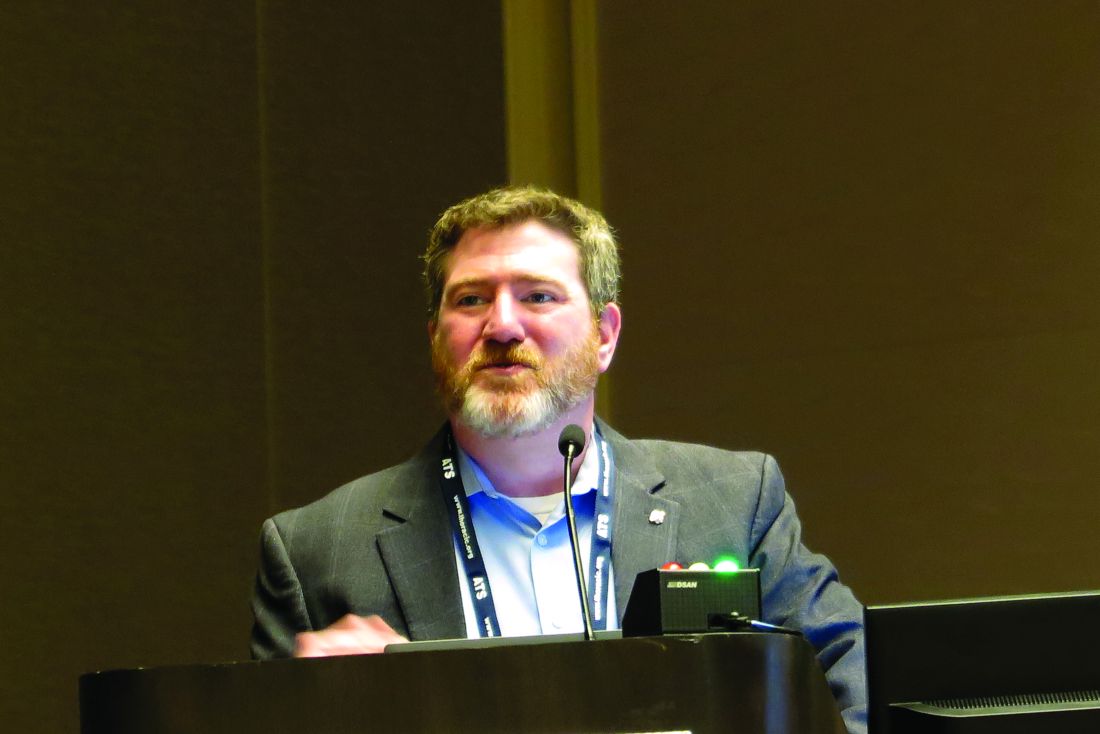
“Every day we download over 25 terabytes of data,” said John Haynes, MS, program manager for Health and Air Quality Applications in the Earth Action Program of the NASA Earth Science Division in Washington.
“Many of the observation data sets are critical for healthy air quality applications: observation of land surface temperature, sea surface temperature, precipitation, fires and thermal anomalies, aerosols, just to name a few, and the really awesome news is this offering from our constellation of satellites is free and open access, available to everyone across the globe,” he said.
The mission of NASA’s Earth Action Program is “to enable people and organizations to apply insights from Earth science to benefit the economy, health, quality of life, and environment.”
Program staff work with both industry and nonprofit environmental advocacy and health groups to help inform their decisions and actions with Earth science information.
NASA supports the use of Earth observations to help monitor and manage infectious diseases and environmental health, toxins and pathogens that affect health, air quality standards, and to assess the effects of climate change on air quality and public health.
Mr. Haynes noted that worldwide, six major cities have incorporated NASA data on fine particulate matter smaller than 2.5 microns (PM2.5) into their climate action plans. These cities include Accra, Ghana; Addis Ababa, Ethiopia; Buenos Aires, Argentina; Guadalajara, Mexico; Lima, Peru; and Johannesburg, South Africa.
Monitoring pollution with TEMPO
There are more than 30 Earth-monitoring systems currently in orbit or soon to be launched, including NASA’s Tropospheric Emissions: Monitoring of Pollution (TEMPO), launched in April 2023, with first operations in August 2023. The instrument is in a geostationary orbit about 22,236 miles above the equator at longitudes that allow it to survey virtually all of North America — from coast to coast, and from southern Mexico, Cuba, Puerto Rico, and the Bahamas to Northern Canada.
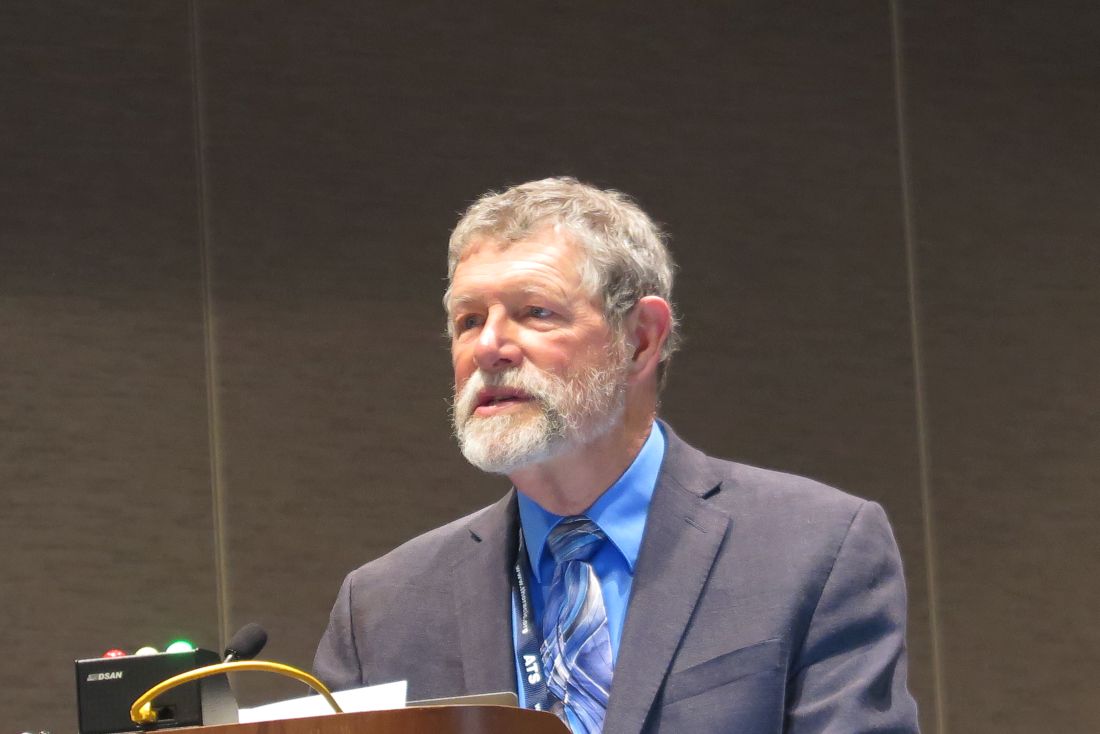
TEMPO is part of a geostationary air quality satellite “constellation” or group that provides daylight observation over the entire Northern Hemisphere, explained Aaron Naeger, PhD, MS, mission applications lead for TEMPO at the NASA Marshall Space Flight Center in Huntsville, Alabama.
Until TEMPO, space-based instruments had relatively low spatial resolution and could only capture one image each day. In contrast, TEMPO can scan east-west each daylight hour across its entire coverage area (known as the Field of Regard), and even more frequently during early morning and late afternoon. This allows researchers to measure volumes of pollution, sources, and how these pollution levels vary over time. The system measures ozone levels, nitrogen dioxide (NO2,) formaldehyde, and aerosols.
More than 100 federal, state, local and tribal air quality agencies use the data captured by TEMPO to inform public health efforts.
Dr. Naeger gave examples of how the system can help identify public health hazards, including scans that showed high NO2 levels from cities, traffic corridors, power plants, oil and gas fields, and fires.
Similarly, the system detected unhealthy ozone and PM2.5 levels during prescribed burns in April 2024, as well as notable differences between weekdays and weekends in NO2 concentrations across California and the Front Range in Colorado. These showed higher levels along traffic corridors during weekdays related to increased traffic volumes and tailpipe emissions.
Fire and heat
Other NASA health and air quality initiatives include the FireAQ project, based at the University of Iowa in Iowa City, which provides free online weekly briefings on fire-related air quality concerns using data from TEMPO and other NASA satellite systems. The FireAQ project was described by Jun Wang, PhD, from the University of Iowa in Iowa City.
NASA also fosters collaborations to reduce health disparities in air quality and respiratory health in urban heat islands and other areas affected by extreme temperatures due to climate change, as discussed by Christopher K. Uejio, PhD, from Florida State University in Tallahassee.
Air pollution expert George D. Thurston, ScD, professor of medicine and population health at the NYU Grossman School of Medicine, who attended the session, said that the PM2.5 standard includes nontoxic particulate matter, such as soil, and misses sub-micron sized particles, and asked Mr. Haynes whether smaller particles were being measured in the studies he described.
Mr. Haynes replied that the systems do not directly measure PM2.5 but instead rely on aerosol optical depth, a measure of the extent to which atmospheric particles absorb or scatter sunlight.
Dr. Thurston, who in 1987 was coauthor of groundbreaking study showing the link between PM2.5 levels and mortality, is now an advocate for a tougher standard of measuring ambient ultrafine particles with an aerodynamic diameter less than .1 microns in size (PM1).
NASA health and climate data are available at https://www.earthdata.nasa.gov/.
Mr. Haynes and Dr. Naeger are NASA employees. Dr. Thurston had no relevant disclosures.
SAN DIEGO – No matter where on earth you live, there’s likely to be an eye in the sky hovering overhead, and that’s a good thing, at least when it comes to satellite monitoring of air quality, said scientists from the National Aeronautics and Space Administration (NASA).
In a special symposium held at the American Thoracic Society’s international conference, NASA health and air quality specialists described the use of space-based systems and earth science applications to improve understanding of respiratory health risks worldwide, and to help enrich pulmonary research with galaxies of data.
“Every day we download over 25 terabytes of data,” said John Haynes, MS, program manager for Health and Air Quality Applications in the Earth Action Program of the NASA Earth Science Division in Washington.
“Many of the observation data sets are critical for healthy air quality applications: observation of land surface temperature, sea surface temperature, precipitation, fires and thermal anomalies, aerosols, just to name a few, and the really awesome news is this offering from our constellation of satellites is free and open access, available to everyone across the globe,” he said.
The mission of NASA’s Earth Action Program is “to enable people and organizations to apply insights from Earth science to benefit the economy, health, quality of life, and environment.”
Program staff work with both industry and nonprofit environmental advocacy and health groups to help inform their decisions and actions with Earth science information.
NASA supports the use of Earth observations to help monitor and manage infectious diseases and environmental health, toxins and pathogens that affect health, air quality standards, and to assess the effects of climate change on air quality and public health.
Mr. Haynes noted that worldwide, six major cities have incorporated NASA data on fine particulate matter smaller than 2.5 microns (PM2.5) into their climate action plans. These cities include Accra, Ghana; Addis Ababa, Ethiopia; Buenos Aires, Argentina; Guadalajara, Mexico; Lima, Peru; and Johannesburg, South Africa.
Monitoring pollution with TEMPO
There are more than 30 Earth-monitoring systems currently in orbit or soon to be launched, including NASA’s Tropospheric Emissions: Monitoring of Pollution (TEMPO), launched in April 2023, with first operations in August 2023. The instrument is in a geostationary orbit about 22,236 miles above the equator at longitudes that allow it to survey virtually all of North America — from coast to coast, and from southern Mexico, Cuba, Puerto Rico, and the Bahamas to Northern Canada.
TEMPO is part of a geostationary air quality satellite “constellation” or group that provides daylight observation over the entire Northern Hemisphere, explained Aaron Naeger, PhD, MS, mission applications lead for TEMPO at the NASA Marshall Space Flight Center in Huntsville, Alabama.
Until TEMPO, space-based instruments had relatively low spatial resolution and could only capture one image each day. In contrast, TEMPO can scan east-west each daylight hour across its entire coverage area (known as the Field of Regard), and even more frequently during early morning and late afternoon. This allows researchers to measure volumes of pollution, sources, and how these pollution levels vary over time. The system measures ozone levels, nitrogen dioxide (NO2,) formaldehyde, and aerosols.
More than 100 federal, state, local and tribal air quality agencies use the data captured by TEMPO to inform public health efforts.
Dr. Naeger gave examples of how the system can help identify public health hazards, including scans that showed high NO2 levels from cities, traffic corridors, power plants, oil and gas fields, and fires.
Similarly, the system detected unhealthy ozone and PM2.5 levels during prescribed burns in April 2024, as well as notable differences between weekdays and weekends in NO2 concentrations across California and the Front Range in Colorado. These showed higher levels along traffic corridors during weekdays related to increased traffic volumes and tailpipe emissions.
Fire and heat
Other NASA health and air quality initiatives include the FireAQ project, based at the University of Iowa in Iowa City, which provides free online weekly briefings on fire-related air quality concerns using data from TEMPO and other NASA satellite systems. The FireAQ project was described by Jun Wang, PhD, from the University of Iowa in Iowa City.
NASA also fosters collaborations to reduce health disparities in air quality and respiratory health in urban heat islands and other areas affected by extreme temperatures due to climate change, as discussed by Christopher K. Uejio, PhD, from Florida State University in Tallahassee.
Air pollution expert George D. Thurston, ScD, professor of medicine and population health at the NYU Grossman School of Medicine, who attended the session, said that the PM2.5 standard includes nontoxic particulate matter, such as soil, and misses sub-micron sized particles, and asked Mr. Haynes whether smaller particles were being measured in the studies he described.
Mr. Haynes replied that the systems do not directly measure PM2.5 but instead rely on aerosol optical depth, a measure of the extent to which atmospheric particles absorb or scatter sunlight.
Dr. Thurston, who in 1987 was coauthor of groundbreaking study showing the link between PM2.5 levels and mortality, is now an advocate for a tougher standard of measuring ambient ultrafine particles with an aerodynamic diameter less than .1 microns in size (PM1).
NASA health and climate data are available at https://www.earthdata.nasa.gov/.
Mr. Haynes and Dr. Naeger are NASA employees. Dr. Thurston had no relevant disclosures.
FROM ATS 2024

New Administration Routes for Adrenaline in Anaphylaxis
PARIS — While anaphylaxis requires immediate adrenaline administration through autoinjection, the use of this treatment is not optimal. Therefore, the development of new adrenaline formulations (such as for intranasal, sublingual, and transcutaneous routes) aims to facilitate the drug’s use and reduce persistent delays in administration by patients and caregivers. An overview of the research was presented at the 19th French-speaking Congress of Allergology.
Anaphylaxis is a severe and potentially fatal immediate hypersensitivity reaction with highly variable and dynamic clinical presentations. It requires prompt recognition for immediate treatment with intramuscular (IM) adrenaline (at the anterolateral aspect of the mid-thigh).
One might think that this reflex is acquired, but in France, while the number of prescribed adrenaline autoinjection (AAI) devices has been increasing for a decade, reaching 965,944 units in 2022, this first-line treatment is underused. Anapen (150, 300, and 500 µg), EpiPen (150 and 300 µg), Jext (150 µg and 300 µg), and Emerade (150, 300, and 500 µg) are the four products marketed in France in 2024.
“Only 17.3% of individuals presenting to the emergency department in the Lorraine region used it in 2015,” said Catherine Neukirch, MD, a pneumologist at Hôpital Bichat–Claude Bernard in Paris, France, with rates of 11.3% for children and 20.3% for adults.
Anaphylaxis Incidence Increasing
Approximately 0.3% (95% CI, 0.1-0.5) of the population will experience an anaphylaxis episode in their lifetime. Incidence in Europe, across all causes, is estimated between 1.5 and 7.9 cases per 100,000 inhabitants per year. Although anaphylaxis is on the rise, its associated mortality remains low, ranging between 0.05 and 0.51 per million per year for drugs, between 0.03 and 0.32 per million per year for foods, and between 0.09 and 0.13 per million per year for hymenopteran venoms.
Data from the European Anaphylaxis Registry indicate that anaphylaxis manifests rapidly after allergen exposure: 55% of cases occur within 10 minutes and 80% within 30 minutes. In addition, a biphasic reaction, which can occur up to 72 hours after exposure, is observed in < 5% of cases.
While a delay in adrenaline use is associated with risk for increased morbidity and mortality, AAI significantly reduces error rates compared with manual treatments involving ampoules, needles, and syringes. It also reduces the associated panic risks. However, there are multiple barriers to adrenaline use. The clinical symptoms of anaphylaxis may be misleading, especially if it occurs without cutaneous and urticarial manifestations but with only acute bronchospasm. It may present as isolated laryngeal edema without digestive involvement, hypotension, or other respiratory problems.
Other limitations to adrenaline use include technical difficulties and the possibility of incorrect administration, the need for appropriate needle sizes for patients with obesity, needle phobia, potential adverse effects of adrenaline injections, failure to carry two autoinjectors, constraints related to storage and bulky transport, as well as the need for training and practice.
“These factors contribute to underuse of adrenaline by patients and caregivers,” said Dr. Neukirch, which results in delays in necessary administration.
Adrenaline Treatment Criteria?
An analysis published in 2023 based on pharmacovigilance data from 30 regional French centers from 1984 to 2022 included 42 reported cases (average age, 33 years; 26% children) of reactions to AAI, which probably is an underestimate. About 40% of AAI uses occurred during anaphylaxis. The remaining 60% were triggered outside of reactions. The main reasons were accidental injections, mainly in the fingers, and cases of not triggering the autoinjector, underlining the importance of patient education.
In 2015, the European Medicines Agency required pharmacological studies for injectable adrenaline on healthy volunteers. These studies include ultrasound measurements of bolus injection, pharmacokinetics (ie, absorption, distribution, metabolism, and excretion), and pharmacodynamics (ie, the effect of the drug and the mechanism of action in the body), with precise evaluation of cardiovascular effects (eg, systolic and diastolic blood pressures and heart rate).
Among the information collected with the different products, ultrasound studies have shown a different localization of the adrenaline bolus (ie, in muscle in patients with normal BMI and mostly in adipose tissue in patients with BMI indicating overweight and obesity). The consequences of this finding are still unknown.
In a study with 500 µg Anapen, women with overweight or obesity showed different pharmacokinetic or pharmacodynamic profiles from those in men with normal weight, with an increase in the area under the curve (0-240 min) and marked changes in the heart rate time curve.
IM administration of 0.5 mg produces rapid pharmacokinetic effects in patients with normal weight, overweight, or obesity, with a delay for the second peak in the latter case. This delay perhaps results from initial local vasoconstriction due to adrenaline.
The early peak plasma concentration occurs at 5-10 minutes for AAI, with a faster speed for Anapen and EpiPen.
Moreover, needle size is not the most important factor. Rather, it is the strength and speed of injection, which can vary depending on the AAI.
Also, the optimal plasma concentration of adrenaline to treat anaphylaxis is not known; studies cannot be conducted during anaphylaxis. In terms of pharmacokinetics, a small series discovered that increased skin or muscle thickness delays the absorption of EpiPen AAI.
Intranasal Adrenaline
To facilitate rapid adrenaline use and convince reluctant patients to carry and use adrenaline, intranasal, sublingual, or transcutaneous forms are under development.
Three intranasal forms of adrenaline are already well advanced, including Neffy from ARS Pharma, epinephrine sprays from Bryn Pharma and Hikma, and Oxero from Oragoo, which contains dry powder.
A comparison of intranasal adrenaline Neffy and AAI shows that the former has satisfactory pharmacokinetic and pharmacodynamic effects.
In a phase 1 randomized crossover study of 42 healthy adults comparing the pharmacokinetic effects of Neffy adrenaline (2 mg) and EpiPen (0.3 mg), as well as IM epinephrine 0.3 mg, several observations were made. For a single dose, the maximum concentration (Cmax) of Neffy was lower than that of EpiPen.
However, with repeated doses administered 10 minutes apart, the Cmax of Neffy was higher than that of EpiPen. At this stage, pharmacodynamic responses to intranasal products are at least comparable with those of approved injectable products.
A comparison of the pharmacodynamic effects, such as systolic and diastolic blood pressures and heart rate, of Neffy adrenaline and AAI concluded that the profile of Neffy is comparable with that of EpiPen and superior to that of IM epinephrine.
In patients with a history of allergic rhinitis, adrenaline Cmax appears to be increased, while time to peak plasma concentration (Tmax) is reduced. Low blood pressure does not prevent Neffy absorption. Neffy is currently under review by the American and European health authorities.
Intranasal absorption of dry powder adrenaline appears to be faster than that of EpiPen, thus offering a clinical advantage in the short therapeutic window for anaphylaxis treatment.
In an open-label trial conducted on 12 adults with seasonal allergic rhinitis without asthma, the pharmacokinetics, pharmacodynamics, and safety of adrenaline were compared between FMXIN002 (1.6 and 3.2 mg), which was administered intranasally with or without nasal allergen challenge, and IM EpiPen 0.3 mg. Pharmacokinetics varied by patient. Nevertheless, nasal FMXIN002 had a shorter Tmax, a doubled Cmax after the allergen challenge peak, and a higher area under the curve in the 8 hours following administration compared with EpiPen. Pharmacodynamic effects comparable with those of EpiPen were noted at 15 minutes to 4 hours after administration. The tolerance was good, with mild and local side effects. The powder seems to deposit slightly better in the nasal cavity. It remains stable for 6 months at a temperature of 40 °C and relative humidity of 75% and for 2 years at a temperature of 25 °C and relative humidity of 60%.
Sublingual Adrenaline Film
AQST-109 is a sublingual film that is intended to allow rapid administration of epinephrine 1, which is a prodrug of adrenaline. The product is the size of a postage stamp, weighs < 30 g, and dissolves on contact with the tongue.
The EPIPHAST II study was a phase 1, multiperiod, crossover study conducted on 24 healthy adults (age, 24-49 years) who were randomly assigned to receive either 12 or 0.3 mg of AQST-109 of manual IM adrenaline in the first two periods. All participants received 0.3 mg of EpiPen in the last period.
EpiPen 0.3 mg resulted in a higher Cmax than AQST-109 12 mg. AQST-109 12 mg had the fastest median Tmax of 12 minutes. The areas under the curve of AQST-109 12 mg fell between those of EpiPen 0.3 mg and manual IM adrenaline 0.3 mg.
Early increases in systolic blood pressure, diastolic blood pressure, and heart rate were observed with AQST-109 12 mg. Changes were more pronounced with AQST-109 12 mg despite a higher Cmax with EpiPen 0.3 mg.
Part 3 of the EPIPHAST study evaluated the impact of food exposure (ie, a peanut butter sandwich) on the pharmacokinetics of AQST-109 12 mg in 24 healthy adults. Oral food residues did not significantly affect pharmacodynamic parameters, and no treatment-related adverse events were reported.
Researchers concluded that AQST-109 12 mg absorption would not be altered by “real” situations if used during meals. “These results suggest that the sublingual adrenaline film could be promising in real situations,” said Dr. Neukirch, especially in cases of food allergy with recent ingestion of the allergenic food.
Transcutaneous Adrenaline
A transcutaneous form of adrenaline that uses the Zeneo device developed by Crossject, a company based in Dijon, France, comes in the form of an AAI that requires no needle. This project, funded by the European Union, uses a gas generator to propel the drug at very high speed through the skin in 50 milliseconds. This method allows for extended drug storage.
Dr. Neukirch reported financial relationships with Viatris, Stallergènes, ALK, Astrazeneca, Sanofi, GSK, and Novartis.
This story was translated from the Medscape French edition using several editorial tools, including AI, as part of the process. Human editors reviewed this content before publication. A version of this article first appeared on Medscape.com.
Debate on pulmonary safety of gas stoves: Is the risk just hot air?
SAN DIEGO — While there is currently no smoking gun definitively showing that indoor nitrogen dioxide (NO2) concentrations from gas appliances are a cause of pulmonary diseases, the circumstantial evidence of the baleful effects of gas stoves on lung function is pretty compelling, said participants in a pro-con debate.
The debate was held at the American Thoracic Society’s international conference.
PRO: Gas stoves cause lung disease
Arguing for the “pro” side, John R. Balmes, MD of the University of California, San Francisco, and a physician member of the California Air Resources Board, began by admitting that “I would never have said gas stoves cause lung disease, but that’s what they assigned me.”
Gamely proceeding anyway, Dr. Balmes noted that natural gas — methane — is a potent greenhouse gas, and that cooking with natural gas leads to generation of NO2 with high peak concentrations in the home, especially in the kitchen, but in other rooms as well.
“We know that NO2 is an irritant gas that can cause bronchoconstriction, airway hyperresponsiveness and inflammation, and there’s increased risk of asthma and COPD exacerbations,” he said.
The US Environmental Protection Agency (EPA) outdoor ambient air standard for NO2 is 100 parts per billion (ppb) or lower, which are the levels needed to prevent asthma exacerbations. In separate meta-analyses there was a 1.05 rise in asthma incidence per every 2 ppb of NO2, and an increase of 1.07 in COPD incidence for every 5 ppb of NO2, Dr. Balmes noted.
The respiratory effects of gas stoves were revealed in a 2013 meta-analysis of 10 studies from North America and Europe, which showed a pooled odds ratio for current asthma of 1.34. Building on these data, authors of a 2022 paper estimated that 13% of childhood asthma could be prevented by elimination gas cooking.
Although the causative link is missing, the evidence is abundant that natural gas isn’t good for anyone, he acknowledged.
Con: More evidence needed
Arguing for the “con” side of the question, Meredith C. McCormack, MD, MHS, professor of medicine in the pulmonary and critical care division at Johns Hopkins University in Baltimore, said that “more definitive evidence is needed to define whether gas stoves cause lung disease.”
But Dr. McCormack didn’t let the natural gas industry off the hook, noting that a systematic review and meta-analysis of cooking with gas in high-, middle-, and low-income countries showed that domestic use of gas fuels vs. electric was associated with increased risk of asthma (1.11 overall), COPD (1.15), and pneumonia (1.26).

The link between gas and risk of asthma was significant only for adults, however, and the data on the risks for COPD and for pneumonia or other respiratory infections came almost exclusively from low-income countries, she noted.
Despite the lack of evidence for a causative link, however, Dr. McCormack pointed to evidence that indoor NO2 is an air pollutant that acts as a respiratory irritant, and that indoor NO2 levels in homes with gas stoves have been shown to be more than twice as high as those in homes with electric stoves.
Other evidence shows that indoor NO2 is associated with increased symptoms and use of rescue medications for children with asthma, and with shortness of breath, nocturnal symptoms, reduction in lung function, and exacerbations in COPD.
Still other studies have shown that exchanging a gas stove for an electric stove can reduce NO2 concentrations in the home by up to 50%, but there is still a need for clinical trial evidence of a health benefit for such an exchange, she said.
And even if a gas stove is swapped out for an electric or induction range, household members with asthma are exposed to other hazards, including second-hand smoke, cooking exhaust, candle or incense burning, outdoor particulate matter that finds its way indoors, mold, and mouse or cockroach allergens, she noted.
On common ground
Environmental interventions that can benefit all members of a household — not just those with obstructive pulmonary disease — include smoking cessation, charcoal filter-equipped air cleaners, stove hoods that vent outdoors, integrated pest management, hypoallergenic pillow and mattress covers, high efficiency particulate air (HEPA) vacuums, and mold and radon abatement.
Both Dr. Balmes and Dr. McCormack agreed in the end that gas stoves contribute to respiratory morbidity, and that both state and national policy changes are needed to support transition to cleaner indoor air, with financial incentives available for households with more modest incomes.
“For everyone, there is a climate-change mitigation imperative to transition away from gas appliances if we want to tackle the climate emergency,” Dr. Balmes said.
End indoor combustion
George D. Thurston, ScD, professor of medicine and population health at the NYU Grossman School of Medicine, who attended the debate, told Chest Physician that the participants talked about NO2 but didn’t touch on particulate pollution generated by gas stoves.
Burning natural gas produces particles that are very similar in composition to those produced by burning coal, oil, or diesel fuel, Dr. Thurston said, and he pointed out that interventions such as range hoods work only if they actually vent outdoors, and aren’t simply fans that recirculate the air within the home. And even when ventilation works as it should to move air out of the house, it only pumps it back into the atmosphere, where it contributes to climate change.
“We need combustion-free homes. That’s the unifying principle. We have to keep our eyes on that prize,” he said.
Dr. Balmes, Dr. McCormack, and Dr. Thurston all reported having no relevant disclosures.
SAN DIEGO — While there is currently no smoking gun definitively showing that indoor nitrogen dioxide (NO2) concentrations from gas appliances are a cause of pulmonary diseases, the circumstantial evidence of the baleful effects of gas stoves on lung function is pretty compelling, said participants in a pro-con debate.
The debate was held at the American Thoracic Society’s international conference.
PRO: Gas stoves cause lung disease
Arguing for the “pro” side, John R. Balmes, MD of the University of California, San Francisco, and a physician member of the California Air Resources Board, began by admitting that “I would never have said gas stoves cause lung disease, but that’s what they assigned me.”
Gamely proceeding anyway, Dr. Balmes noted that natural gas — methane — is a potent greenhouse gas, and that cooking with natural gas leads to generation of NO2 with high peak concentrations in the home, especially in the kitchen, but in other rooms as well.
“We know that NO2 is an irritant gas that can cause bronchoconstriction, airway hyperresponsiveness and inflammation, and there’s increased risk of asthma and COPD exacerbations,” he said.
The US Environmental Protection Agency (EPA) outdoor ambient air standard for NO2 is 100 parts per billion (ppb) or lower, which are the levels needed to prevent asthma exacerbations. In separate meta-analyses there was a 1.05 rise in asthma incidence per every 2 ppb of NO2, and an increase of 1.07 in COPD incidence for every 5 ppb of NO2, Dr. Balmes noted.
The respiratory effects of gas stoves were revealed in a 2013 meta-analysis of 10 studies from North America and Europe, which showed a pooled odds ratio for current asthma of 1.34. Building on these data, authors of a 2022 paper estimated that 13% of childhood asthma could be prevented by elimination gas cooking.
Although the causative link is missing, the evidence is abundant that natural gas isn’t good for anyone, he acknowledged.
Con: More evidence needed
Arguing for the “con” side of the question, Meredith C. McCormack, MD, MHS, professor of medicine in the pulmonary and critical care division at Johns Hopkins University in Baltimore, said that “more definitive evidence is needed to define whether gas stoves cause lung disease.”
But Dr. McCormack didn’t let the natural gas industry off the hook, noting that a systematic review and meta-analysis of cooking with gas in high-, middle-, and low-income countries showed that domestic use of gas fuels vs. electric was associated with increased risk of asthma (1.11 overall), COPD (1.15), and pneumonia (1.26).
The link between gas and risk of asthma was significant only for adults, however, and the data on the risks for COPD and for pneumonia or other respiratory infections came almost exclusively from low-income countries, she noted.
Despite the lack of evidence for a causative link, however, Dr. McCormack pointed to evidence that indoor NO2 is an air pollutant that acts as a respiratory irritant, and that indoor NO2 levels in homes with gas stoves have been shown to be more than twice as high as those in homes with electric stoves.
Other evidence shows that indoor NO2 is associated with increased symptoms and use of rescue medications for children with asthma, and with shortness of breath, nocturnal symptoms, reduction in lung function, and exacerbations in COPD.
Still other studies have shown that exchanging a gas stove for an electric stove can reduce NO2 concentrations in the home by up to 50%, but there is still a need for clinical trial evidence of a health benefit for such an exchange, she said.
And even if a gas stove is swapped out for an electric or induction range, household members with asthma are exposed to other hazards, including second-hand smoke, cooking exhaust, candle or incense burning, outdoor particulate matter that finds its way indoors, mold, and mouse or cockroach allergens, she noted.
On common ground
Environmental interventions that can benefit all members of a household — not just those with obstructive pulmonary disease — include smoking cessation, charcoal filter-equipped air cleaners, stove hoods that vent outdoors, integrated pest management, hypoallergenic pillow and mattress covers, high efficiency particulate air (HEPA) vacuums, and mold and radon abatement.
Both Dr. Balmes and Dr. McCormack agreed in the end that gas stoves contribute to respiratory morbidity, and that both state and national policy changes are needed to support transition to cleaner indoor air, with financial incentives available for households with more modest incomes.
“For everyone, there is a climate-change mitigation imperative to transition away from gas appliances if we want to tackle the climate emergency,” Dr. Balmes said.
End indoor combustion
George D. Thurston, ScD, professor of medicine and population health at the NYU Grossman School of Medicine, who attended the debate, told Chest Physician that the participants talked about NO2 but didn’t touch on particulate pollution generated by gas stoves.
Burning natural gas produces particles that are very similar in composition to those produced by burning coal, oil, or diesel fuel, Dr. Thurston said, and he pointed out that interventions such as range hoods work only if they actually vent outdoors, and aren’t simply fans that recirculate the air within the home. And even when ventilation works as it should to move air out of the house, it only pumps it back into the atmosphere, where it contributes to climate change.
“We need combustion-free homes. That’s the unifying principle. We have to keep our eyes on that prize,” he said.
Dr. Balmes, Dr. McCormack, and Dr. Thurston all reported having no relevant disclosures.
FROM ATS 2024
Does More Systemic Treatment for Advanced Cancer Improve Survival?
This conclusion of a new study published online May 16 in JAMA Oncology may help reassure oncologists that giving systemic anticancer therapy (SACT) at the most advanced stages of cancer will not improve the patient’s life, the authors wrote. It also may encourage them to instead focus more on honest communication with patients about their choices, Maureen E. Canavan, PhD, at the Cancer and Outcomes, Public Policy and Effectiveness Research (COPPER) Center at the Yale School of Medicine in New Haven, Connecticut, and colleagues, wrote in their paper.
How Was the Study Conducted?
Researchers used Flatiron Health, a nationwide electronic health records database of academic and community practices throughout the United State. They identified 78,446 adults with advanced or metastatic stages of one of six common cancers (breast, colorectal, urothelial, non–small cell lung cancer [NSCLC], pancreatic and renal cell carcinoma) who were treated at healthcare practices from 2015 to 2019. They then stratified practices into quintiles based on how often the practices treated patients with any systemic therapy, including chemotherapy and immunotherapy, in their last 14 days of life. They compared whether patients in practices with greater use of systemic treatment at very advanced stages had longer overall survival.
What Were the Main Findings?
“We saw that there were absolutely no survival differences between the practices that used more systemic therapy for very advanced cancer than the practices that use less,” said senior author Kerin Adelson, MD, chief quality and value officer at MD Anderson Cancer Center in Houston, Texas. In some cancers, those in the lowest quintile (those with the lowest rates of systemic end-of-life care) lived fewer years compared with those in the highest quintiles. In other cancers, those in the lowest quintiles lived more years than those in the highest quintiles.
“What’s important is that none of those differences, after you control for other factors, was statistically significant,” Dr. Adelson said. “That was the same in every cancer type we looked at.”
An example is seen in advanced urothelial cancer. Those in the first quintile (lowest rates of systemic care at end of life) had an SACT rate range of 4.0-9.1. The SACT rate range in the highest quintile was 19.8-42.6. But the median overall survival (OS) rate for those in the lowest quintile was 12.7 months, not statistically different from the median OS in the highest quintile (11 months.)
How Does This Study Add to the Literature?
The American Society of Clinical Oncology (ASCO) and the National Quality Forum (NQF) developed a cancer quality metric to reduce SACT at the end of life. The NQF 0210 is a ratio of patients who get systemic treatment within 14 days of death over all patients who die of cancer. The quality metric has been widely adopted and used in value-based care reporting.
But the metric has been criticized because it focuses only on people who died and not people who lived longer because they benefited from the systemic therapy, the authors wrote.
Dr. Canavan’s team focused on all patients treated in the practice, not just those who died, Dr. Adelson said. This may put that criticism to rest, Dr. Adelson said.
“I personally believed the ASCO and NQF metric was appropriate and the criticisms were off base,” said Otis Brawley, MD, associate director of community outreach and engagement at the Sidney Kimmel Comprehensive Cancer Center at Johns Hopkins University School of Medicine in Baltimore. “Canavan’s study is evidence suggesting the metrics were appropriate.”
This study included not just chemotherapy, as some other studies have, but targeted therapies and immunotherapies as well. Dr. Adelson said some think that the newer drugs might change the prognosis at end of life. But this study shows “even those drugs are not helping patients to survive with very advanced cancer,” she said.
Could This Change Practice?
The authors noted that end-of life SACT has been linked with more acute care use, delays in conversations about care goals, late enrollment in hospice, higher costs, and potentially shorter and poorer quality life.
Dr. Adelson said she’s hoping that the knowledge that there’s no survival benefit for use of SACT for patients with advanced solid tumors who are nearing the end of life will lead instead to more conversations about prognosis with patients and transitions to palliative care.
“Palliative care has actually been shown to improve quality of life and, in some studies, even survival,” she said.
“I doubt it will change practice, but it should,” Dr. Brawley said. “The study suggests that doctors and patients have too much hope for chemotherapy as patients’ disease progresses. In the US especially, there is a tendency to believe we have better therapies than we truly do and we have difficulty accepting that the patient is dying. Many patients get third- and fourth-line chemotherapy that is highly likely to increase suffering without realistic hope of prolonging life and especially no hope of prolonging life with good quality.”
Dr. Adelson disclosed ties with AbbVie, Quantum Health, Gilead, ParetoHealth, and Carrum Health. Various coauthors disclosed ties with Roche, AbbVie, Johnson & Johnson, Genentech, the National Comprehensive Cancer Network, and AstraZeneca. The study was funded by Flatiron Health, an independent member of the Roche group. Dr. Brawley reports no relevant financial disclosures.
FROM JAMA ONCOLOGY
Former UCLA Doctor Receives $14 Million in Gender Discrimination Retrial
A California jury has awarded $14 million to a former University of California, Los Angeles (UCLA) oncologist who claimed she was paid thousands less than her male colleagues and wrongfully terminated after her complaints of gender-based harassment and intimidation were ignored by program leadership.
The decision comes after a lengthy 8-year legal battle in which an appellate judge reversed a previous jury decision in her favor.
Lauren Pinter-Brown, MD, a hematologic oncologist, was hired in 2005 by the University of California, Los Angeles School of Medicine — now called UCLA’s David Geffen School of Medicine. As the school’s lymphoma program director, she conducted clinical research alongside other oncology doctors, including Sven de Vos, MD.
She claimed that her professional relationship with Dr. de Vos became contentious after he demonstrated “oppositional” and “disrespectful” behavior at team meetings, such as talking over her and turning his chair so Dr. Pinter-Brown faced his back. Court documents indicated that Dr. de Vos refused to use Dr. Pinter-Brown’s title in front of colleagues despite doing so for male counterparts.
Dr. Pinter-Brown argued that she was treated as the “butt of a joke” by Dr. de Vos and other male colleagues. In 2016, she sued Dr. de Vos, the university, and its governing body, the Board of Regents, for wrongful termination.
She was awarded a $13 million verdict in 2018. However, the California Court of Appeals overturned it in 2020 after concluding that several mistakes during the court proceedings impeded the school’s right to a fair and impartial trial. The case was retried, culminating in the even higher award of $14 million issued on May 9.
“Two juries have come to virtually identical findings showing multiple problems at UCLA involving gender discrimination,” Dr. Pinter-Brown’s attorney, Carney R. Shegerian, JD, told this news organization.
A spokesperson from UCLA’s David Geffen School of Medicine said administrators are carefully reviewing the new decision.
The spokesperson told this news organization that the medical school and its health system remain “deeply committed to maintaining a workplace free from discrimination, intimidation, retaliation, or harassment of any kind” and fostering a “respectful and inclusive environment ... in research, medical education, and patient care.”
Gender Pay Disparities Persist in Medicine
The gender pay gap in medicine is well documented. The 2024 Medscape Physician Compensation Report found that male doctors earn about 29% more than their female counterparts, with the disparity growing larger among specialists. In addition, a recent JAMA Health Forum study found that male physicians earned 21%-24% more per hour than female physicians.
Dr. Pinter-Brown, who now works at the University of California, Irvine, alleged that she was paid $200,000 less annually, on average, than her male colleagues.
That’s not surprising, says Martha Gulati, MD, professor and director of preventive cardiology at Cedars-Sinai Smidt Heart Institute, Los Angeles. She coauthored a commentary about gender disparities in JAMA Network Open. Dr. Gulati told this news organization that even a “small” pay disparity of $100,000 annually adds up.
“Let’s say the [male physician] invests it at 3% and adds to it yearly. Even without a raise, in 20 years, that is approximately $3 million,” Dr. Gulati explained. “Once you find out you are paid less than your male colleagues, you are upset. Your sense of value and self-worth disappears.”
Eileen Barrett, MD, MPH, president-elect of the American Medical Women’s Association, said that gender discrimination is likely more prevalent than research indicates. She told this news organization that self-doubt and fear of retaliation keep many from exposing the mistreatment.
Although more women are entering medicine, too few rise to the highest positions, Dr. Barrett said.
“Unfortunately, many are pulled and pushed into specialties and subspecialties that have lower compensation and are not promoted to leadership, so just having numbers isn’t enough to achieve equity,” Dr. Barrett said.
Dr. Pinter-Brown claimed she was repeatedly harassed and intimidated by Dr. de Vos from 2008 to 2015. Despite voicing concerns multiple times about the discriminatory behavior, the only resolutions offered by the male-dominated program leadership were for her to separate from the group and conduct lymphoma research independently or to avoid interacting with Dr. de Vos, court records said.
Even the school’s male Title IX officer, Jan Tillisch, MD, who handled gender-based discrimination complaints, reportedly made sexist comments. When Dr. Pinter-Brown sought his help, he allegedly told her that she had a reputation as an “angry woman” and “diva,” court records showed.
According to court documents, Dr. Pinter-Brown endured nitpicking and research audits as retaliation for speaking out, temporarily suspending her research privileges. She said she was subsequently removed from the director position and replaced by Dr. de Vos.
Female physicians who report discriminatory behavior often have unfavorable outcomes and risk future career prospects, Dr. Gulati said.
To shift this dynamic, she said institutions must increase transparency and practices that support female doctors receiving “equal pay for equal work.”
A version of this article appeared on Medscape.com.
Novel ENV-101 associated with improved lung function in IPF
SAN DIEGO —
Early efficacy data from a phase 2a safety trial suggest that the novel oral agent, dubbed ENV-101, is associated with improvements in forced vital capacity (FVC) and other measures of lung function, and may be a disease-modifying therapy for IPF, according to Toby M. Maher, MD, PhD, director of the interstitial lung disease program at Keck School of Medicine, University of Southern California, Los Angeles. Dr. Maher presented the results at the American Thoracic Society’s international conference.
“Historically we’ve not been seeing improvements in FVC, which is what we’ve been seeing [with ENV-101], and I think it’s conceivable that you can get remodeling of early areas of fibrosis in the lung,” Dr. Maher said in an interview with Chest Physician.
“We know from histology studies that if you look at IPF lungs you’ll see areas of end-stage fibrosis, but even in advanced disease you’ll see areas where the lung is relatively well preserved and there’s early fibrosis, so I think it’s conceivable that there is remodeling of some of those early areas of fibrosis,” he said.
Vital pathway
The Hedgehog pathway is highly conserved in evolution. The cell-signaling pathway is active embryogenesis, tissue proliferation, and organ development. There is also evidence to suggest that in adult the pathway becomes reactivated following tissue injury, as can occur in lung epithelia, Dr. Maher explained.
Although as the word “idiopathic” in IPF indicates the etiology of the disease is unknown, investigators have found that in IPF repetitive epithelial injury to lung tissue leads to activation of the Hedgehog pathway. Hedgehog signaling in turn induces formation and activation of myofibroblasts that lay down fibrotic matrix and contract lung tissue, leading to significant impairments in gas exchange, Dr. Maher said.
ENV-101 blocks Hedgehog from binding to the PTCH1 receptor, preventing release of the zinc-finger protein GLI1 from the kinase complex into the cell cytoplasm. With signaling blocked, myofibroblasts undergo apoptosis instead of initiating wound repair as they normally would, thereby eliminating an evident mechanism of IPF pathology, he explained.
Study details
In the phase 2a trial, investigators enrolled patients with IPF who were not taking antifibrotic agents and who had a percent predicted FVC greater than 50%, percent predicted diffusing capacity for carbon monoxide (DLCO) of at least 35%, and life expectancy of more than 1 year.
The patients were randomized to receive 200 mg oral ENV-101 daily (18 patients) or placebo (15 patients) for 12 weeks.
The primary endpoint of the trial was safety of the experimental agent. A previous phase 1b study of a different Hedgehog inhibitor — vismodegib (Erivedge), in combination with the antifibrotic agent pirfenidone (Pirespa) — in patients with IPF was discontinued because of poor tolerability.
In the current study, the most common treatment-related adverse events were dysgeusia in 57% of patients who received the drug, alopecia in 52%, and muscle spasms in 43%. The spasms were generally less severe than those seen in the vismodegib/pirfenidone trial mentioned above.
Seven patients (33%) had treatment-emergent events leading to dose interruption. Five patients discontinued treatment: one who withdrew because of taste alterations, one who was lost to follow-up after an IPF exacerbation, and three who withdrew consent.
There were no treatment-related deaths, and no clinically significant findings on labs, vital signs, electrocardiograms, or physical exam.
Efficacy endpoints
An analysis of the secondary efficacy endpoints showed a 1.9% mean improvement in FVC from baseline among patients assigned to ENV-101, compared with a mean decline of 1.3% of patients assigned to placebo (P = .035).
Patients on the active drug also had a 200-mL mean increase in total lung capacity, compared with a mean decline of 56 mL for patients on placebo (P = .005).
In addition, high-resolution CR studies showed a 9.4% absolute decrease from baseline in quantitative interstitial lung disease with ENV-101, vs. a 1.1% increase among controls, a 2% absolute decline from baseline in quantitative lung fibrosis compared with a 0.87% increase with placebo, and a 4.6% absolute decrease from baseline in quantitative ground glass, compared with an increase of 0.29% with placebo.
Bad taste a good sign?
Reinoud Gosens PhD, University of Groningen, the Netherlands, who co-moderated the session but was not involved in the study, questioned whether the dysgeusia seen in patients who received ENV-101 might be related to the dysgeusia seen in clinical trials of P2X3 receptor antagonists for cough.
“I was wondering if there would be a mechanistic overlap between Hedgehog inhibition and cough, which would be quite relevant for IPF,” he said in an interview.
The increase in FVC seen with ENV-101 and with the investigational agent buloxibutid, a novel angiotensin II type 2 receptor agonist described in a separate presentation by Dr. Maher, suggests that these drugs may have the ability to help remodel damaged lungs, Dr. Gosens said.
Investigators are currently planning a phase 2 dose-ranging trial (WHISTLE-PF) in patients with IPF or progressive pulmonary fibrosis.
The phase 2a trial was supported by Endeavor BioMedicines. Dr. Maher disclosed consultancy or speaker fees from Endeavor and others. Dr. Gosens had no relevant disclosures.
FROM ATS 2024
How Physician Mortgage Loans Work for Doctors With Debt
Tell someone you’re a doctor, and the reaction is often: “You must be rich.” But physicians who are just finishing medical school or are in their early careers might feel far from it. The average medical school debt is more than $200,000, with total debts including undergrad climbing well north of $250,000.
That leaves house-hunting physicians in a predicament. A key factor for lending institutions is the “debt to income” ratio, a calculation which indicates if you already have too much debt to pay your mortgage. That single equation could eliminate you from lenders’ mortgage requirements.
But young doctors are also in a unique situation. Yes, they carry above-average levels of debt, but they are on a path to substantial income in future years. That’s where the physician mortgage loan (PML) becomes a useful option.
What Is a Physician Mortgage Loan?
, according to Stephen Chang, MD, a radiologist, and a managing director at Acts Financial Advisors in McLean, Virginia.
The key features, according to James M. Dahle, MD, an emergency physician and founder of The White Coat Investor, include:
- No required down payment, which is typically 20% with a conventional loan.
- No private mortgage insurance (PMI). This is often a requirement of traditional loans, designed to protect the lender if the buyer misses payments. PMLs don’t involve PMI even if you don’t put down 20%.
- No pay stubs. With a conventional loan, pay stubs are often required to prove income level and reliability. PMLs will often allow an employment contract in place of those.
- Different consideration of the student loan burden.
Those are the upsides, of course, but there may be downsides. Dr. Dahle said a PML might involve slightly higher rates and fees than a conventional mortgage does but not always.
Who Is Best Suited for a Physician Mortgage Loan?
Financial advisers caution that everyone should first consider their full financial picture before applying for a mortgage, PML or otherwise. “If you don’t have the money saved for a down payment, one can ask if you are financially prepared to purchase a home,” says Cobin Soelberg, MD, an anesthesiologist and owner of Greeley Wealth Management, a financial planning firm serving physician families in Bend, Oregon.
If your savings are slim, you might need to build those accounts further before pursuing home ownership and the expenses that come along with it.
Your credit score can contribute to the equation. “With any loan product, we always recommend working to optimize your personal credit score as soon as possible before applying for a loan,” said Mark P. Eid, MD, a dermatologist and co–managing director (with Dr. Chang) at Act Financial Advisors. “Once you get into the high 700s, you’ve typically qualified for the best interest rates, so while that perfect 850 is nice to achieve, it’s by no means necessary.”
Also, assess your reasons for purchasing a home and whether it will fit your lifestyle in the coming years. “The main reason that [my wife and I] wanted to buy a home was for stability,” said Jordan Frey, MD, founder of The Prudent Plastic Surgeon. “After living in apartments for years, we wanted a place that was truly our own. We definitely felt disappointed and frustrated when worrying that our student debt may limit our ability to do this.”
Like many physicians, Dr. Frey had taken on a huge amount of debt, to the tune of half a million dollars in student loans and credit card debt when he finished training in 2020. The question Dr. Frey and his wife wrestled with was: “How much debt should we take on in addition to what we already have?”
What Are the Risks? What’s in the Fine Print?
The eased limitations of PMLs come with potential pitfalls, and physicians should not imagine that they have unlimited buying power.
“Many physicians buy more expensive or bigger houses than they need simply because banks are willing to lend physicians money,” Dr. Soelberg warns. “So, the doctor gets locked into a large mortgage and cannot build wealth, save for retirement, and repay their student loans.”
As you shop around, beware of omissions and scams. When meeting with lenders, Dr. Frey recalled that some didn’t even present PMLs as an option, and others presented them with unfavorable terms. He was careful to look for disadvantages hidden in the fine print, such as a potential “big hike in the rate a year later.”
But sometimes, a scam is not outright deception but is more like temptation. So it’s important to have your own best interests in mind without relying on lenders’ advice.
“When we were shopping around, some mortgage lenders would [offer] $1.5 million, and we thought ‘that makes no sense,’ ” said Dr. Frey. “[Physicians] have big future income, which makes us attractive to these lenders. No one in their right mind would give a mortgage like this to anyone else. They aren’t worried about whether it’s a smart decision for you or not.”
What Other Red Flags Should You Look Out for?
Dr. Frey recommends medical professionals beware of these red flags when shopping for PMLs:
- A request for any type of collateral, including your medical practice
- A rate that is much higher than others
- A lender is pushing you to borrow a higher amount than you’re comfortable with
- A lender attempts to influence your decision about the size of your down payment
Remember, if you are choosing an adjustable-rate mortgage (ARM), your rate will recalibrate on the basis of the market’s rates — for better or worse. This means that your payment might be higher or lower, taking current interest rates into account, based on the market.
Looking back, Dr. Frey said he might reconsider his decision to use a 10-year ARM. He and his wife chose it because the rate was low at the time, and they planned to pay off the mortgage quickly or move before it went up. But the uncertainty added an element of pressure.
How Can PMLs Contribute to Overall Financial Health?
Dr. Frey says his physician mortgage was “a huge advantage,” allowing him and his wife to put 0% down on their home without PMI. But most importantly, it fit within their overall financial plan, which included investing. “The money that we would have potentially used for a down payment, we used to buy a rental property, which then got us more income,” he says.
Of course, buying a rental property is not the only path to financial health and freedom. Many people approach a home as an investment that will eventually become fully their own. Others might put that down payment toward building a safety net of savings accounts.
Used strategically and intentionally, PMLs can put you on a more predictable financial path. And with less money stress, buying a home can be an exciting milestone as you plan your future and put down roots in a community.
A version of this article appeared on Medscape.com.
New Data to Change Practice on BP Control in Acute Stroke: INTERACT4
BASEL, SWITZERLAND — Early reduction of blood pressure has a beneficial effect in hemorrhagic stroke but a detrimental effect in ischemic stroke, new trial data show. The findings could shake up recommendations on control of blood pressure in acute stroke patients.
“This is the first time that we have randomized evidence of blood pressure control prior to reperfusion in ischemic stroke patients, and our data will challenge the current guidelines that recommend lowering blood pressure to below 180 mm Hg systolic in these patients,” said study coauthor Craig Anderson, MD, George Institute for Global Health, Sydney, Australia.
“And this study also clearly shows for the first time that getting blood pressure under control in hemorrhagic stroke patients in the first couple of hours has definitive benefits,” he added.
The findings were presented on May 16 at the European Stroke Organization Conference (ESOC) annual meeting and published online simultaneously in The New England Journal of Medicine.
A Test of Early BP Control
The trial was conducted to test the strategy of very early blood pressure control during patient transport in an ambulance after acute stroke, which investigators suspected could benefit patients with both types of stroke.
The hypothesis was that this would reduce bleeding in the brain for those with hemorrhagic stroke. For ischemic stroke patients, it was thought this strategy would speed up administration of thrombolysis, because guidelines recommend bringing blood pressure under control before thrombolysis.
For the INTERACT4 trial, which was conducted in China, 2404 patients with suspected acute stroke and elevated systolic blood pressure (≥ 150 mm Hg) who were assessed in the ambulance within 2 hours after symptom onset were randomized to receive immediate treatment with intravenous urapidil to lower the systolic blood pressure or usual blood pressure management (usual care group).
The median time between symptom onset and randomization was 61 minutes, and the mean blood pressure at randomization was 178/98 mm Hg.
Stroke was subsequently confirmed by imaging in 2240 patients, of whom 46% had a hemorrhagic stroke and 54% an ischemic stroke.
At the time of arrival at the hospital, the mean systolic blood pressure in the intervention group was 158 mm Hg, compared with 170 mm Hg in the usual care group.
The primary efficacy outcome was functional status as assessed by modified Rankin scale score at 90 days.
Overall, there was no difference between the two groups in terms of functional outcome scores (common odds ratio [OR], 1.00; 95% CI, 0.87-1.15), and the incidence of serious adverse events was similar.
But the study showed very different results in patients with hemorrhagic stroke vs those with ischemic stroke.
‘Slam-Dunk’ Effect
Anderson has led several previous trials of blood pressure control in stroke patients, some of which have suggested benefit of lowering blood pressure in those with hemorrhagic stroke, but he says the results of the current trial are more clear-cut.
“We have never seen such a slam-dunk effect as there was in INTERACT4,” Dr. Anderson said. “Not only did we show that early reduction of blood pressure in hemorrhagic stroke patients improved functional outcome, it also reduced bleeding in the brain, improved survival and quality of life, and reduced surgery and infection complications. That’s quite remarkable.”
The findings offer “clear evidence that for patients with hemorrhagic stroke, we must get the blood pressure under control as soon as possible and introduce systems of care to ensure this happens,” he added.
The reason for the clear findings in the current trial is probably the treatment time, Dr. Anderson said.
“This is the first trial in which blood pressure has been controlled in the ambulance and occurred much earlier than in the previous trials.”
Challenging Ischemic Stroke Guidelines
The INTERACT4 results in ischemic stroke patients are likely to be more controversial.
“Our results are clearly challenging longstanding beliefs around blood pressure control in ischemic stroke prior to thrombolysis,” Dr. Anderson said.
Current guidelines recommend a blood pressure < 185 mm Hg systolic before initiation of thrombolysis because of concerns about intracerebral hemorrhage, he noted. Often, blood pressure is lowered rapidly down to much lower levels in order give thrombolysis quickly.
“Our results suggest this may not be a good idea,” Dr. Anderson said. “I think these data will shake us up a bit and make us more cautious about reducing blood pressure in these patients. Personally, I wouldn’t touch the blood pressure at all in ischemic stroke patients after these results.”
He said the mechanisms behind the different stroke types would explain the results.
“If a patient is bleeding, it makes sense that higher blood pressure would make that worse,” Dr. Anderson said. “But when a patient has a blocked artery and ischemia in the brain, it seems likely that the extra pressure is needed to keep oxygen delivery to the ischemic tissue.”
Accurate Diagnosis Necessary
Because it is not possible to make an accurate diagnosis between ischemic and hemorrhagic stroke without a CT scan, Dr. Anderson stressed that at the present time, no action on blood pressure can be taken in the ambulance.
“There is a lot of interest in developing a lightweight brain scanner to be used in ambulances, but this won’t be routinely available for several years,” he said. “So for now, quick diagnosis of the type of stroke that is occurring on the patient’s arrival at the emergency department and, for hemorrhagic stroke patients, swift action to control blood pressure at this point is critical to preserving brain function.”
Commenting on the INTERACT4 results at the ESOC meeting, Simona Sacco, MD, professor of neurology at the University of L’Aquila, Italy, said this was a very important trial that would impact clinical practice.
“The data really reinforce that hemorrhagic stroke patients must have their blood pressure reduced as soon as possible,” she stated.
Dr. Sacco said the trial emphasizes the need to be able to distinguish between a hemorrhagic and ischemic stroke in a prehospital setting and supports the introduction of more mobile stroke units carrying CT scanners and calls for the development of biomarkers that can allow rapid differentiation between the two conditions.
In an accompanying editorial, Jonathan Edlow, MD, Department of Emergency Medicine, Beth Israel Deaconess Medical Center, Boston, points out several aspects of the trial that may potentially limit the generalizability of the findings. These include use of urapidil as the antihypertensive agent, which is unavailable in the United States; all patients being of Han Chinese ethnicity; and an unusually high sensitivity of initial CT scans in detecting visible signs of ischemia or infarction in patients in acute ischemic stroke.
“These findings should be considered hypothesis-generating, and they make the case for validation of the trial results in other settings,” Dr. Edlow wrote.
The INTERACT4 trial was funded by the National Health and Medical Research Council of Australia, the George Institute for Global Health, several Chinese healthcare institutions, and Takeda Pharmaceuticals China. Disclosures for study and editorial authors are provided in the original articles.
A version of this article appeared on Medscape.com.
‘Big Breakthrough’: New Low-Field MRI Is Safer and Easier
For years, researchers and medical companies have explored low-field MRI systems (those with a magnetic field strength of less than 1 T) — searching for a feasible alternative to the loud, expensive machines requiring special rooms with shielding to block their powerful magnetic field.
Most low-field scanners in development are for brain scans only. In 2022, the US Food and Drug Administration (FDA) cleared the first portable MRI system — Hyperfine’s Swoop, designed for use at a patient’s bedside — for head and brain scans. But the technology has not been applied to whole-body MRI — until now.
In a new study published in Science, researchers from Hong Kong described a whole-body, ultra low–field MRI.
The device uses a 0.05 T magnet — one sixtieth the magnetic field strength of the standard 3 T MRI model common in hospitals today, said lead author Ed Wu, PhD, professor of biomedical engineering at The University of Hong Kong.
Because the field strength is so low, no protective shielding is needed. Patients and bystanders can safely use smart phones . And the scanner is safe for patients with implanted devices, like a cochlear implant or pacemaker, or any metal on their body or clothes. No hearing protection is required, either, because the machine is so quiet.
If all goes well, the technology could be commercially available in as little as a few years, Dr. Wu said.
But first, funding and FDA approval would be needed. “A company is going to have to come along and say, ‘This looks fantastic. We’re going to commercialize this, and we’re going to go through this certification process,’ ” said Andrew Webb, PhD, professor of radiology and the founding director of the C.J. Gorter MRI Center at the Leiden University Medical Center, Leiden, the Netherlands. (Dr. Webb was not involved in the study.)
Improving Access to MRI
One hope for this technology is to bring MRI to more people worldwide. Africa has less than one MRI scanner per million residents, whereas the United States has about 40.
While a new 3 T machine can cost about $1 million, the low-field version is much cheaper — only about $22,000 in materials cost per scanner, according to Dr. Wu.
A low magnetic field means less electricity, too — the machine can be plugged into a standard wall outlet. And because a fully shielded room isn’t needed, that could save another $100,000 in materials, Dr. Webb said.
Its ease of use could improve accessibility in countries with limited training, Dr. Webb pointed out.
“To be a technician is 2-3 years training for a regular MRI machine, a lot of it to do safety, a lot of it to do very subtle planning,” said Webb. “These [low-field] systems are much simpler.”
Challenges and the Future
The prototype weighs about 1.5 tons or 3000 lb. (A 3 T MRI can weigh between 6 and 13 tons or 12,000 and 26,000 lb.) That might sound like a lot, but it’s comparable to a mobile CT scanner, which is designed to be moved from room to room. Plus, “its weight can be substantially reduced if further optimized,” Dr. Wu said.
One challenge with low-field MRIs is image quality, which tends to be not as clear and detailed as those from high-power machines. To address this, the research team used deep learning (artificial intelligence) to enhance the image quality. “Computing power and large-scale data underpin our success, which tackles the physics and math problems that are traditionally considered intractable in existing MRI methodology,” Dr. Wu said.
Dr. Webb said he was impressed by the image quality shown in the study. They “look much higher quality than you would expect from such a low-field system,” he said. Still, only healthy volunteers were scanned. The true test will be using it to view subtle pathologies, Dr. Webb said.
That’s what Dr. Wu and his team are working on now — taking scans to diagnose various medical conditions. His group’s brain-only version of the low-field MRI has been used for diagnosis, he noted.
A version of this article appeared on Medscape.com.
Crossing State Lines: PA Licensure Compact Coming Soon
For decades, physicians and nurses who ventured across state lines to practice, particularly in locum tenens roles, have reaped the benefits of medical licensure compacts. Yet, the same courtesy has eluded physician assistants (PAs), until now.
In April, Virginia Governor Glenn Youngkin signed the bill enacting the PA Compact making Virginia the seventh state to join. The legislation opens a cross-state agreement with seven states and finally allows locum tenens PAs to practice across these state’s borders.
How the PA Compact Works
The interstate arrangement recognizes valid, unencumbered PA licenses issued by other states in the compact. PAs working within the seven states won’t need a separate license from any of those states to practice.
The states include Delaware, Nebraska, Utah, Washington, West Virginia, Wisconsin, and Virginia. While the compact has been approved, the American Academy of Physician Associates said it could take an additional 18-24 months for the states to execute it, giving PAs the access they need to work in the compact states.
How the PA Compact Helps
The PA Compact holds the promise of alleviating some of the travel barriers that PAs often encounter, especially when they work locum tenens or in telehealth and must traverse state lines to deliver essential healthcare. This agreement not only enhances healthcare access but also empowers facilities to recruit new PAs, thereby bridging gaps in their healthcare staffing and addressing public health emergencies more effectively.
PAs will also gain increased flexibility and additional opportunities to earn and benefit from the right to practice in more states without requiring a time-consuming and expensive licensure from each state.
One motivating factor behind developing an interstate compact for physician assistants is that the same types of compacts for physicians and nurses are highly successful. The Nurse Licensure Compact and the Interstate Medical Licensure Compact for physicians encompass 37 and 41 states, respectively. While the seven-state PA Compact is in its earliest stages, it will likely be equally beneficial for PAs.
A survey by Barton Associates found that 95% of PAs said they would be more likely to consider working in a different state if the PA Compact made it more accessible.
Other states have begun legislation to enact a PA Compact, including Colorado, New Hampshire, Maine, Michigan New York, Ohio, Oklahoma, Rhode Island, Tennessee, and Vermont.
If your state still needs to enact a compact or file for compact legislation, let your elected officials know that the PAs in your state want to join a compact.
A version of this article appeared on Medscape.com .