User login
Official Newspaper of the American College of Surgeons
VIDEO: Despite privacy concerns, some physicians optimistic about Google Glass
ORLANDO – Google Glass will soon be available to the public, but its future role in medicine is still hazy.
Glass, which is among the growing number of wearable technologies, has several applications to health care settings, experts say. Just last year, a surgeon at Ohio State University’s Wexner Medical Center in Columbus live-streamed an ACL procedure via his Google Glass. And most recently, Rhode Island Hospital’s emergency department began using the device’s video capabilities to consult with off-site specialists.
But Glass is not HIPAA compliant, raising patient privacy concerns and dissuading hospitals from incorporating it into their growing list of mobile technologies. Still, technology enthusiasts are charging ahead, envisioning a future in which Glass could be yet another piece in physicians’ toolkits.
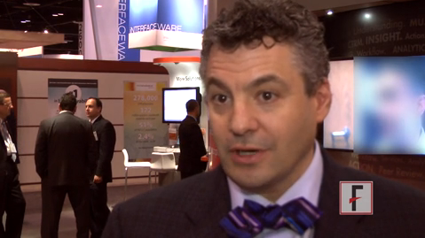
At the annual meeting of the Healthcare Information and Management Systems Society, Dr. Jonathan A. Handler, chief medical information officer for M*Modal, spoke about Glass's potential in medicine.
The video associated with this article is no longer available on this site. Please view all of our videos on the MDedge YouTube channel
On Twitter @naseemsmiller
ORLANDO – Google Glass will soon be available to the public, but its future role in medicine is still hazy.
Glass, which is among the growing number of wearable technologies, has several applications to health care settings, experts say. Just last year, a surgeon at Ohio State University’s Wexner Medical Center in Columbus live-streamed an ACL procedure via his Google Glass. And most recently, Rhode Island Hospital’s emergency department began using the device’s video capabilities to consult with off-site specialists.
But Glass is not HIPAA compliant, raising patient privacy concerns and dissuading hospitals from incorporating it into their growing list of mobile technologies. Still, technology enthusiasts are charging ahead, envisioning a future in which Glass could be yet another piece in physicians’ toolkits.
At the annual meeting of the Healthcare Information and Management Systems Society, Dr. Jonathan A. Handler, chief medical information officer for M*Modal, spoke about Glass's potential in medicine.
The video associated with this article is no longer available on this site. Please view all of our videos on the MDedge YouTube channel
On Twitter @naseemsmiller
AT HIMSS14
Fluorescent cholangiography as effective as standard, but cheaper
MIAMI BEACH – Intraoperative fluorescent cholangiography is just as effective as traditional cholangiography but costs hundreds of dollars less and is significantly faster to perform.
It’s also a great teaching tool, Dr. Fernando Dip said at the annual meeting of the Americas Hepato-Pancreato-Biliary Association. In just one session, all of the third- and fourth-year surgical residents were able to correctly identify 100% of the biliary structures.
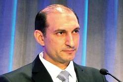
"It appears to be an additional tool for the laparoscopic surgeon," said Dr. Dip, chief of surgical research at the Cleveland Clinic in Weston, Fla. "It’s quick, inexpensive, real-time, there are no adverse events, and it’s an inciscionless procedure."
Common bile duct injury is the most frequent injury seen in laparoscopic cholecystectomy, he said, and although the overall incidence is low, the number of injuries each year is not inconsiderable, since more than 750,000 laparoscopic cholecystectomies are performed in the United States annually.
"Only 3% of these injuries are due to problems with technical skill," Dr. Dip said. "The other 97% are problems of visual perception – illusions of where the ducts are."
Intraoperative cholangiography helps surgeons visualize this anatomy, but its true usefulness is somewhat controversial. Dr. Dip cited a recent study of almost 93,000 patients – 40% of whom underwent the procedure. It showed that intraoperative cholangiography is not effective as a preventive strategy against common duct injury during cholecystectomy.
Intraoperative fluorescent cholangiography is sometimes used to identify biliary anatomy in extrahepatic surgery. Dr. Dip and his colleagues examined its usefulness in 45 patients undergoing laparoscopic cholecystectomy. Senior residents performed all of the procedures under the supervision of experienced laparoscopic surgeons. All patients underwent the investigational procedure, followed by standard cholangiography.
The patients had a mean age of 49 years and were evenly split between men and women. The mean body mass index was 28 kg/m2. Surgical indications were cholelithiasis (22), acute cholecystitis (17), chronic cholecystitis (5), and polyp (1). About 1 hour before surgery, patients received an infusion of indocyanine green 0.5 mg/kg. During the laparoscopic exploration, surgeons used near-infrared light to make the marker fluoresce as it was excreted by the liver.
In a picture review before surgery, all residents correctly identified 100% of the anatomic structures visualized by the fluorescent procedure. They were able to complete the fluorescent procedure in all of the patients. The completion rate for cholangiography was 93% (42 patients). In three patients, cholangiography failed because the cystic duct could not be cannulated.
The residents identified the cystic duct in 44 of the patients (98%), the common bile duct in 36 (80%), and the common hepatic ducts in 27 (60%). Neither technique identified any aberrant or accessory ducts.
Because fluorescent cholangiography is a real-time surgical procedure, it allowed checking of the transection and resection of the gallbladder pedicle before smooth dissection in all of the patients.
The procedure was significantly quicker than standard cholangiography (0.71 minutes vs. 7.15 minutes). It was also significantly cheaper, costing a mean of $14 vs. $778. There were no adverse surgical events, and no adverse reactions to the dye or at the infusion site, Dr. Dip said. Additionally, he noted, fluorescent cholangiography does not rely on x-rays, and so spared the patients any radiation exposure.
He added that the Cleveland Clinic, in conjunction with the University of Tokyo, will soon launch a randomized clinical trial comparing the two methods. An ongoing trial from another institution is evaluating fluorescent cholangiography compared with critical view technique in visualizing anatomy during laparoscopic cholecystectomy. It was set to wrap up in January, but according to the trial record on clinicaltrials.gov, is still recruiting patients.
Dr. Dip had no financial disclosures.
On Twitter @alz_gal
MIAMI BEACH – Intraoperative fluorescent cholangiography is just as effective as traditional cholangiography but costs hundreds of dollars less and is significantly faster to perform.
It’s also a great teaching tool, Dr. Fernando Dip said at the annual meeting of the Americas Hepato-Pancreato-Biliary Association. In just one session, all of the third- and fourth-year surgical residents were able to correctly identify 100% of the biliary structures.
"It appears to be an additional tool for the laparoscopic surgeon," said Dr. Dip, chief of surgical research at the Cleveland Clinic in Weston, Fla. "It’s quick, inexpensive, real-time, there are no adverse events, and it’s an inciscionless procedure."
Common bile duct injury is the most frequent injury seen in laparoscopic cholecystectomy, he said, and although the overall incidence is low, the number of injuries each year is not inconsiderable, since more than 750,000 laparoscopic cholecystectomies are performed in the United States annually.
"Only 3% of these injuries are due to problems with technical skill," Dr. Dip said. "The other 97% are problems of visual perception – illusions of where the ducts are."
Intraoperative cholangiography helps surgeons visualize this anatomy, but its true usefulness is somewhat controversial. Dr. Dip cited a recent study of almost 93,000 patients – 40% of whom underwent the procedure. It showed that intraoperative cholangiography is not effective as a preventive strategy against common duct injury during cholecystectomy.
Intraoperative fluorescent cholangiography is sometimes used to identify biliary anatomy in extrahepatic surgery. Dr. Dip and his colleagues examined its usefulness in 45 patients undergoing laparoscopic cholecystectomy. Senior residents performed all of the procedures under the supervision of experienced laparoscopic surgeons. All patients underwent the investigational procedure, followed by standard cholangiography.
The patients had a mean age of 49 years and were evenly split between men and women. The mean body mass index was 28 kg/m2. Surgical indications were cholelithiasis (22), acute cholecystitis (17), chronic cholecystitis (5), and polyp (1). About 1 hour before surgery, patients received an infusion of indocyanine green 0.5 mg/kg. During the laparoscopic exploration, surgeons used near-infrared light to make the marker fluoresce as it was excreted by the liver.
In a picture review before surgery, all residents correctly identified 100% of the anatomic structures visualized by the fluorescent procedure. They were able to complete the fluorescent procedure in all of the patients. The completion rate for cholangiography was 93% (42 patients). In three patients, cholangiography failed because the cystic duct could not be cannulated.
The residents identified the cystic duct in 44 of the patients (98%), the common bile duct in 36 (80%), and the common hepatic ducts in 27 (60%). Neither technique identified any aberrant or accessory ducts.
Because fluorescent cholangiography is a real-time surgical procedure, it allowed checking of the transection and resection of the gallbladder pedicle before smooth dissection in all of the patients.
The procedure was significantly quicker than standard cholangiography (0.71 minutes vs. 7.15 minutes). It was also significantly cheaper, costing a mean of $14 vs. $778. There were no adverse surgical events, and no adverse reactions to the dye or at the infusion site, Dr. Dip said. Additionally, he noted, fluorescent cholangiography does not rely on x-rays, and so spared the patients any radiation exposure.
He added that the Cleveland Clinic, in conjunction with the University of Tokyo, will soon launch a randomized clinical trial comparing the two methods. An ongoing trial from another institution is evaluating fluorescent cholangiography compared with critical view technique in visualizing anatomy during laparoscopic cholecystectomy. It was set to wrap up in January, but according to the trial record on clinicaltrials.gov, is still recruiting patients.
Dr. Dip had no financial disclosures.
On Twitter @alz_gal
AT AHPBA 2014
Major finding: Fluorescent cholangiography identified 100% of relevant anatomy during laparoscopic cholecystectomy, while costing less than standard cholangiography ($14 vs. $778).
Data source: Prospective study of 45 patients.
Disclosures: Dr. Dip had no financial disclosures.

Two most common vaginal prolapse surgeries yield similar outcomes
Two-year outcomes were similar between women who underwent transvaginal sacrospinous ligament fixation and those who underwent transvaginal uterosacral ligament suspension to correct apical vaginal or uterine prolapse and stress incontinence in the first randomized trial to compare the two approaches.
The 5-year OPTIMAL (Operations and Pelvic Muscle Training in the Management of Apical Support Loss) trial at nine U.S. medical centers participating in the Pelvic Floor Disorders Network involved 374 women randomly assigned to transvaginal surgery for stage 2 through stage 4 prolapse. A total of 186 underwent sacrospinous ligament fixation (SSLF), while 188 patients underwent uterosacral ligament suspension (ULS).
The women also were separately randomized to receive perioperative behavioral therapy with pelvic floor muscle training or usual perioperative care, wrote Dr. Matthew D. Barber of the department of obstetrics and gynecology and the Women’s Health Institute at the Cleveland Clinic. The report appears in the March 11 issue of JAMA.
At 2-year follow-up, neither surgical approach was superior to the other in the composite primary outcome of the percentage of patients with surgical success: 60.5% for SSLF and 59.2% for ULS. The investigators defined successful surgery as the absence of descent of the vaginal apex more than one-third into the vaginal canal; anterior or posterior vaginal wall descent beyond the hymen; bothersome vaginal bulge symptoms; and further treatment for prolapse. The overall rates of failure were 18% for bothersome vaginal bulge, 17.5% for anterior and/or posterior prolapse, and 5.1% for further treatment such as more surgery or use of a pessary.
The two groups were no different in any secondary outcomes such as blood loss, duration of surgery, length of hospitalization, or postoperative treatment. Serious adverse events including bladder perforation and the formation of vaginal granulation tissue occurred in 16.7% of the SSLF group and 16.5% of the ULS group. There were no significant differences between women who received the behavioral intervention and those who did not, the investigators said (JAMA 2014;311:1023-34).
They noted that 4.3% of the women who underwent SSLF developed persistent pain, confirming previous reports that the procedure may cause acute neurologic pain, particularly buttock pain that may result from gluteal nerve entrapment. This highlights "the need to provide preoperative counseling to patients about this potential risk," Dr. Barber and his associates wrote.
This study was supported by the Eunice Kennedy Shriver National Institute of Child Health and Human Development and the National Institutes of Health Office of Research on Women’s Health. Some of Dr. Barber’s coauthors reported ties to Astellas, Pfizer, GlaxoSmithKline, Uromedica, IDEO, Xanodyne, Renew Medical, American Medical Systems, Ethicon/Johnson & Johnson, Boston Scientific, and Ferring Pharmaceuticals.
FROM JAMA
Major Finding: At 2-year follow-up, neither surgical approach was superior to the other in the composite primary outcome of the percentage of patients with surgical success: 60.5% for sacrospinous ligament fixation and 59.2% for uterosacral ligament suspension.
Data Source: A randomized trial comparing outcomes between 186 women who had SSLF and 188 who had ULS to correct apical vaginal or uterine prolapse.
Disclosures: This study was supported by the Eunice Kennedy Shriver National Institute of Child Health and Human Development and the National Institutes of Health Office of Research on Women’s Health. Some of Dr. Barber’s coauthors reported ties to Astellas, Pfizer, GlaxoSmithKline, Uromedica, IDEO, Xanodyne, Renew Medical, American Medical Systems, Ethicon/Johnson & Johnson, Boston Scientific, and Ferring Pharmaceuticals.
High posthepatectomy bilirubin bodes ill for patients
MIAMI BEACH – An elevated bilirubin level on postoperative day 3 after major hepatectomy may be a harbinger of hepatic insufficiency that leads to poor outcomes – including an increased risk of death.
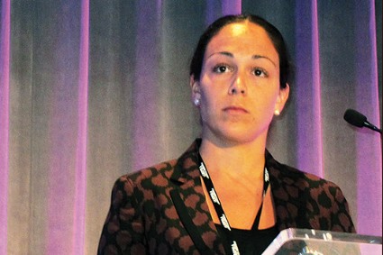
Compared with patients who had lower bilirubin levels, a level of 3 mg/dL or higher was associated with an eightfold increase in the risk of both a major complication and of dying within 90 days of surgery, Dr. Joanna W. Etra said at the annual meeting of the Americas Hepato-Pancreato-Biliary Association.
Unfortunately, said Dr. Etra of the Winship Cancer Institute of Emory University, Atlanta, there seems to be no way to predict before surgery who will develop the elevated levels, and no preemptive treatment. Still, she said, the finding could be a good way to be alert to the possibility of a problem.
She presented a retrospective study of 535 patients who underwent a major hepatectomy at the center from 2000 to 2012. Their mean age was 55 years. Most (73%) had cancer; 39% had undergone preoperative chemotherapy. About a third (38%) had colorectal metastases in the liver. The average preoperative bilirubin level was 0.7 mg/dL. Most of the procedures (83%) were open, with a right hepatectomy most common (44%)
Postoperatively, 10% of the group developed hepatic insufficiency. Postoperative complications developed in 58%; of these, of which 22% were major. Death within 90 days occurred in 4.5% of the entire group.
Dr. Etra and her colleagues divided the group by postoperative day 3 bilirubin levels: lower than 3 mg/dL and 3 mg/dL or higher. They examined outcomes among the two groups.
Postoperative complications were significantly more common among those with the higher bilirubin levels (76% vs. 54%), as were major complications (46% vs. 18%), and 90-day mortality (16% vs. 2%).
A multivariate analysis found that the higher level doubled the risk of any complication, and tripled the risk of both a major complication and 90-day mortality,
"Having identified this association with outcomes, we refocused on the dichotomized bilirubin groups to see if we could also identify any pre- or intraoperative factors that might predict this elevated bilirubin," she said. "But in a multifactorial analysis, we found that no single factor – age, gender, cancer, preoperative platelets, MELD [model for end-stage liver disease] score, blood loss or transfusion – was a significant predictor."
Dr. Etra had no financial disclosures.
MIAMI BEACH – An elevated bilirubin level on postoperative day 3 after major hepatectomy may be a harbinger of hepatic insufficiency that leads to poor outcomes – including an increased risk of death.
Compared with patients who had lower bilirubin levels, a level of 3 mg/dL or higher was associated with an eightfold increase in the risk of both a major complication and of dying within 90 days of surgery, Dr. Joanna W. Etra said at the annual meeting of the Americas Hepato-Pancreato-Biliary Association.
Unfortunately, said Dr. Etra of the Winship Cancer Institute of Emory University, Atlanta, there seems to be no way to predict before surgery who will develop the elevated levels, and no preemptive treatment. Still, she said, the finding could be a good way to be alert to the possibility of a problem.
She presented a retrospective study of 535 patients who underwent a major hepatectomy at the center from 2000 to 2012. Their mean age was 55 years. Most (73%) had cancer; 39% had undergone preoperative chemotherapy. About a third (38%) had colorectal metastases in the liver. The average preoperative bilirubin level was 0.7 mg/dL. Most of the procedures (83%) were open, with a right hepatectomy most common (44%)
Postoperatively, 10% of the group developed hepatic insufficiency. Postoperative complications developed in 58%; of these, of which 22% were major. Death within 90 days occurred in 4.5% of the entire group.
Dr. Etra and her colleagues divided the group by postoperative day 3 bilirubin levels: lower than 3 mg/dL and 3 mg/dL or higher. They examined outcomes among the two groups.
Postoperative complications were significantly more common among those with the higher bilirubin levels (76% vs. 54%), as were major complications (46% vs. 18%), and 90-day mortality (16% vs. 2%).
A multivariate analysis found that the higher level doubled the risk of any complication, and tripled the risk of both a major complication and 90-day mortality,
"Having identified this association with outcomes, we refocused on the dichotomized bilirubin groups to see if we could also identify any pre- or intraoperative factors that might predict this elevated bilirubin," she said. "But in a multifactorial analysis, we found that no single factor – age, gender, cancer, preoperative platelets, MELD [model for end-stage liver disease] score, blood loss or transfusion – was a significant predictor."
Dr. Etra had no financial disclosures.
AT AHPBA 2014
Major finding: A high posthepatectomy bilirubin level was associated with an 8-fold increase in the risk of postoperative complications and 90-day mortality.
Data source: A retrospective study of 535 patients.
Disclosures: Dr. Joanna Etra had no financial disclosures.
Team planning cuts pancreatectomy readmissions
MIAMI BEACH – A combination of teamwork and leadership led to a 50% reduction in readmission after pancreatectomy in a large academic facility.
The readmission rate at Indiana University Hospital fell from a high of 23% to just over 11% over 5 years – even though length of stay and mortality remained stable, Dr. Eugene Ceppa reported at the annual meeting of the Americas Hepato-Pancreato-Biliary Association.

The multifaceted project made some progress during the first few years of implementation, said Dr. Ceppa of Indiana University Hospital, Indianapolis. But the biggest changes really came in years 4 and 5, after the team adopted its own version of a national readmission prevention plan, and created a "discharge coach" – a staff member dedicated to ensuring that patients were ready to leave the hospital, with plenty of support at home.
Studies generally show that pancreatectomy has a very high readmission rate, hovering around 18%. The situation was tolerated over the years, according to Dr. Henry Pitt, who coauthored the paper. But in 2008, the Centers for Medicare & Medicaid Services introduced the idea that hospital readmissions were eating away at health care dollars.
In a landmark paper, Dr. Brian Jack and colleagues noted that only 13% of discharged patients needed repeat hospitalizations, but these patients used up to 60% of the $753 billion spent on discharge in 2003.
Project RED (ReEngineering Discharge), originating from Boston University Medical Center, suggested that reducing readmissions could save $5 billion each year. Originally focused on reducing readmissions for heart failure, Project RED has been successfully adapted to multiple models – including surgery.
Changes have not come about overnight, said Dr. Pitt, now chief quality officer for Temple University Health System in Philadelphia. "There was denial at first that readmission was a problem," he said in an interview. "Then there was a period of time when there was acceptance but no idea of how to fix it. Now we are beginning to do so."
As pay-for-performance became ever more important, pancreatic surgeons at Indiana University Hospital decided to attack the problem of readmission for their pancreatectomy patients. Over a 5-year period, from 2007 to 2012, they implemented a number of reforms, beginning with renewed efforts to decrease surgical morbidity – especially their 24% rate of surgical site infections after pancreatectomy. Dr. Pitt and his colleagues had already shown that these infections were a leading cause of surgical morbidity, and reducing them was a logical first step toward reducing readmission.
The next step was to create a discharge team that would work cooperatively to make sure patients were in optimal condition to leave the hospital. The Readmission Quality Improvement Team consisted of physicians, nurses, physical and occupational therapists, case managers, pharmacists, and dietitians. Because of their efforts, the number of pancreatectomy patients discharged with some kind of home health care support increased from 20% to 50%.
At the same time, the surgeons made a policy change: There would be no readmissions without the approval of the attending surgeon. Because patients often traveled far to the university hospital, they would frequently go to their local hospitals when problems arose after they went home.
"A lot of the calls to us from patients would go to residents," Dr. Pitt said. "The default was to send them to their local emergency department, because the patients were so far away. And then we would get calls for transfers. We said that house staff would no longer have the authority to make those admissions. If they thought readmission was necessary, they had to call the attending surgeon. Just improving that decision-making process made a big difference, with fewer people going to the ED in the first place. Often it was just a matter of reassuring the patient."
By 2010, readmissions had dropped from 24% to 16%. In 2011, the team employed its own adaptation of Project RED. Each discharge included an 11-point checklist of things that had to be completed before a patient could leave. Those tasks include the following:
• Reconcile medications.
• Reconcile discharge plan with national guidelines.
• Make follow-up appointments.
• Follow up on any outstanding tests.
• Arrange for postdischarge services.
• Explain to the patient what do if a problem arises.
• Conduct patient education.
• Communicate discharge information to primary care physician.
• Make a follow-up call within 3 days of discharge.
The final puzzle piece was the discharge coach, Dr. Pitt said. The coach is a highly experienced nurse whose job it is to make sure each patient receives consistent discharge care and follow-through.
Last year, the team reviewed the project’s results, which Dr. Ceppa presented during the meeting. From 2007 to 2012, 1,147 patients underwent pancreatectomy at the facility. The mean age was 58 years. Pancreatic adenocarcinoma was the most common indication for surgery.
During the study period, neither mortality (2.7%) nor mean length of stay (10 days) changed. But readmission steadily decreased from the 2007 high of 23%. From 2008 through 2011, the readmission ranged from 18% to 15%. But after the changes implemented in 2011, the rate dropped to 11% – a significant decrease from baseline. Dr. Ceppa and Dr. Pitt attributed this to a combination of factors: stepped up discharge planning, attending-only readmission, and the discharge coach.
"I think the major thing we learned is that improving something like this isn’t simple or quick," Dr Pitt said. "You have to be persistent and really work on all the puzzle pieces until they fit into place."
Neither Dr. Pitt nor Dr. Ceppa had any financial disclosures.
MIAMI BEACH – A combination of teamwork and leadership led to a 50% reduction in readmission after pancreatectomy in a large academic facility.
The readmission rate at Indiana University Hospital fell from a high of 23% to just over 11% over 5 years – even though length of stay and mortality remained stable, Dr. Eugene Ceppa reported at the annual meeting of the Americas Hepato-Pancreato-Biliary Association.
The multifaceted project made some progress during the first few years of implementation, said Dr. Ceppa of Indiana University Hospital, Indianapolis. But the biggest changes really came in years 4 and 5, after the team adopted its own version of a national readmission prevention plan, and created a "discharge coach" – a staff member dedicated to ensuring that patients were ready to leave the hospital, with plenty of support at home.
Studies generally show that pancreatectomy has a very high readmission rate, hovering around 18%. The situation was tolerated over the years, according to Dr. Henry Pitt, who coauthored the paper. But in 2008, the Centers for Medicare & Medicaid Services introduced the idea that hospital readmissions were eating away at health care dollars.
In a landmark paper, Dr. Brian Jack and colleagues noted that only 13% of discharged patients needed repeat hospitalizations, but these patients used up to 60% of the $753 billion spent on discharge in 2003.
Project RED (ReEngineering Discharge), originating from Boston University Medical Center, suggested that reducing readmissions could save $5 billion each year. Originally focused on reducing readmissions for heart failure, Project RED has been successfully adapted to multiple models – including surgery.
Changes have not come about overnight, said Dr. Pitt, now chief quality officer for Temple University Health System in Philadelphia. "There was denial at first that readmission was a problem," he said in an interview. "Then there was a period of time when there was acceptance but no idea of how to fix it. Now we are beginning to do so."
As pay-for-performance became ever more important, pancreatic surgeons at Indiana University Hospital decided to attack the problem of readmission for their pancreatectomy patients. Over a 5-year period, from 2007 to 2012, they implemented a number of reforms, beginning with renewed efforts to decrease surgical morbidity – especially their 24% rate of surgical site infections after pancreatectomy. Dr. Pitt and his colleagues had already shown that these infections were a leading cause of surgical morbidity, and reducing them was a logical first step toward reducing readmission.
The next step was to create a discharge team that would work cooperatively to make sure patients were in optimal condition to leave the hospital. The Readmission Quality Improvement Team consisted of physicians, nurses, physical and occupational therapists, case managers, pharmacists, and dietitians. Because of their efforts, the number of pancreatectomy patients discharged with some kind of home health care support increased from 20% to 50%.
At the same time, the surgeons made a policy change: There would be no readmissions without the approval of the attending surgeon. Because patients often traveled far to the university hospital, they would frequently go to their local hospitals when problems arose after they went home.
"A lot of the calls to us from patients would go to residents," Dr. Pitt said. "The default was to send them to their local emergency department, because the patients were so far away. And then we would get calls for transfers. We said that house staff would no longer have the authority to make those admissions. If they thought readmission was necessary, they had to call the attending surgeon. Just improving that decision-making process made a big difference, with fewer people going to the ED in the first place. Often it was just a matter of reassuring the patient."
By 2010, readmissions had dropped from 24% to 16%. In 2011, the team employed its own adaptation of Project RED. Each discharge included an 11-point checklist of things that had to be completed before a patient could leave. Those tasks include the following:
• Reconcile medications.
• Reconcile discharge plan with national guidelines.
• Make follow-up appointments.
• Follow up on any outstanding tests.
• Arrange for postdischarge services.
• Explain to the patient what do if a problem arises.
• Conduct patient education.
• Communicate discharge information to primary care physician.
• Make a follow-up call within 3 days of discharge.
The final puzzle piece was the discharge coach, Dr. Pitt said. The coach is a highly experienced nurse whose job it is to make sure each patient receives consistent discharge care and follow-through.
Last year, the team reviewed the project’s results, which Dr. Ceppa presented during the meeting. From 2007 to 2012, 1,147 patients underwent pancreatectomy at the facility. The mean age was 58 years. Pancreatic adenocarcinoma was the most common indication for surgery.
During the study period, neither mortality (2.7%) nor mean length of stay (10 days) changed. But readmission steadily decreased from the 2007 high of 23%. From 2008 through 2011, the readmission ranged from 18% to 15%. But after the changes implemented in 2011, the rate dropped to 11% – a significant decrease from baseline. Dr. Ceppa and Dr. Pitt attributed this to a combination of factors: stepped up discharge planning, attending-only readmission, and the discharge coach.
"I think the major thing we learned is that improving something like this isn’t simple or quick," Dr Pitt said. "You have to be persistent and really work on all the puzzle pieces until they fit into place."
Neither Dr. Pitt nor Dr. Ceppa had any financial disclosures.
AT AHPBA 2014
Major finding: Readmission after pancreatectomy dropped from 24% to 11% after implementation of a multifaceted effort to reduce infections and improve discharge planning,
Data source: A retrospective study of 1,147 patients.
Disclosures: Neither Dr. Ceppa nor Dr. Pitt had any financial disclosures.

Intraoperative ultrasound can change approach to liver resection
MIAMI BEACH – Intraoperative ultrasound during resection of colorectal liver metastases changed operative management in 43% of cases, according to a retrospective study.
Ultrasound employed during surgery identified new lesions not seen on preoperative imaging, and gave additional details about known lesions, said Dr. Sarah Knowles, a surgical resident at the University of Western Ontario, London (Canada).
"Intraoperative ultrasound provided new information about the number, size, location, and appearance of the lesions," she said at the annual meeting of the Americas Hepato-Pancreato-Biliary Association. The procedure was associated with significantly more blood loss, however, and there were no differences in negative surgical margins between patients who had ultrasound during surgery and those who did not.

Dr. Knowles presented a retrospective study of 103 patients who, from 2009 to 2012, underwent liver resection of colorectal cancer metastases. Mean age of the patients was 62 years. Most (94%) had undergone preoperative computerized tomography; the rest had undergone magnetic resonance imaging. The mean time from preoperative imaging to surgery was 66 days. Most of the patients (72) had intraoperative ultrasound imaging.
There were 45 anatomic resections and 27 nonanatomic resections in the group that had intraoperative ultrasound. This was significantly different from the nonultrasound group, which had 25 anatomic and 6 nonanatomic resections.
Ultrasound changed surgical strategy in 43% (31) of those who had it. Surgical strategy changed in 10% of those who had no intraoperative imaging – a significant difference (P = less than .001). Blood loss was significantly greater in the ultrasound group (650 mL vs. 350 mL).
The bulk of the strategic changes in the ultrasound group (17) were due to additional information about the location of lesions – either deeper or more superficial than preoperative imaging suggested. Other reasons for a shift in strategy were the discovery of new lesions (13), a disappearing lesion (1), larger-than-expected lesions (5), smaller-than-expected lesions (2), or a difference in the lesions’ appearance (3).
Resection margins were similar in the two groups. In the ultrasound group, 85% had R0 margins and 15% had R1 margins. In the group without ultrasound, 87% had R0 margins and 13% had R1 margins. There was no significant difference in disease-free 5-year survival.
Dr. Knowles had no financial disclosures.
MIAMI BEACH – Intraoperative ultrasound during resection of colorectal liver metastases changed operative management in 43% of cases, according to a retrospective study.
Ultrasound employed during surgery identified new lesions not seen on preoperative imaging, and gave additional details about known lesions, said Dr. Sarah Knowles, a surgical resident at the University of Western Ontario, London (Canada).
"Intraoperative ultrasound provided new information about the number, size, location, and appearance of the lesions," she said at the annual meeting of the Americas Hepato-Pancreato-Biliary Association. The procedure was associated with significantly more blood loss, however, and there were no differences in negative surgical margins between patients who had ultrasound during surgery and those who did not.
Dr. Knowles presented a retrospective study of 103 patients who, from 2009 to 2012, underwent liver resection of colorectal cancer metastases. Mean age of the patients was 62 years. Most (94%) had undergone preoperative computerized tomography; the rest had undergone magnetic resonance imaging. The mean time from preoperative imaging to surgery was 66 days. Most of the patients (72) had intraoperative ultrasound imaging.
There were 45 anatomic resections and 27 nonanatomic resections in the group that had intraoperative ultrasound. This was significantly different from the nonultrasound group, which had 25 anatomic and 6 nonanatomic resections.
Ultrasound changed surgical strategy in 43% (31) of those who had it. Surgical strategy changed in 10% of those who had no intraoperative imaging – a significant difference (P = less than .001). Blood loss was significantly greater in the ultrasound group (650 mL vs. 350 mL).
The bulk of the strategic changes in the ultrasound group (17) were due to additional information about the location of lesions – either deeper or more superficial than preoperative imaging suggested. Other reasons for a shift in strategy were the discovery of new lesions (13), a disappearing lesion (1), larger-than-expected lesions (5), smaller-than-expected lesions (2), or a difference in the lesions’ appearance (3).
Resection margins were similar in the two groups. In the ultrasound group, 85% had R0 margins and 15% had R1 margins. In the group without ultrasound, 87% had R0 margins and 13% had R1 margins. There was no significant difference in disease-free 5-year survival.
Dr. Knowles had no financial disclosures.
AT AHPBA 2014
Major finding: Intraoperative ultrasound changed surgical strategy in 43% of patients undergoing liver resections of colorectal cancer metastases.
Data source: A retrospective study of 103 patients.
Disclosures: Dr. Knowles had no financial disclosures.
Two studies: Preimplant kidney biopsy doesn’t predict organ viability
Preimplant biopsy of donor kidneys doesn’t accurately predict the organs’ viability and leads to many acceptable kidneys being discarded, according to two separate reports published online in the Clinical Journal of the American Society of Nephrology.
Biopsies are obtained routinely from donor kidneys in the United States, and unfavorable biopsy findings are the most frequently cited reason for discarding donor kidneys as unacceptable. Ideally, such samples would be obtained by core needle biopsy, would not be frozen, would be thoroughly examined by a pathologist with special training in reading kidney biopsies, and would be assessed meticulously for chronic tubular atrophy, arteriolar hyalinosis, interstitial inflammation, interstitial fibrosis, and the presence and severity of acute tubular necrosis.
In actual practice, however, these samples are almost always obtained by wedge biopsy, are frozen, and are rushed through a perfunctory examination by whatever pathologist is available so that the organ can be transplanted as quickly as possible if it is found to be acceptable. So many clinicians have questioned whether the results of such biopsies actually assess the organs’ viability and predict graft failure, both groups of researchers noted.
In what they described as the largest cohort study to date on this issue, one team analyzed data regarding 651 consecutive kidney transplants performed during a 2-year period, for which four organ procurement organizations obtained the kidneys from 369 deceased donors. The four organizations performed wedge biopsies immediately after procurement, and different pathology services evaluated frozen sections from these organs and reported their findings to potential transplant centers.
Patient outcomes were tracked using information in the United Network for Organ Sharing (UNOS) database, said Dr. Isaac E. Hall of the section of nephrology and the program of applied translational research, Yale University, New Haven, Conn., and his associates.
They assessed whether a biopsy finding of acute tubular necrosis correlated with the graft’s performance after transplantation. Acute tubular necrosis was reported in 110 biopsies (17%) overall. The four procurement organizations varied widely in their reported rates of the abnormality, from a low of zero cases to a high of 25% of cases. This variation suggests that the process of obtaining and interpreting these biopsies is, at best, not uniform among procurement groups, according to the investigators.
During a median follow-up of 1 year, the primary outcome of interest – delayed graft function – occurred in 45% of kidneys that were reported to have tubular necrosis and in 39% of those reportedly free of such necrosis. This is a nonsignificant difference. There also was no significant difference in the secondary outcome of graft failure between recipients of organs with acute tubular necrosis, compared with recipients of organs without it.
In summary, there was no significant association between biopsy reports of acute tubular necrosis and graft viability. "It is reasonable to question whether acute tubular necrosis, or acute kidney injury in general for that matter, truly causes important allograft outcomes," Dr. Hall and his colleagues wrote (Clin. J. Am. Soc. Nephrol. 2014 [doi:10.2215/CJN.08270813]).
In the second study, Dr. Bertram L. Kasiske of the Scientific Registry of Transplant Recipients, Minneapolis Medical Research Foundation, and his associates assessed the records for 83 kidneys that were discarded because of unfavorable biopsy findings (cases) and 83 contralateral kidneys from the same donors that were transplanted (contralateral controls). They compared these findings with those for 151 transplanted kidneys from 83 deceased donors who were matched for the index cases’ donor profiles.
"Ours is the first controlled study to compare biopsy findings between discarded kidneys and matched transplanted kidneys," Dr. Kasiske and his colleagues noted.
They found that most of the biopsy reports were of low quality, and very few indicated the amounts of tubular atrophy, interstitial inflammation, arteriolar hyalinosis, or acute tubular necrosis. The percentage of glomerulosclerosis was often the only finding upon which to base the decision of whether to use or discard the allograft.
But the percentage of glomerulosclerosis overlapped substantially between cases (discarded kidneys) and controls (transplanted kidneys). This suggests that "information obtained from procurement biopsies is of low quality and may lead to unnecessary discard of transplantable kidneys," Dr. Kasiske and his associates wrote (Clin. J. Am. Soc. Nephrol. 2014 [doi:10.2215/CJN.07610713]).
"A reasonable conclusion from this and other studies is that the widespread practice of routinely obtaining procurement biopsies should be abandoned, as has been successfully done in Europe," they noted.
Dr. Hall’s study was supported by the American Heart Association, the Roche Organ Transplantation Research Foundation, the National Institute of Diabetes and Digestive and Kidney Diseases, and the U.S. Health Resources and Services Administration. Dr. Kasiske’s study was supported by the Minneapolis Medical Research Foundation, the Organ Procurement and Transplantation Network, the Scientific Registry of Transplant Recipients, and the United Network for Organ Sharing. Both research groups reported no potential financial conflicts of interest.
The decision to transplant a particular kidney is extremely complex "and dependent on a multitude of factors, including donor age, clinical history, anatomic abnormalities, terminal creatinine, and biopsy findings," wrote Dr. Sayeed Khan Malek.
"When the biopsy findings are consistent with the clinical evaluation of the donor, they are useful in making the determination about transplanting the kidney. However, biopsy findings considered in isolation are of limited value and should be interpreted with caution when making the decision to turn down a potentially transplantable kidney," he said.
Dr. Sayeed Khan Malek is clinical director of transplant surgery at Brigham and Women’s Hospital, Boston. He reported no potential financial conflicts of interest. These remarks were taken from his editorial accompanying the two reports (Clin. J. Am. Soc. Nephrol;2014 [doi:10.2215/CJN.00470114]).
Preimplant biopsy of donor kidneys doesn’t accurately predict the organs’ viability and leads to many acceptable kidneys being discarded, according to two separate reports published online in the Clinical Journal of the American Society of Nephrology.
Biopsies are obtained routinely from donor kidneys in the United States, and unfavorable biopsy findings are the most frequently cited reason for discarding donor kidneys as unacceptable. Ideally, such samples would be obtained by core needle biopsy, would not be frozen, would be thoroughly examined by a pathologist with special training in reading kidney biopsies, and would be assessed meticulously for chronic tubular atrophy, arteriolar hyalinosis, interstitial inflammation, interstitial fibrosis, and the presence and severity of acute tubular necrosis.
In actual practice, however, these samples are almost always obtained by wedge biopsy, are frozen, and are rushed through a perfunctory examination by whatever pathologist is available so that the organ can be transplanted as quickly as possible if it is found to be acceptable. So many clinicians have questioned whether the results of such biopsies actually assess the organs’ viability and predict graft failure, both groups of researchers noted.
In what they described as the largest cohort study to date on this issue, one team analyzed data regarding 651 consecutive kidney transplants performed during a 2-year period, for which four organ procurement organizations obtained the kidneys from 369 deceased donors. The four organizations performed wedge biopsies immediately after procurement, and different pathology services evaluated frozen sections from these organs and reported their findings to potential transplant centers.
Patient outcomes were tracked using information in the United Network for Organ Sharing (UNOS) database, said Dr. Isaac E. Hall of the section of nephrology and the program of applied translational research, Yale University, New Haven, Conn., and his associates.
They assessed whether a biopsy finding of acute tubular necrosis correlated with the graft’s performance after transplantation. Acute tubular necrosis was reported in 110 biopsies (17%) overall. The four procurement organizations varied widely in their reported rates of the abnormality, from a low of zero cases to a high of 25% of cases. This variation suggests that the process of obtaining and interpreting these biopsies is, at best, not uniform among procurement groups, according to the investigators.
During a median follow-up of 1 year, the primary outcome of interest – delayed graft function – occurred in 45% of kidneys that were reported to have tubular necrosis and in 39% of those reportedly free of such necrosis. This is a nonsignificant difference. There also was no significant difference in the secondary outcome of graft failure between recipients of organs with acute tubular necrosis, compared with recipients of organs without it.
In summary, there was no significant association between biopsy reports of acute tubular necrosis and graft viability. "It is reasonable to question whether acute tubular necrosis, or acute kidney injury in general for that matter, truly causes important allograft outcomes," Dr. Hall and his colleagues wrote (Clin. J. Am. Soc. Nephrol. 2014 [doi:10.2215/CJN.08270813]).
In the second study, Dr. Bertram L. Kasiske of the Scientific Registry of Transplant Recipients, Minneapolis Medical Research Foundation, and his associates assessed the records for 83 kidneys that were discarded because of unfavorable biopsy findings (cases) and 83 contralateral kidneys from the same donors that were transplanted (contralateral controls). They compared these findings with those for 151 transplanted kidneys from 83 deceased donors who were matched for the index cases’ donor profiles.
"Ours is the first controlled study to compare biopsy findings between discarded kidneys and matched transplanted kidneys," Dr. Kasiske and his colleagues noted.
They found that most of the biopsy reports were of low quality, and very few indicated the amounts of tubular atrophy, interstitial inflammation, arteriolar hyalinosis, or acute tubular necrosis. The percentage of glomerulosclerosis was often the only finding upon which to base the decision of whether to use or discard the allograft.
But the percentage of glomerulosclerosis overlapped substantially between cases (discarded kidneys) and controls (transplanted kidneys). This suggests that "information obtained from procurement biopsies is of low quality and may lead to unnecessary discard of transplantable kidneys," Dr. Kasiske and his associates wrote (Clin. J. Am. Soc. Nephrol. 2014 [doi:10.2215/CJN.07610713]).
"A reasonable conclusion from this and other studies is that the widespread practice of routinely obtaining procurement biopsies should be abandoned, as has been successfully done in Europe," they noted.
Dr. Hall’s study was supported by the American Heart Association, the Roche Organ Transplantation Research Foundation, the National Institute of Diabetes and Digestive and Kidney Diseases, and the U.S. Health Resources and Services Administration. Dr. Kasiske’s study was supported by the Minneapolis Medical Research Foundation, the Organ Procurement and Transplantation Network, the Scientific Registry of Transplant Recipients, and the United Network for Organ Sharing. Both research groups reported no potential financial conflicts of interest.
FROM THE CLINICAL JOURNAL OF THE AMERICAN SOCIETY OF NEPHROLOGY
Major finding: In one study, biopsy findings from donor kidneys did not correlate with performance of the allografts after transplantation, and in another study, the biopsy findings for kidneys that were discarded as unacceptable were basically the same as those for contralateral kidneys that were successfully transplanted.
Data source: A multicenter cohort study involving 651 kidney transplantations during a 2-year period, and a case-control study involving 83 donor kidneys that were discarded because of unfavorable biopsy findings plus 83 contralateral kidneys from the same donors that were successfully transplanted.
Disclosures: Dr. Hall’s study was supported by the American Heart Association, the Roche Organ Transplantation Research Foundation, the National Institute of Diabetes and Digestive and Kidney Diseases, and the U.S. Health Resources and Services Administration. Dr. Kasiske’s study was supported by the Minneapolis Medical Research Foundation, the Organ Procurement and Transplantation Network, the Scientific Registry of Transplant Recipients, and UNOS. Both research groups reported no potential financial conflicts of interest.
Administration to allow noncompliant health policies through 2017
The Obama Administration said that it will allow through 2016 renewals of health insurance policies that do not meet the minimum criteria established by the Affordable Care Act.
Each state has the final say on whether such policies can continue to be sold, but the move by the White House will help remove some of the hurdles, a senior administration official said March 5.
In November, President Obama said that, to avoid cancellations, the federal government would let insurers renew, for 1 year, policies that did not contain the essential health benefits outlined by the ACA. That would give policyholders who did renew time to find new coverage.

About half of the states agreed to allow renewals after the policy change, but some states with the largest number of such policyholders – including California, Washington, New York, and Maryland – refused to allow renewals.
Now, in a wide-ranging rule that covers many aspects of the ACA for 2015, the administration said that noncompliant policies can be renewed as late as Oct. 1, 2016. No new policies can be issued. States that chose not to allow the renewals before also can change their decisions, senior administration officials said.
In 2015, the administration also is giving consumers an additional month to shop for coverage on the federal and state health insurance exchanges for 2015: Open enrollment will run from Nov. 15, 2014, to Feb. 15, 2015.
The rule also reduces out-of-pocket limits for individuals and families who buy coverage through the exchanges. Depending on income, individuals will pay a maximum of $2,250 to $5,200, and families will pay $4,500 to $10,400.
"These policies implement the health care law in a common-sense way by continuing to smooth the transition for consumers and stakeholders and fixing problems wherever the law provides flexibility," HHS Secretary Kathleen Sebelius said in a statement. "This comprehensive guidance will help ensure that consumers, employers and insurers have the information they need to plan for next year and make it easier for families to make decisions to access quality, affordable coverage."
The administration said that it had worked on the guidance for 2015 with input from consumers, states, businesses, health professionals, insurance commissioners, insurers, and members of Congress.
On Twitter @aliciaault
The Obama Administration said that it will allow through 2016 renewals of health insurance policies that do not meet the minimum criteria established by the Affordable Care Act.
Each state has the final say on whether such policies can continue to be sold, but the move by the White House will help remove some of the hurdles, a senior administration official said March 5.
In November, President Obama said that, to avoid cancellations, the federal government would let insurers renew, for 1 year, policies that did not contain the essential health benefits outlined by the ACA. That would give policyholders who did renew time to find new coverage.
About half of the states agreed to allow renewals after the policy change, but some states with the largest number of such policyholders – including California, Washington, New York, and Maryland – refused to allow renewals.
Now, in a wide-ranging rule that covers many aspects of the ACA for 2015, the administration said that noncompliant policies can be renewed as late as Oct. 1, 2016. No new policies can be issued. States that chose not to allow the renewals before also can change their decisions, senior administration officials said.
In 2015, the administration also is giving consumers an additional month to shop for coverage on the federal and state health insurance exchanges for 2015: Open enrollment will run from Nov. 15, 2014, to Feb. 15, 2015.
The rule also reduces out-of-pocket limits for individuals and families who buy coverage through the exchanges. Depending on income, individuals will pay a maximum of $2,250 to $5,200, and families will pay $4,500 to $10,400.
"These policies implement the health care law in a common-sense way by continuing to smooth the transition for consumers and stakeholders and fixing problems wherever the law provides flexibility," HHS Secretary Kathleen Sebelius said in a statement. "This comprehensive guidance will help ensure that consumers, employers and insurers have the information they need to plan for next year and make it easier for families to make decisions to access quality, affordable coverage."
The administration said that it had worked on the guidance for 2015 with input from consumers, states, businesses, health professionals, insurance commissioners, insurers, and members of Congress.
On Twitter @aliciaault

Truly connected health care
For years, we have all been promised interconnected electronic patient records – the concept that information would follow patients and be available across all care settings. Unfortunately, this reality has yet to materialize.
Regional Health Information Organizations (RHIOs) exist all over (these are hospitals, physician practices, and health networks that exchange data on a local level), but they typically present a "brick wall" at their borders where the information sharing stops. Thankfully, that is poised to change, with a new push toward meaningful interconnectedness.
Finally, health information technology (HIT) thought leaders and electronic health records (EHR) vendors are homing in on standards that will allow physicians and other healthcare providers to exchange information seamlessly regardless of EHR system or geographical location. The goal, as usual, is to improve patient care and outcomes. In this column, we felt it would be useful to highlight some of the ideas driving this effort, as there is good reason to be optimistic about the possibility of true interoperability.
A view from the top
Recently, at the annual meeting of the Health Information Management Systems Society (HIMSS) in Orlando, Dr. Karen DeSalvo, national health IT coordinator, made a strong plea for interoperability by 2017. She said "We can do national health care exchange in 3 years. ... I want this as a doctor, I want this as patient, I want this as a daughter."
Dr. DeSalvo’s words resonate because this is a concept we all expected from the earliest days of electronic records. Unfortunately, barriers – both real and artificial – have stood in the way. First and foremost are patient privacy and information security. It is essential that patient data are protected and maintained as the data pass from one system to the next, but often, even the basic function of matching patients with the right records can be challenging. Many identifiers exist, including various medical record numbers and insurance codes. If a name is misspelled or middle initial omitted, this can also create significant chaos and allow sensitive data to file into the wrong place, creating HIPAA violations and distrust among patients.
In addition to concerns over data security, the lack of standard formatting for storing and sharing records has been a huge hurdle for EHR vendors. Not surprisingly, the simultaneous development of dozens of competing EHR products has created significant fragmentation throughout the market. Instead of starting from a common set of data elements, each EHR was essentially designed "in a vacuum," and only now are their creators attempting to find a way to get them to communicate. A number of cross-platform projects currently underway highlight this effort.
VHS or Betamax?
On Feb. 25, 2014, NextGen Healthcare and Cerner announced bilateral certification of their EHR products and affirmed their commitment to interoperability. According to the press release, they are committed to "creating proactive working relationships, expanding the boundaries of current interoperability ... and supporting the efforts of [their] clients to advance the health of their communities." These may not be novel goals, but at least both vendors have demonstrated their best attempt to achieve them. Also, both companies have a huge footprint in the market, so their collaboration is certain to raise the level of attention being paid to this topic. But this isn’t the only attempt at broad-based interoperability.
EClinicalWorks (eCW), another large player in the ambulatory EHR market, has developed a product know as P2P, or Provider to Provider. Originally conceived as a way to communicate between different users of the eCW platform, it has now been expanded to all providers, regardless of their EHR, as a secure communication tool. This new product is known as P2POpen and is free to any health care professional wishing to join "the network." According to their website, P2POpen now boasts close to 22,000 users and facilitates "improved quality, safety, and efficiency of care." Like the collaboration mentioned above, P2POpen has a shot at success, but both of these efforts may also portend more debate over standardization moving forward. Will the disparity in how each company addresses the issue of interoperability lead to further fragmentation? And how can we be sure that embracing one of these ideas won’t severely limit us in the future?
Setting the standard
The only way to answer these and other looming concerns over developing a consistent interoperability protocol is to start at the top and rapidly build consensus. In other words, Dr. DeSalvo’s words need to be quickly translated into action, as she and other thought leaders agree – not only on a standard – but also on a way to properly motivate EHR vendors to adopt that standard.
One possible opportunity to enforce this as Meaningful Use stage III. The requirements for stage III EHR certification, which have yet to be finalized, should include a necessity for every vendor to adopt a consistent format for information exchange and data transmission. Making this a certification requirement will ensure that all EHR products remain cooperative if they plan to stay competitive.
Another solution being considered involves the use of centralized "Health Information Exchanges," which could collect and warehouse data from multiple sources and then retransmit those data in a universally readable format. Either way, if a consensus can be reached and issues over privacy and security addressed, it will only spell a victory for physicians and patients and truly get us on the road to improved patient care and outcomes. We tend to agree with Dr. DeSalvo, and believe that victory is well within reach.
Dr. Notte is a family physician and clinical informaticist for Abington (Pa.) Memorial Hospital. He is also a partner in EHR Practice Consultants, a firm that aids physicians in adopting electronic health records. An avid programmer, he has published software for handheld devices in partnership with national organizations, and he is always looking for new ways to bring evidence-based medicine to the point of care. Dr. Skolnik is associate director of the family medicine residency program at Abington Memorial Hospital and professor of family and community medicine at Temple University, Philadelphia. He is also editor-in-chief of Redi-Reference Inc., a software company that creates mobile apps.
Pediatric appendicitis outcomes similar for ultrasound/MRI and CT imaging
Using ultrasonography and magnetic resonance imaging instead of computed tomography to diagnose children’s acute appendicitis resulted in similar clinical outcomes, a study found.
The retrospective study found no differences in the time to receive antibiotics or an appendectomy, the negative appendectomy rate, the perforation rate, or the length of stay among children undergoing either diagnostic method, reported Dr. Gudrun Aspelund and her colleagues at Columbia University Medical Center, New York (Pediatrics 2014;133:1-8).

CT scans have historically been used to diagnose appendicitis because of their high sensitivity and specificity, but they expose children to radiation, raising concerns about cumulative exposure and later cancer risk. Ultrasonography requires more operator expertise (with a sensitivity range of 44%-100%), but it involves no radiation and has been used with MRI to effectively diagnose appendicitis in adults.
Among 662 patients under age 18 with suspected appendicitis at Morgan Stanley Children’s Hospital, New York, 265 (group A) were assessed between November 2008 and October 2010, including 224 (84.5%) with CT, 40 with ultrasonography, and 1 with MRI. After the hospital prioritized ultrasonography/MRI for diagnostic imaging for appendicitis, 397 (group B) were assessed between November 2010 and October 2012, including 365 receiving ultrasounds, 142 receiving MRI, and 35 receiving CT scans (including those receiving multiple imaging).
Among group A patients (primarily CT scans), 51% had positive imaging for appendicitis; group B included 41% with positive imaging (P = .007). Patients with appendicitis were treated with an appendectomy, percutaneous drainage, or antibiotics. While the 291 patients with negative studies were confirmed true negatives, 74 others (11%) were lost to follow-up but believed to be true negatives.
Despite the significantly higher positive imaging rate in group A than in group B, no false negative imaging was identified in either group, and negative appendectomy rates were similar: 2.5% in group A and 1.4% in group B (P = .7). "Sensitivity, specificity, and positive and negative predictive value of the imaging pathways for the diagnosis of appendicitis were similar between study periods," the researchers reported.
Appendectomy rates were 45% in group A and 37% in group B (P = .0003). The researchers reported that perforation rates were not significantly different between the groups, but they did not provide numbers for the rates.
Similarly, there was no difference in time to antibiotics (8.7 hours in group A and 8.2 hours in group B) or time to appendectomy (13.2 and 13.9 hours, respectively). Overall length of stay did not differ between the groups (52.2 and 43.3 hours), nor did length of stay for image-positive appendicitis (82.2 and 76.6 hours).
"Use of ultrasonography and MRI is possible and effective for diagnosis in most cases of pediatric appendicitis," Dr. Aspelund and her associates reported. "These data support the notion that use of CT could be limited."
The study was internally funded, and the authors reported no disclosures.
Using ultrasonography and magnetic resonance imaging instead of computed tomography to diagnose children’s acute appendicitis resulted in similar clinical outcomes, a study found.
The retrospective study found no differences in the time to receive antibiotics or an appendectomy, the negative appendectomy rate, the perforation rate, or the length of stay among children undergoing either diagnostic method, reported Dr. Gudrun Aspelund and her colleagues at Columbia University Medical Center, New York (Pediatrics 2014;133:1-8).
CT scans have historically been used to diagnose appendicitis because of their high sensitivity and specificity, but they expose children to radiation, raising concerns about cumulative exposure and later cancer risk. Ultrasonography requires more operator expertise (with a sensitivity range of 44%-100%), but it involves no radiation and has been used with MRI to effectively diagnose appendicitis in adults.
Among 662 patients under age 18 with suspected appendicitis at Morgan Stanley Children’s Hospital, New York, 265 (group A) were assessed between November 2008 and October 2010, including 224 (84.5%) with CT, 40 with ultrasonography, and 1 with MRI. After the hospital prioritized ultrasonography/MRI for diagnostic imaging for appendicitis, 397 (group B) were assessed between November 2010 and October 2012, including 365 receiving ultrasounds, 142 receiving MRI, and 35 receiving CT scans (including those receiving multiple imaging).
Among group A patients (primarily CT scans), 51% had positive imaging for appendicitis; group B included 41% with positive imaging (P = .007). Patients with appendicitis were treated with an appendectomy, percutaneous drainage, or antibiotics. While the 291 patients with negative studies were confirmed true negatives, 74 others (11%) were lost to follow-up but believed to be true negatives.
Despite the significantly higher positive imaging rate in group A than in group B, no false negative imaging was identified in either group, and negative appendectomy rates were similar: 2.5% in group A and 1.4% in group B (P = .7). "Sensitivity, specificity, and positive and negative predictive value of the imaging pathways for the diagnosis of appendicitis were similar between study periods," the researchers reported.
Appendectomy rates were 45% in group A and 37% in group B (P = .0003). The researchers reported that perforation rates were not significantly different between the groups, but they did not provide numbers for the rates.
Similarly, there was no difference in time to antibiotics (8.7 hours in group A and 8.2 hours in group B) or time to appendectomy (13.2 and 13.9 hours, respectively). Overall length of stay did not differ between the groups (52.2 and 43.3 hours), nor did length of stay for image-positive appendicitis (82.2 and 76.6 hours).
"Use of ultrasonography and MRI is possible and effective for diagnosis in most cases of pediatric appendicitis," Dr. Aspelund and her associates reported. "These data support the notion that use of CT could be limited."
The study was internally funded, and the authors reported no disclosures.
FROM PEDIATRICS
Major finding: Among 265 pediatric patients with suspected appendicitis primarily imaged with CT and 397 primarily imaged with ultrasonography and/or magnetic resonance imaging, 51% and 41%, respectively, had positive imaging (P = .007), with similar diagnostic accuracy.
Data source: A retrospective analysis of 662 children less than 18 years old, assessed for possible appendicitis.
Disclosures: The study was internally funded, and the authors reported no disclosures.