User login
Vaccine + chemo induce robust T-cell responses in late-stage cervical cancer
ORLANDO – In patients with advanced cervical cancer, combining chemotherapy with a vaccine against human papillomavirus (HPV) type 16 resulted in a robust, T-cell–mediated immune response and long duration survival for a large proportion of patients, reported investigators from the Netherlands.
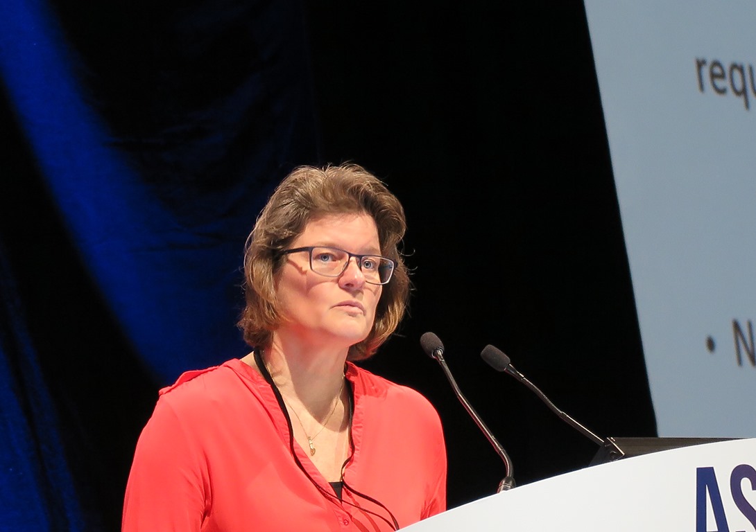
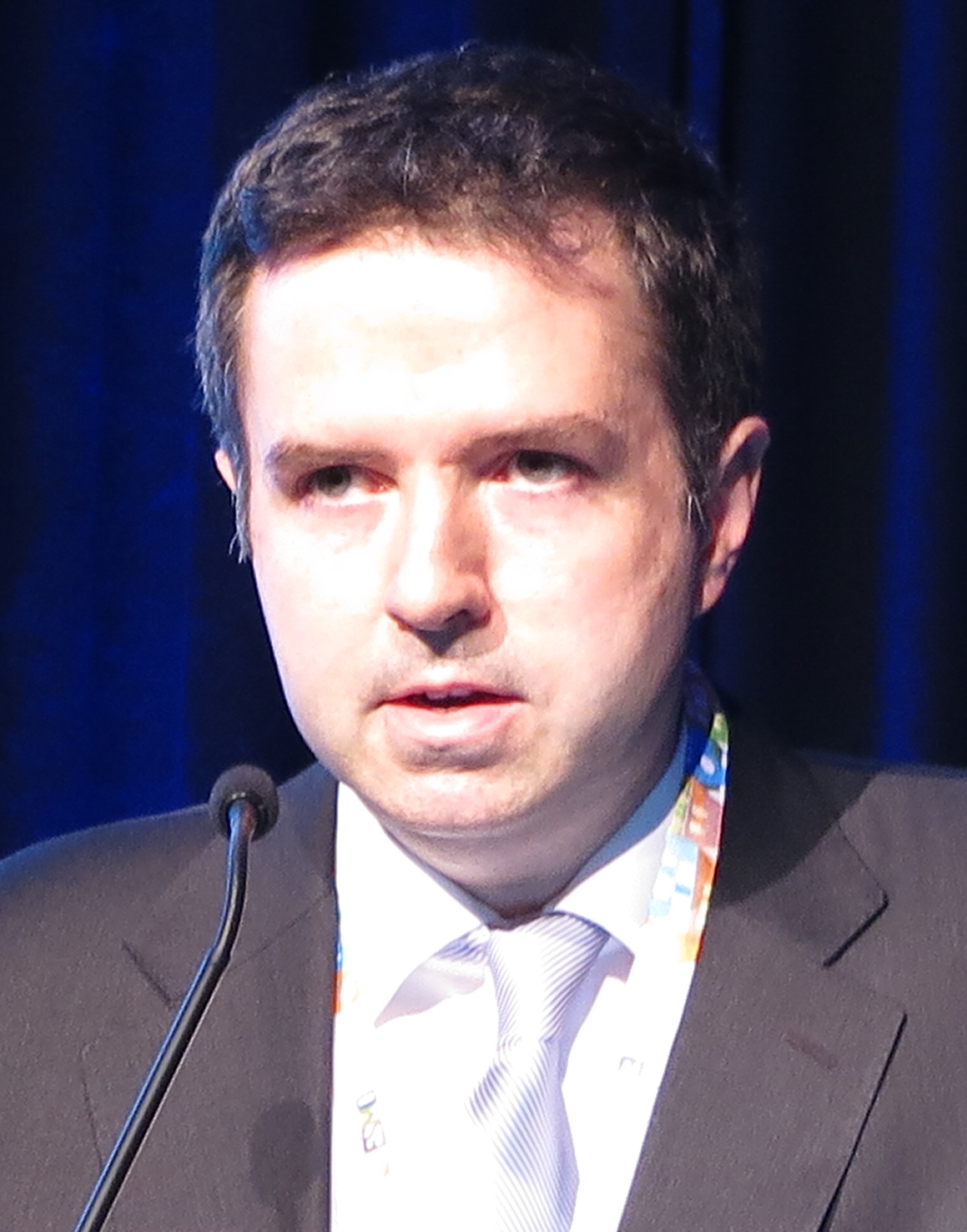
Patients with advanced, HPV16-positive cervical cancer who were treated with standard chemotherapy and vaccinated with HPV16 synthetic long peptides (HPV16-SLP) had substantially increased T-cell responses, and these responses correlated with survival, said Marij Welters, PhD, from Leiden (the Netherlands) University Medical Center.
Data from this study provide “a strong rationale to conduct a randomized phase II trial in which a combination with checkpoint inhibitors might be attractive,” she added.
Combination required
Although therapeutic vaccination with HPV16-SLP has been shown to evoke T-cell–mediated shrinkage of HPV16-induced cervical neoplasia, the investigators found in a previous study that vaccination did not result in either tumor regression or prolonged progression-free survival of patients with advanced or recurrent HPV16-induced cervical cancer.
In the phase I trial reported here, the investigators explored whether combination HPV16-SLP with chemotherapy could potentiate T-cell responses.
In a second study, Welters et al. also showed that vaccination with HPV16-SLP after the second round of chemotherapy, when myeloid cells were at their nadir, resulted in robust T-cell responses in both mice and humans.
In the phase I trial reported here, the authors reported on the feasibility and efficacy of the technique in a larger cohort of patients with late-stage, HPV16-positive cervical cancer.
The investigators enrolled cohorts of 12 patients each and delivered one HPV16-SLP vaccine dose 2 weeks after the second, third, and fourth cycles of a total of six chemotherapy cycles with carboplatin and paclitaxel. The vaccine was test dosed at levels of 20, 40, 100, and 300 mcg/peptide, with or without 1 mcg/kg of pegylated interferon-alpha at each peptide dose level. The peptides covered the length of the HPV16 E6 and E7 proteins.
“Upon vaccination, we see a very strong T-cell response induced by the vaccine,” Dr. Welters said.
They also tested general immune responses to various microbial antigens and saw no significant differences in responses among the various dose cohorts.
Early response data was available for a total of 59 patients, 35 of whom received the vaccine/chemotherapy combination as first-line therapy and 24 of whom received it in the second line.
Two of the first-line patients had a complete response, as did one patient who received the combination as second-line therapy. Respective rates of partial responses were 22 and 5, stable disease was seen in 9 and 13 patients, and disease progression in 2 and 5 patients.
The overall response rate for patients treated in the first line was 69%, and the combined overall response and stable-disease rates were 94%. Among patients treated in the second line, the respective rates were 25% and 79%.
Median overall survival (OS) from the time of the first chemotherapy dose was 16.8 months among the first-line patients vs. 7.9 months among second-line patients (P = .0110)
At the data cutoff, median OS had not been reached for the two highest peptide dose levels (100 and 300 mcg).
The investigators also found that OS was independent of general immune status among the patients, suggesting that the benefit was derived specifically from induced T-cell responses.
‘Provocative and promising’
Invited discussant Heather McArthur, MD, MPH, from Cedars-Sinai Medical Center, Los Angeles, called the finding “provocative and promising.”
“So, it doesn’t matter if your immune system is suppressed overall. It’s the quality of players on the field and the focus and specificity of those players that matter, and that’s what they were able to demonstrate, which I think is incredibly powerful,” she said.
She noted, however, that although safety was listed as a study endpoint, Dr. Welters did not provide data on toxicities.
The trial was supported by the Dutch Cancer Society and ISA Pharmaceuticals BV. Dr. Welters reported no conflicts of interest. Dr. McArthur has previously disclosed participation in advisory boards for Celgene, Merck, Spectrum Pharmaceuticals, OBI Pharma, Peregrine Pharmaceuticals, and Syndax Pharmaceuticals, and research support from Bristol-Myers Squibb, MedImmune/AstraZeneca, Eli Lilly, ZIOPHARM Oncology, and Merck.
AT THE CLINICAL IMMUNO-ONCOLOGY SYMPOSIUM
Key clinical point: An HPV16 peptide vaccine, combined with chemotherapy, induced immune response in patients with advanced cervical cancer.
Major finding: The overall response rate for chemotherapy-naive patients treated with the combination of chemotherapy and an HPV16 synthetic long peptide vaccine was 69%.
Data source: Phase I dose-finding trial with response data on 59 patients with advanced cervical cancer.
Disclosures: The trial was supported by the Dutch Cancer Society and ISA Pharmaceuticals BV. Dr. Welters reported no conflicts of interest. Dr. McArthur has previously disclosed participation in advisory boards for Celgene, Merck, Spectrum Pharmaceuticals, OBI Pharma, Peregrine Pharmaceuticals, and Syndax Pharmaceuticals and research support from Bristol-Myers Squibb, MedImmune/AstraZeneca, Eli Lilly, ZIOPHARM Oncology, and Merck.
Genomic differences seen in mRCC during first- and second-line therapy
ORLANDO – In the largest assessment to date of circulating tumor DNA (ctDNA) in patients with metastatic renal cell carcinoma (mRCC), the majority of patients were found to have clinically relevant genomic alterations.
The most frequently occurring alterations for the entire cohort were TP53, VHL, NF1, EGFR, and ARID1A, but, importantly, the genetic profiles differed between patients receiving first-line therapy and those receiving second-line treatments.
“Compared to patients receiving first-line therapy, patients receiving post–first-line agents had increased genomic alterations in TP53, NF1, EGFR, and PIK3CA,” said lead study author Sumanta K. Pal, MD, a urologic oncologist at City of Hope, Duarte, Calif.
“These alterations underscore potential mechanisms of resistance,” said Dr. Pal, who presented the findings of his study in a press briefing held at the 2017 Genitourinary Cancers Symposium sponsored by the American Society of Clinical Oncology, ASTRO, and the Society of Urologic Oncology..
Several targeted therapies have been approved for mRCC, including vascular endothelial growth factor–targeted therapies, mammalian target of rapamycin inhibitors, and checkpoint inhibitors. However, treatment of mRCC is generally distinctly different for the first- and second-line settings.
Dr. Pal noted that, while “efforts such as the TCGA [The Cancer Genome Atlas] have shed some light on the tumor biology, it is important to keep in mind that these datasets reflect earlier stages of disease. Certainly, there may be an evolution of tumor biology as patients progress toward metastasis.”
Circulating tumor markers represent a practical means of serially assessing tumor biology, and ctDNA can account for tumor heterogeneity. In this study, the authors sought to determine the mutational landscape of mRCC as well as to assess changes across patients receiving first-line and subsequent therapies by using ctDNA.
Data were obtained from 224 patients who received ctDNA profiling at progression as part of routine clinical care using Guardant360, a CLIA-certified comprehensive plasma assay that evaluated 70 genes. Of this group, 64 and 56 patients were coded as receiving frontline and post–first-line agents, respectively.
Genomic alterations were pooled for the entire group, and first and second (subsequent) therapies were compared, based on conventional practice patterns (first-line regimens included sunitinib, pazopanib and bevacizumab, and second line included everolimus, axitinib, cabozantinib, and nivolumab).
Genomic alterations were found in 78.6% of patients, with an average of 3.3 genomic alterations per patient. For patients receiving first-line therapy, the average number of ctDNA alterations was 2.9, compared with 3.7 for those in the cohort who were receiving second-line therapy. The median (range) ctDNA variant allele fractions were 0.23 (0.05-9.92) in the first-line group and 0.24 (0.04-47.14) in second line.
The authors observed that there were disparities in genomic alterations between both patient cohorts, with the highest disparity seen in (second vs. first line) TP53 (49% vs. 25%), VHL (29% vs. 25%), NF1 (20% vs. 15%), EGFR (17% vs. 21%), and PIK3CA (17% vs. 8%).
“These alterations underscore potential mechanisms of resistance,” said Dr. Pal.
He also pointed out that there were significant differences between the current dataset and other published reports, which may reflect the advanced state of the disease of the patients in this study.
Efforts are also ongoing to add detailed data on demographics and clinical outcomes to the current dataset, Dr. Pal added.
Acting as the paper’s discussant, Primo N. Lara Jr., MD, of the University of California, Davis, Comprehensive Cancer Center pointed out that as there are no validated biomarkers of drug resistance or tumor evolution and that liquid biopsy offers a potential platform.
The use of ctDNA is a “convenient technology that offers new means to assess RCC biology,” said Dr. Lara, but the caveat is that it is “still in its infancy and has no immediate clinical application.”
The current study is “hypothesis generating only.” ctDNA changes need to be related to outcome following treatment, and the functional role of genomic alterations in RCC biology must be validated, he said.
AT THE GENITOURINARY CANCERS SYMPOSIUM
Key clinical point: The genetic profile of tumors in mRCC differed in patients receiving first-line and second-line therapies.
Major finding: Genomic alterations were identified in 78.6% of patients, with an average of 3.3 genomic alterations per patient.
Data source: Experimental study that used circulating tumor DNA to assess the mutational landscape of metastatic renal cell carcinoma.
Disclosures: The funding source is not disclosed. Dr. Pal and his coauthors all report relationships with multiple pharmaceutical companies. Dr. Lara reports financial ties to multiple pharmaceutical companies.
Six Open Clinical Trials That Are Expanding Our Understanding of Immunotherapies
Using the immune system to help fight cancer is one of newest and most promising directions in cancer research. While many of the findings so far remain preliminary, a number of new studies are being developed or are already underway. Not surprisingly, federal oncologists and hematologists are leading the way with ground-breaking research. Importantly, a number of trials are recruiting patients at VA facilities. Here are a few of the studies already underway:
Study: Vaccine Therapy in Treating Patients With Newly Diagnosed Advanced Colon Polyps
Sponsor: National Cancer Institute
This randomized phase II clinical trial studies how well MUC1 peptide-poly-ICLC adjuvant vaccine works in treating patients with newly diagnosed advanced colon polyps (adenomatous polyps). Adenomatous polyps are growths in the colon that may develop into colorectal cancer over time. Vaccines made from peptides may help the body build an effective immune response to kill polyp cells. MUC1 peptide-poly-ICLC adjuvant vaccine may also prevent the recurrence of adenomatous polyps and may prevent the development of colorectal cancer.
Federal Study Locations (7 total): Kansas City VAMC
Study: Nivolumab and Ipilimumab With or Without Sargramostim in Treating Patients With Stage III-IV Melanoma That Cannot Be Removed by Surgery
Sponsor: National Cancer Institute
This randomized phase II/III trial studies the side effects and best dose of nivolumab and ipilimumab when given together with or without sargramostim and to see how well these drugs work in treating patients with stage III-IV melanoma that cannot be removed by surgery. Monoclonal antibodies, such as ipilimumab and nivolumab, may kill tumor cells by blocking blood flow to the tumor, by stimulating white blood cells to kill the tumor cells, or by attacking specific tumor cells and stop them from growing or kill them. Colony-stimulating factors, such as sargramostim, may increase the production of white blood cells. It is not yet known whether nivolumab and ipilimumab are more effective with or without sargramostim in treating patients with melanoma.
Federal Study Locations (311 total): Little Rock (Arkansas) VAMC
Study: Lenalidomide or Observation in Treating Patients With Asymptomatic High-Risk Smoldering Multiple Myeloma (NCT01169337)
Sponsor: National Cancer Institute
This randomized phase II/III trial studies how well lenalidomide works in treating patients with asymptomatic high-risk asymptomatic (smoldering) multiple myeloma. Biological therapies, such as lenalidomide, may stimulate the immune system in different ways and stop cancer cells from growing. Sometimes the cancer may not need treatment until it progresses. In this case, observation may be sufficient. It is not yet known whether lenalidomide is effective in treating patients with high-risk smoldering multiple myeloma than observation alone.
Federal Study Locations (600 total): Kansas City VAMC, VA New Jersey Health Care System, East Orange
Study: Blinatumomab and Combination Chemotherapy or Dasatinib, Prednisone, and Blinatumomab in Treating Older Patients With Acute Lymphoblastic Leukemia (NCT02143414)
Sponsor: National Cancer Institute
This phase II trial studies the side effects and how well blinatumomab and combination chemotherapy or dasatinib, prednisone, and blinatumomab work in treating older patients with acute lymphoblastic leukemia. Monoclonal antibodies, such as blinatumomab, find cancer cells and help kill them. Drugs used in chemotherapy, such as prednisone, vincristine sulfate, methotrexate, and mercaptopurine, work in different ways to stop the growth of cancer cells, either by killing the cells, by stopping them from dividing, or halting the cells’ ability to spread. Dasatinib may stop the growth of cancer cells by blocking some of the enzymes needed for cell growth. Giving blinatumomab with combination chemotherapy or dasatinib and prednisone may kill more cancer cells.
Federal Study Locations (180 total): Little Rock (Arkansas) VAMC
Study: Rituximab, Bendamustine Hydrochloride, and Bortezomib Followed by Rituximab and Lenalidomide in Treating Older Patients With Previously Untreated Mantle Cell Lymphoma (NCT01415752)
Sponsor: Eastern Cooperative Oncology Group
Monoclonal antibodies, such as rituximab, can block cancer growth in different ways. Some find cancer cells and help kill them or carry cancer-killing substances to them. Others interfere with the ability of cancer cells to grow and spread. Drugs used in chemotherapy, such as bendamustine hydrochloride, also work in different ways to kill cancer cells or stop them from dividing. Bortezomib may stop the growth of cancer cells by blocking some of the enzymes needed for cell growth. Lenalidomide may stop the growth of mantle cell lymphoma by blocking blood flow to the cancer. It is not yet known whether giving rituximab together with bendamustine and bortezomib is more effective than rituximab and bendamustine, followed by rituximab alone or with lenalidomide in treating mantle cell lymphoma.
Federal Study Locations (426 total): Kansas City VAMC, VA New Jersey Health Care System, East Orange
Study: Rituximab and Combination Chemotherapy With or Without Lenalidomide in Treating Patients With Newly Diagnosed Stage II-IV Diffuse Large B Cell Lymphoma (NCT01856192)
This randomized phase II trial studies how well rituximab and combination chemotherapy with or without lenalidomide work in treating patients with newly diagnosed stage II-IV diffuse large B cell lymphoma. Monoclonal antibodies, such as rituximab, may interfere with the ability of cancer cells to grow and spread. Drugs used in chemotherapy, such as cyclophosphamide, doxorubicin hydrochloride, vincristine sulfate, and prednisone, work in different ways to stop the growth of cancer cells, either by killing the cells, by stopping them from dividing, or by stopping them from spreading. Lenalidomide may stimulate the immune system in different ways and stop cancer cells from growing. It is not yet known whether rituximab and combination chemotherapy are more effective when given with or without lenalidomide in treating patients with diffuse large B cell lymphoma.
Federal Study Locations (511 total): Little Rock (Arkansas) VAMC
Nivolumab + ipilimumab induced fulminant, fatal myocarditis
Two patients taking the immune checkpoint inhibitors nivolumab and ipilimumab for metastatic melanoma developed fulminant, fatal myocarditis, investigators reported in the New England Journal of Medicine.
Even though this adverse effect is rare, “clinicians should be vigilant for immune-mediated myocarditis, particularly because of its early onset, nonspecific symptomatology, and fulminant progression,” said Douglas B. Johnson, MD, of Vanderbilt University Medical Center, Nashville, and his associates.
The first case involved a 65-year-old woman with no cardiac risk factors who was admitted to the hospital with chest pain, dyspnea, and fatigue 12 days after she received her first dose of the combination therapy. She was found to have myocarditis and myositis with rhabdomylysis. Despite treatment with high-dose glucocorticoids, she developed intraventricular conduction delay within 24 hours, followed by complete heart block. She died from multisystem organ failure and refractory ventricular tachycardia.
The second case involved a 63-year-old man with no cardiac risk factors who was admitted with fatigue and myalgias 15 days after he received his first dose of the combination therapy. He showed profound ST-segment depression, an intraventricular conduction delay, myocarditis, and myositis. He also was treated with high-dose glucocorticoids but developed complete heart block and died from cardiac arrest.
Both patients had “strikingly elevated troponin levels and refractory conduction-system abnormalities with preserved cardiac function,” the investigators noted. Postmortem assessments showed intense lymphocytic infiltrates only in striated cardiac and skeletal muscle and in metastases; adjacent smooth muscle and other tissues were unaffected. Pathology results “were reminiscent of those observed in patients with acute allograft rejection after cardiac transplantation,” Dr. Johnson and his associates said (N Engl J Med. 2016 Nov 3. doi: 10.1056/NEJMoa1609214).
To assess the frequency of myocarditis and myositis in patients receiving immune checkpoint inhibitors for many different cancers, the investigators searched Bristol-Myers Squibb safety databases. They found 18 drug-related cases of severe myocarditis among 20,594 patients, for a frequency of 0.09%. Patients who received combined nivolumab and ipilimumab had more frequent and more severe myocarditis than those who took either agent alone.
“There are no known data regarding what monitoring strategy may be of value; in our practice, we are performing baseline ECG and weekly testing of troponin levels during weeks 1-3 for patients receiving combination immunotherapy,” the researchers noted.
This work was supported by the Bready Family Foundation, the National Cancer Institute, Vanderbilt-Ingram Cancer Center Ambassadors, the Breast Cancer Specialized Program of Research Excellence, the National Comprehensive Cancer Network, the National Institutes of Health, the Howard Hughes Medical Institute, and Gilead Life Sciences. Dr. Johnson reported receiving personal fees from Genoptix and Bristol-Myers Squibb, and his associates reported ties to numerous industry sources.
FROM THE NEW ENGLAND JOURNAL OF MEDICINE
Key clinical point: Two patients taking the immune checkpoint inhibitors nivolumab and ipilimumab for metastatic melanoma developed fulminant, fatal myocarditis.
Major finding: A search of Bristol-Myers Squibb safety databases found 18 drug-related cases of severe myocarditis among 20,594 patients, for a frequency of 0.09%.
Data source: Two case reports of a rare adverse effect of treatment with immune checkpoint inhibitors.
Disclosures: This work was supported by the Bready Family Foundation, the National Cancer Institute, Vanderbilt-Ingram Cancer Center Ambassadors, the Breast Cancer Specialized Program of Research Excellence, the National Comprehensive Cancer Network, the National Institutes of Health, the Howard Hughes Medical Institute, and Gilead Life Sciences. Dr. Johnson reported receiving personal fees from Genoptix and Bristol-Myers Squibb, and his associates reported ties to numerous industry sources.
Pembrolizumab ‘new standard of care’ in advanced PD-L1-rich NSCLC
COPENHAGEN – Chalk up another one for immunotherapy: the PD-1 checkpoint inhibitor pembrolizumab cut the risk of disease progression or death in half among select patients with non–small cell lung cancer (NSCLC), compared with standard platinum doublet chemotherapy, in the first-line setting.
Among 305 patients with non–small cell lung cancers with 50% or greater expression of the programmed death ligand 1 (PD-L1), median progression-free survival (PFS) for patients treated with pembrolizumab (Keytruda) was 10.3 months, compared with 6 months for patients assigned to receive platinum-based chemotherapy at the investigators discretion (hazard ratio, 0.50, P less than .001), reported Martin Reck, MD, from the department of thoracic oncology at the Lung Clinic Grosshansdorf, in Germany.
Results of the KEYNOTE-024 study were also published online in the New England Journal of Medicine (2016 Oct 9. doi: 10.1056/NEJMoa1606774). Approximately 23%-30% of patients with advanced non–small cell lung cancers have tumors that express PD-L1 on the membrane of at least 50% of tumor cells, making them attractive targets for pembrolizumab, which is a monoclonal antibody directed against programmed death 1 (PD-1). Pembrolizumab disengages the brake on the immune system caused by the interaction of receptor PD-1 with the PD-L1 and PD-L2 ligands.
The study was conducted to compare upfront pembrolizumab with platinum-based chemotherapy in patients with newly diagnosed advanced NSCLC that did not carry targetable EGFR-activating mutations or ALK translocations.
A total of 305 patients from 16 countries with untreated stage IV NSCLC, good performance status, and tumors with a 50% or greater expression of PD-L1 were enrolled and randomized to either pembrolizumab 200 mg intravenously every 3 weeks for up to 2 years. Or four to six cycles of platinum-doublet chemotherapy at the investigator’s discretion. The combinations included carboplatin plus pemetrexed, cisplatin plus pemetrexed, carboplatin plus gemcitabine, cisplatin plus gemcitabine, or carboplatin plus paclitaxel.
At a median follow-up of 11.2 months, 48.1% of patients assigned to pembrolizumab were still on treatment, as were 10% of those assigned to standard chemotherapy.
As noted before, PFS, the primary endpoint, was significantly better with pembrolizumab, as was the secondary endpoint of overall survival at 6 months. In all, 80% of patients treated with pembrolizumab were still alive at 6 months, compared with 72% of patients on chemotherapy (HR, 0.60; P = .005).
The confirmed response rate was also higher in the pembrolizumab arm, at 44.8% vs. 27.8%(P = .0011), and the median duration of response was longer (not reached vs. 6.3 months). There were six complete responses in the pembrolizumab arm.
Pembrolizumab also demonstrated a generally more favorable safety profile, with adverse events of any grade occurring in 73.4% of patients, compared with 90% of those treated with chemotherapy.
Grade 3 or 4 adverse events and treatment-related deaths were also lower in the pembrolizumab arm, at 26.6% vs. 53.3%.
Jean-Charles Soria, MD, chair of drug development at Gustave Roussy Cancer Center in Paris, the invited discussant, noted that the “45% objective response rate in first-line non–small cell lung cancer is unheard of, and is achieved with a monotherapy.”
“Pembrolizumab clearly leads to a higher objective response, a longer duration of response, a lower frequency of adverse events, better PFS, better OS, compared to chemotherapy.”
“We have, probably, a new standard of care” for patients with high PD-L1 expression and no targetable mutations,” he said.
AT ESMO 2016
Key clinical point: Pembrolizumab was superior to chemotherapy in stage IV NSCLC with PD-L1 expression of 50% or more.
Major finding: The hazard ratio for progression-free survival was 0.50 for pembrolizumab vs. platinum-based chemotherapy.
Data source: Randomized phase III trial in 305 patients with untreated stage IV NSCLC with 50% or more of tumor cells expressing PD-L1
Disclosures: The study was sponsored by Merck, Sharp & Dohme. Dr. Reck and Dr. Soria disclosed financial relationships (consulting/honoraria, research funding, etc.) with several companies, but not Merck.
Change in end-of-life cancer care imperative
With the passage of the Medicare Access and CHIP Reauthorization Act, changes to how cancer care is delivered are fast approaching. This legislation aims to reward value-based care and incentivize alternative payment models that prize quality. The shift from quantity-based to value-based reimbursement is motivated in part by the rising cost of health care as well as the growing demand from patients, employers, and payers to better understand the quality of care being delivered. In cancer care, one area of high-cost and questionable value being examined is aggressive care at the end of life.
The scientific pace of progress in cancer care is exciting, with 19 therapies approved or granted a new indication in 2015. New categories of drugs, such as immunotherapies, are changing how we treat patients. It is also a time of great change in how cancer care is being delivered in our clinics, hospitals, and academic institutions. We must be vigilant in learning from these experiments in care delivery to ensure that they deliver on their promise of value to patients.
Dr. Bobby Daly, Dr. Andrew Hantel, and Dr. Blase Polite are with the University of Chicago.
Pembrolizumab boosts response but not survival in small study of advanced NSCLC
COPENHAGEN – Adding the PD-1 checkpoint inhibitor pembrolizumab (Keytruda) to a standard platinum-doublet chemotherapy regimen nearly doubled response rates among patients with previously untreated advanced nonsquamous non–small cell lung cancer, but did not result in an overall survival advantage, results of a phase II trial show.
After a median follow-up of 10.6 months, the objective response rate among patients randomized to receive carboplatin and pemetrexed plus pembrolizumab was 55%, compared with 29% for patients treated with the platinum doublet alone, reported Corey J. Langer, MD, from the Abramson Cancer Center of the University of Pennsylvania, Philadelphia.
Dr. Langer presented results on one cohort in the KEYNOTE 021 trial, a phase II, randomized, open-label multicohort study looking at pembrolizumab in combination with chemotherapy or immunotherapy.
In this cohort, 123 patients with untreated stage IIIB or IV nonsquamous non–small cell lung cancer with no activating EGFR mutations or ALK translocations were randomly assigned to receive either pembrolizumab 200 mg every 3 weeks for 2 years plus carboplatin dosed to the area under the curve and infused at 5 mg/mL per min plus pemetrexed 500 mg/m2 every 3 weeks for four cycles, or to chemotherapy alone.
Following completion of the trial, patients randomized to chemotherapy could be switched over to pembrolizumab at the same dose and scheduled for up to 2 years.
As noted, for the primary endpoint of confirmed objective response rates, the rate in the pembrolizumab/chemo group was nearly double that of the chemo-alone group (55% vs. 29%, P = .0016).
Among 33 patients on the pembrolizumab/chemo combination and 18 on chemo alone who had clinical responses according to independent central review, the median time to response was 1.5 months vs. 2.7 months, respectively. The median duration of response had not been reached in either trial arm at the time of data cutoff, and 88% and 78% of patients, respectively, had ongoing treatment responses.
Progression-free survival, a secondary endpoint, was also significantly better with the combo, with a hazard ratio of 0.53 (P = .0102).
There was no difference in overall survival, however: 75% of patients on the combination were alive at 1 year, compared with 72% of the patients on chemo alone.
Grade 3 or greater treatment-related adverse events were seen in 39% of patients on pembrolizumab, compared with 26% of patients on chemotherapy.
The most common grade 3 or greater adverse events in the combination arm were anemia, decreased neutrophil count, acute kidney injury, decreased lymphocyte count, fatigue, neutropenia, sepsis, and thrombocytopenia. In the chemotherapy-alone group, the most common grade 3 or greater events were anemia, decreased neutrophil count, pancytopenia, and thrombocytopenia.
There were three deaths, one from sepsis each in the pembrolizumab-treated group and chemotherapy alone group, and one from pancytopenia in the chemo alone group.
One (2%) of 59 patients in the pembrolizumab plus chemotherapy group experienced treatment-related death because of sepsis, compared with two (3%) of 62 patients in the chemotherapy group.
Invited discussant Jean-Charles Soria, MD, chair of drug development at Gustave Roussy Cancer Center in Paris, said that although the findings of the trial are “intriguing,” there were not enough patients to allow for drawing significant conclusions about the potential use of the combination in clinical practice.
Key clinical point: Adding pembrolizumab to platinum-based chemotherapy for upfront therapy of advanced NSCLC nearly doubled response rates.
Major finding: The overall response rate in the pembrolizumab/chemo group was 55% vs. 29% for chemotherapy alone (P = .0016)
Data source: Phase II randomized, open-label trial in 123 patients with untreated stage IIIB or IV nonsquamous NSCLC.
Disclosures: The study was funded by Merck, Sharp, and Dohme. Dr. Langer disclosed research funding from the company. Dr. Soria disclosed financial relationships (consulting/honoraria, research funding) with several companies, but not Merck.
Glimmer of promise for nivolumab in neoadjuvant NSCLC therapy
COPENHAGEN – Neoadjuvant therapy with the PD-1 checkpoint inhibitor nivolumab is safe, does not delay surgery, and may offer clinical benefit in some patients with early-stage non–small cell lung cancer, preliminary results of a clinical trial show.
Among 17 patients with previously untreated stage I-IIIA non–small cell lung cancer (NSCLC) who had two courses of nivolumab (Opdivo) followed by surgical resection, 12 had pathologic evidence of tumor regression, including 7 who had what investigators termed a “major pathologic response,” defined as less than 10% residual viable tumor, reported Patrick M. Forde, MBBCh, of the Sidney Kimmel Comprehensive Cancer Center at Johns Hopkins University in Baltimore.
To see whether immunotherapy could be safe and practical in the neoadjuvant setting, the investigators enrolled 18 patients with untreated, resectable disease and performed pretreatment tumor biopsies. Patients then received doses of nivolumab 3 mg/kg delivered at 4 weeks and 2 weeks prior to scheduled surgery. Following surgery, patients received adjuvant chemotherapy at the investigator’s discretion.
Treatment related adverse events included one grade 3 to 4 toxicity and one event leading to discontinuation. There were no adverse events requiring delay of surgery.
The investigators also conducted an exploratory analysis of response to treatment among 17 who had sufficient follow-up data for evaluation of the primary endpoints of safety and feasibility.
As noted, 12 patients had a measurable tumor response, and 7 had a major pathologic response. Of this latter group, three had no radiographic evidence of response, but tumor specimens from all 7 showed evidence of “substantial” T-cell infiltration, indicating an enhanced immune response, Dr. Forde said.
In all, 9 of the 17 patients had tumor regression of more than 50%, and 7 patients had pathologic downstaging from their pretreatment clinical stage.
Four of the seven tumors with the major pathologic response were tested with a programmed death–1 ligand immunohistochemical assay, and three were positive for the ligand.
The investigators also isolated both unique and shared T-cell clones from peripheral blood, and detected new infiltration of T-cell clones that were not seen in the tumor specimens taken prior to nivolumab therapy.
Based on these early results, the investigators plan to expand the study, with one cohort planned to receive a third presurgical dose of nivolumab, and a second cohort scheduled to receive both nivolumab and ipilimumab (Yervoy).
It remains to be seen, he said, whether patients should also receive maintenance therapy, and whether combined checkpoint inhibitors might be more effective.
However, Pieter Postmus, chair of thoracic oncology at the University of Liverpool (England), who was not involved in the study, said that he is reserving judgment on efficacy until further data are available.
“There is a potential for bias when comparing a small biopsy, which might not represent the whole tumor, with the resected tumor,” he said in a statement. “This is not a validated way to measure response to a treatment. It describes a biological effect but whether that has any clinical impact on survival is unproven.”
“Although we do not know for the time being if a major pathological response is correlated with improved survival, this method could first be validated in a cohort of patients with advanced disease by comparing the percentages of viable tumor cells in tumor biopsies taken before and 4 to 8 weeks after immunotherapy,” he added. “If in this way regression – as defined in the preoperative study – correlates with survival in patients with advanced cancer, it is likely to hold true in less advanced or resectable patients. Long-term survival data will be the ultimate test for these neoadjuvant immunotherapy strategies.”
AT ESMO 2016
Key clinical point: Checkpoint inhibitors have shown good efficacy for treatment of advanced NSCLC, but use in the neoadjuvant setting is still investigational.
Major finding: Seven of 17 patients had a major pathologic response (less than 10% viable tumor remaining).
Data source: Safety and feasibility study in 18 patients with stage I-IIIA non–small cell lung cancer.
Disclosures: The study was supported by the American Association for Cancer Research, the Cancer Research Institute, LUNGevity, and Stand Up to Cancer. Bristol-Myers Squibb donated nivolumab and provided funding for PD-L1 testing. Dr. Forde disclosed institution research grants from Bristol-Myers Squibb, Kyowa, Novartis, and uncompensated consulting for AstraZeneca and Celgene. Dr. Baas disclosed grants and research support from Bristol-Myers Squibb. Dr. Postmus had no disclosures.
10 recommendations for the Cancer Moonshot
Responding to the Cancer Moonshot initiative, a panel of scientists, clinicians, patient advocates, and industry representatives has issued 10 recommendations for accelerating cancer research in an article published in Science.
The recommendations address:
• Development of a patient engagement network.
• Precise cataloging of tumor molecular changes.
• Analysis of samples already available from patients who have received the standard of care.
• Improvements for data sharing, access, and analysis.
• Development of models to understand how childhood cancers develop.
• Research to describe how fusion oncoproteins drive cancer development.
• Creation of a cancer immunotherapy clinical trials network.
• Systematic efforts to gather information on patient-reported outcomes.
• Implementation of evidence-based approaches to prevention.
The panel’s recommendations were presented to the National Cancer Advisory Board, the adviser to the National Cancer Institute. The ability to conduct research stemming from the panel’s recommendations will depend on whether, and how much, funding is approved by Congress.
Read the article here: http://science.sciencemag.org/content/early/2016/09/07/science.aai7862.full.
Gene profile predicts RCC response to nivolumab
Many patients with advanced renal cell carcinoma have tumors that do not respond to immune checkpoint inhibitors targeted against the programmed death-1 (PD-1) pathway, despite expression of the target PD ligand 1 (PD-L1) on their tumors. Now investigators think they know why, and hope to use the information to predict which patients are likely to benefit and identify potential new therapies or combinations.
A study of renal cell carcinoma (RCC) samples from tumors with both good and poor clinical responses to treatment with the anti–PD-1 agent nivolumab (Opdivo) showed that a tumor gene–expression profile tipped more toward genes for controlling metabolic functions rather than immune functions was associated with a lack of response to anti-PD-1 therapy, reported Suzanne L. Topalian, MD, and her colleagues from Johns Hopkins University and the Sidney Kimmel Comprehensive Cancer Center, both in Baltimore.
“These findings suggest that tumor cell–intrinsic metabolic factors may contribute to treatment resistance in RCC, thus serving as predictive markers for treatment outcomes and potential new targets for combination therapy regimens with anti–PD-1,” they wrote in a study published online in Cancer Immunology Research.
The investigators obtained tumor samples from 13 patients with unresectable metastatic RCC treated in one of four clinical trials. They used radiographic staging to classify each patient as either a responder or nonresponder to anti–PD-1 therapy according to RECIST (Response Evaluation Criteria in Solid Tumors). The samples were evaluated with whole genome microarray and multiplex quantitative reverse-transcription polymerase chain reaction (qRT-PCR) profiling and analysis, and the results were compared with those from eight renal cell carcinoma cell lines.
They looked for expression of nearly 30,000 gene targets in samples from responders and nonresponders and found a pattern of differential expression of genes encoding for metabolic pathways and immune functions.
Specifically, they found that the expression of genes involved in metabolic and solute transport functions (for example, UGT1A) were associated with poor response to nivolumab, whereas overexpression of genes for immune markers involved in T-cell differentiation (BACH2) and leukocyte migration (CCL3) were associated with a good response.
The investigators acknowledge that the study was retrospective and limited by the analysis of only a small number of tumor samples but suggest that their findings point the way to further investigations in larger groups of patients with RCC tumors, including those both positive and negative for PD-L1 expression.
“The general approach to identifying biomarkers of clinical response to PD-1–targeted therapies has so far focused on immunologic factors in the [tumor microenvironment]. However, a deeper level of investigation may be warranted for individual tumor types, and intersections of tumor cell–intrinsic factors with immunologic factors may be particularly revealing,” they wrote.
The study was supported by research grants from the Bloomberg-Kimmel Institute for Cancer Immunotherapy at Johns Hopkins, Bristol-Myers Squibb, the National Cancer Institute, and Stand Up To Cancer. Dr. Topalian has served as a consultant/advisory board member for Five Prime Therapeutics, MedImmune, Merck, and Pfizer, and has an ownership interest in Bristol-Myers Squibb, Five Prime Therapeutics,and Potenza Therapeutics. Other coauthors reported similar potential conflicts of interest.
FROM CANCER IMMUNOLOGY RESEARCH
Key clinical point: Many renal cell carcinoma tumors do not respond to therapy with an anti-PD-1 agent, despite being positive for the PD-L1 target.
Major finding: A gene expression profile favoring genes associated with metabolic and solute transport functions was associated with poor response to nivolumab.
Data source: Retrospective study of tumor gene expression in 13 patients with advanced RCC and 8 RCC cell lines.
Disclosures: The study was supported by research grants from the Bloomberg-Kimmel Institute for Cancer Immunotherapy at Johns Hopkins, Baltimore; Bristol-Myers Squibb; the National Cancer Institute; and Stand Up To Cancer. Dr. Topalian has served as a consultant/advisory board member for Five Prime Therapeutics, MedImmune, Merck, and Pfizer, and has an ownership interest in Bristol-Myers Squibb, Five Prime Therapeutics,and Potenza Therapeutics. Other coauthors reported similar potential conflicts of interest.

