User login
Onychomycosis: Not just for adults
COEUR D’ALENE, IDAHO – Conventional wisdom holds that onychomycosis is rare in children. Not so.
Fully one-third of children and adolescents who presented with a nail complaint to a prominent dermatologic nail disorders center were diagnosed with mycologically confirmed onychomycosis, Dr. Julie Jefferson reported at the annual meeting of the Society for Pediatric Dermatology.
That’s a substantially higher prevalence than the 15.5% figure reported by Spanish investigators in a 20-year retrospective study (Mycoses 2011;54:450-3). It’s also well below the 47% prevalence recently reported in Denver in a 5-year retrospective study, where Trychophyton rubrum was the most common pathogen, and the highest prevalence of onychomycosis in the pediatric population was seen in 6- to 10-year-olds (Pediatr. Dermatol. 2014;31:106-8), noted Dr. Jefferson of Johns Hopkins University, Baltimore.
The investigators in both Span and Denver observed that the prevalence of pediatric onychomycosis appears to be increasing in recent years.
Dr. Jefferson presented a retrospective study that included 917 patients up to age 18 years who presented to the Oregon Dermatology and Research Center, Portland, in a recent 6-year period. One or more nail disorders were diagnosed in 11%. The mean age at presentation was 9.4 years, with a mean 2.4-year duration of the condition prior to presentation. Toenails were affected in 47 patients, fingernails in 37, and both in 18. Fourteen patients had two nail disorders, 9 had three, and 2 had four distinct nail disorders.
The etiologies ranged widely, from infections to congenital and hereditary malformations, tumors, inflammatory processes, and systemic diseases.
The most common nail condition was onychomycosis, diagnosed in 34 patients. Thus, 3.7% of all pediatric patients presenting to the dermatology center for any reason were diagnosed with onychomycosis, a higher rate than previously reported by others.
Other conditions included 13 cases of longitudinal melanonychia, 11 of disappearing nail bed, 9 of retronychia, 8 cases of congenital malalignment, 8 cases of trachyonychia, 7 of paronychia, 5 cases of psoriasis, and 2 of lichen planus.
Three of four patients with an ingrown toenail also had congenital malalignment of the affected great toenail, supporting the notion put forth by other investigators that congenital malalignment of the great toenail predisposes to ingrown toenails, according to Dr. Jefferson.
She reported having no financial conflicts related to this study.
AT THE SPD ANNUAL MEETING
Key clinical point: Onychomycosis appears to be rising in children and adolescents, and needs to be considered in the differential diagnosis of pediatric nail abnormalities.
Major finding: Onychomycosis was diagnosed in 3.7% of all children and adolescents who presented for any reason to a tertiary dermatology center, and in 33% of the 102 who presented for evaluation of a nail disorder.
Data source: This was a 6-year, single-center, retrospective study of 917 patients up to 18 years of age.
Disclosures: The presenter reported having no financial conflicts with regard to this study.

Maintenance therapy options for childhood alopecia areata
COEUR D’ALENE, IDAHO – Systemic corticosteroids usually bring an impressive initial response for children with alopecia areata. The big question is, ‘What do you try next?’
Children can’t safely remain on long-term systemic steroids. And the great majority of responders will relapse once steroids are discontinued. The challenge is to find a safe, well-tolerated drug with long-term effectiveness as a maintenance therapy.

"For me, when I look at the list of drugs for maintenance therapy in alopecia areata, which includes cyclosporine, azathioprine, and sulfasalazine, the drug I’m most comfortable giving to children for a prolonged time is methotrexate. That’s why this drug has really become my go-to maintenance therapy for patients with alopecia areata," Dr. Ruth Ann Vleugels said at the annual meeting of the Society for Pediatric Dermatology.
"I will give them 2-3 months of systemic steroids as a boost while their methotrexate kicks in; but after that, it’s maintenance therapy with methotrexate at 1 mg/kg/week," added Dr. Vleugels, director of the Autoimmune Skin Disease Program at Brigham and Women’s Hospital, Boston.
Of course, methotrexate doesn’t work as maintenance therapy for all children with alopecia areata. Patients who don’t respond to the initial several months of combination therapy with systemic steroids won’t respond to maintenance therapy with methotrexate alone.
For those few patients who need second-line maintenance therapy, Dr. Vleugels’ drug of choice is the transplant anti-rejection medication mycophenolate mofetil (CellCept), which is used off-label for alopecia areata. Mycophenolate mofetil also is her rescue agent as maintenance therapy for children with systemic lupus erythematosus, cutaneous lupus erythematosus, juvenile dermatomyositis, and linear morphea with progressive hemifacial atrophy.
In patients with progressive juvenile systemic sclerosis, Dr. Vleugels and her Boston colleagues now use mycophenolate mofetil, rather than methotrexate, as their first-line therapy based upon favorable results in their practice and prospective studies in adults (J. Rheumatol. 2012;39:1241-7).
The mycophenolate mofetil dosing in children with alopecia areata is 600 mg/m2 given twice daily, or 1 g twice daily in patients bigger than 1.5 m2. She uses half the dose for the first 2-3 weeks, then titrates up to full-dose therapy in order to minimize GI side effects, which she said are far and away the most common adverse effects. Smaller children do best with the liquid formulation.
A common clinical dilemma involves the child who experiences intolerable GI upset on mycophenolate mofetil. Dr. Vleugels offered two suggestions: One is to tell the parents to ignore the package insert and have their child take the drug with meals.
"No one can handle this drug on an empty stomach," she said.
A caveat: mycophenolate mofetil cannot be taken with antacids.
If mealtime dosing doesn’t solve the GI upset problem, the alternative is to switch to mycophenolic acid (Myfortic), which is enteric-coated, has better bioavailability, and is better tolerated. It’s also more expensive than mycophenolate mofetil and will require prior authorization from payers. The dose conversion is as follows: 500 mg mycophenolate mofetil equals 360 mg of mycophenolic acid.
Dr. Vleugels reported having no financial conflicts with regard to her presentation.
COEUR D’ALENE, IDAHO – Systemic corticosteroids usually bring an impressive initial response for children with alopecia areata. The big question is, ‘What do you try next?’
Children can’t safely remain on long-term systemic steroids. And the great majority of responders will relapse once steroids are discontinued. The challenge is to find a safe, well-tolerated drug with long-term effectiveness as a maintenance therapy.
"For me, when I look at the list of drugs for maintenance therapy in alopecia areata, which includes cyclosporine, azathioprine, and sulfasalazine, the drug I’m most comfortable giving to children for a prolonged time is methotrexate. That’s why this drug has really become my go-to maintenance therapy for patients with alopecia areata," Dr. Ruth Ann Vleugels said at the annual meeting of the Society for Pediatric Dermatology.
"I will give them 2-3 months of systemic steroids as a boost while their methotrexate kicks in; but after that, it’s maintenance therapy with methotrexate at 1 mg/kg/week," added Dr. Vleugels, director of the Autoimmune Skin Disease Program at Brigham and Women’s Hospital, Boston.
Of course, methotrexate doesn’t work as maintenance therapy for all children with alopecia areata. Patients who don’t respond to the initial several months of combination therapy with systemic steroids won’t respond to maintenance therapy with methotrexate alone.
For those few patients who need second-line maintenance therapy, Dr. Vleugels’ drug of choice is the transplant anti-rejection medication mycophenolate mofetil (CellCept), which is used off-label for alopecia areata. Mycophenolate mofetil also is her rescue agent as maintenance therapy for children with systemic lupus erythematosus, cutaneous lupus erythematosus, juvenile dermatomyositis, and linear morphea with progressive hemifacial atrophy.
In patients with progressive juvenile systemic sclerosis, Dr. Vleugels and her Boston colleagues now use mycophenolate mofetil, rather than methotrexate, as their first-line therapy based upon favorable results in their practice and prospective studies in adults (J. Rheumatol. 2012;39:1241-7).
The mycophenolate mofetil dosing in children with alopecia areata is 600 mg/m2 given twice daily, or 1 g twice daily in patients bigger than 1.5 m2. She uses half the dose for the first 2-3 weeks, then titrates up to full-dose therapy in order to minimize GI side effects, which she said are far and away the most common adverse effects. Smaller children do best with the liquid formulation.
A common clinical dilemma involves the child who experiences intolerable GI upset on mycophenolate mofetil. Dr. Vleugels offered two suggestions: One is to tell the parents to ignore the package insert and have their child take the drug with meals.
"No one can handle this drug on an empty stomach," she said.
A caveat: mycophenolate mofetil cannot be taken with antacids.
If mealtime dosing doesn’t solve the GI upset problem, the alternative is to switch to mycophenolic acid (Myfortic), which is enteric-coated, has better bioavailability, and is better tolerated. It’s also more expensive than mycophenolate mofetil and will require prior authorization from payers. The dose conversion is as follows: 500 mg mycophenolate mofetil equals 360 mg of mycophenolic acid.
Dr. Vleugels reported having no financial conflicts with regard to her presentation.
EXPERT ANALYSIS FROM THE SPD ANNUAL MEETING

Pull the hair for pediatric alopecia diagnosis
COEUR D’ALENE, IDAHO – The most important test in determining the cause of diffuse hair loss in children and adolescents is a gentle hair pull, Dr. Elise A. Olsen said at the annual meeting of the Society for Pediatric Dermatology.
"This is something you should do in all patients with alopecia, accompanied by looking under the microscope at the hair you’ve pulled. Grasp a small clump of hair close to the scalp and gently pull through to the ends. It’s important that you do it not just in one area but all over, in various places on the scalp. Coming away with three or four hairs per pull is abnormal. Be especially gentle in young children because you can actually induce what looks like a loose anagen syndrome, confusing the picture," explained Dr. Olsen, professor of dermatology and medicine and director of the hair disorders research and treatment center at Duke University in Durham, N.C.

She described how to use the hair pull and other tools to differentiate between telogen effluvium, alopecia areata, androgenetic alopecia, loose anagen syndrome, and short anagen syndrome.
Telogen effluvium: This condition is characterized by a global decrease in hair density. This global reduction can be confirmed by performing a midline part on the back and top of the scalp, which should show a similarly widened, thinned part.
Microscopic examination of the proximal end of all hairs that come off with hair pulls in an adolescent with telogen effluvium should show them to be telogen hairs.
"If you see any anagen hairs, that’s abnormal, and I would urge you to think about another condition, like loose anagen syndrome or alopecia areata," Dr. Olsen said.
Potential etiologies of telogen effluvium include stress, thyroid disease, medication side effects, vitamin A intake in excess of 15,000 IU/day, and numerous diet or nutritional deficits.
"Diet is incredibly important in figuring out the cause of telogen effluvium, particularly in children and adolescents, where you might be dealing with bulimia, anorexia nervosa, or another abnormal diet," according to the dermatologist.
The relationship between hair loss and iron deficiency is a matter of long-standing controversy. Low iron levels have often been linked to hair loss. As yet, however, there is no well-controlled clinical trial showing that iron replacement improves telogen effluvium in an iron-deficient patient.
Isotretinoin tops the list of medications that can cause telogen effluvium in pediatric patients. Other drugs that need to be considered include sodium valproate, antidepressants, lithium, and medications for attention-deficit/hyperactivity disorder.
Vitamin supplements should be stopped for at least 24 hours before conducting screening laboratory testing in a patient with telogen effluvium. Dr. Olsen recommended ordering a CBC with differential; thyroid-stimulating hormone and free thyroxine; serum ferritin; total iron binding capacity; and an erythrocyte sedimentation rate to screen for occult inflammatory conditions, which would skew the ferritin results. While a serum ferritin less than 40 ng/mL ordinarily has 98% sensitivity and specificity for iron deficiency, the bar rises to less than 70 ng/mL in patients with any kind of underlying systemic inflammation.
Alopecia areata: This form of diffuse hair loss can look clinically just like telogen effluvium. The key distinguishing feature is a positive hair pull showing not only telogen hairs but dystrophic, "exclamation point" anagen hairs as well.
"These anagen hairs are broken off or bayonet-like in appearance, and there’s usually a distortion of the hair shaft diameter as well," she explained.
Scalp dermoscopy will show yellow dots of keratinaceous debris in the empty follicles of patients with alopecia areata, an uncommon condition in children.
Androgenetic alopecia: The most useful clue in differentiating this condition from telogen effluvium is that a midline part will be widened on the central scalp but never over the occiput. The gentle hair pull typically doesn’t yield any hairs except in affected areas on the top portion of the scalp. Any hairs produced via the hair pull will be telogen hairs, and they will typically vary in diameter. Dermoscopy may show perifollicular pigmentation in areas of hair loss.
"If you diagnose alopecia areata in an adolescent, there are some key things you need to discuss with the parents," Dr. Olsen stressed.
For example, their child is likely to have a more rapidly progressive course of hair loss. And affected girls are at increased risk for underlying hyperandrogenemia-related symptoms, including hirsutism, insulin resistance, polycystic ovarian syndrome, and metabolic syndrome. A finding of acanthosis nigricans in a nonobese teen is a very good indicator that they may have underlying insulin resistance.
Loose anagen syndrome: This condition, whose onset is usually before 8 years of age, is characterized by short, very slow-growing hair. The hair pull yields anagen hairs, which under the microscope have a distinctive appearance marked by a rolled-up proximal tip.
Short anagen syndrome: The hallmarks here are decreased hair density, increased shedding, and an inability to grow hair long. On the hair pull, affected patients have an increased number of telogen hairs. The telogen hairs, which are newly regrowing in response to the truncated anagen cycle, will have a tapered tip at the distal end.
Scalp biopsy is of variable utility in diagnosing the cause of diffuse hair loss in young patients. It can be used to make the diagnosis of certain conditions, including acute alopecia areata, trichotillomania, connective tissue disease, infection, tumor, and scarring disorders. However, scalp biopsy can’t be used to make a definitive diagnosis of telogen effluvium, androgenetic alopecia, or long-standing alopecia areata – it can only be suggestive.
Pathologic standards dictate that the scalp biopsy should always be 4 mm. Normal white adults have roughly 36 follicles per 4-mm biopsy. African American adults have about 22. While no standard numbers have been established for children, the number of follicles per biopsy is higher than in adults because of the child’s smaller head size. After all, the number of follicles present at birth is what an individual will carry throughout life, Dr. Olsen explained.
It’s important that patients and parents understand the need for patience regarding hair regrowth, which takes 6-12 months after the cause of alopecia has been identified and eliminated. That means, for example, that in a patient with thyroid disease the clock for regrowth starts ticking not at the time treatment begins, but when the patient becomes euthyroid.
Dr. Olsen reported serving as a consultant to Canfield Scientific and Allergan.
COEUR D’ALENE, IDAHO – The most important test in determining the cause of diffuse hair loss in children and adolescents is a gentle hair pull, Dr. Elise A. Olsen said at the annual meeting of the Society for Pediatric Dermatology.
"This is something you should do in all patients with alopecia, accompanied by looking under the microscope at the hair you’ve pulled. Grasp a small clump of hair close to the scalp and gently pull through to the ends. It’s important that you do it not just in one area but all over, in various places on the scalp. Coming away with three or four hairs per pull is abnormal. Be especially gentle in young children because you can actually induce what looks like a loose anagen syndrome, confusing the picture," explained Dr. Olsen, professor of dermatology and medicine and director of the hair disorders research and treatment center at Duke University in Durham, N.C.
She described how to use the hair pull and other tools to differentiate between telogen effluvium, alopecia areata, androgenetic alopecia, loose anagen syndrome, and short anagen syndrome.
Telogen effluvium: This condition is characterized by a global decrease in hair density. This global reduction can be confirmed by performing a midline part on the back and top of the scalp, which should show a similarly widened, thinned part.
Microscopic examination of the proximal end of all hairs that come off with hair pulls in an adolescent with telogen effluvium should show them to be telogen hairs.
"If you see any anagen hairs, that’s abnormal, and I would urge you to think about another condition, like loose anagen syndrome or alopecia areata," Dr. Olsen said.
Potential etiologies of telogen effluvium include stress, thyroid disease, medication side effects, vitamin A intake in excess of 15,000 IU/day, and numerous diet or nutritional deficits.
"Diet is incredibly important in figuring out the cause of telogen effluvium, particularly in children and adolescents, where you might be dealing with bulimia, anorexia nervosa, or another abnormal diet," according to the dermatologist.
The relationship between hair loss and iron deficiency is a matter of long-standing controversy. Low iron levels have often been linked to hair loss. As yet, however, there is no well-controlled clinical trial showing that iron replacement improves telogen effluvium in an iron-deficient patient.
Isotretinoin tops the list of medications that can cause telogen effluvium in pediatric patients. Other drugs that need to be considered include sodium valproate, antidepressants, lithium, and medications for attention-deficit/hyperactivity disorder.
Vitamin supplements should be stopped for at least 24 hours before conducting screening laboratory testing in a patient with telogen effluvium. Dr. Olsen recommended ordering a CBC with differential; thyroid-stimulating hormone and free thyroxine; serum ferritin; total iron binding capacity; and an erythrocyte sedimentation rate to screen for occult inflammatory conditions, which would skew the ferritin results. While a serum ferritin less than 40 ng/mL ordinarily has 98% sensitivity and specificity for iron deficiency, the bar rises to less than 70 ng/mL in patients with any kind of underlying systemic inflammation.
Alopecia areata: This form of diffuse hair loss can look clinically just like telogen effluvium. The key distinguishing feature is a positive hair pull showing not only telogen hairs but dystrophic, "exclamation point" anagen hairs as well.
"These anagen hairs are broken off or bayonet-like in appearance, and there’s usually a distortion of the hair shaft diameter as well," she explained.
Scalp dermoscopy will show yellow dots of keratinaceous debris in the empty follicles of patients with alopecia areata, an uncommon condition in children.
Androgenetic alopecia: The most useful clue in differentiating this condition from telogen effluvium is that a midline part will be widened on the central scalp but never over the occiput. The gentle hair pull typically doesn’t yield any hairs except in affected areas on the top portion of the scalp. Any hairs produced via the hair pull will be telogen hairs, and they will typically vary in diameter. Dermoscopy may show perifollicular pigmentation in areas of hair loss.
"If you diagnose alopecia areata in an adolescent, there are some key things you need to discuss with the parents," Dr. Olsen stressed.
For example, their child is likely to have a more rapidly progressive course of hair loss. And affected girls are at increased risk for underlying hyperandrogenemia-related symptoms, including hirsutism, insulin resistance, polycystic ovarian syndrome, and metabolic syndrome. A finding of acanthosis nigricans in a nonobese teen is a very good indicator that they may have underlying insulin resistance.
Loose anagen syndrome: This condition, whose onset is usually before 8 years of age, is characterized by short, very slow-growing hair. The hair pull yields anagen hairs, which under the microscope have a distinctive appearance marked by a rolled-up proximal tip.
Short anagen syndrome: The hallmarks here are decreased hair density, increased shedding, and an inability to grow hair long. On the hair pull, affected patients have an increased number of telogen hairs. The telogen hairs, which are newly regrowing in response to the truncated anagen cycle, will have a tapered tip at the distal end.
Scalp biopsy is of variable utility in diagnosing the cause of diffuse hair loss in young patients. It can be used to make the diagnosis of certain conditions, including acute alopecia areata, trichotillomania, connective tissue disease, infection, tumor, and scarring disorders. However, scalp biopsy can’t be used to make a definitive diagnosis of telogen effluvium, androgenetic alopecia, or long-standing alopecia areata – it can only be suggestive.
Pathologic standards dictate that the scalp biopsy should always be 4 mm. Normal white adults have roughly 36 follicles per 4-mm biopsy. African American adults have about 22. While no standard numbers have been established for children, the number of follicles per biopsy is higher than in adults because of the child’s smaller head size. After all, the number of follicles present at birth is what an individual will carry throughout life, Dr. Olsen explained.
It’s important that patients and parents understand the need for patience regarding hair regrowth, which takes 6-12 months after the cause of alopecia has been identified and eliminated. That means, for example, that in a patient with thyroid disease the clock for regrowth starts ticking not at the time treatment begins, but when the patient becomes euthyroid.
Dr. Olsen reported serving as a consultant to Canfield Scientific and Allergan.
EXPERT ANALYSIS FROM THE SPD ANNUAL MEETING

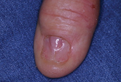
Shedding of the Fingernails
The Diagnosis: Onychomadesis
The nail changes were characteristic of onychomadesis. Systemic illness in this patient most likely resulted in temporary arrest of nail matrix activity, leading to separation of the proximal nail plate from the proximal nail fold, which gave rise to a deep transverse sulcus.1 Conversely, Beau lines are characterized by transverse grooves that move distally as the nail grows. Onychomadesis also is seen in pemphigus vulgaris, which could be due to an autoimmune disease inhibiting normal nail plate growth and development of blisters beneath the nail causing detachment of the nail plate.2 Drug-induced Beau lines or onychomadesis are most frequently caused by chemotherapeutic agents (taxanes) and retinoids, which reflect an arrest in epithelial proliferation.3 Familial cases also have been described.4 Management of the nail abnormality should focus on the underlying medical problem or triggering factor. In our patient, hypertension and kidney disease were managed by a low-salt diet, oral antihypertensives, and iron replacement.
1. Wolff K, Goldsmith LA, Katz SI, et al, eds. Fitzpatrick’s Dermatology in General Medicine. 7th ed. New York, NY: McGraw-Hill; 2008.
2. Engineer L, Norton LA, Ahmed AR. Nail involvement in pemphigus vulgaris. J Am Acad Dermatol. 2000;43:529-535.
3. Minisini AM, Tosti A, Sobrero AF, et al. Taxane-induced nail changes: incidence, clinical presentation and outcome. Ann Oncol. 2003;14:333-337.
4. Mehra A, Murphy RJ, Wilson BB. Idiopathic familial onychomadesis. J Am Acad Dermatol. 2000;43(2, pt 2):349-350.
The Diagnosis: Onychomadesis
The nail changes were characteristic of onychomadesis. Systemic illness in this patient most likely resulted in temporary arrest of nail matrix activity, leading to separation of the proximal nail plate from the proximal nail fold, which gave rise to a deep transverse sulcus.1 Conversely, Beau lines are characterized by transverse grooves that move distally as the nail grows. Onychomadesis also is seen in pemphigus vulgaris, which could be due to an autoimmune disease inhibiting normal nail plate growth and development of blisters beneath the nail causing detachment of the nail plate.2 Drug-induced Beau lines or onychomadesis are most frequently caused by chemotherapeutic agents (taxanes) and retinoids, which reflect an arrest in epithelial proliferation.3 Familial cases also have been described.4 Management of the nail abnormality should focus on the underlying medical problem or triggering factor. In our patient, hypertension and kidney disease were managed by a low-salt diet, oral antihypertensives, and iron replacement.
1. Wolff K, Goldsmith LA, Katz SI, et al, eds. Fitzpatrick’s Dermatology in General Medicine. 7th ed. New York, NY: McGraw-Hill; 2008.
2. Engineer L, Norton LA, Ahmed AR. Nail involvement in pemphigus vulgaris. J Am Acad Dermatol. 2000;43:529-535.
3. Minisini AM, Tosti A, Sobrero AF, et al. Taxane-induced nail changes: incidence, clinical presentation and outcome. Ann Oncol. 2003;14:333-337.
4. Mehra A, Murphy RJ, Wilson BB. Idiopathic familial onychomadesis. J Am Acad Dermatol. 2000;43(2, pt 2):349-350.
A 70-year-old woman was referred to the dermatology department with abnormal-appearing fingernails of 6 months’ duration. Clinical examination showed complete shedding of the proximal nail plate and separation from the nail bed involving all the fingernails. There also was thickening of the distal nail plate. The patient also had diffuse thinning of the hair on the scalp. She had chronic kidney disease, likely from hypertensive nephrosclerosis, that was complicated by iron-deficient anemia. No new systemic medication had been given.

New guidelines proposed for nail involvement in psoriatic arthritis
NEW YORK – A consensus group has developed evidence-based treatment recommendations for patients with psoriatic arthritis and nail involvement and raised the possibility of issuing them independent of recommendations for the skin, according to a report on the deliberations.
"The suggestion has been made to separate out skin from nail because the assessment is quite different to the point where you can potentially have patients with severe nail disease but limited skin disease," reported Dr. April W. Armstrong, director of the psoriasis program at the University of Colorado, Denver.

The proposal was made in the course of presenting the preliminary evidence-based recommendations on managing psoriatic arthritis (PsA) nail involvement to the full membership of the Group for Research and Assessment of Psoriasis and Psoriatic Arthritis. GRAPPA is in the process of preparing a new set of PsA treatment recommendations.
The decision to address nails separately from skin was not a unanimous recommendation within the committee, and no conclusion was reached, but Dr. Armstrong brought the issue forward "to be clear about our ambiguity" regarding this specific aspect of how to best outline treatment options useful to clinicians.
Other recommendations, based on evidence, are more assured of making the final version, because they are evidence based. When presented at GRAPPA, which convened its most recent annual meeting jointly with the Spondyloarthritis Research & Treatment Network, each recommendation was graded for the quality of the evidence and strength of the consensus.
For example, the committee found little controlled evidence to support a significant benefit from topical therapies, including those commonly used, such as steroids and vitamin D analogs. Although the consensus committee concluded that the risk of adverse events is low and the costs are low to moderate, the efficacy is only modest. The strength for recommending topical therapies overall was characterized as "weak."
For procedural therapies, such as pulsed-dye laser or intralesional injections, no placebo-controlled trials could be identified by the committee, and the expert opinion of its members was that the efficacy is relatively low in general even if benefit is achieved in some individuals. The cost was characterized as low to moderate. The committee concluded that only a "weak" recommendation should be conferred to these types of interventions for nail involvement.
The proposed recommendation for oral therapies, such a methotrexate, cyclosporine, leflunomide, and acitretin, was considered "stronger than that for topical therapies" when nail involvement is moderate, but the committee also found supportive evidence of benefit to be of "low quality." Again, although recognizing that some PsA patients may benefit, the recommendation for oral therapies overall for nail involvement was characterized as weak.
For biologics, including both tumor necrosis factor (TNF) inhibitors and the interleukin 12/23 inhibitor ustekinumab, the consensus committee reported that there are good quality data showing efficacy in nails, including some studies showing superiority of TNF inhibitors to methotrexate and cyclosporine. For patients with moderate to severe nail involvement warranting the potential risks of these therapies, the preliminary recommendation for these agents was "strong" even if the costs of these therapies were characterized as "high to crazy."
One unresolved issue, however, is which strategy to recommend for placing nail involvement into "mild," "moderate," or "severe" categories. Objective scores, such as Nail Psoriasis Severity Index (NAPSI), were considered by the consensus committee to be helpful but insufficient.
"Perhaps in addition to objective scoring of nail disease, we should also take into account [the impact] on quality of life as well as function," Dr. Armstrong said.
The final GRAPPA treatment recommendations for nail involvement, as well as other aspects of PsA management, will be made after a discussion of the consensus group proposals over the next several months, followed by voting among the GRAPPA membership. As GRAPPA is an international organization, it is intended that final recommendations will be broadly applicable.
Dr. Armstrong reported financial relationships with AbbVie, Amgen, Janssen Biotech, Merck, Novartis, and Pfizer.
NEW YORK – A consensus group has developed evidence-based treatment recommendations for patients with psoriatic arthritis and nail involvement and raised the possibility of issuing them independent of recommendations for the skin, according to a report on the deliberations.
"The suggestion has been made to separate out skin from nail because the assessment is quite different to the point where you can potentially have patients with severe nail disease but limited skin disease," reported Dr. April W. Armstrong, director of the psoriasis program at the University of Colorado, Denver.
The proposal was made in the course of presenting the preliminary evidence-based recommendations on managing psoriatic arthritis (PsA) nail involvement to the full membership of the Group for Research and Assessment of Psoriasis and Psoriatic Arthritis. GRAPPA is in the process of preparing a new set of PsA treatment recommendations.
The decision to address nails separately from skin was not a unanimous recommendation within the committee, and no conclusion was reached, but Dr. Armstrong brought the issue forward "to be clear about our ambiguity" regarding this specific aspect of how to best outline treatment options useful to clinicians.
Other recommendations, based on evidence, are more assured of making the final version, because they are evidence based. When presented at GRAPPA, which convened its most recent annual meeting jointly with the Spondyloarthritis Research & Treatment Network, each recommendation was graded for the quality of the evidence and strength of the consensus.
For example, the committee found little controlled evidence to support a significant benefit from topical therapies, including those commonly used, such as steroids and vitamin D analogs. Although the consensus committee concluded that the risk of adverse events is low and the costs are low to moderate, the efficacy is only modest. The strength for recommending topical therapies overall was characterized as "weak."
For procedural therapies, such as pulsed-dye laser or intralesional injections, no placebo-controlled trials could be identified by the committee, and the expert opinion of its members was that the efficacy is relatively low in general even if benefit is achieved in some individuals. The cost was characterized as low to moderate. The committee concluded that only a "weak" recommendation should be conferred to these types of interventions for nail involvement.
The proposed recommendation for oral therapies, such a methotrexate, cyclosporine, leflunomide, and acitretin, was considered "stronger than that for topical therapies" when nail involvement is moderate, but the committee also found supportive evidence of benefit to be of "low quality." Again, although recognizing that some PsA patients may benefit, the recommendation for oral therapies overall for nail involvement was characterized as weak.
For biologics, including both tumor necrosis factor (TNF) inhibitors and the interleukin 12/23 inhibitor ustekinumab, the consensus committee reported that there are good quality data showing efficacy in nails, including some studies showing superiority of TNF inhibitors to methotrexate and cyclosporine. For patients with moderate to severe nail involvement warranting the potential risks of these therapies, the preliminary recommendation for these agents was "strong" even if the costs of these therapies were characterized as "high to crazy."
One unresolved issue, however, is which strategy to recommend for placing nail involvement into "mild," "moderate," or "severe" categories. Objective scores, such as Nail Psoriasis Severity Index (NAPSI), were considered by the consensus committee to be helpful but insufficient.
"Perhaps in addition to objective scoring of nail disease, we should also take into account [the impact] on quality of life as well as function," Dr. Armstrong said.
The final GRAPPA treatment recommendations for nail involvement, as well as other aspects of PsA management, will be made after a discussion of the consensus group proposals over the next several months, followed by voting among the GRAPPA membership. As GRAPPA is an international organization, it is intended that final recommendations will be broadly applicable.
Dr. Armstrong reported financial relationships with AbbVie, Amgen, Janssen Biotech, Merck, Novartis, and Pfizer.
AT THE 2014 GRAPPA AND SPARTAN ANNUAL MEETINGS

Arthritis drug restores hair in man with alopecia universalis and psoriasis
A 25-year-old man with plaque psoriasis and virtually no hair of any sort now sports a full head of hair plus body hair after treatment with the arthritis drug tofacitinib, according to Dr. Brittany G. Craiglow and Dr. Brett A. King of Yale University, New Haven, Conn.
The treatment has been so successful that Dr. King has submitted a proposal for a clinical trial involving a cream form of tofacitinib as a treatment for alopecia areata, according to a statement from the university.
Tofacitinib is approved only for the treatment of adult patients with moderately to severely active rheumatoid arthritis who have had an inadequate response or intolerance to methotrexate, but it is in clinical development for the treatment of psoriasis.
Dr. Craiglow and Dr. King began treating the patient with 10 mg oral tofacitinib (Xeljanz) daily. At baseline, the patient had been diagnosed with plaque psoriasis and alopecia universalis. His only body hair was a small amount of hair within the psoriasis plaques on his head, the researchers said (J. Invest. Dermatol. 2014 June 18 [doi:10.1038/jid.2014.260]).
After 2 months of tofacitinib dosed at 5 mg twice daily, the psoriasis on the patient’s scalp, torso, and elbows showed some improvement, and there was some hair growth on his face and scalp. The researchers increased the dose to 10 mg in the morning and 5 mg at night. After 3 more months, the patient had complete regrowth of scalp hair, as well as some growth of eyebrows, eyelashes, armpit hair, and pubic hair. After 8 months, the patient had full regrowth of all body hair, with the exception of hair on the arms and legs (which had been sparse prior to his alopecia diagnosis, the researchers said).
Although the hair growth has been dramatic, improvements in the patient’s psoriasis have been slower, likely because of the dosage.
“While we considered increasing the dose of tofacitinib, the patient is so pleased with the regrowth of his hair (and is not particularly bothered by the remaining psoriasis) that he has chosen to continue at the present dose,” the researchers noted.
The researchers considered using tofacitinib to treat the patient’s alopecia universalis based on the research of Angela M. Christiano, Ph.D., of Columbia University, New York, in which the drug reversed hair loss in a mouse model of alopecia areata.
The patient has reported no side effects, and lab testing has shown no abnormalities in complete blood count, serum creatinine, electrolytes, liver function, glucose, or lipids, the researchers noted.
The researchers had no financial conflicts to disclose.
[email protected]
Tender Thumbnail Papule
The Diagnosis: Myxoid Cyst
Myxoid cysts, often called ganglion cysts or mucous cysts, are smooth translucent nodules that arise between the dorsal aspect of the distal interphalangeal joint and proximal nail fold.1 Lesions often have an associated distal depression or groove on the affected fingernail and typically appear on the middle and index fingers between the fourth and seventh decades of life.2,3 Acute lesions may present as tender nodules, whereas gradually developing lesions tend to be painless.3 Trauma to the distal interphalangeal joint, degenerative processes, and osteoarthritis may increase hyaluronic acid production and allow synovial fluid to escape the joint space, accumulating in the surrounding tissue. Subungual localization of a mucous cyst is rare and may be difficult to diagnose.4 The differential diagnosis includes benign and malignant tumors such as periungual fibroma, glomus tumor, basal cell carcinoma, squamous cell carcinoma, and amelanotic melanoma.5 Our patient declined treatment with cryotherapy or intralesional steroid injection and drainage. He was referred to the orthopedic surgery department for surgical removal of the cyst and imaging studies. Radiographs of the right thumb revealed severe osteoarthritis of the distal interphalangeal joint, demonstrating the association of digital mucous cysts.
1. Karrer S, Hohenleutner U, Szeimies RM, et al. Treatment of a digital mucous cyst with a carbon dioxide laser. Acta Derm Venereol. 1999;79:224-225.
2. Brown RE, Zook EG, Russell RC. Fingernail deformities secondary to ganglions of the distal interphalangeal joint (mucous cysts). Plast Reconstr Surg. 1991;87:718-725.
3. de Berker D, Goettman S, Baran R. Subungual myxoid cysts: clinical manifestations and response to therapy. J Am Acad Dermatol. 2002;46:394-398.
4. Lin YC, Wu TH, Scher RK. Nail changes and association of osteoarthritis in digital myxoid cyst. Dermatol Surg. 2008;34:364-369.
5. Kivanc-Altunay I, Kumbasar E, Gokdemir G. Unusual localization of multiple myxoid (mucous) cysts of toes. Dermatol Online J. 2004;10:23.
The Diagnosis: Myxoid Cyst
Myxoid cysts, often called ganglion cysts or mucous cysts, are smooth translucent nodules that arise between the dorsal aspect of the distal interphalangeal joint and proximal nail fold.1 Lesions often have an associated distal depression or groove on the affected fingernail and typically appear on the middle and index fingers between the fourth and seventh decades of life.2,3 Acute lesions may present as tender nodules, whereas gradually developing lesions tend to be painless.3 Trauma to the distal interphalangeal joint, degenerative processes, and osteoarthritis may increase hyaluronic acid production and allow synovial fluid to escape the joint space, accumulating in the surrounding tissue. Subungual localization of a mucous cyst is rare and may be difficult to diagnose.4 The differential diagnosis includes benign and malignant tumors such as periungual fibroma, glomus tumor, basal cell carcinoma, squamous cell carcinoma, and amelanotic melanoma.5 Our patient declined treatment with cryotherapy or intralesional steroid injection and drainage. He was referred to the orthopedic surgery department for surgical removal of the cyst and imaging studies. Radiographs of the right thumb revealed severe osteoarthritis of the distal interphalangeal joint, demonstrating the association of digital mucous cysts.
1. Karrer S, Hohenleutner U, Szeimies RM, et al. Treatment of a digital mucous cyst with a carbon dioxide laser. Acta Derm Venereol. 1999;79:224-225.
2. Brown RE, Zook EG, Russell RC. Fingernail deformities secondary to ganglions of the distal interphalangeal joint (mucous cysts). Plast Reconstr Surg. 1991;87:718-725.
3. de Berker D, Goettman S, Baran R. Subungual myxoid cysts: clinical manifestations and response to therapy. J Am Acad Dermatol. 2002;46:394-398.
4. Lin YC, Wu TH, Scher RK. Nail changes and association of osteoarthritis in digital myxoid cyst. Dermatol Surg. 2008;34:364-369.
5. Kivanc-Altunay I, Kumbasar E, Gokdemir G. Unusual localization of multiple myxoid (mucous) cysts of toes. Dermatol Online J. 2004;10:23.

A 71-year-old man presented to the dermatology clinic with mild tenderness and disfigurement of the right thumbnail of 6 months’ duration. The patient reported trauma to his thumb from closing a window on it during the time between onset of symptoms and presentation to the dermatology clinic. On physical examination the right thumbnail was atrophic with a flesh-colored papule involving the proximal nail bed. The nail plate overlying the papule was thinned by the underlying growth and there was a linear groove extending from the papule to the end of the nail. A biopsy was recommended for diagnosis and lidocaine was injected into the proximal aspect of the nail fold for local anesthesia. The lidocaine filled the papule, resulting in increased subungual pressure that caused the lesion to rupture through the nail plate, extruding a clear mucoid substance.