User login
Systolic, diastolic BP each tied to adverse CV outcomes
Both systolic and diastolic hypertension independently predict myocardial infarction and strokes, but systolic blood pressure is more strongly linked to adverse outcomes.
That’s according to a study of more than 1 million patients and 36 million outpatient blood pressure measurements published in the New England Journal of Medicine.
Systolic and diastolic hypertension predicted adverse outcomes at cutpoints of 140/90 and 130/80 mm Hg in the large retrospective cohort study, supporting the recent guideline changes that made blood pressure targets more stringent for higher-risk patients, said lead investigator Alexander C. Flint, MD, of Kaiser Permanente Northern California (KPNC) in Oakland.
“While systolic does count for more, in the fact that it is a stronger driver of the risk of heart attack and stroke, diastolic absolutely does as well, and it does so independently. So we ignore our diastolic hypertension at our own peril,” Dr. Flint said in an interview.
Systolic hypertension began to overshadow diastolic after the Framingham Heart Study and others that suggested it is a more important predictor of adverse cardiovascular outcomes, Dr. Flint and coauthors said in a report on their study.
Those findings caused some to say diastole should be abandoned, and led to a “near-exclusive focus” on systolic hypertension in a 2000 advisory statement from the National High Blood Pressure Education Program, they say in their report.
While current guidelines emphasize the importance of both systolic and diastolic targets, many clinicians today often assign little importance to diastolic blood pressure values, the report adds.
“The pendulum needs to swing back, right down in the middle,” Dr. Flint said in the interview.
The study by Dr. Flint and colleagues comprised a cohort of approximately 1.3 million outpatients from KPNC who had at least one baseline blood pressure reading in during 2007-2008, and two or more follow-up measurements between 2009 and 2016, for a total of about 36.8 million data points.
Systolic hypertension burden was linked to the composite of MI or stroke, with a hazard ratio of 1.18 (95% confidence interval, 1.17-1.18; P less than .001) per unit increase in z score, according to results of a multivariable regression analysis. Likewise, diastolic hypertension burden was linked to those adverse outcomes, with a hazard ratio of 1.06 (95% CI, 1.06-1.07; P less than .001).
Put in terms of estimated risk of MI or stroke, patients with a systolic blood pressure around 160 mm Hg – 3 standard deviations from the mean – was 4.8%, compared to a predicted risk of just 1.9% for a systolic blood pressure near 136 mm Hg, the investigators said in their report.
Similarly, predicted risk was 3.6% for a diastolic pressure of about 96 mm Hg, also 3 standard deviations from the mean, and 1.9% for a diastolic BP near 81 mm Hg.
“The two are not that separate,” Dr. Flint said of the risks associated with systolic and diastolic hypertension at that 3-standard-deviation point. Beyond that, increased systolic blood pressure is associated with more risk relative to increased diastolic blood pressure, the logistic regression modeling shows.
Taken together, findings from this large cohort study emphasize the importance of making lifestyle modifications and adjusting medication to ensure that both systolic and diastolic targets are met, according to Dr. Flint.
“Rises in systolic blood pressure count for more in influencing the risk of heart attack and stroke,” he said, “but diastolic independently counts for quite a lot. It’s a close second.”
Dr. Flint reported no disclosures. Senior author Deepak L. Bhatt, MD, MPH, reported disclosures with Amarin, AstraZeneca, Bristol-Myers Squibb, Eisai, Ethicon, Medtronic, Sanofi Aventis, Takeda, The Medicines Company, and others. The remaining authors had no disclosures.
SOURCE: Flint AC et al. N Engl J Med. 2019 Jul 18. doi: 10.1056/NEJMoa1803180.
FROM THE NEW ENGLAND JOURNAL OF MEDICINE
Key clinical point: .
Major finding: Systolic and diastolic hypertension burden were linked to the composite endpoint with hazard ratios of 1.18 and 1.06 per unit increase in z score, respectively.
Study details: A retrospective cohort study of roughly 1.3 million outpatients with 36.8 million BP measurements.
Disclosures: The senior author of the study reported disclosures with Amarin, AstraZeneca, Bristol Myers Squibb, Eisai, Ethicon, Medtronic, Sanofi Aventis, Takeda, The Medicines Company, and others. The remaining authors had no disclosures.
Source: Flint AC et al. N Engl J Med. 2019 Jul 18. doi: 10.1056/NEJMoa1803180.
Almost half of sudden cardiac deaths linked to prior silent MI
Almost half of individuals who died of sudden cardiac death (SCD) had a myocardial scar at autopsy, indicating a prior silent myocardial infarction (SMI), in a case-controlled study.
The research team led by Juha H. Vähätalo, MD, from the University of Oulu (Finland), compared autopsy findings, clinical characteristics, and ECG markers associated with SMI in 5,869 people in Northern Finland who had sudden cardiac deaths during 1998-2017.

Overall, 75% of the deaths were caused by coronary artery disease (CAD), and of them, 71% had no previous diagnosis of CAD. Of these latter individuals, 42% had a myocardial scar at autopsy (detected by macroscopic and microscopic evaluation of myocardium), a finding that the authors said indicated a previous, unrecognized MI.
The analysis showed that individuals with SMI were slightly older, at 69.9 years, than were those with no SMI, at 65.5 years, and were more likely to be male (83.4% vs. 75.5%).
The group with prior SMI also died during physical activity at a greater rate than did those without (18.2% vs. 12.4%), the study authors reported in their paper published in JAMA Cardiology.
The research team obtained 438 ECGs prior to SCD; 187 in individuals with SMI and 251 in the group previously diagnosed with CAD.
Of the premortem ECGs in the individuals who had had an SCD after an SMI, 67% were abnormal, the researchers reported.
The SMI group had more frequently inverted T waves (16.6% vs. 8.4%) and pathologic Q waves (12.8% vs. 6.8%), compared with the non-SMI group. Both differences were statistically significant.
Fragmented QRS was the most common marker of a scar in the SMI group, however the authors noted that the fQRS complex was “probably a sensitive marker of myocardial scarring, but its specificity is not very high”.
Overall, having at least one of the following ECG abnormalities – fQRS, Q wave, T-wave inversion, or QRS of at least 110 msec – was more common in the SMI group (66.8%) compared with the non-SMI group (55.4%).
“Among patients in whom SCD without a prior MI is the first sign of cardiac disease, a previous ECG result is likely to be normal. ... ECGs were available only in 187 individuals with SMI, so the data are not sufficient to draw definite conclusions. Rather, they support motivation for further studies on this question,” the study authors noted.
“In the future, other, more efficient methods might be useful for diagnosing SMI, in addition to standard ECGs,” such as cardiac magnetic resonance imaging, but the cost-effectiveness “is likely to be unreasonable. Therefore, screening high-risk populations with ECG to identify individuals for further examinations would probably be reasonable,” they wrote.
The research team noted some limitations of the study such as the autopsy data not revealing the size of the scar detected in the myocardium and not all individuals had an ECG recorded prior to death.
SOURCE: JAMA Cardiol. 2019 Jul 10; doi: 10.1001/jamacardio.2019.2210
Almost half of individuals who died of sudden cardiac death (SCD) had a myocardial scar at autopsy, indicating a prior silent myocardial infarction (SMI), in a case-controlled study.
The research team led by Juha H. Vähätalo, MD, from the University of Oulu (Finland), compared autopsy findings, clinical characteristics, and ECG markers associated with SMI in 5,869 people in Northern Finland who had sudden cardiac deaths during 1998-2017.
Overall, 75% of the deaths were caused by coronary artery disease (CAD), and of them, 71% had no previous diagnosis of CAD. Of these latter individuals, 42% had a myocardial scar at autopsy (detected by macroscopic and microscopic evaluation of myocardium), a finding that the authors said indicated a previous, unrecognized MI.
The analysis showed that individuals with SMI were slightly older, at 69.9 years, than were those with no SMI, at 65.5 years, and were more likely to be male (83.4% vs. 75.5%).
The group with prior SMI also died during physical activity at a greater rate than did those without (18.2% vs. 12.4%), the study authors reported in their paper published in JAMA Cardiology.
The research team obtained 438 ECGs prior to SCD; 187 in individuals with SMI and 251 in the group previously diagnosed with CAD.
Of the premortem ECGs in the individuals who had had an SCD after an SMI, 67% were abnormal, the researchers reported.
The SMI group had more frequently inverted T waves (16.6% vs. 8.4%) and pathologic Q waves (12.8% vs. 6.8%), compared with the non-SMI group. Both differences were statistically significant.
Fragmented QRS was the most common marker of a scar in the SMI group, however the authors noted that the fQRS complex was “probably a sensitive marker of myocardial scarring, but its specificity is not very high”.
Overall, having at least one of the following ECG abnormalities – fQRS, Q wave, T-wave inversion, or QRS of at least 110 msec – was more common in the SMI group (66.8%) compared with the non-SMI group (55.4%).
“Among patients in whom SCD without a prior MI is the first sign of cardiac disease, a previous ECG result is likely to be normal. ... ECGs were available only in 187 individuals with SMI, so the data are not sufficient to draw definite conclusions. Rather, they support motivation for further studies on this question,” the study authors noted.
“In the future, other, more efficient methods might be useful for diagnosing SMI, in addition to standard ECGs,” such as cardiac magnetic resonance imaging, but the cost-effectiveness “is likely to be unreasonable. Therefore, screening high-risk populations with ECG to identify individuals for further examinations would probably be reasonable,” they wrote.
The research team noted some limitations of the study such as the autopsy data not revealing the size of the scar detected in the myocardium and not all individuals had an ECG recorded prior to death.
SOURCE: JAMA Cardiol. 2019 Jul 10; doi: 10.1001/jamacardio.2019.2210
FROM JAMA CARDIOLOGY
Risk of cardiac events jumps after COPD exacerbation
particularly in older individuals, new research has found.
In Respirology, researchers report the outcomes of a nationwide, register-based study involving 118,807 patients with chronic obstructive pulmonary disease (COPD) who experienced a major adverse cardiac event after an exacerbation.
They found that the risk of any major cardiac adverse event increased 270% in the 4 weeks after the onset of an exacerbation (95% confidence interval, 3.60-3.80). The strongest association was seen for cardiovascular death, for which there was a 333% increase in risk, but there was also a 257% increase in the risk of acute MI and 178% increase in the risk of stroke.
The risk of major adverse cardiac events was even higher among individuals who were hospitalized because of their COPD exacerbation (odds ratio, 5.92), compared with a 150% increase in risk among those who weren’t hospitalized but were treated with oral corticosteroids and 108% increase among those treated with amoxicillin with enzyme inhibitors.
The risk of a major cardiac event after a COPD exacerbation also increased with age. Among individuals younger than 55 years, there was a 131% increase in risk, but among those aged 55-69 years there was a 234% increase, among those aged 70-79 years the risk increased 282%, and among those aged 80 years and older it increased 318%.
Mette Reilev, from the department of public health at the University of Southern Denmark, Odense, and coauthors suggested that acute exacerbations were associated with elevated levels of systemic inflammatory markers such as fibrinogen and interleukin-6, which were potently prothrombotic and could potentially trigger cardiovascular events.
“Additionally, exacerbations may trigger type II myocardial infarctions secondary to an imbalance in oxygen supply and demand,” they wrote.
The authors raised the question of whether cardiovascular prevention strategies should be part of treatment recommendations for people with COPD, and suggested that prevention of COPD exacerbations could be justified even on cardiovascular grounds alone.
“Studies investigating the effect of cardiovascular treatment on the course of disease among COPD exacerbators are extremely scarce,” they wrote. “Thus, it is currently unknown how to optimize treatment and mitigate the increased risk of [major adverse cardiovascular events] following the onset of exacerbations.”
However, they noted that prednisolone treatment for more severe exacerbations may have a confounding effect, as oral corticosteroids could induce dyslipidemia, hypertension, and hyperglycemia, and increase long-term cardiovascular risk.
Six authors declared funding from the pharmaceutical industry – three of which were institutional support – unrelated to the study.
SOURCE: Reilev M et al. Respirology. 2019 Jun 21. doi: 10.1111/resp.13620.
FROM RESPIROLOGY
Medicare may best Medicare Advantage at reducing readmissions
Although earlier research may suggest otherwise, traditional new research suggests.

Researchers used what they described as “a novel data linkage” comparing 30-day readmission rates after hospitalization for three major conditions in the Hospital Readmissions Reduction Program for patients using traditional Medicare versus Medicare Advantage. Those conditions included acute MI, heart failure, and pneumonia.
“Our results contrast with those of previous studies that have reported lower or statistically similar readmission rates for Medicare Advantage beneficiaries,” Orestis A. Panagiotou, MD, of Brown University, Providence, R.I., and colleagues wrote in a research report published in Annals of Internal Medicine.
In this retrospective cohort study, the researchers linked data from 2011 to 2014 from the Medicare Provider Analysis and Review (MedPAR) file to the Healthcare Effectiveness Data and Information Set (HEDIS).
The novel linkage found that HEDIS data underreported hospital admissions for acute MI, heart failure, and pneumonia, the researchers stated. “Plans incorrectly excluded hospitalizations that should have qualified for the readmission measure, and readmission rates were substantially higher among incorrectly excluded hospitalizations.”
Despite this, in analyses using the linkage of HEDIS and MedPAR, “Medicare Advantage beneficiaries had higher 30-day risk-adjusted readmission rates after [acute MI, heart failure, and pneumonia] than did traditional Medicare beneficiaries,” the investigators noted.
Patients in Medicare Advantage had lower unadjusted readmission rates compared with those in traditional Medicare (16.6% vs. 17.1% for acute MI; 21.4% vs. 21.7% for heart failure; and 16.3% vs. 16.4% for pneumonia). After standardization, Medicare Advantage patients had higher readmission rates, compared with those in traditional Medicare (17.2% vs. 16.9% for acute MI; 21.7% vs. 21.4% for heart failure; and 16.5% vs. 16.0% for pneumonia).
The study authors added that, while unadjusted readmission rates were higher for traditional Medicare beneficiaries, “the direction of the difference reversed after standardization. This occurred because Medicare Advantage beneficiaries have, on average, a lower expected readmission risk [that is, they are ‘healthier’].” Prior studies have documented that Medicare Advantage plans enroll beneficiaries with fewer comorbid conditions and that high-cost beneficiaries switch out of Medicare Advantage and into traditional Medicare.
The researchers suggested four reasons for the differences between the results in this study versus others that compared patients using Medicare with those using Medicare Advantage. These were that the new study included a more comprehensive data set, analyses with comorbid conditions “from a well-validated model applied by CMS [Centers for Medicare & Medicaid Services],” national data focused on three conditions included in the Hospital Readmissions Reduction Program, and patients discharged to places other than skilled nursing facilities and inpatient rehabilitation facilities.
Authors of an accompanying editorial called for caution to be used in interpreting Medicare Advantage enrollment as causing an increased readmission risk.
“[The] results are sensitive to adjustment for case mix,” wrote Peter Huckfeldt, PhD, of the University of Minnesota, Minneapolis, and Neeraj Sood, PhD, of the University of Southern California, Los Angeles, in the editorial published in Annals of Internal Medicine (2019 June 25. doi:10.7326/M19-1599) “Using diagnosis codes on hospital claims for case-mix adjustments may be increasingly perilous. ... To our knowledge, there is no recent evidence comparing the intensity of diagnostic coding between clinically similar [traditional Medicare] and [Medicare Advantage] hospital admissions, but if [traditional Medicare] enrollees were coded more intensively than [Medicare Advantage] enrollees, this could lead to [traditional Medicare] enrollees having lower risk-adjusted readmission rares due to coding practices.”
The editorialists added that using a cross-sectional comparison of Medicare Advantage and traditional Medicare patients is concerning because a “key challenge in estimating the effect of [Medicare Advantage] is that enrollment is voluntary,” which can lead to a number of analytical concerns.
The researchers concluded that their findings “are concerning because CMS uses HEDIS performance to construct composite quality ratings and assign payment bonuses to Medicare Advantage plans.
“Our study suggests a need for improved monitoring of the accuracy of HEDIS data,” they noted.
The National Institute on Aging provided the primary funding for this study. A number of the authors received grants from the National Institutes of Health during the conduct of the study. No other relevant disclosures were reported.
SOURCE: Panagiotou OA et al. Ann Intern Med. 2019 Jun 25. doi: 10.7326/M18-1795.
Although earlier research may suggest otherwise, traditional new research suggests.
Researchers used what they described as “a novel data linkage” comparing 30-day readmission rates after hospitalization for three major conditions in the Hospital Readmissions Reduction Program for patients using traditional Medicare versus Medicare Advantage. Those conditions included acute MI, heart failure, and pneumonia.
“Our results contrast with those of previous studies that have reported lower or statistically similar readmission rates for Medicare Advantage beneficiaries,” Orestis A. Panagiotou, MD, of Brown University, Providence, R.I., and colleagues wrote in a research report published in Annals of Internal Medicine.
In this retrospective cohort study, the researchers linked data from 2011 to 2014 from the Medicare Provider Analysis and Review (MedPAR) file to the Healthcare Effectiveness Data and Information Set (HEDIS).
The novel linkage found that HEDIS data underreported hospital admissions for acute MI, heart failure, and pneumonia, the researchers stated. “Plans incorrectly excluded hospitalizations that should have qualified for the readmission measure, and readmission rates were substantially higher among incorrectly excluded hospitalizations.”
Despite this, in analyses using the linkage of HEDIS and MedPAR, “Medicare Advantage beneficiaries had higher 30-day risk-adjusted readmission rates after [acute MI, heart failure, and pneumonia] than did traditional Medicare beneficiaries,” the investigators noted.
Patients in Medicare Advantage had lower unadjusted readmission rates compared with those in traditional Medicare (16.6% vs. 17.1% for acute MI; 21.4% vs. 21.7% for heart failure; and 16.3% vs. 16.4% for pneumonia). After standardization, Medicare Advantage patients had higher readmission rates, compared with those in traditional Medicare (17.2% vs. 16.9% for acute MI; 21.7% vs. 21.4% for heart failure; and 16.5% vs. 16.0% for pneumonia).
The study authors added that, while unadjusted readmission rates were higher for traditional Medicare beneficiaries, “the direction of the difference reversed after standardization. This occurred because Medicare Advantage beneficiaries have, on average, a lower expected readmission risk [that is, they are ‘healthier’].” Prior studies have documented that Medicare Advantage plans enroll beneficiaries with fewer comorbid conditions and that high-cost beneficiaries switch out of Medicare Advantage and into traditional Medicare.
The researchers suggested four reasons for the differences between the results in this study versus others that compared patients using Medicare with those using Medicare Advantage. These were that the new study included a more comprehensive data set, analyses with comorbid conditions “from a well-validated model applied by CMS [Centers for Medicare & Medicaid Services],” national data focused on three conditions included in the Hospital Readmissions Reduction Program, and patients discharged to places other than skilled nursing facilities and inpatient rehabilitation facilities.
Authors of an accompanying editorial called for caution to be used in interpreting Medicare Advantage enrollment as causing an increased readmission risk.
“[The] results are sensitive to adjustment for case mix,” wrote Peter Huckfeldt, PhD, of the University of Minnesota, Minneapolis, and Neeraj Sood, PhD, of the University of Southern California, Los Angeles, in the editorial published in Annals of Internal Medicine (2019 June 25. doi:10.7326/M19-1599) “Using diagnosis codes on hospital claims for case-mix adjustments may be increasingly perilous. ... To our knowledge, there is no recent evidence comparing the intensity of diagnostic coding between clinically similar [traditional Medicare] and [Medicare Advantage] hospital admissions, but if [traditional Medicare] enrollees were coded more intensively than [Medicare Advantage] enrollees, this could lead to [traditional Medicare] enrollees having lower risk-adjusted readmission rares due to coding practices.”
The editorialists added that using a cross-sectional comparison of Medicare Advantage and traditional Medicare patients is concerning because a “key challenge in estimating the effect of [Medicare Advantage] is that enrollment is voluntary,” which can lead to a number of analytical concerns.
The researchers concluded that their findings “are concerning because CMS uses HEDIS performance to construct composite quality ratings and assign payment bonuses to Medicare Advantage plans.
“Our study suggests a need for improved monitoring of the accuracy of HEDIS data,” they noted.
The National Institute on Aging provided the primary funding for this study. A number of the authors received grants from the National Institutes of Health during the conduct of the study. No other relevant disclosures were reported.
SOURCE: Panagiotou OA et al. Ann Intern Med. 2019 Jun 25. doi: 10.7326/M18-1795.
FROM ANNALS OF INTERNAL MEDICINE
Novel cardiogenic shock, PCI protocol nets 72% acute survival
LAS VEGAS – A novel protocol for acute management of patients in cardiogenic shock secondary to an acute MI that started hemodynamic support prior to coronary revascularization produced an unprecedented in-hospital survival rate of 72% in 171 patients treated at any of 35 U.S. centers.
The 72% acute survival compares with historical rates of roughly 50% starting with the landmark SHOCK trial from 1999 (N Engl J Med. 1999 Aug 26;341[9]:625-34) and continuing in much more recent reports (J Am Coll Cardiol. 2017 Jan 24;69[3]:278-87)
“This is a first step toward reducing the futility in treating a disease where management has not changed for more than 20 years,” Mir B. Basir, D.O., said at the annual scientific sessions of the Society for Cardiovascular Angiography and Interventions. While Dr. Basir acknowledged that the new protocol needs further testing, as well as further improvement, a need exists to immediately implement changes in the routine management of cardiogenic shock caused by an acute MI because “50% in-hospital survival is no longer acceptable,” said Dr. Basir, an interventional cardiologist at the Henry Ford Health System in Detroit.
The National Cardiogenic Shock Initiative operates as a single-arm study with no control group. The novel management protocol used by the Initiative in the current study included the following five key best-practice steps, Dr. Basir said in a video interview:
1. Begin hemodynamic support before increasing dosages of vasopressors or inotropes.
2. Use right-heart catheterization to monitor the patient’s hemodynamics, which shows the efficacy of the hemodynamic support and guides tapering down of vasopressor and inotrope drugs.
3. Apply hemodynamic support before starting percutaneous coronary intervention (PCI).
4. Act fast, with a goal of less than 90 minutes from door to hemodynamic support. In the 171 patients that Dr. Basir reviewed, the average door-to-support time was 85 minutes.
5. Mitigate complications from the devices and vascular access.
This protocol started at four hospitals in the Detroit region, and then expanded to the National Cardiogenic Shock Initiative that now includes 68 U.S. sites and more than 200 patients treated, with another 23 U.S. hospitals about to join. The 68 active sites include 26 academic centers and 42 community hospitals. The initiative has enrolled patients who match the enrollment criteria of the SHOCK trial so that historical comparisons are possible. The initiative’s patients would be classified as class C, D, or E patients based on the society’s newly published cardiogenic shock classification scheme (Catheter Cardiovasc Interv. 2019 May 19. doi: 10.1002/ccd.28329).
“The numbers that Dr. Basir has reported are very encouraging and provocative,” commented Chandanreddy M. Devireddy, MD, an interventional cardiologist at Emory Healthcare in Atlanta. “In light of the fact that we have had few solutions for these patients, this will accelerate the discussion.”
Several barriers exist for widespread adoption of the initiative’s protocol, Dr. Basir said. The protocol requires a lot of resources and the ability to deliver this care 24/7. Currently, hemodynamic support is “greatly underused,” and right-heart catheterization is not standard of care for these patients at many U.S. centers, he noted.
A few weeks before Dr. Basir’s report at the meeting, his data from the National Cardiogenic Shock Initiative appeared in an article published online (Catheter Cardiovasc Interv. 2019 Apr 25. doi: 10.1002/ccd.28307).
REPORTING FROM SCAI 2019
Ankylosing spondylitis patients taking COX-2 inhibitors may see fewer cardiovascular events
MADISON, WISC. – Patients with ankylosing spondylitis had a small but significant reduction in risk for cardiovascular events if they were taking cyclooxygenase-2 (COX-2) inhibitors, according to a new systematic review and meta-analysis.
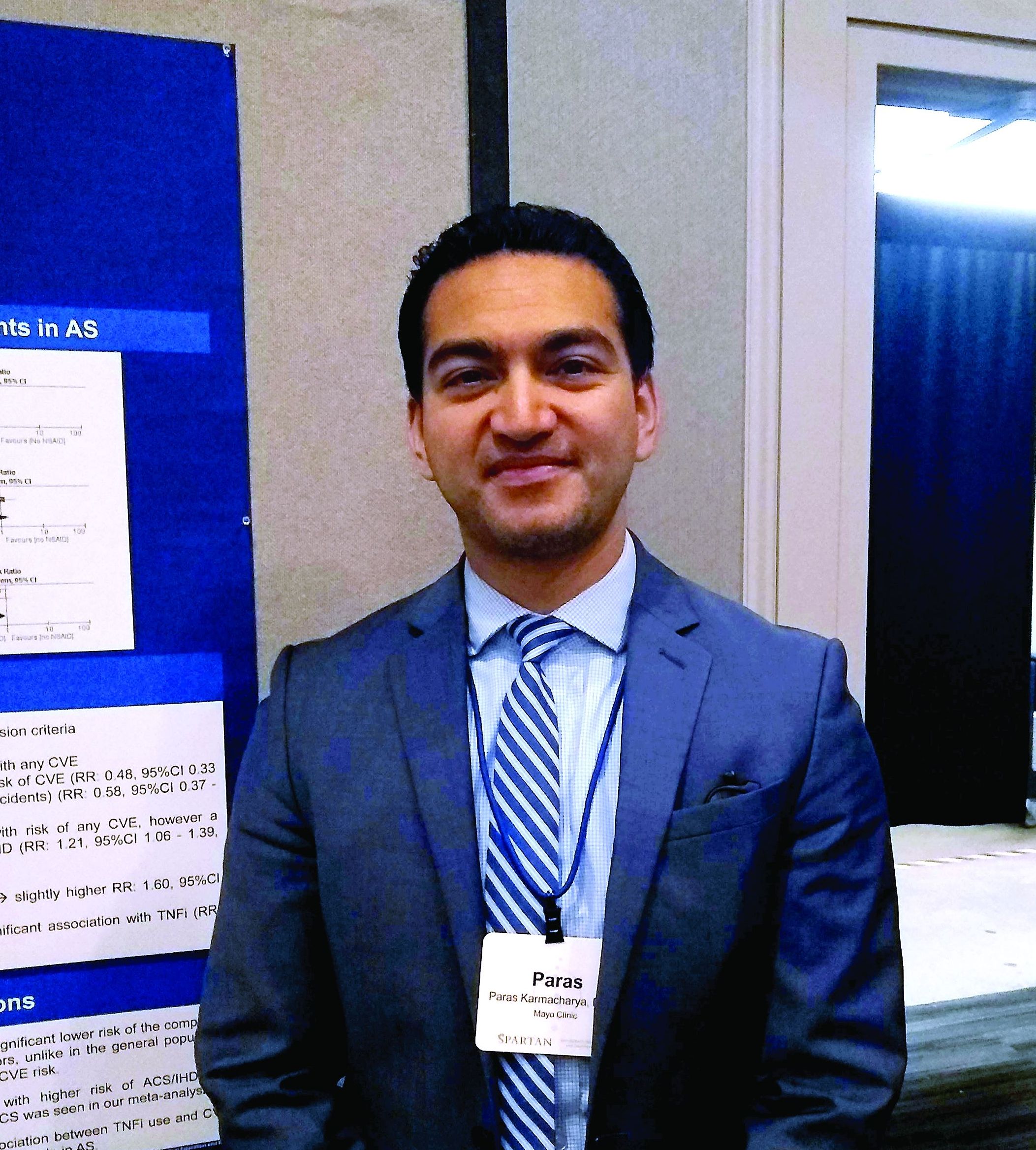
The reduced risk observed in this analysis (risk ratio, 0.48; 95% confidence interval, 0.33-0.70) contrasts with the increased risk for cardiovascular events seen with COX-2 inhibitor use in the general population, said Paras Karmacharya, MBBS, speaking in an interview at the annual meeting of the Spondyloarthritis Research and Treatment Network (SPARTAN). The overall effect was highly statistically significant (P = .0001), and the finding provides “reassuring” data for a population that’s known to have an elevated risk for cardiovascular events, he said.
“[W]e found in the subgroup analysis that COX-2 inhibitors were associated with a reduced risk of cardiovascular events as a whole,” an association also seen when looking just at ischemic stroke, Dr. Karmacharya said. “So that was sort of surprising; in the general population, there are some concerns about using COX-2 inhibitors.”
Looking at data for the nine studies that met criteria for inclusion in the meta-analysis, Dr. Karmacharya, a rheumatology fellow at the Mayo Clinic, Rochester, Minn., and his collaborators calculated risk ratios for a composite outcome of all cardiovascular events (CVE) for all individuals taking NSAIDs, compared with individuals with ankylosing spondylitis (AS) who were not taking NSAIDs. Here, they found a relative risk of 0.94 (95% CI, 0.50-1.75; P = .84).
Next, the investigators calculated a relative risk of 0.78 for the composite CVE outcome just for those taking nonselective NSAIDs (95% CI, 0.44-1.38; P = .40).
Along with NSAIDs, Dr. Karmacharya and his coauthors also looked at the relationship between tumor necrosis factor inhibitors (TNFIs) and cardiovascular events. They found a significantly increased risk for the composite endpoint among AS patients taking TNFIs (RR, 1.60; 95% CI, 1.05-2.41; P = .03), but the comparison was limited to only one study.
In their analysis, the investigators also broke out risk of acute coronary syndrome (ACS)/ischemic heart disease. “The only place where we found some increased risk was ACS and ischemic heart disease, and that was with nonselective NSAIDS,” Dr. Karmacharya said (RR, 1.21; 95% CI, 1.06-1.39; P = .005). No significant changes in relative risk for ACS and ischemic heart disease were seen for the total group of NSAID users, for the subgroups taking COX-2 inhibitors, or for those taking TNFIs.
Finally, the investigators found a relative risk of 0.58 for stroke among the full group of NSAID users and a relative risk of 0.59 for those taking COX-2 inhibitors, but no reduced risk for the subgroup taking nonselective NSAIDs (P = .02, .04, and .37, respectively).
“While NSAIDs are known to be associated with an increased risk of CVE in the general population, whether the anti-inflammatory effects of NSAIDs reduce or modify the CVE risk in AS is controversial,” Dr. Karmacharya and his collaborators wrote. In this context, the meta-analysis provides a useful perspective for rheumatologists who care for AS patients, Dr. Karmacharya said: “I think it’s important, because most of these patients are on NSAIDs long-term.”
However, all of the studies included in the meta-analysis were observational, with no randomized, controlled trials meeting inclusion criteria. Also, some analyses presented in the poster involved as few as two studies, so findings should be interpreted with caution, he added. “We don’t have a lot of studies included in the analysis. ... so we need more data for sure, but I think what data we have so far look reassuring.”
Dr. Karmacharya reported that he had no conflicts of interest, and reported no outside sources of funding.
MADISON, WISC. – Patients with ankylosing spondylitis had a small but significant reduction in risk for cardiovascular events if they were taking cyclooxygenase-2 (COX-2) inhibitors, according to a new systematic review and meta-analysis.
The reduced risk observed in this analysis (risk ratio, 0.48; 95% confidence interval, 0.33-0.70) contrasts with the increased risk for cardiovascular events seen with COX-2 inhibitor use in the general population, said Paras Karmacharya, MBBS, speaking in an interview at the annual meeting of the Spondyloarthritis Research and Treatment Network (SPARTAN). The overall effect was highly statistically significant (P = .0001), and the finding provides “reassuring” data for a population that’s known to have an elevated risk for cardiovascular events, he said.
“[W]e found in the subgroup analysis that COX-2 inhibitors were associated with a reduced risk of cardiovascular events as a whole,” an association also seen when looking just at ischemic stroke, Dr. Karmacharya said. “So that was sort of surprising; in the general population, there are some concerns about using COX-2 inhibitors.”
Looking at data for the nine studies that met criteria for inclusion in the meta-analysis, Dr. Karmacharya, a rheumatology fellow at the Mayo Clinic, Rochester, Minn., and his collaborators calculated risk ratios for a composite outcome of all cardiovascular events (CVE) for all individuals taking NSAIDs, compared with individuals with ankylosing spondylitis (AS) who were not taking NSAIDs. Here, they found a relative risk of 0.94 (95% CI, 0.50-1.75; P = .84).
Next, the investigators calculated a relative risk of 0.78 for the composite CVE outcome just for those taking nonselective NSAIDs (95% CI, 0.44-1.38; P = .40).
Along with NSAIDs, Dr. Karmacharya and his coauthors also looked at the relationship between tumor necrosis factor inhibitors (TNFIs) and cardiovascular events. They found a significantly increased risk for the composite endpoint among AS patients taking TNFIs (RR, 1.60; 95% CI, 1.05-2.41; P = .03), but the comparison was limited to only one study.
In their analysis, the investigators also broke out risk of acute coronary syndrome (ACS)/ischemic heart disease. “The only place where we found some increased risk was ACS and ischemic heart disease, and that was with nonselective NSAIDS,” Dr. Karmacharya said (RR, 1.21; 95% CI, 1.06-1.39; P = .005). No significant changes in relative risk for ACS and ischemic heart disease were seen for the total group of NSAID users, for the subgroups taking COX-2 inhibitors, or for those taking TNFIs.
Finally, the investigators found a relative risk of 0.58 for stroke among the full group of NSAID users and a relative risk of 0.59 for those taking COX-2 inhibitors, but no reduced risk for the subgroup taking nonselective NSAIDs (P = .02, .04, and .37, respectively).
“While NSAIDs are known to be associated with an increased risk of CVE in the general population, whether the anti-inflammatory effects of NSAIDs reduce or modify the CVE risk in AS is controversial,” Dr. Karmacharya and his collaborators wrote. In this context, the meta-analysis provides a useful perspective for rheumatologists who care for AS patients, Dr. Karmacharya said: “I think it’s important, because most of these patients are on NSAIDs long-term.”
However, all of the studies included in the meta-analysis were observational, with no randomized, controlled trials meeting inclusion criteria. Also, some analyses presented in the poster involved as few as two studies, so findings should be interpreted with caution, he added. “We don’t have a lot of studies included in the analysis. ... so we need more data for sure, but I think what data we have so far look reassuring.”
Dr. Karmacharya reported that he had no conflicts of interest, and reported no outside sources of funding.
REPORTING FROM SPARTAN 2019
Key clinical point: Individuals with ankylosing spondylitis (AS) who took cyclooxygenase 2 (COX-2) inhibitors had a reduced risk of cardiovascular events, compared with AS patients who were not using COX-2 inhibitors.
Major finding: Individuals with AS taking COX-2 inhibitors had a risk ratio of 0.48 for cardiovascular events (95% CI, 0.33-0.70; P = .001).
Study details: Systematic review and meta-analysis of nine observational studies that variably examined the association between NSAID use and tumor necrosis factor inhibitor use and cardiovascular events among individuals with AS.
Disclosures: The authors reported no conflicts of interest and no outside sources of funding.
Source: Karmacharya P. et al. SPARTAN 2019.
Ticagrelor doesn’t beat clopidogrel in postfibrinolysis STEMI
NEW ORLEANS – In STEMI patients who aren’t able to undergo primary PCI, ticagrelor after fibrinolytic therapy offered no advantages over clopidogrel – a less potent and less costly antiplatelet agent – in rates of cardiovascular events or bleeding though 12 months of follow-up in the TREAT trial.
“In terms of efficacy, it is appropriate to interpret TREAT statistically as a neutral trial,” Otavio Berwanger, MD, PhD, advised at the annual meeting of the American College of Cardiology.
TREAT (Ticagrelor in patients with ST-elevation myocardial infarction treated with pharmacological thrombolysis) was a 10-country, 152-site, randomized, open-label clinical trial of 3,799 STEMI (ST-elevation MI) patients treated with fibrinolytic therapy followed an average of 11 hours later by a loading dose of either ticagrelor (Brilinta) or clopidogrel, then 12 months of standard-dose maintenance therapy of their designated potent antiplatelet drug. The adherence rate was 90% at 12 months. Participating countries included Russia, China, Brazil, Australia, and Canada, but not the United States.
The primary efficacy endpoint was the 12-month composite of death from a vascular cause, MI, stroke, severe recurrent ischemia, TIA, or another arterial thrombotic event. The rate was 8% in the ticagrelor group and 9.1% with clopidogrel, a 12% relative risk reduction in favor of ticagrelor that was not statistically significant. But then, TREAT was underpowered to show a difference in efficacy. However, the 12% relative risk reduction mirrors that seen in the earlier PLATO trial of 18,624 patients with acute coronary syndrome who were randomized to ticagrelor or clopidogrel in conjunction with primary PCI, a difference that was statistically significant because of PLATO’s much larger size (N Engl J Med. 2009 Sep 10;361[11]:1045-57).
TREAT was sufficiently powered to assess safety. There was no significant between-group difference in TIMI major bleeding, the primary safety endpoint. However, the rate of total bleeding events was significantly higher in the ticagrelor arm, by a margin of 10.25% versus 6.15%. Moreover, the rate of TIMI clinically significant bleeding requiring medical attention was also higher in the ticagrelor group – 5.2% versus 3.8% – and the TIMI minimal bleeding rate of 5.85% in the ticagrelor group was more than double that in the clopidogrel arm, reported Dr. Berwanger of the Heart Hospital Research Institute in São Paolo.
These 12-month outcomes echo those previously reported at the 30-day mark in TREAT (JAMA Cardiol. 2018 May 1;3[5]:391-9).
Discussant C. Michael Gibson, MD, put TREAT in perspective: “Here we’re looking to see if there are differences between two thienopyridine inhibitors. There’s nothing really that important on the efficacy side, although there was 1.5% missingness in the study. And there was a higher number of total bleeds with ticagrelor.
“Some of the junior members of the audience may not be all that familiar with fibrinolysis. In the era where it was more prominent, reocclusion occurred in 5%-8% of patients. When it did occur, it led to a tripling of mortality. It’s important to note that the first study of a thienopyridine inhibitor added to lytics was CLARITY, almost 15 years ago, showing a reduction in death, MI, or reocclusion down from about 15% to 7% [N Engl J Med 2005; 352:1179-89]. So it should be very clear to the audience that reocclusion is a problem and the addition of a thienopyridine inhibitor improves that,” explained Dr. Gibson, professor of medicine at Harvard Medical School, Boston.
The TREAT trial was funded by AstraZeneca. Dr. Berwanger reported receiving research grants from and serving as a consultant to that company and half a dozen others.
Simultaneously with the presentation, the TREAT study was published online (J Am Coll Cardiol. 2019 Mar 12. doi: 10.1016/j.jacc.2019.03.011).
REPORTING FROM ACC 19
Despite failed primary endpoint, MI alert device has predictive value
WASHINGTON –Although an implantable device for detecting myocardial infarction missed the primary composite outcome endpoint in a controlled trial, a newly completed extended analysis associated the device with a higher positive predictive value and a lower false positive rate when compared to sham control, according to data presented at CRT 2019, sponsored by MedStar Heart & Vascular Institute.
“Among high risk patients, this system may be beneficial in the identification of both symptomatic and asymptomatic coronary events,” reported C. Michael Gibson, MD, chief of clinical research in the cardiology division at Beth Israel Deaconess Hospital, Boston.
The implantable device (AngelMed Guardian System), which received Food and Drug Administration approval in April 2018, is designed to identify MI through detection of ST-segment elevations in the absence of an elevated heart rate. When the system detects an event during continuous monitoring, it sends a signal (internal vibration and auditory signal to an external monitor) designed to tell the patient to seek medical care.
The previously published multicenter and randomized ALERTS (AngelMed for Early Recognition and Treatment of STEMI) trial that tested this device was negative for primary composite endpoint of cardiac or unexplained death, new Q-wave MI, or presentation at the emergency department (ED) more than 2 hours after symptom onset (J Am Coll Cardiol. 2019 Feb 25. pii: S0735-1097[19]30237-2). In that trial 907 patients were fitted with the device and then randomized to having the device switched on or left off.
At 7 days, a primary endpoint was reached by 3.8% of those in the device-on group versus 4.9% of those in the device-off group, which was not significantly different.
Although the primary endpoint was not met, there were promising results. For example, in those who did have an occlusive event, patients in the device-on group had better preserved left ventricular function when evaluated after the event, a result consistent with earlier presentation in the ED and earlier treatment. In fact, 85% of patients with an MI in the device-on group presented to a hospital within 2 hours, compared with just 5% of those in the device-off control group during the initial study period.
More evidence of a potential clinical role for the device has now been generated in a new extended analysis. This analysis was made possible because patients in both of the randomized groups continue to wear the device, including those in the device-off group who had their devices activated after 6 months. There are now 3 more years of data of follow-up from those initially in the device-on group and those switched from the device-off group.
“So we started the clock over with a new statistical analysis plan and new endpoints,” Dr. Gibson explained. The FDA was consulted in selecting endpoints, particularly regarding evidence that the device did not increase false-positive ED visits.
There were numerous encouraging findings. One was that 42 silent MIs, which would otherwise have been missed, were detected over the extended follow-up. Another was that the annualized false-positive rate was lower in those with an activated device (0.164/year) when compared to the original device-off group (0.678/year; P less than .001). Lastly, the positive predictive value of an alarm during the extended follow-up was higher than that of symptoms alone among the original device-off group (25.8% vs. 18.2%).
The device was found safe. The rate of system-related complications was under 4%, which Dr. Gibson said is noninferior to that associated with pacemakers.
One of the potential explanations for the failure of the device to achieve the primary endpoint in the original trial was an unexpectedly low event rate, according to Dr. Gibson.
Even before this extended analysis, the FDA had accepted the potential benefits of this device as demonstrated in the approval last year. In the labeling, the device is called “a more accurate predictor of acute coronary syndrome events when compared to patient recognized symptoms alone and demonstrates a reduced rate over time of patient presentations without ACS events.”
“About 50% of patients wait more than 3 hours after the onset of symptoms before reaching an emergency room,” observed Dr. Gibson. Emphasizing the evidence that delay is an important predictor of adverse outcomes, he suggested the alarm device might be useful in accelerating care in some high risk groups.
REPORTING FROM CRT 2019
30-day readmissions after STEMI with cardiogenic shock 13%
WASHINGTON – and remain in hospital for an average of 6 days, according to an analysis from the National Readmission Database presented at CRT 2019 sponsored by MedStar Heart & Vascular Institute.

“About one in four of the readmissions was for heart failure,” reported Karan Sud, MD, a cardiology resident at the Mount Sinai St. Luke’s West Hospital, New York.
Despite gains in acute survival among STEMI patients in cardiogenic shock, little attention has been paid to the risk of readmissions, according to Dr. Sud. According to data collected from the National Readmissions Database for 2010-2014, these rates are high enough to deserve attention, he said.
“Our goal is now to develop a scoring system based on our predictive model to identify patients at the index admission who are at risk for readmission,” Dr. Sud reported. On the basis of these predictors, it might be possible to implement strategies to optimize management and improve access to care.
In the years studied, there were 94,991 patients with STEMI and cardiogenic shock captured in the National Readmissions Database, of whom 43,205 survived and were followed for readmission. Of the 5,503 readmissions within 30 days, 12% were considered unplanned.
Half of the readmissions were for noncardiovascular causes, including sepsis, respiratory failure, and major bleeding. Of those related to cardiovascular disease, about half, or nearly 25% of the total, were for heart failure.
The predictors of readmission included female sex, age older than 75 years, average length of stay longer than 10 days, and more than three comorbidities, such as diabetes or chronic kidney disease, according to Dr. Sud.
“Those sent home from the index admission were more likely than those discharged to an extended care facility to be readmitted,” he added. He also noted that lower socioeconomic status was a risk factor for readmission, a phenomenon that he attributed to access issues regarding follow-up care.
“We are now conducting a prospective study to look at readmissions at 6 months,” reported Dr. Sud, who believes that efforts to understand the risk of readmission following STEMI complicated by cardiogenic shock might uncover opportunities for better management.
WASHINGTON – and remain in hospital for an average of 6 days, according to an analysis from the National Readmission Database presented at CRT 2019 sponsored by MedStar Heart & Vascular Institute.
“About one in four of the readmissions was for heart failure,” reported Karan Sud, MD, a cardiology resident at the Mount Sinai St. Luke’s West Hospital, New York.
Despite gains in acute survival among STEMI patients in cardiogenic shock, little attention has been paid to the risk of readmissions, according to Dr. Sud. According to data collected from the National Readmissions Database for 2010-2014, these rates are high enough to deserve attention, he said.
“Our goal is now to develop a scoring system based on our predictive model to identify patients at the index admission who are at risk for readmission,” Dr. Sud reported. On the basis of these predictors, it might be possible to implement strategies to optimize management and improve access to care.
In the years studied, there were 94,991 patients with STEMI and cardiogenic shock captured in the National Readmissions Database, of whom 43,205 survived and were followed for readmission. Of the 5,503 readmissions within 30 days, 12% were considered unplanned.
Half of the readmissions were for noncardiovascular causes, including sepsis, respiratory failure, and major bleeding. Of those related to cardiovascular disease, about half, or nearly 25% of the total, were for heart failure.
The predictors of readmission included female sex, age older than 75 years, average length of stay longer than 10 days, and more than three comorbidities, such as diabetes or chronic kidney disease, according to Dr. Sud.
“Those sent home from the index admission were more likely than those discharged to an extended care facility to be readmitted,” he added. He also noted that lower socioeconomic status was a risk factor for readmission, a phenomenon that he attributed to access issues regarding follow-up care.
“We are now conducting a prospective study to look at readmissions at 6 months,” reported Dr. Sud, who believes that efforts to understand the risk of readmission following STEMI complicated by cardiogenic shock might uncover opportunities for better management.
REPORTING FROM CRT 2019
More chest compression–only CPR leads to increased survival rates
according to a Swedish study of out-of-hospital cardiac arrests and subsequent CPR.

“These findings support continuous endorsement of chest compression–only CPR as an option in future CPR guidelines because it is associated with higher CPR rates and survival in out-of-hospital cardiac arrests,” wrote Gabriel Riva, MD, of the Karolinska Institutet, Stockholm, and his coauthors. The study was published in Circulation.
To determine changes in the rate and type of CPR performed before emergency medical services (EMS) arrival, the researchers compared all bystander-witnessed out-of-hospital cardiac arrests (OHCAs) reported in Sweden between 2000 and 2017. In all, 30,445 patients were included; the time periods compared were 2000-2005, 2006-2010, and 2011-2017. Patients were categorized as receiving either no CPR (NO-CPR), standard CPR (S-CPR), or chest compression–only CPR (CO-CPR). In 2005, CO-CPR was introduced in national CPR guidelines as an option for bystanders; in 2010, it was recommended for anyone untrained in CPR.
The proportion of patients who received CPR in general increased from 41% in 2000-2005 to 59% in 2006-2010 to 68% in 2011-2017. S-CPR changed from 35% to 45% to 38% over the three periods, while CO-CPR increased from 5% to 14% to 30%. In regard to 30-day survival rates, the S-CPR group saw an increase from 9% to 13% to 16% and the CO-CPR group increased from 8% to 12% to 14%, compared with 4% to 6% to 7% for the NO-CPR group.
The authors noted the limitations of their study, including the results being based on register data and therefore subject to misclassification and missing data. In addition, missing data negated any reporting on the neurological function of survivors; analyzing witnessed OHCAs only also meant the findings could not be validated for nonwitnessed OHCA.
The Swedish Heart and Lung Foundation funded the study. The authors made no disclosures.
SOURCE: Riva G et al. Circulation. 2019 Apr 1. doi: 10.1161/CIRCULATIONAHA.118.038179.
according to a Swedish study of out-of-hospital cardiac arrests and subsequent CPR.
“These findings support continuous endorsement of chest compression–only CPR as an option in future CPR guidelines because it is associated with higher CPR rates and survival in out-of-hospital cardiac arrests,” wrote Gabriel Riva, MD, of the Karolinska Institutet, Stockholm, and his coauthors. The study was published in Circulation.
To determine changes in the rate and type of CPR performed before emergency medical services (EMS) arrival, the researchers compared all bystander-witnessed out-of-hospital cardiac arrests (OHCAs) reported in Sweden between 2000 and 2017. In all, 30,445 patients were included; the time periods compared were 2000-2005, 2006-2010, and 2011-2017. Patients were categorized as receiving either no CPR (NO-CPR), standard CPR (S-CPR), or chest compression–only CPR (CO-CPR). In 2005, CO-CPR was introduced in national CPR guidelines as an option for bystanders; in 2010, it was recommended for anyone untrained in CPR.
The proportion of patients who received CPR in general increased from 41% in 2000-2005 to 59% in 2006-2010 to 68% in 2011-2017. S-CPR changed from 35% to 45% to 38% over the three periods, while CO-CPR increased from 5% to 14% to 30%. In regard to 30-day survival rates, the S-CPR group saw an increase from 9% to 13% to 16% and the CO-CPR group increased from 8% to 12% to 14%, compared with 4% to 6% to 7% for the NO-CPR group.
The authors noted the limitations of their study, including the results being based on register data and therefore subject to misclassification and missing data. In addition, missing data negated any reporting on the neurological function of survivors; analyzing witnessed OHCAs only also meant the findings could not be validated for nonwitnessed OHCA.
The Swedish Heart and Lung Foundation funded the study. The authors made no disclosures.
SOURCE: Riva G et al. Circulation. 2019 Apr 1. doi: 10.1161/CIRCULATIONAHA.118.038179.
FROM CIRCULATION
Key clinical point: Since chest compression-only CPR was introduced and recommended as an alternative for bystanders witnessing a cardiac arrest, CPR rates and survival rates have increased.
Major finding: From 2001-2005 to 2011-2017, 30-day survival rates increased from 9% to 16% for the standard CPR group and from 8% to 14% for the chest compression–only group, compared with 4%-7% for the no CPR group.
Study details: An observational nationwide cohort study of 30,445 Swedish patients who suffered out-of-hospital cardiac arrest.
Disclosures: The Swedish Heart and Lung Foundation funded the study. The authors made no disclosures.
Source: Riva G et al. Circulation. 2019 Apr 1. doi: 10.1161/CIRCULATIONAHA.118.038179.