User login
First Paycheck Equals Investment Decisions
Hospitalists setting out on their careers are used to life as struggling students. Once they start earning a sizeable salary, they’re hit with some tough choices: How fast should they pay off medical school loans? Can they afford to give in to the temptation of an expensive reward? How much savings do they really need?
“It is a bit of a shock to start your first job as an attending physician,” says Margaret C. Fang, MD, MPH, assistant adjunct professor of medicine at the University of California Hospital Medicine Group. “Your bank account seems to grow so much faster!” No matter how big that account may seem, it can dwindle away every month if you don’t practice good money management skills.
Sameer Badlani, MD, hospitalist and instructor at the University of Chicago, is faculty advisor to a medical student interest group at his university called Money and Medicine. “It’s all about delayed gratification,” he says of the effect a physician’s salary has on a new hospitalist. “I say, just wait one year in your new job to see what your expenses are before you buy that big house or that expensive convertible.”
Do Your Research
Residents and early career hospitalists—and anyone who is having trouble saving some salary—would do well to tackle the subject of money management as if it were a clinical course. “What you do with your money deserves a lot of attention,” Dr. Fang says. “Vigilance about finances is important, but many physicians are not as prepared to deal with money management as they are to care for patients.”
When you’re about to start a job, find out the financial options before you’re faced with a mountain of forms and a heap of decisions. “When I started here, I had all this paperwork: I had to sign up for health insurance, disability, long-term disability, 401(k) and 403(b) plans, and more. It’s really daunting,” Dr. Fang says. “A little upfront research is important, so you can make good decisions about these things.”
When it comes to figuring out how much to contribute to retirement accounts, savings accounts and investments, consider enlisting some outside help. “Many institutions that hire young hospitalists offer financial counseling,” Dr. Fang points out. “I’ve done a lot of independent reading. But if your finances are more challenging—say you’re carrying a lot of debt—it’s reasonable to work with a financial consultant.”
Deal with Debt
Before you start investing your money, take a look at your debt. What to do with it—for instance, should you pay off all of your loans and bills—will be one of the most important decisions a new hospitalist will make.
“The first thing you should do is clear any credit card debt, because it carries such high interest rates,” Dr. Badlani advises. “Do this before investing in a Roth IRA or anything.”
As for student loans, “I recommend taking your time paying off fixed APR loans and federally subsidized loans,” Dr. Badlani says “These typically carry low interest and they are tax deductible. You’re better off investing your money in indexed mutual funds because the stock market averages an 8% to 10% return (long-term), versus loans that are maybe 6% interest, or 4% when you calculate the tax deduction.”
However, if your student loan comes from a private bank, it may carry a higher interest rate and variable APRs. In this case, says Dr. Badlani, “you need to pay off those loans first” or look into consolidating or refinancing high-interest student loans.
Build Your Savings
Once you’ve cleared or reduced any high-interest debt, it’s time to focus on saving some money—no matter how much you’re making. “Residents say they have no money to save,” Dr. Badlani says. “But you need to look at your lifestyle and look for what is called ‘the latte factor.’ If you work hard, you may feel you deserve a treat, so you buy a $4 latte every day—maybe two a day. If you work 27 days a month, this adds up to $200 a month or nearly $3,000 a year. If you put that money in your Roth IRA every year of your residency and your fellowship, that adds up to a cool $150,000—based on 8% to 9% annual return, compounded annually for 30 years and adjusted for investment fees.”
—Sameer Badlani, MD
Starting a money market account or putting money in a certificate of deposit (CD) will keep it liquid. “You should have three to six months’ expenses, in case you lose your job or get sick,” Dr. Badlani says. “But be sure to put this ‘cushion money’ in a money market account, which these days are typically returning 3.5% APR versus regular checking accounts that offer a measly .25%, to offset inflation eating into your savings.”
Invest Early for Retirement
Residents and low-income hospitalists are in an excellent position to start investing for retirement. “While you’re a resident or intern, you most likely qualify for a Roth IRA,” Dr. Badlani explains. “This is the only time you’ll be able to invest in this. It’s a good choice for people with potential for increasing their income. Roth contributions are made after taxes and the account grows tax-free; you never have to pay tax on that money in retirement. For 2008, you’re eligible to contribute to a Roth IRA if you’re single or file as head of household with a modified adjusted gross income of below $114,000, or if you file jointly with income below $166,000.”
Another smart retirement investment for just about any hospitalist is employer-matched contributions. “If your employer will match your [retirement] contribution, that’s free money,” Dr. Badlani points out. “If you’re not taking advantage of that, you’re making a big mistake. Institutions will typically match up to about 5% of an employee’s contribution to a 401(k) or 403(b). Plus, by putting money in a retirement account, you’re reducing your tax burden.”
Online calculators can help you figure out how much to save—including matching funds—for a comfortable retirement. “The $1 million retirement has been the American dream for a long time, but that’s increased now to $1.5 to $1.8 million,” Dr. Badlani says. “But for a comfortable lifestyle and accounting for spiraling healthcare costs, I would recommend aiming for $5 million. That takes a lot of discipline over a long stretch of time.” He recommends the online calculator at www.dinkytown.net, which shows that a 29-year-old earning an annual income of $150,000 can retire at 65 with $5,868,264—if he or she contributes 15% to a 403(b) retirement account with a 5% employer match. “But you have to stick to this every month for the next 36 years,” Dr. Badlani warns. “That takes discipline.”
Regardless of how old you are or how much income you currently have, it’s wise to start practicing smart habits with spending, saving and investing your money. As Dr. Badlani says, “Good money habits last forever.” TH
Jane Jerrard also writes “Public Policy” for The Hospitalist.
Alternative Medications
Complementary and alterative medicine (CAM) is defined as any product, including herbal remedies/foods/teas, vitamins, minerals, and natural products, that can be purchased without a prescription at a health food store, supermarket, from a magazine/newspaper or online, for self-treatment.1
Taylor et al. evaluated CAM perceptions of emergency department patients in Australia. They determined 44% of patients felt that by using CAM they were “drug free,” with 29% of patients agreeing (or strongly agreeing) CAM use is always safe to take with prescription medications. In an earlier study, Eisenberg et al. evaluated CAM use perceptions in the United States and found 79% of patients felt that, combined with prescription drugs, CAM was superior to either modality alone.2 They also found 63% to 72% of CAM-using patients that had seen a medical doctor in the prior year did not disclose the therapy.
The two most common reasons cited by patients were “they felt it wasn’t important for the doctor to know (61%)” and “the doctor didn’t ask (60%).” Overall, national CAM-use surveys have revealed that about 80% of adults typically do not disclose CAM use to medical doctors.
It is, therefore, imperative that physicians ask patients about their CAM use. It also is important to remember there are more foods and beverages that contain some of these “natural” ingredients, and patients need to be queried about the use of these products. CAM products can complicate traditional patient management, either when used alone or in combination with prescription drugs.
A clinically significant drug interaction that bears its own warning is that of warfarin and chondroitin/glucosamine. Patients might not tell you that they are taking chondroitin/glucosamine, so you have to ask. Functional foods and beverages that include “natural” ingredients, such as glucosamine and chondroitin, and other CAM abound. Examples include Joint Juice (1,500 mg glucosamine), Vitamin Water, and others.3
Two case reports, and a report from the World Health Organization (WHO) Collaborating Center for International Drug Monitoring, and the MedWatch database point to a potentially serious drug interaction between glucosamine and warfarin.4-7 Although not FDA approved for joint supplementation, the usual daily dose should generally not exceed 1.5 grams of glucosamine and 1.2 grams of chondroitin. Chondroitin may have anti-coagulant activity, which would explain the increase in International Normalized Ratio (INR) seen in patients using it in combination with warfarin. The WHO database identified at least 34 cases of concomitant use, with most cases of increased INR resolving upon glucosamine discontinuation. Nine cases required physician intervention, and in one case a positive rechallenge was documented. In June 2007, there were 81 cases of a possible interaction from the MedWatch database, of these, 61 cases had potential alternate etiologies. Of the 20 possibly-related cases, five led to patient hospitalization due to bleeding complications; the median patient age was 62 years and there were no deaths reported.
Ramsay et al. reported from a United Kingdom survey that 92% of patients admitted to taking herbal medicines while receiving warfarin, noting that significant numbers of patients are in need of close monitoring. They also note that other CAM can interact with warfarin to increase bleeding time or act as anti-platelet agents (e.g., high dose vitamin E [> 400 IU], fish oils, garlic, St. John’s Wort, etc.) They particularly note that all patients receiving warfarin or that will be commencing warfarin be specifically asked about their use of CAM. The mechanism for this interaction still is unclear, but may be due to the inhibition of warfarin metabolism by the CYP2C9 or CYP3A4 enzymes, or a pharmacodynamic interaction between glucosamine and warfarin.
It is critical all patients, particularly those taking warfarin, have a full drug history at all hospitalizations. Make sure you inquire about vitamins and other supplements/CAM. If someone else is doing the intake medication history, make sure they get this information. A nurse might not think to ask. TH
Michele B. Kaufman, PharmD, BSc, RPh, is a registered pharmacist based in New York City.
References
1. Taylor DM, Walsham N, Taylor SE, Wong LF. Complementary and alternative medicines versus prescription drugs: perceptions of emergency department patients. Emerg Med J. 2006;23:266-268.
2. Eisenberg DM, Kessler RC, Van Rompay MI, et al.. Perceptions about complementary therapies relative to conventional therapies among adults how use both: results from a national survey. Ann Intern Med. 2001;135:344-351.
3. What is Joint Juice? Joint Juice, Inc. www.jointjuice.com/jointjuice.php. Published 1997. Accessed August 1, 2008.
4. Rozenfeld V, Crain JL, Callahan AK. Possible augmentation of warfarin effect by glucosamine-chondroitin. Am J Health-Syst Pharm. 2004;61:306-307.
5. Knudsen JF, Sokol GH. Potential glucosamine-warfarin interaction resulting in increased international normalized ratio: case report and review of the literature and medwatch database. Pharmacother. 2008;28(4):540-548.
6. Ramsay NA, Kenny MW, Davies G, Patel JP. Complimentary and alternative medicine use among patients starting warfarin. Br J Haematology. 2005;130:777-780.
7. Yue Q-Y, Strandell J, Myrberg O. Concomitant use of glucosamine may potentiate the effect of warfarin. The Uppsalla Monitoring Centre Web site. www.who-umc.org/graphics/9722.pdf; Accessed August 1, 2008.
In the Literature
Literature at a Glance
- Postoperative prophylactic LMWH should be considered for arthroscopic knee surgery patients.
- Individualized therapy is required for DVT prophylaxis in the neurosurgical patient.
- SMART-COP is a reasonable screening tool for ICU admission.
- Mediterranean and low-carb diets are safe and effective alternatives to low-fat diets.
- Admissions for ACS in both smokers and non-smokers decrease after implementation of smoking ban.
- Beta-blockers should be continued whenever possible in patients hospitalized for heart failure with LVSD.
- Non-invasive ventilation improved dyspnea, but did not improve short-term mortality rates in patients with acute cardiogenic pulmonary edema.
- COX-2 inhibitors should be used with caution in patients with increased cerebrovascular disease risk.
LMWH after Arthroscopic Knee Surgery May Prevent VTE Compared to Graduated Compression Stockings
Clinical question: Does low molecular weight heparin (LMWH) prevent venous thromboembolism (VTE) compared to compression stockings without increasing bleeding complications in arthroscopic knee surgery?
Background: Knee arthroscopy is a common orthopedic surgery and postoperative venous thromboprophylaxis is not routinely recommended.
Study design: Randomized, controlled trial with blinding of the investigators.
Setting: Single orthopedic clinic in Italy, with followup at a university hospital.
Synopsis: 1,761 consecutive patients undergoing knee arthroscopy were randomly assigned to full-length graduated compression stockings (CS) for seven days postoperatively, subcutaneous LMWH (nadoparin 3800 units daily) for seven or 14 days postoperatively. The primary outcome of asymptomatic proximal deep venous thrombosis (DVT), symptomatic VTE, and all-cause mortality within three months of surgery was higher with CS (3.2%) than with LMWH for seven or 14 days (0.9% in each group) (P=0.005). There was no significant difference in bleeding events between groups.
The study was underpowered to detect differences in bleeding risk. Furthermore, almost half the events making up the primary outcome were distal DVTs of uncertain clinical significance. Notably, the 14-day LMWH group was discontinued early because of unspecified safety concerns related to longer exposure to LMWH.
Bottom line: Postoperative prophylactic LMWH for seven days may prevent some thromboses after knee surgery and should be considered in higher-risk patients.
Citation: Camporese G, Bernardi E, Prandoni P, et al. Low-molecular-weight heparin versus compression stockings for thrombophylaxis after knee arthroscopy. Ann Intern Med. 2008;14(9):73-82.
Heparins and Compression Devices are Effective in Preventing VTE in a Mixed Neurosurgical Population
Clinical question: What is the efficacy and safety of LMWH, unfractionated heparin, and mechanical devices in preventing VTE in neurosurgical patients?
Background: Neurosurgical patients are at high risk for VTE, but concerns remain regarding the risk of bleeding complications with the use of LMWH or unfractionated heparin (UFH).
Study design: Meta-analysis of 18 randomized trials and 12 cohort studies.
Setting: Patients undergoing spinal surgery or craniotomy.
Synopsis: Among all patients, the pooled DVT rate was 15.5/100. Use of sequential compression devices (SCD) significantly reduced the risk of DVT compared with placebo (relative risk [RR] 0.41, 95% confidence interval [CI] 0.21-0.78). Subcutaneous LMWH was associated with a significantly reduced risk of DVT compared with CS (RR 0.60, 95% CI 0.44-0.81). No other head-to-head comparisons were associated with significant reductions in VTE risk. After adjusting for potential risk factors for DVT and study design, use of heparins or SCDs was associated with a lower risk of DVT. Intracranial hemorrhage (ICH), minor bleeding, major bleeding, or death was not statistically different between any of the groups, although, after adjustment, LMWH was associated with a slightly increased risk of ICH.
The quality of included studies varied considerably and inter-rater agreement on study quality was low, raising the possibility of study selection bias. Potential publication bias was not addressed. Bleeding complications were rare, so the estimates of risk may be imprecise.
Bottom line: Individualized therapy is required for DVT prophylaxis in the neurosurgical patient; SCDs reduce VTE risk and both pharmacologic and mechanical prophylaxis may be indicated in patients with increased VTE risk.
Citation: Collen JF, Jackson JL, Shorr AF, Moores LK. Prevention of venous thromboembolism in neurosurgery: A metaanalysis. Chest. 2008;13(4):237-249.
SMART-COP Predicts Need for ICU Care in CAP
Clinical question: Can a clinical tool predict the need for critical care in community acquired pneumonia (CAP)?
Background: Clinical tools predicting 30-day mortality in community acquired pneumonia (CAP) exist, but do not accurately identify who will require intensive care unit-level care, such as intensive respiratory or vasosuppressor support (IRVS).
Study design: Prospective multi-center observational study.
Setting: Six hospitals in Australia participating in the Australian Community Acquired Pneumonia Study (ACAPS).
Synopsis: Multivariate analysis of a dataset of 882 episodes of CAP identified eight factors that were associated with the need for IRVS, summarized by the mnemonic “SMART-COP” (Systolic blood pressure, Multilobar chest radiography involvement, low Albumin level, high Respiratory rate, Tachycardia, Confusion, poor Oxygenation, and low arterial pH). Assigning one point for five factors and two points for three factors (systolic blood pressure, poor oxygenation, and low arterial pH) a SMART-COP score >3 identified 92.3% (95% CI 84.8-96.9%) of patients who required IRVS, including 84% who did not initially require ICU care. Specificity was 62.3% (CI 58.8-65.7%). Test characteristics for predicting IRVS were superior to existing prediction rules (PSI and CURB-65).
Most patients were drawn from large, urban teaching hospitals in Australia, so the results may not be generalizable. The authors also presented a modification of SMART-COP, using pulse oximetry rather than blood gas results; this may be even more useful in the pre-hospital setting.
Bottom line: SMART-COP is a reasonable screening tool for predicting need for ICU-level care in patients admitted with CAP.
Citation: Charles PGP, Wolfe, R, Whitby, M, et. al. SMART-COP: a tool for predicting the need for intensive respiratory or vasopressor support in community-acquired pneumonia. Clin Infect Dis. 2008;47(3):375-384.
Mediterranean and Low-Carbohydrate Diets are Effective for Weight Loss
Clinical question: Are dietary intervention with low-fat, Mediterranean or low-carbohydrate diets effective?
Background: Obesity is a growing, worldwide problem. Past trials comparing the effectiveness and safety of various dietary interventions have been limited by short follow up and high dropout rates.
Study design: Prospective randomized trial.
Setting: Employees of a research center in Israel.
Synopsis: 322 subjects (average BMI 31) were randomized to a low-fat/restricted-calorie, Mediterranean/ restricted-calorie, or a low-carbohydrate/non-restricted calorie diet. Diet adherence was 84.6% at two years and all groups lost significant amounts of weight. The Mediterranean and low-carbohydrate diets showed similar aver∆age weight loss of 4.4 kg and 4.7 kg, respectively. The low-fat diet group on average lost 2.9 kg. Diabetic patients had improved glycemic control and lower insulin levels with the Mediterranean diet. Subjects assigned to the low-carbohydrate diet had the greatest improvement in lipid profile (20% relative decrease of total cholesterol to HDL ratio).
The trial took place at a single site (a scientific research center in Israel) and included only 14% women, so its generalization is uncertain. The study was based on self-reported dietary intake and may be subject to reporting bias.
Bottom line: Mediterranean and low-carbohydrate diets are safe and effective alternatives to low-fat diets with favorable effects on glycemic control in diabetics and lipid metabolism, respectively.
Citation: Shai I, Schwarz-fuchs D, Henkin Y, et al. Weight loss with a low-carbohydrate, Mediterranean, or low-fat diet. NEJM. 2008;359(3):229-241.
Admissions for Acute Coronary Syndrome Decreased after Implementation of Smoke-free Legislation
Clinical question: Is there a reduction in admissions for acute coronary syndrome (ACS) after enactment of smoke-free legislation?
Background: Multiple, small, retrospective studies have shown a decrease in ACS after implementation of smoke-free legislation.
Study design: Prospective observational multi-center cohort study.
Setting: Nine hospitals in Scotland.
Synopsis: Data was collected on all patients admitted with ACS 10 months before and after implementation of smoke-free legislation, which prohibited smoking in all enclosed public and work places in Scotland. After the smoking ban, the number of ACS admissions fell by 17% (95% CI 16-18) in Scotland as a whole, compared with a 4% reduction in England during the same period (England does not have similar smoke-free legislation). Among smokers, former smokers and non-smokers, the number of ACS admissions decreased by 14% (95% CI 12-16), 19% (95% CI 17-21), and 21% (95% CI 18-24), respectively. Among non-smokers, self-reported exposure to second-hand smoke decreased significantly; these reductions were confirmed by measured reductions in serum cotinine levels, even among those who never smoked.
Results were limited by the observational nature of the study, although the authors did attempt to carefully match comparison cohorts by season and geography. Also, secular trends other than legislation may have reduced prevalent smoking in Scotland during the study period.
Bottom line: Admissions for ACS for both smokers and non-smokers decreased after implementation of smoke-free legislation.
Citation: Pell JP, Haw S, Cobbe S, et al. Smoke-free legislation and hospitalizations for acute coronary syndrome. NEJM. 2008;359(5):482-491.
Continuation of Beta-blockers in Patients Hospitalized for Heart Failure Improves Mortality
Clinical question: Does the withdrawal or continuation of beta-blockers in patients hospitalized with decompensated heart failure have any effect on clinical outcomes?
Background: Previous clinical trials have demonstrated mortality benefit with the use of beta-blockers in patients with symptomatic chronic heart failure and left ventricular systolic dysfunction (LVSD), however, controversy exists whether to continue these medications in acute decompensated heart failure.
Study design: Prospective cohort analysis from the OPTIMIZE-HF registry (Organized Program to Initiate Lifesaving Treatment in Hospitalized Patients with Heart Failure).
Setting: 91 academic and community hospitals in the United States.
Synopsis: Data was analyzed on 2,373 registry patients with documented LVSD (EF <40) eligible for beta-blocker therapy. During hospitalization, 1,350 patients were continued on beta-blockers, 79 had therapy withdrawn, 303 were not started, and 632 had beta-blockade initiated. Compared with no beta-blocker treatment, adjusted hazard ratio (HR) for death at 60 and 90 days following discharge was lower in patients who were continued on beta-blockade (HR 0.60, 95% CI 0.37–0.99). Compared with continuation of beta-blockade, withdrawal of beta-blockade increased the risk of death (HR 2.3, 95% CI 1.2–4.6).
Results were limited by the observational nature of the study and short follow up. The reason for discontinuation or not starting beta-blockade was not captured in the database, so it is possible sicker patients had beta-blockers discontinued during hospitalization (although the authors attempted to control for this).
Bottom line: Beta-blockers should be continued whenever possible in patients hospitalized for heart failure with LVSD.
Citation: Fonarow GC, Abraham WT, Albert NM, et al. Influence of beta-blocker continuation or withdrawal on outcomes in patients hospitalized with heart failure. J Am Coll Cardiol. 2008;52(3):190-199.
Non-invasive Ventilation Does Not Improve Short-term Mortality in Acute Cardiogenic Pulmonary Edema
Clinical question: Does non-invasive ventilation reduce mortality in patients with acute cardiogenic pulmonary edema and are there differences in outcome between use of continuous positive airway pressure (CPAP) or non-invasive positive pressure ventilation (NIPPV)?
Background: In patients with acute cardiogenic pulmonary edema, noninvasive ventilation improves physiologic variables and symptoms, decreases rates of invasive ventilation, and may improve mortality.
Study design: Randomized multi center controlled trial.
Setting: 26 district and regional hospitals in the United Kingdom.
Synopsis: 1,156 patients admitted with acute cardiogenic pulmonary edema between July 2003 and April 2007 were randomized to standard oxygen therapy, versus CPAP or NIPPV. There were no significant differences in seven- or 30-day mortality rates between the standard oxygen therapy versus noninvasive ventilation. Mortality at seven days was 9.8% in the standard oxygen group versus 9.5% in the noninvasive ventilation group (P=0.87); 30-day mortality was 16% in the standard oxygen group and 15% in the non-invasive ventilation group (P=0.64). There were no major differences in treatment outcome with NIPPV compared to CPAP.
Although mortality was not decreased, non-invasive ventilation did improve dyspnea and tachycardia within one hour of therapy.
Bottom line: In patients admitted with acute cardiogenic pulmonary edema, noninvasive ventilation improved dyspnea and some physiological parameters, but did not improve short-term mortality rates.
Citation: Gray A, Goodacre S, Newby D, Masson M, Sampson F, Nicholl J. Noninvasive ventilation in acute cardiogenic pulmonary edema. NEJM. 2008;359(2):142-151.
Cyclooxygenase 2 Inhibitors May Increase the Risk of Ischemic Stroke
Clinical question: Do NSAIDs and COX-2 inhibitors increase the risk of ischemic or hemorrhagic stroke?
Background: Selected cyclooxygenase 2 (COX-2) inhibitors have been shown to increase cardiovascular morbidity in a dose-dependent manner and are now used with caution in patients at risk for cardiovascular disease. Little is known about the safety of these medications and non-aspirin, non-steroidal anti-inflammatory drugs (NSAIDS) in those at risk for cerebrovascular disease.
Study design: Retrospective observational cohort study.
Setting: Tennessee Medicaid Program enrollees.
Synopsis: Data was collected from the medical records of 336,906 subjects. Non-users had a baseline stroke rate of 4.51 strokes/1000 person-years. The rate increased to 5.15/1,000 person-years and 5.95/1,000 person-years for rofecoxib and valdecoxib, respectively. Celecoxib and other NSAIDs did not significantly increase the risk of stroke. Analysis of new users of rofexocib and valdecoxib yielded a similarly increased risk of stroke. Most strokes were ischemic.
Limitations include the ready availability of NSAIDs raising the possibility that some patients classified as non-users were actually users of NSAIDs. Other potential confounders may not have been measured and, therefore, not available for analysis.
Bottom line: COX-2 inhibitors should be used with caution in patients with increased cerebrovascular disease risk.
Citation: Roumie CL, Mitchel EF, Kaltenback L, Arbogast PG, Gideon P Griffen MR. Nonaspirin NSAIDs, cyclooxygenase 2 inhibitors, and the risk for stroke. Stroke. 2008;39:1037-2045.
3 ... 2 ... 1 ... Moving Day

SHM is growing, changing, evolving and advancing. If you have been a member or been engaged with the society for the past few years, this isn’t news to you. Our membership is growing; the products, publications and services we offer are expanding; attendance at our annual meeting is increasing; and we are continuing to create new and valuable online resources. These are tangible signs of growth that many of you see and touch on a regular basis. On a day-to-day basis, I see the same things, but because I work for SHM, I have the opportunity to see the growth and change from within the organization.
When I signed on with SHM more than three years ago, I walked through the door and into a small office, approximately 3,000 square feet in size with about 13 full-time staff members. Since then, we have grown steadily, consistently adding new faces to the SHM team and expanding into new places by breaking through a wall into an adjacent space. Flash forward to the present day. Between April and July 2008, SHM has added 13 new faces to the staff. At the end of September 2008, we broke ground on construction of our new corporate headquarters, a 16,000-square-foot office in downtown Philadelphia.
Since its inception 12 years ago, SHM has called 190 Independence Mall home, but just as the hospital medicine movement has grown, so has SHM and the staff supporting the society. This winter, SHM will be moving our corporate headquarters to the new facility at 1500 Spring Garden. The process to find our new headquarters has been an extensive one. We began the search for a new office approximately one year ago, and as I am writing this, final construction documents have been sent to a list of general contractors.
During the past six months, SHM has been working with projects managers, architects, engineers, and consultants to take our new office from a “blank slate” to a finished and fully operational office before the end of 2008. As you read this article, construction on the new headquarters is fully underway. Workers are putting up drywall, running cables, laying carpet, and installing equipment that will be the supporting foundation for the staff and society for the next decade.
So, by now you are probably asking, “What does this mean to me? I don’t see these people on a daily basis, and I don’t work at SHM headquarters.” At a very basic level it means SHM will have a new address and new phone numbers. Your letters, applications, registrations, and anything addressed to SHM will be routed to our new home. Additionally, as part of our move, SHM will implement a new phone system. Our toll-free, 1-800 number will remain the same, however, all of the people who work for SHM will have new office phone numbers.
It is important you know how to reach SHM in our new home, but even more important is to know that this move is a significant milestone in the evolution of the society and the next step in providing you, our members, with ever-improving and enhanced levels of service and support. In creating a new facility, we are further equipping staff with the tools they need to serve you, creating technical capacities to meet current and future needs, and setting a stage for SHM’s continued growth in support of the growing hospital medicine movement.
During the weeks and months ahead, the SHM team will be preparing for the launch of the new One Day Hospitalist University, opening of the new Fellowship in Hospital Medicine and[Add Another New Program Here. In addition to all of these new and exciting initiatives, we will be organizing files, packing boxes and preparing for our move. As we transition to new desks, new phones, new commutes and a new environment, we would like to take a moment to thank you for your support and understanding while we take another significant step in the history of the Society of Hospital Medicine.
Behind the Scenes
Change is in the air
By Geri Barnes

It’s autumn and there is a bite to the air. Every year around this time, I vacillate between being depressed about the pending winter and energized by the change of season. This year, I definitely am excited and energized.
As weather is one of those environmental dynamics that impacts daily life, so do changes in the healthcare arena impact on SHM and its life. We’ve seen “never events” come into being, an expansion of CMS’ Hospitals Compare, and an increasing focus on pay-for-performance. All of these factors are designed to improve patient care, particularly care of the hospitalized patient. SHM staff needs to be ready to support the hospital medicine community.


SHM long has been focused on defining and providing hospitalists with the education and resources needed for every day practice, as well as for imple- menting cutting-edge quality improvement interventions. To support these focus areas, our staff members were organized in one department, Education and Quality Initiatives. During the last year, we decided our efforts would be better served by creating two departments: Education and Meetings and Quality Initiatives. Last summer, we hired two new staff members to lead the department and move the quality efforts forward. Jane Kelly-Cummings, RN, CPHQ, senior director, Quality Initiatives, has more than 20 years of experience in clinical practice, quality improvement, patient safety, healthcare informatics and quality improvement education. Linda Boclair, MT (ASCP), MEd, MBA, brings to SHM 25 years of management in the healthcare industry and serves as the Quality Initiatives Department director. You will be hearing more about the Quality Initiatives Department in the near future.
I am heading up the newly organized Education and Meetings Department. I am joined by Erica Pearson, director, Meetings; Theresa Jones, education project manager; Meghan Pitzer, meetings coordinator; and Carolyn Brennan, director, Research Program Development. We are charged with managing SHM’s Education Enterprise, which includes meetings and all other educational activities that support our members.
For meetings, we focus on leading our volunteers in the development of relevant program and educational content, ensuring we meet the requirements for continuing medical education (CME) programs. We design and implement meeting logistics with a common goal: the attendees leave the meeting feeling nothing could have been better organized. The Education and Meetings staff has focused their energies on the following meetings:
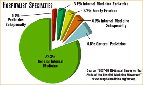
- The cornerstone of our meetings is the SHM annual meeting. Hospital Medicine 2009 will take place May 14-17, 2009, in Chicago at the Hyatt Regency. The planning of the program and logistics began in March 2008, and the organizational effort will continue through the end of the meeting. This comprehensive program includes annual meeting education sessions over the course of two and a half days and another full day of seven concurrent pre-courses.
- An important educational event is SHM’s Leadership Academy. Established in 2005, the Level I Academy has been presented semi-annually, with the eighth event taking place in Los Angeles this past September. Based on a need for the next level of leadership skills, Level II started in 2006 and recently presented for the third time. All events have basically sold out, and their popularity continues to grow.
- SHM instituted the One-Day Hospitalist University (ODHU) series this year, presenting four of our best pre-courses on a regional basis. The goal is to present ODHU in four different locations during the course of the year. The first ODHU takes place this month in Baltimore; the next is Feb. 3-4 in Atlanta.
- Pediatric Hospital Medicine 2009 was held in July in Denver. As the lead sponsor, SHM organized this successful conference, which was co-sponsored by the American Pediatric Association and the American Academy of Pediatrics.
- Expert Training Sessions is a new series of educational events that provide the opportunity to learn quality improvement strategies for glycemic control, VTE prevention, or transitions of care directly from an expert and interact on a personal basis. Presented in Boston and Nashville and planned for St. Louis, this initiative already is proving successful and we are hoping to expand in the near future.
The other major focus area for the Education and Meetings Department lies in meeting the educational needs of the hospital medicine community. Staff, working with the Education Committee, are exploring new and exciting ways to identify needs and define strategies to deliver relevant programming. The efforts, which will lead to a comprehensive education plan that will drive the activities the next few years, are focused on the following:
- Life-long learning has become the standard for physicians in general and hospitalists in particular. SHM is in the early stages of identifying and developing resources that will be readily accessible on the SHM Web site, such as a hospital medicine reading list on clinical and healthcare-systems topics based on the Core Competencies.
- The Education Committee is exploring the possibility of developing an evidence-based medicine (EBM) primer, which can be used to practice and teach EBM. It will be designed for the practicing hospitalist in a community hospital setting and will define how to research, read, and use EBM journal articles.
- SHM is exploring the use of Web 2.0 to continually assess needs, deliver educational programs, and communicate with members and faculty.
- The needs of academic hospitalists are unique and SHM is dedicated to support this important segment of our constituency. Joining with the Society of General Internal Medicine (SGIM), SHM is planning an Academic Boot Camp that will focus on education skills, research, mentoring, and career pathways.
- SHM is developing a comprehensive communication and education program to become the main resource for hospitalists as they engage in Maintenance of Certification.
So, the welcome winds of change blow, bringing the energy and organization needed to accomplish our education and quality goals. We are confident our internal changes will result in moving our agenda forward in ways previously only imagined.
Volunteer Search
Interested in being a part of an SHM Committee or Task Force? Now is your chance! Nominations are open for SHM Committees and Task Forces. This is your opportunity to shape the future of SHM and the hospital medicine movement.
To nominate yourself, visit www.hospitalmedicine.org and click on “About SHM,” then click on “Committees.” Here, you will see a full list of committees, as well as task forces and current members. For each committee you would like to serve on, please submit your name and a one- to two-paragraph statement about why you are qualified and interested. E-mail this information to Joi Seabrooks at [email protected] by Dec. 5. Appointments will be made in February, take affect in May and last one year. TH
SHM is growing, changing, evolving and advancing. If you have been a member or been engaged with the society for the past few years, this isn’t news to you. Our membership is growing; the products, publications and services we offer are expanding; attendance at our annual meeting is increasing; and we are continuing to create new and valuable online resources. These are tangible signs of growth that many of you see and touch on a regular basis. On a day-to-day basis, I see the same things, but because I work for SHM, I have the opportunity to see the growth and change from within the organization.
When I signed on with SHM more than three years ago, I walked through the door and into a small office, approximately 3,000 square feet in size with about 13 full-time staff members. Since then, we have grown steadily, consistently adding new faces to the SHM team and expanding into new places by breaking through a wall into an adjacent space. Flash forward to the present day. Between April and July 2008, SHM has added 13 new faces to the staff. At the end of September 2008, we broke ground on construction of our new corporate headquarters, a 16,000-square-foot office in downtown Philadelphia.
Since its inception 12 years ago, SHM has called 190 Independence Mall home, but just as the hospital medicine movement has grown, so has SHM and the staff supporting the society. This winter, SHM will be moving our corporate headquarters to the new facility at 1500 Spring Garden. The process to find our new headquarters has been an extensive one. We began the search for a new office approximately one year ago, and as I am writing this, final construction documents have been sent to a list of general contractors.
During the past six months, SHM has been working with projects managers, architects, engineers, and consultants to take our new office from a “blank slate” to a finished and fully operational office before the end of 2008. As you read this article, construction on the new headquarters is fully underway. Workers are putting up drywall, running cables, laying carpet, and installing equipment that will be the supporting foundation for the staff and society for the next decade.
So, by now you are probably asking, “What does this mean to me? I don’t see these people on a daily basis, and I don’t work at SHM headquarters.” At a very basic level it means SHM will have a new address and new phone numbers. Your letters, applications, registrations, and anything addressed to SHM will be routed to our new home. Additionally, as part of our move, SHM will implement a new phone system. Our toll-free, 1-800 number will remain the same, however, all of the people who work for SHM will have new office phone numbers.
It is important you know how to reach SHM in our new home, but even more important is to know that this move is a significant milestone in the evolution of the society and the next step in providing you, our members, with ever-improving and enhanced levels of service and support. In creating a new facility, we are further equipping staff with the tools they need to serve you, creating technical capacities to meet current and future needs, and setting a stage for SHM’s continued growth in support of the growing hospital medicine movement.
During the weeks and months ahead, the SHM team will be preparing for the launch of the new One Day Hospitalist University, opening of the new Fellowship in Hospital Medicine and[Add Another New Program Here. In addition to all of these new and exciting initiatives, we will be organizing files, packing boxes and preparing for our move. As we transition to new desks, new phones, new commutes and a new environment, we would like to take a moment to thank you for your support and understanding while we take another significant step in the history of the Society of Hospital Medicine.
Behind the Scenes
Change is in the air
By Geri Barnes
It’s autumn and there is a bite to the air. Every year around this time, I vacillate between being depressed about the pending winter and energized by the change of season. This year, I definitely am excited and energized.
As weather is one of those environmental dynamics that impacts daily life, so do changes in the healthcare arena impact on SHM and its life. We’ve seen “never events” come into being, an expansion of CMS’ Hospitals Compare, and an increasing focus on pay-for-performance. All of these factors are designed to improve patient care, particularly care of the hospitalized patient. SHM staff needs to be ready to support the hospital medicine community.
SHM long has been focused on defining and providing hospitalists with the education and resources needed for every day practice, as well as for imple- menting cutting-edge quality improvement interventions. To support these focus areas, our staff members were organized in one department, Education and Quality Initiatives. During the last year, we decided our efforts would be better served by creating two departments: Education and Meetings and Quality Initiatives. Last summer, we hired two new staff members to lead the department and move the quality efforts forward. Jane Kelly-Cummings, RN, CPHQ, senior director, Quality Initiatives, has more than 20 years of experience in clinical practice, quality improvement, patient safety, healthcare informatics and quality improvement education. Linda Boclair, MT (ASCP), MEd, MBA, brings to SHM 25 years of management in the healthcare industry and serves as the Quality Initiatives Department director. You will be hearing more about the Quality Initiatives Department in the near future.
I am heading up the newly organized Education and Meetings Department. I am joined by Erica Pearson, director, Meetings; Theresa Jones, education project manager; Meghan Pitzer, meetings coordinator; and Carolyn Brennan, director, Research Program Development. We are charged with managing SHM’s Education Enterprise, which includes meetings and all other educational activities that support our members.
For meetings, we focus on leading our volunteers in the development of relevant program and educational content, ensuring we meet the requirements for continuing medical education (CME) programs. We design and implement meeting logistics with a common goal: the attendees leave the meeting feeling nothing could have been better organized. The Education and Meetings staff has focused their energies on the following meetings:
- The cornerstone of our meetings is the SHM annual meeting. Hospital Medicine 2009 will take place May 14-17, 2009, in Chicago at the Hyatt Regency. The planning of the program and logistics began in March 2008, and the organizational effort will continue through the end of the meeting. This comprehensive program includes annual meeting education sessions over the course of two and a half days and another full day of seven concurrent pre-courses.
- An important educational event is SHM’s Leadership Academy. Established in 2005, the Level I Academy has been presented semi-annually, with the eighth event taking place in Los Angeles this past September. Based on a need for the next level of leadership skills, Level II started in 2006 and recently presented for the third time. All events have basically sold out, and their popularity continues to grow.
- SHM instituted the One-Day Hospitalist University (ODHU) series this year, presenting four of our best pre-courses on a regional basis. The goal is to present ODHU in four different locations during the course of the year. The first ODHU takes place this month in Baltimore; the next is Feb. 3-4 in Atlanta.
- Pediatric Hospital Medicine 2009 was held in July in Denver. As the lead sponsor, SHM organized this successful conference, which was co-sponsored by the American Pediatric Association and the American Academy of Pediatrics.
- Expert Training Sessions is a new series of educational events that provide the opportunity to learn quality improvement strategies for glycemic control, VTE prevention, or transitions of care directly from an expert and interact on a personal basis. Presented in Boston and Nashville and planned for St. Louis, this initiative already is proving successful and we are hoping to expand in the near future.
The other major focus area for the Education and Meetings Department lies in meeting the educational needs of the hospital medicine community. Staff, working with the Education Committee, are exploring new and exciting ways to identify needs and define strategies to deliver relevant programming. The efforts, which will lead to a comprehensive education plan that will drive the activities the next few years, are focused on the following:
- Life-long learning has become the standard for physicians in general and hospitalists in particular. SHM is in the early stages of identifying and developing resources that will be readily accessible on the SHM Web site, such as a hospital medicine reading list on clinical and healthcare-systems topics based on the Core Competencies.
- The Education Committee is exploring the possibility of developing an evidence-based medicine (EBM) primer, which can be used to practice and teach EBM. It will be designed for the practicing hospitalist in a community hospital setting and will define how to research, read, and use EBM journal articles.
- SHM is exploring the use of Web 2.0 to continually assess needs, deliver educational programs, and communicate with members and faculty.
- The needs of academic hospitalists are unique and SHM is dedicated to support this important segment of our constituency. Joining with the Society of General Internal Medicine (SGIM), SHM is planning an Academic Boot Camp that will focus on education skills, research, mentoring, and career pathways.
- SHM is developing a comprehensive communication and education program to become the main resource for hospitalists as they engage in Maintenance of Certification.
So, the welcome winds of change blow, bringing the energy and organization needed to accomplish our education and quality goals. We are confident our internal changes will result in moving our agenda forward in ways previously only imagined.
Volunteer Search
Interested in being a part of an SHM Committee or Task Force? Now is your chance! Nominations are open for SHM Committees and Task Forces. This is your opportunity to shape the future of SHM and the hospital medicine movement.
To nominate yourself, visit www.hospitalmedicine.org and click on “About SHM,” then click on “Committees.” Here, you will see a full list of committees, as well as task forces and current members. For each committee you would like to serve on, please submit your name and a one- to two-paragraph statement about why you are qualified and interested. E-mail this information to Joi Seabrooks at [email protected] by Dec. 5. Appointments will be made in February, take affect in May and last one year. TH
Changing of the Guard

Starting a new job—any new job—brings with it trepidations. What’s the boss going to be like? Is there a strict dress code? Am I in over my head? When is payday? Did I make the right choice in taking it?
The nervous energy can be overwhelming. Only the strong survive the first day of human resources training, the endless line of personal questions from new co-workers, and the information technology grunts explaining your computer will be ready in a few days.
Fortunately, I made it through the first day, the second day, and the first week as the new editor of The Hospitalist. Whew! With newbie formalities out of the way, my attention is focused on settling into a routine and learning how best to lead the editorial charge for this news magazine. To that end, I want to say I’m excited to be on board!
Let’s start with a little background about me: I’ve been a writer, copy editor, and section editor for nearly two decades. The majority of my professional experience has been in the newspaper industry where I’ve worked in news, sports, and business departments at papers big and small.
To clear this up right away: I am not a doctor and—admittedly—not an expert in hospital medicine. In fact, except for the births of my two sons, I have spent very little time in or around hospitals the past 20 years. I do know, however, hospital medicine is a growing field, and I am thrilled to jump on this train as it leaves the station.
Some of you may believe a solid knowledge of medicine is a pre-requisite to edit a hospital medicine-focused magazine. For me, though, news is news. An editor may not understand every nuance of the subject matter, but he sure knows a good story when he sees one. My expertise is in developing a story, from concept through research and writing, all the way to presentation and publication. I hope my lack of medical background, in some ways, actually will allow me to bring a fresh set of eyes and editing to the content.
All of this said, my best asset in this new role is my editorial partner: Physician Editor Jeffrey Glasheen, MD. As a practicing hospitalist and leader of a hospital medicine group, Dr. Glasheen is on the front lines of hospital medicine, possesses a strong knowledge of all things SHM, and deals directly with the issues facing all of you dear readers. His passion for his role as physician editor inspires me. No doubt I will lean on him as I learn the ins and outs of hospital medicine.
Others I will look to for guidance include Editorial Director Lisa Dionne here at Wiley-Blackwell; the staff at SHM; and members of Team Hospitalist. Finally, I look to you for ideas and feedback. Who better to inform The Hospitalist pages? This magazine has a narrow focus (hospital medicine), but its readership is diverse and the trends are plentiful.
Because this is the November issue of an election year, I would like to commit to a non-partisan platform of fairness in reporting. I also believe in accurate, straightforward writing, and will maintain high standards at all levels when editing the magazine.
Now, for my first big announcement as editor: In addition to receiving the monthly magazine, you now receive our recently launched weekly electronic publication: the TH eWire. In the eWire, Associate Editor Stephanie Cajigal and I take the “pulse” of hospital medicine and offer up critical news and information we think you will value.
Once again, I am thrilled to be here and eager to make contact with as many hospitalists as possible in the coming months. I encourage you to share your opinions, offer up a story idea, or impart constructive criticism about anything and everything you see in The Hospitalist.
Regardless of the reason, I look forward to hearing from you. TH
Jason Carris is editor of The Hospitalist magazine. Send questions and comments to [email protected].
HM Group Economics 101
If ignorance is bliss, then a substantial slice of hospital medicine group (HMG) leaders are in heaven—about finance, that is. Perhaps they didn’t become physicians to solve the sub-prime mortgage mess or take their companies public, as Dr. Adam Singer, MD, CEO of IPC: The Hospitalist Company, did in January 2008.
Dr. Singer, recently named 2008 Physician Entrepreneur of the Year by Modern Physician, always has insisted HMGs can be financially self-supporting. In 2002, he wrote in Physician Executive “when revenue (fees divided by number of patient encounters) exceeds expenses, the practice becomes economically viable.” He calculated that factoring in malpractice insurance, general expenses, 10 percent of revenue for billing and collection, two patient encounters a day per full-time equivalent employee was an HMGs’ break-even point. “Even at a ridiculously minimum encounter volume of two, a hospitalist practice is viable without any outside subsidy,” he concluded. To drive home his point, Dr. Singer suggested HMGs augment their revenues by serving nursing homes and rehab facilities, teaching, securing research grants, medical directorships, and taking on contracts to manage ER unassigned patients.
Nevertheless, a recent Society of Hospital Medicine survey shows a number of HMG leaders lack the basic financial acumen to get and keep their practices fiscally healthy. Data from SHM’s 2007-2008 Bi-Annual Survey on the State of the Hospital Medicine Movement reveals the vast majority of HMGs fall short of economic independence. Survey results show many HMG leaders’ grasp of basic finance leaves room for improvement.
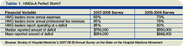
Key findings include:
- 37% of HMG leaders didn’t know their annual expenses;
- 35% didn’t know their annual professional fee revenues; and
- Among those that didn’t know their expenses and revenues, 85% were operating at a deficit.
Additionally, a negative correlation exists between HMG financial performance and compensation. HMG production remained flat, while average hospitalist compensation increased 13% to $193,000 annually. Cushiony subsidies persist; the survey showed 91% of HMGs receive some form of financial support, an average of $954,000 per group or $97,375 for each full-time physician.
Drilling down into the survey’s financial data, analysis by Joe Miller, SHM Executive Advisor to the CEO, points to an ominous monetary “perfect storm” brewing for HMG leaders. Deficits are growing as hospitalist leaders’ knowledge about their groups’ finances are declining.
The trend data also suggest subsidies obscure declining financial support in several areas. Compared to the 2005-06 SHM survey, the latest survey shows the percentage of HMGs receiving financial support dropped in all five categories in which HMGs receive subsidies. Results from the two most recent surveys showed hospital financial support dropped from 60% to 51%; other support from 17% to 12%; income guarantees fell from 72% to 61%; services in-kind declined from 71% to 60%; and case rates slid from 12% to 11%.
Fuzzy Math?
An oft-cited rationale for hospital medicine’s generous subsidies from other stakeholders is that it “creates value,” in terms of reducing hospital length of stay, costs per case and improved outcomes. Throughout the years, such savings have received increased scrutiny. Blogger and SHM member Dr. Robert Wachter, MD, professor and associated chairman of the Department of Medicine at the University of California, San Francisco, said in May “research on these metrics is disappointing. … The move toward hospitalist shift work and the resulting fragmentation of care during hospitalization may be eroding any slim advantage the model had in cost savings.” He concluded, “the bubble will burst the morning CEOs wake up and realize that there is no longer a shortage of hospitalists.”
Burke Kealey, MD, assistant medical director, Hospital Medicine Division of HealthPartners Medical Group in St. Paul, Minn., offers powerful reasons for why hospitalists struggle with Finance 101:
- Hospitalists have little incentive to meet financial performance targets;
- HMG leaders are too busy with recruiting, scheduling and retention to focus on finance;
- Difficulties obtaining useful data from hospital administrators; and
- Too few business models that centralize data from multiple hospitals, making financial analysis of an individual group difficult.
To address the needs, Dr. Kealey, who chairs SHM’s Practice Analysis Committee, has spearheaded SHM’s development of dashboards that utilize performance metrics, such as volume data, case mix, length of stay, resource utilization, productivity, and readmission rates, to help leaders guide a practice to economic health.
Steve Liu, MD, the CEO and founder of Ingenious Med, is a hospitalist in one of the nation’s largest HMGs, with 100,000-plus patient encounters annually at Emory Healthcare in Atlanta. A member of SHM’s Benchmarks Committee, Liu puts HMG leaders’ financial ignorance in the context of a growth industry. “Hospital medicine is young, it’s moving very fast, and it is filled with a youngish crowd who don’t feel they have enough time to focus on financial deficits and inefficiencies,” Dr. Liu says.
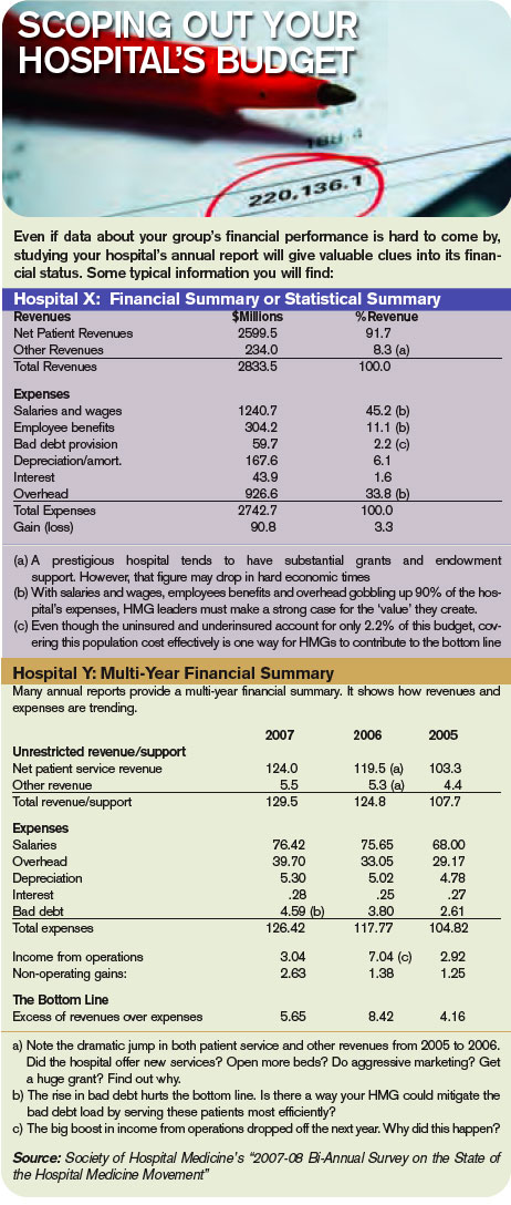
Still, a lack of financial knowledge can trip up the best of physician. Dr. Liu advises hospitalist leaders to focus on professional fee revenues, particularly on fees collected per full-time equivalent employee (FTE); eliminating high expenses from inefficiencies; and checking local and national compensation packages to address what he estimates as 30 percent of HMGs who fail to pay physicians appropriately. Other financial mistakes, he says, are HMG leaders throwing warm bodies at problems without enough back-office support; agreeing to see more patients than they can handle in a day; overspending on recruiters; and failure to understand each full-time employee’s return on investment.
Numerous hospitalists, according to Dr. Liu, believe they don’t have enough time to build partnerships with administrators that would lead to more sharing of financial data. “They may even be in a power struggle with administrators over access to financial data. … If you can’t measure it, you can’t manage it. That’s the essence of Finance 101,” Dr. Liu says, “and it’s very frustrating to hospitalists who don’t have the business skill sets and tools to succeed financially.” Since data is power, it’s imperative to ask the chief financial officer (CFO) for data; that way the HMG can work toward profitability.
Making sense of financial data is critical, says Leslie Flores, director of SHM’s Practice Management Institute. “HMG leaders have to learn to take data, such as number of encounters, relative value units, average daily census, number of days worked per year, the total subsidy for the practice, and average subsidy per hospitalist, and make sense of it. Where do the numbers come from and what do they mean are critical questions,” she says.
Big Picture
SHM’s most recent survey reflects the average hospitalist medicine group’s business model. Production is flat; compensation is up 13%; and 91% of HMGs receive subsidies nearing $1 million each. From a dollars-and-sense vantage point, the business case for hospital medicine doesn’t work. So why do hospitals go along with a broken business model, especially at a time when a hospital’s cost curve is growing faster than its revenue curve, making it even more difficult to justify HMG subsidies?
The short answer is that most hospital medicine programs are invited in by hospital administrators and serve a specific purpose related to overall revenue generation and/or cost controls. Common reasons for creating hospitalist programs are improving Emergency Department throughput, relieving community-based physicians from hospital duties, boosting subspecialist revenues with medical co-management, and cost cutting through reduced length of stay and improved resource utilization. Each has different financial metrics associated with it, and HMG leaders should understand each metric thoroughly.
If, for example, relieving the pressure on community-based doctors is the HMG’s primary goal, an HMG leader’s strategy might be to attract the best hospitalists available, offering a high compensation package and recruiting bonus. That would help cover the increased patient load, but it might pressure the hospital to shoulder a steep subsidy.
Alternatively, relying on financial and performance metrics might yield better rules about when to justify a new hire. Seasoned HMG leaders would rely on a group’s average daily census of 50 to 70 patients as the tipping point for adding a FTE hospitalist. Similarly, night admissions of 10 to 14 patients should trigger the hiring of a nocturnist, with his or her premium pay.
Brian Bossard, MD, founder of Inpatient Physicians Associates in Lincoln, Neb., relies on that type of data and other “lean” management concepts. To optimize each physicians’ patient loads and productivity, he has patients assigned by physical proximity, so his hospitalists won’t waste time running around the hospital seeing patients. Saving time and manpower contributes to an HMG’s financial health.
Once HMGs satisfy the primary service goals set by their hospitals, leaders should focus on enhancing their hospitals’ revenue generation from various sources. The typical mix of hospital revenue streams are, in size order—clinical services, research, philanthropic grants, interest income on cash assets, and royalties from intellectual property. Clinical services and research account for about 95% of the average hospital revenues, with the balance split among the other three.
Hospitals try to boost revenue by adding product lines, enhancing the value of current products, increasing market share, and capitalizing on production efficiencies. Translating business concepts from widgets to wards, increasing both volume and acuity of patient encounters, should be commonplace and a revenue booster. Research revenues are another story, particularly with slowdowns in National Institutes of Health funding. Recruiting hospitalists who want to do research leaves hospitals covering their salaries and labs for long periods of time without revenue generation.
Focusing on proven revenue generators—enhancing the value of current products, increasing market share, and boosting production efficiencies—can help HMG leaders carve out a niche that truly creates value for the hospital.
It may take a lot for an HMG leader to build solid financial performance on a strong clinical foundation, however, it is doable. Dr. Liu sums up how HMG finances will improve: “As hospital medicine matures, expect its leaders to mature as well,” he says. “They will become more business savvy and learn to speak the language of business, even if they have to force themselves to learn.” TH
If ignorance is bliss, then a substantial slice of hospital medicine group (HMG) leaders are in heaven—about finance, that is. Perhaps they didn’t become physicians to solve the sub-prime mortgage mess or take their companies public, as Dr. Adam Singer, MD, CEO of IPC: The Hospitalist Company, did in January 2008.
Dr. Singer, recently named 2008 Physician Entrepreneur of the Year by Modern Physician, always has insisted HMGs can be financially self-supporting. In 2002, he wrote in Physician Executive “when revenue (fees divided by number of patient encounters) exceeds expenses, the practice becomes economically viable.” He calculated that factoring in malpractice insurance, general expenses, 10 percent of revenue for billing and collection, two patient encounters a day per full-time equivalent employee was an HMGs’ break-even point. “Even at a ridiculously minimum encounter volume of two, a hospitalist practice is viable without any outside subsidy,” he concluded. To drive home his point, Dr. Singer suggested HMGs augment their revenues by serving nursing homes and rehab facilities, teaching, securing research grants, medical directorships, and taking on contracts to manage ER unassigned patients.
Nevertheless, a recent Society of Hospital Medicine survey shows a number of HMG leaders lack the basic financial acumen to get and keep their practices fiscally healthy. Data from SHM’s 2007-2008 Bi-Annual Survey on the State of the Hospital Medicine Movement reveals the vast majority of HMGs fall short of economic independence. Survey results show many HMG leaders’ grasp of basic finance leaves room for improvement.
Key findings include:
- 37% of HMG leaders didn’t know their annual expenses;
- 35% didn’t know their annual professional fee revenues; and
- Among those that didn’t know their expenses and revenues, 85% were operating at a deficit.
Additionally, a negative correlation exists between HMG financial performance and compensation. HMG production remained flat, while average hospitalist compensation increased 13% to $193,000 annually. Cushiony subsidies persist; the survey showed 91% of HMGs receive some form of financial support, an average of $954,000 per group or $97,375 for each full-time physician.
Drilling down into the survey’s financial data, analysis by Joe Miller, SHM Executive Advisor to the CEO, points to an ominous monetary “perfect storm” brewing for HMG leaders. Deficits are growing as hospitalist leaders’ knowledge about their groups’ finances are declining.
The trend data also suggest subsidies obscure declining financial support in several areas. Compared to the 2005-06 SHM survey, the latest survey shows the percentage of HMGs receiving financial support dropped in all five categories in which HMGs receive subsidies. Results from the two most recent surveys showed hospital financial support dropped from 60% to 51%; other support from 17% to 12%; income guarantees fell from 72% to 61%; services in-kind declined from 71% to 60%; and case rates slid from 12% to 11%.
Fuzzy Math?
An oft-cited rationale for hospital medicine’s generous subsidies from other stakeholders is that it “creates value,” in terms of reducing hospital length of stay, costs per case and improved outcomes. Throughout the years, such savings have received increased scrutiny. Blogger and SHM member Dr. Robert Wachter, MD, professor and associated chairman of the Department of Medicine at the University of California, San Francisco, said in May “research on these metrics is disappointing. … The move toward hospitalist shift work and the resulting fragmentation of care during hospitalization may be eroding any slim advantage the model had in cost savings.” He concluded, “the bubble will burst the morning CEOs wake up and realize that there is no longer a shortage of hospitalists.”
Burke Kealey, MD, assistant medical director, Hospital Medicine Division of HealthPartners Medical Group in St. Paul, Minn., offers powerful reasons for why hospitalists struggle with Finance 101:
- Hospitalists have little incentive to meet financial performance targets;
- HMG leaders are too busy with recruiting, scheduling and retention to focus on finance;
- Difficulties obtaining useful data from hospital administrators; and
- Too few business models that centralize data from multiple hospitals, making financial analysis of an individual group difficult.
To address the needs, Dr. Kealey, who chairs SHM’s Practice Analysis Committee, has spearheaded SHM’s development of dashboards that utilize performance metrics, such as volume data, case mix, length of stay, resource utilization, productivity, and readmission rates, to help leaders guide a practice to economic health.
Steve Liu, MD, the CEO and founder of Ingenious Med, is a hospitalist in one of the nation’s largest HMGs, with 100,000-plus patient encounters annually at Emory Healthcare in Atlanta. A member of SHM’s Benchmarks Committee, Liu puts HMG leaders’ financial ignorance in the context of a growth industry. “Hospital medicine is young, it’s moving very fast, and it is filled with a youngish crowd who don’t feel they have enough time to focus on financial deficits and inefficiencies,” Dr. Liu says.
Still, a lack of financial knowledge can trip up the best of physician. Dr. Liu advises hospitalist leaders to focus on professional fee revenues, particularly on fees collected per full-time equivalent employee (FTE); eliminating high expenses from inefficiencies; and checking local and national compensation packages to address what he estimates as 30 percent of HMGs who fail to pay physicians appropriately. Other financial mistakes, he says, are HMG leaders throwing warm bodies at problems without enough back-office support; agreeing to see more patients than they can handle in a day; overspending on recruiters; and failure to understand each full-time employee’s return on investment.
Numerous hospitalists, according to Dr. Liu, believe they don’t have enough time to build partnerships with administrators that would lead to more sharing of financial data. “They may even be in a power struggle with administrators over access to financial data. … If you can’t measure it, you can’t manage it. That’s the essence of Finance 101,” Dr. Liu says, “and it’s very frustrating to hospitalists who don’t have the business skill sets and tools to succeed financially.” Since data is power, it’s imperative to ask the chief financial officer (CFO) for data; that way the HMG can work toward profitability.
Making sense of financial data is critical, says Leslie Flores, director of SHM’s Practice Management Institute. “HMG leaders have to learn to take data, such as number of encounters, relative value units, average daily census, number of days worked per year, the total subsidy for the practice, and average subsidy per hospitalist, and make sense of it. Where do the numbers come from and what do they mean are critical questions,” she says.
Big Picture
SHM’s most recent survey reflects the average hospitalist medicine group’s business model. Production is flat; compensation is up 13%; and 91% of HMGs receive subsidies nearing $1 million each. From a dollars-and-sense vantage point, the business case for hospital medicine doesn’t work. So why do hospitals go along with a broken business model, especially at a time when a hospital’s cost curve is growing faster than its revenue curve, making it even more difficult to justify HMG subsidies?
The short answer is that most hospital medicine programs are invited in by hospital administrators and serve a specific purpose related to overall revenue generation and/or cost controls. Common reasons for creating hospitalist programs are improving Emergency Department throughput, relieving community-based physicians from hospital duties, boosting subspecialist revenues with medical co-management, and cost cutting through reduced length of stay and improved resource utilization. Each has different financial metrics associated with it, and HMG leaders should understand each metric thoroughly.
If, for example, relieving the pressure on community-based doctors is the HMG’s primary goal, an HMG leader’s strategy might be to attract the best hospitalists available, offering a high compensation package and recruiting bonus. That would help cover the increased patient load, but it might pressure the hospital to shoulder a steep subsidy.
Alternatively, relying on financial and performance metrics might yield better rules about when to justify a new hire. Seasoned HMG leaders would rely on a group’s average daily census of 50 to 70 patients as the tipping point for adding a FTE hospitalist. Similarly, night admissions of 10 to 14 patients should trigger the hiring of a nocturnist, with his or her premium pay.
Brian Bossard, MD, founder of Inpatient Physicians Associates in Lincoln, Neb., relies on that type of data and other “lean” management concepts. To optimize each physicians’ patient loads and productivity, he has patients assigned by physical proximity, so his hospitalists won’t waste time running around the hospital seeing patients. Saving time and manpower contributes to an HMG’s financial health.
Once HMGs satisfy the primary service goals set by their hospitals, leaders should focus on enhancing their hospitals’ revenue generation from various sources. The typical mix of hospital revenue streams are, in size order—clinical services, research, philanthropic grants, interest income on cash assets, and royalties from intellectual property. Clinical services and research account for about 95% of the average hospital revenues, with the balance split among the other three.
Hospitals try to boost revenue by adding product lines, enhancing the value of current products, increasing market share, and capitalizing on production efficiencies. Translating business concepts from widgets to wards, increasing both volume and acuity of patient encounters, should be commonplace and a revenue booster. Research revenues are another story, particularly with slowdowns in National Institutes of Health funding. Recruiting hospitalists who want to do research leaves hospitals covering their salaries and labs for long periods of time without revenue generation.
Focusing on proven revenue generators—enhancing the value of current products, increasing market share, and boosting production efficiencies—can help HMG leaders carve out a niche that truly creates value for the hospital.
It may take a lot for an HMG leader to build solid financial performance on a strong clinical foundation, however, it is doable. Dr. Liu sums up how HMG finances will improve: “As hospital medicine matures, expect its leaders to mature as well,” he says. “They will become more business savvy and learn to speak the language of business, even if they have to force themselves to learn.” TH
Navigate the Winds of Change
When a Catholic group moved to buy Lutheran Medical Center in Wheat Ridge, Colo., just outside of Denver, hospitalist Steven Krebs, MD, had strong objections. Sisters of Charity of Leavenworth Health System already was part-owner of the 400-bed medical center. As the sole owner, it would require the hospital to follow Catholic rules regarding end-of-life care and reproductive health, meaning some medical services would be prohibited.
“It’s really the last hospital before you go into the mountains. There’s no real hospital facility until Vail, almost 100 miles up the road,” Dr. Krebs says. Patients who wanted a tubal ligation, an emergency contraceptive, or other medical services typically not offered in a Catholic hospital would have to travel to receive them—or not receive them at all.
After negotiations failed to produce a satisfactory outcome, Dr. Krebs took the drastic step of becoming part of a lawsuit to stop the sale. In May 2008, Colorado’s governor signed into law a bill that allows the state attorney general to review how the sale of a nonprofit hospital affects patient care. If he believes care will be affected, the attorney general may ask for more information from the sponsors of the transaction or require a public hearing be held before determining whether to approve the transaction.
The sale is pending.
—Steven Krebs, MD, Lutheran Medical Center, Wheat Ridge, Colo.
In an ideal world, patients would get the same excellent care, no matter who owns or runs a hospital. A sale or a change in executive leadership wouldn’t alter the tone of a hospital. As the Colorado case shows, ownership and leadership matter.
Hospitalists who have been through a hospital sale or a change of leadership say it’s possible to influence the process from within, to benefit the hospital, the patients, and the hospitalists themselves, often through simple negotiation and clear communication.
Become a Resource, Partner for Leadership
Established hospitalist programs are in an especially strong bargaining position. “We have a track record we can point to,” says Brian Bossard, MD, director of Inpatient Physician Associates, whose medical center, BryanLGH in Lincoln, Neb., went through a search for a new CEO in early 2008. That track record includes a strong relationship between the hospitalist program and hospital administrators.
When the medical center considered formalizing its hospitalist program in 2002, for example, Dr. Bossard went with administrators to regional and national meetings. “Instead of having competing perspectives on what the costs of the program should be and what the value equation is, we came from a similar perspective,” he explains. “Since that time, that trust has been maintained by coming through on promises, whether it’s being able to manage the volumes or get good results.”
A strong foundation can lead to a well-integrated hospitalist program and positive relationships with the CEO and COO, Dr. Bossard adds. Though he considers the change in CEOs at BryanLGH “a little unsettling,” he says he’s confident his hospitalist program has the support of the hospital community. Perhaps as evidence of that, the hospital hiring committee considered input from hospitalists before making a final selection.
Dr. Bossard plans to present to the new CEO the hospitalist program’s accomplishments, goals, and potential challenges early on, so the CEO can get to know the program. “The [hospitalists] really should try to position themselves, in my view anyway, as sort of an insider with useful information, a leader they can go to to ask what’s really going on in the hospital,” Dr. Bossard says. “Hospitalist programs will grow so rapidly within hospital systems, taking care of 50% to 75%, to sometimes close to 100% of patients. They’re really great sources of information.”
Keep Lines of Communication Open
Strong relationships with hospital leadership creates a stable environment where hospitalists feel valued. It also helps ensure a program can weather almost any storm, says Julia Wright, MD, University of Wisconsin Hospital and Clinics hospitalist director and the director of hospital medicine at UW School of Medicine and Public Health in Madison, Wis. “Once you get to the point where you have value, a mission, a system of operations, and a delivery-of-care plan, then that might translate into some personal ownership in the program that could withstand a change in administration,” she says.
Dr. Wright, who took a five-person hospitalist program and has enlarged it to 13, says she understands the value of communication with hospital executives. She also has a game plan, should executive leadership at her hospital change. “The first thing I would do is sit down and discuss what my mission is,” she says. “Continued dialogue after that is really important.” The dialogue would include meeting with hospital administration regularly, as well as talking about objectives and ways to meet them. “I just know what’s worked to keep our program on track,” she says, “and it’s been very successful.”
Leverage a Change in Administration
At Meriter Hospital in Madison, Wis., Cate Ranheim, MD, director of the hospitalist program, found a change in administration actually benefitted her hospitalists. “Our previous administration was essentially a chief operating officer (COO) who approved—or more often—refused, every request for even the simplest things, from sinks to filing cabinets to call rooms,” she explains. “The former CEO was here for 30-plus years, as was the COO, and was virtually invisible within the institution.”
The COO was a strong advocate for the hospitalist group, but Dr. Ranheim says she still had to go through tough negotiations to get what the group needed. When a new CEO came in and announced an open-door policy, Dr. Ranheim jumped at the opportunity to be heard. “Whenever I need something for the group, I go directly to him, and I have never been refused anything because he knows I am not game-playing or negotiating,” she says. “If I say the group needs something, it really does.”
Dr. Ranheim’s experience actually is fairly common. Executive leadership or ownership changes often create “a window of opportunity to further some agendas,” Dr. Wright points out. “If there’s a group that’s been saying, ‘We really want to start this new initiative and just haven’t been able to get there yet,’ this might be the time to do that.”
Whether the transition is a good time can depend on how comfortable a hospitalist group feels with the new executive. “In my own experience, it’s all about personality,” Dr. Ranheim says. “As hospitalist director, I instantly trusted my new CEO, and that trust has never been violated in either direction.” She adds, “Of course, I still do get everything in writing.”
Pay Attention to National Standards
Whether hospitalists can bring about improvements during a hospital change of ownership or leadership isn’t just based on how much the program improves care or reduces costs at the facility. National standards also should come into play, Dr. Bossard says. “The hospitalist negotiator and the administration need to be comfortable with their command of what the data shows and what their standards are,” he cautions.
At Lutheran Medical Center in Colorado, Dr. Krebs felt strongly the change in ownership would cause patients hardship and go against national standards. Though both sides attempted to collect feedback and reach a compromise, neither came up with a solution that satisfied Dr. Krebs. He felt he had no choice but to seek legal action. (Exempla Healthcare, which manages the medical center, also sued to stop the sale).
“If the parent organization of Jehovah’s Witnesses took over a hospital and declared that no one could have a transfusion, no one would allow that,” Dr. Krebs argues. “If I have a patient that’s a Jehovah’s Witness, and they say, ‘I won’t have a transfusion,’ that’s very important. However, that person doesn’t have the right to impose their belief system on someone else.”
Dr. Krebs says he has the support of his medical community and his hospital, largely because, as a hospitalist, he’s been involved with the community all along. He says any hospitalist who hopes to influence standards of care should get involved, too, by sitting on or chairing a hospital committee, becoming an integral part of the medical community early on, and, above all, providing great care.
“If you leverage the facility you’re in to the Nth degree for income, you’re not going to be viewed as a partner, but as a vendor commodity, and you’ll have very little influence,” Dr. Krebs says. “There’s a benefit to being a hospitalist. You are in a unique position to exert influence on the hospital, because in some ways, both parties need the other to do well.” TH
A young pregnant woman with shortness of breath
A 21-year-old woman who is 12 weeks pregnant according to the date of her last menstrual period comes to the emergency department with shortness of breath and chest pain.
One week ago she began experiencing pre-syncope and shortness of breath on minimal exertion and then even at rest on most days. The shortness of breath worsened throughout the week, eventually limiting her daily activities to such a degree that she restricted herself to bed rest.
Her chest pain started today while she was sitting in church, without any apparent provocation. It is right-sided, sharp, and focal, and it does not radiate. At the same time, her shortness of breath was more severe than before, so she immediately came to the emergency department.
This is her third pregnancy; she has had one live birth and one abortion. Her last pregnancy was full-term, with routine prenatal care and no complications. However, so far during this pregnancy, she has had no prenatal care, she has not taken prenatal vitamins, and she has been unable to maintain adequate nutrition because of persistent emesis, which began early in her pregnancy and continues to occur as often as two or three times daily. She has lost 20 pounds over the past 12 weeks.
She says she has no close contacts who are sick, and she has had no fever, diarrhea, dysuria, urinary frequency or urgency, palpitations, swelling of the legs or feet, blurry vision, or increase in neck girth. She says she does not smoke or use alcohol or illicit substances. Her only previous surgery was laser-assisted in situ keratoplasty (LASIK) eye surgery in 1998. She is allergic to seafood only. She has not eaten at any new places recently. She is up to date with her childhood vaccinations. She has no family history of hypercoagulability or venous thrombotic events.
PHYSICAL EXAMINATION
She is breathing rapidly—as fast as 45 breaths per minute. Her temperature is 37.2°C (98.9°F), blood pressure 95/60 mm Hg, oxygen saturation 100% while on 10 L of oxygen using a nonrebreather mask, pulse 102 beats per minute, and weight 55.9 kg (123.2 pounds). She appears alert, oriented, and comfortable, with a thin body habitus. She has no jugular venous distention, neck mass, or thyromegaly. Her lungs are clear to auscultation, with no wheezes or rales. The cardiovascular examination is normal. She has a regular heart rate and rhythm, normal S1 and S2 sounds, and no rubs, clicks, or murmurs. Pulses in the extremities are normal, and she has no peripheral edema. The neurologic examination is normal.
Electrocardiography shows sinus tachycardia with first-degree atrioventricular block.
DIFFERENTIAL DIAGNOSIS
1. At this point, which is the most probable cause of her symptoms?
- Pulmonary embolism
- Peripartum cardiomyopathy
- Acute coronary syndrome
- Aortic dissection
- Expected physiologic changes of pregnancy
Pulmonary embolism would be the most probable diagnosis, given the patient’s pregnancy, shortness of breath, and tachycardia and the pleuritic quality of her chest pain.
Peripartum cardiomyopathy is also a possible cause, as it may present with profound shortness of breath and markedly decreased cardiac function. But it is much less likely in this patient because she is early in her pregnancy, and peripartum cardiomyopathy usually is seen during the last month of gestation or the first months after delivery.
Acute coronary syndrome is unlikely, given her young age and the lack of significant risk factors or a supporting history.
Aortic dissection is unlikely in view of her medical history.
Physiologic changes of pregnancy. Many pregnant women experience a sensation of not being able to catch their breath or expand their lungs fully, as the diaphragm is limited by the gravid abdomen. They also present with dyspnea, fatigue, reduced exercise capacity, peripheral edema, or volume overload.1 However, these changes tend to occur gradually and worsen over time. This patient’s degree of shortness of breath and its sudden onset do not seem like normal physiologic changes of pregnancy.
Other possible causes of dyspnea in a pregnant woman include asthma, pleural empyema, pneumonia, and severe anemia. Asthma should be considered in anyone with a history of wheezing, cough, and dyspnea. Fever and sputum production would support a diagnosis of pneumonia or empyema. In addition, maternal heart disease (eg, endocarditis, pulmonary hypertension) complicates 0.2% to 3% of pregnancies.1
CASE CONTINUED
The emergency department staff decide to evaluate the patient for heart failure and pulmonary embolism.
Bedside echocardiography reveals an ejection fraction of 55% (normal range 50%–75%), normal heart function and size, and no valvular abnormalities.
Chest radiography is normal.
Lower-extremity duplex ultrasonography is negative for deep-vein thrombosis.
The D-dimer level is 380 ng/mL (normal range < 500 ng/mL).
The medical intensive care unit is consulted about the patient’s continued tachypnea and the possible need for intubation. A ventilation-perfusion scan is performed to screen for pulmonary embolism, and it is negative.
An obstetric team performs Doppler ultrasonography at the bedside; a fetal heartbeat can be heard, thus confirming a viable pregnancy.
The patient has normal serum levels of the cardiac enzymes troponin T and creatine kinase-MB fraction, thus all but ruling out myocardial ischemia.
The patient is admitted to the hospital the next day, and a cardiology consult is obtained.
RULING OUT PULMONARY EMBOLISM
2. Has pulmonary embolism been definitively ruled out at this point?
- Yes
- No
The answer is no. The negative ventilation-perfusion scan and normal D-dimer test in this patient are not enough to rule out pulmonary embolism. The diagnosis of pulmonary embolism should be based on the clinician’s estimation of the pretest probability of pulmonary embolism (which is based on presenting signs and symptoms), as well as on a variety of tests, including spiral computed tomography (CT), ventilation-perfusion lung scanning, and serum D-dimer testing. Signs and symptoms that may guide the clinician are chest pain (present in 70% of patients with pulmonary embolism), tachypnea (70%), cough (40%), shortness of breath (25%), and tachycardia (33%).2 A history of pregnancy, malignancy, immobility, or recent surgery may also increase the pretest probability of pulmonary embolism. In many cases, one’s clinical suspicion is highly predictive and is useful in diagnosing pulmonary embolism.
The accuracy of the tests varies widely, depending on the pretest probability of pulmonary embolism. For instance, in a patient with a high pretest probability but a low-probability ventilation-perfusion scan, the true probability of pulmonary embolism is 40%, but in a patient with a low pretest probability and a low-probability scan, the probability is only 4%.
The Wells criteria can be used to calculate the pretest probability of pulmonary embolism. Given this patient’s tachycardia and clinical presentation, her pretest probability according to the Wells criteria indicates increased risk. However, because her D-dimer test, lower-extremity Doppler test, and ventilation-perfusion scan were normal, pulmonary embolism is less likely.3
However, if one’s clinical suspicion is high enough, further investigation of pulmonary embolism would proceed despite the encouraging test results.
CASE CONTINUED
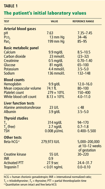
The cardiology consult team notes that her beta human chorionic gonadotropin (beta-hCG) level is much higher than would be expected at 12 weeks of pregnancy, and so they are concerned about the possibility of a molar pregnancy. In addition, her level of thyroid-stimulating hormone (TSH, or thyrotropin) is markedly low.
HYPERTHYROIDISM IN PREGNANCY
3. Which of the following would not explain this patient’s markedly low TSH level?
- Graves disease
- Molar pregnancy
- TSH-secreting pituitary adenoma
- Gestational transient thyrotoxicosis
- Twin pregnancy
Hyperthyroidism (also called thyrotoxicosis) has many causes, including but not limited to Graves disease, pituitary adenoma, struma ovarii (teratoma), hCG-secreting hydatidiform mole, and thyroid carcinoma (which is rare).4 In most of these disorders, the TSH level is low while the levels of thyroxine (T4), triiodothyronine (T3), or both are high.
Symptoms of hyperthyroidism are the effect of elevated T4 and T3 levels on the target organs themselves. Common symptoms include fever, tachycardia, tremor, stare, sweating, and lid lag. Other symptoms include nervousness, delirium, hypersensitivity to heat, flushing, palpitations, fatigue, weight loss, dyspnea, weakness, increased appetite, swelling of the legs, nausea, vomiting, diarrhea, goiter, tremor, atrial fibrillation, and cardiac failure.4 In its extreme form, called thyroid storm, thyrotoxicosis can be life-threatening. The likelihood of an impending thyroid storm can be assessed by clinical variables such as the patient’s temperature and heart rate and whether he or she has heart failure or gastrointestinal manifestations.5
Graves disease, the most common cause of hyperthyroidism in pregnancy, is due to stimulation of TSH receptors by antibodies against these receptors. Graves disease is possible in this patient, but a subsequent TSH receptor antibody test is negative.
Pituitary adenomas are one of the few causes of hyperthyroidism in which the TSH level is high, not low. Therefore, this is the correct answer.
Gestational transient thyrotoxicosis is a nonautoimmune condition that results in transient hyperthyroidism of variable severity.6 Usually, it occurs in otherwise normal pregnancies without complications, but the initial manifestation is hyper- emesis.6 It can be differentiated from Graves disease by the absence of TSH receptor antibodies and by no history of thyroid disorder.7 Common symptoms of gestational transient thyrotoxicosis include weight loss (or failure to gain weight), tachycardia, and fatigue.
The reason for the transient rise in T4 may be that beta-hCG is structurally similar to TSH (and also to luteinizing hormone and follicle-stimulating hormone), so that it has mild thyroid-stimulating effects.7 Sustained high levels of beta-hCG may in time give rise to the manifestations of thyrotoxicosis.
Molar pregnancy also can cause hyper-thyroidism via elevated levels of beta-hCG. However, twin pregnancy is more common and can produce sustained levels of beta-hCG above 100,000 IU/L. In most cases of twin pregnancy, the TSH level is decreased and the T4 level transiently elevated.6 The elevated beta-hCG and the subsequent thyrotropic manifestations are thought to be directly related, and symptoms resolve when beta-hCG levels go down.6
In most cases of hyperthyroidism in pregnancy, the acute condition can be managed by a short (≤ 2-month) course of a beta-blocker. In rare cases, propylthiouracil treatment may be required. Gestational transient thyrotoxicosis is not associated with detrimental outcomes.
Case continued
Our patient’s TSH level is low and her free T4 and T3 levels are elevated. Her high beta-hCG level may be stimulating the thyroid gland and may account for the low TSH value, as well as for her tachycardia, emesis, shortness of breath, and weight loss.
After an obstetric consult, it is determined that our patient has a viable pregnancy. However, further investigation with transvaginal ultrasonography reveals that she has two viable, single-placenta, intrauterine gestations, separated by a thin chorionic membrane.
Beta-hCG and free T4 levels are significantly higher in twin pregnancies than in single pregnancies, especially in the early stages.6 In our patient, the twin pregnancy led to the elevated beta-hCG, which eventually manifested as thyrotoxicosis, which caused the shortness of breath, hyperemesis, weight loss, tachycardia, and nausea.
Shortness of breath in patients with thyrotoxicosis is well recognized but not well explained. It may be caused by decreased lung compliance, engorged capillaries in the lung, or left ventricular failure, as well as by chest pain due to increased myocardial demand or coronary artery vasospasm.4 The dyspnea is present at rest and during exertion, and the high metabolic rate is thought to lead to an inappropriate response of the ventilatory system.3,8
WHAT TREATMENT?
4. How would you treat this patient at this point?
- No drug therapy, just supportive care
- Propranolol (Inderal)
- Levothyroxine
- Propylthiouracil
Several types of drugs are used to manage hyperthyroidism.
Antithyroid drugs such as propylthiouracil, methimazole (Northyx, Tapazole), and carbimazole block thyroid hormone synthesis by inhibiting thyroid peroxidase. Propylthiouracil also blocks peripheral conversion of T4 to T3. Side effects of these agents include abnormal sense of taste, pruritus, urticaria, agranulocytosis, and hepatotoxicity.4
Usually, hyperthyroidism is treated with propylthiouracil at the smallest effective dose. This has been proven to be safe to the fetus and mother during pregnancy.9 Propylthiouracil and the other drugs in its class cross the placenta, but propylthiouracil crosses at one-quarter the rate of the other two.9
Beta-blockers are effective in the acute phase of thyrotoxicosis against tachycardia, hypertension, and atrial fibrillation. They also decrease conversion of T4 to T3, which is an added benefit. Beta-blockers can be tapered as thyroid hormone levels decrease.
A short course of a short-acting beta-blocker would be an option for our patient and would decrease her symptoms, although she does not have the typical markedly elevated T4 or T3 levels. In the long term, a beta-blocker would present a fetal risk, but short courses can be tolerated without incident.9
Radioactive iodine 131 is used in patients with Graves disease. 131Iodine therapy is safe for most adults, but in pregnancy its use is contraindicated. Fetal thyroid tissue is thought to be present after 10 weeks of gestation and could be damaged by the use of radioactive iodine. Another warning with the use of radioactive iodine is that patients should avoid close contact with other adults for a few days after treatment, and should avoid close contact with children and pregnant women for 2 to 3 weeks after treatment because of the risk of exposure to radiation emanating from the thyroid gland.
Levothyroxine is a treatment for hypothyroidism, not hyperthyroidism.
CASE CONTINUED
Our patient is treated with propranolol and monitored for several days in the hospital, during which her symptoms markedly improve. She is discharged without complications.
TAKE-HOME POINTS
The evaluation of shortness of breath in adult patients can be difficult, given the many possible causes. It is especially challenging in pregnant patients, since normal physiologic changes of pregnancy may produce these symptoms.
In many instances, cardiomyopathy must be suspected if a pregnant patient complains of shortness of breath. However, it is not the only possible cause.
- Dobbenga-Rhodes YA, Prive AM. Assessment and evaluation of the woman with cardiac disease during pregnancy. J Perinat Neonatal Nurs 2006; 20:295–302.
- Carman TL, Deitcher SR. Advances in diagnosing and excluding pulmonary embolism: spiral CT and D-dimer measurement. Cleve Clin J Med 2002; 69:721–729.
- Wells PS, Anderson DR, Rodger M, et al. Derivation of a simple clinical model to categorize patients’ probability of pulmonary embolism: increasing the model’s utility with the SimpliRED D-dimer. Thromb Haemost 2000; 83:416–420.
- Nayak B, Burman K. Thyrotoxicosis and thyroid storm. Endocrinol Metab Clin North Am 2006; 35:663–686.
- Burch HB, Wartofsky L. Life-threatening thyrotoxicosis. Thyroid storm. Endocrinol Metab Clin North Am 1993; 22:263–277.
- Grün JP, Meuris S, De Nayer P, Glinoer D. The thyrotrophic role of human chorionic gonadotrophin (hCG) in the early stages of twin (versus single) pregnancies. Clin Endocrinol (Oxf) 1997; 46:719–725.
- Glinoer D, De Nayer P, Robyn C, Lejeune B, Kinthaert J, Meuris S. Serum levels of intact human chorionic gonadotropin (HCG) and its free alpha and beta subunits, in relation to maternal thyroid stimulation during normal pregnancy. J Endocrinol Invest 1993; 16:881–888.
- Small D, Gibbons W, Levy RD, de Lucas P, Gregory W, Cosio MG. Exertional dyspnea and ventilation in hyper-thyroidism. Chest 1992; 101:1268–1273.
- Atkins P, Cohen SB, Phillips BJ. Drug therapy for hyper-thyroidism in pregnancy: safety issues for mother and fetus. Drug Saf 2000; 23:229–244.
A 21-year-old woman who is 12 weeks pregnant according to the date of her last menstrual period comes to the emergency department with shortness of breath and chest pain.
One week ago she began experiencing pre-syncope and shortness of breath on minimal exertion and then even at rest on most days. The shortness of breath worsened throughout the week, eventually limiting her daily activities to such a degree that she restricted herself to bed rest.
Her chest pain started today while she was sitting in church, without any apparent provocation. It is right-sided, sharp, and focal, and it does not radiate. At the same time, her shortness of breath was more severe than before, so she immediately came to the emergency department.
This is her third pregnancy; she has had one live birth and one abortion. Her last pregnancy was full-term, with routine prenatal care and no complications. However, so far during this pregnancy, she has had no prenatal care, she has not taken prenatal vitamins, and she has been unable to maintain adequate nutrition because of persistent emesis, which began early in her pregnancy and continues to occur as often as two or three times daily. She has lost 20 pounds over the past 12 weeks.
She says she has no close contacts who are sick, and she has had no fever, diarrhea, dysuria, urinary frequency or urgency, palpitations, swelling of the legs or feet, blurry vision, or increase in neck girth. She says she does not smoke or use alcohol or illicit substances. Her only previous surgery was laser-assisted in situ keratoplasty (LASIK) eye surgery in 1998. She is allergic to seafood only. She has not eaten at any new places recently. She is up to date with her childhood vaccinations. She has no family history of hypercoagulability or venous thrombotic events.
PHYSICAL EXAMINATION
She is breathing rapidly—as fast as 45 breaths per minute. Her temperature is 37.2°C (98.9°F), blood pressure 95/60 mm Hg, oxygen saturation 100% while on 10 L of oxygen using a nonrebreather mask, pulse 102 beats per minute, and weight 55.9 kg (123.2 pounds). She appears alert, oriented, and comfortable, with a thin body habitus. She has no jugular venous distention, neck mass, or thyromegaly. Her lungs are clear to auscultation, with no wheezes or rales. The cardiovascular examination is normal. She has a regular heart rate and rhythm, normal S1 and S2 sounds, and no rubs, clicks, or murmurs. Pulses in the extremities are normal, and she has no peripheral edema. The neurologic examination is normal.
Electrocardiography shows sinus tachycardia with first-degree atrioventricular block.
DIFFERENTIAL DIAGNOSIS
1. At this point, which is the most probable cause of her symptoms?
- Pulmonary embolism
- Peripartum cardiomyopathy
- Acute coronary syndrome
- Aortic dissection
- Expected physiologic changes of pregnancy
Pulmonary embolism would be the most probable diagnosis, given the patient’s pregnancy, shortness of breath, and tachycardia and the pleuritic quality of her chest pain.
Peripartum cardiomyopathy is also a possible cause, as it may present with profound shortness of breath and markedly decreased cardiac function. But it is much less likely in this patient because she is early in her pregnancy, and peripartum cardiomyopathy usually is seen during the last month of gestation or the first months after delivery.
Acute coronary syndrome is unlikely, given her young age and the lack of significant risk factors or a supporting history.
Aortic dissection is unlikely in view of her medical history.
Physiologic changes of pregnancy. Many pregnant women experience a sensation of not being able to catch their breath or expand their lungs fully, as the diaphragm is limited by the gravid abdomen. They also present with dyspnea, fatigue, reduced exercise capacity, peripheral edema, or volume overload.1 However, these changes tend to occur gradually and worsen over time. This patient’s degree of shortness of breath and its sudden onset do not seem like normal physiologic changes of pregnancy.
Other possible causes of dyspnea in a pregnant woman include asthma, pleural empyema, pneumonia, and severe anemia. Asthma should be considered in anyone with a history of wheezing, cough, and dyspnea. Fever and sputum production would support a diagnosis of pneumonia or empyema. In addition, maternal heart disease (eg, endocarditis, pulmonary hypertension) complicates 0.2% to 3% of pregnancies.1
CASE CONTINUED
The emergency department staff decide to evaluate the patient for heart failure and pulmonary embolism.
Bedside echocardiography reveals an ejection fraction of 55% (normal range 50%–75%), normal heart function and size, and no valvular abnormalities.
Chest radiography is normal.
Lower-extremity duplex ultrasonography is negative for deep-vein thrombosis.
The D-dimer level is 380 ng/mL (normal range < 500 ng/mL).
The medical intensive care unit is consulted about the patient’s continued tachypnea and the possible need for intubation. A ventilation-perfusion scan is performed to screen for pulmonary embolism, and it is negative.
An obstetric team performs Doppler ultrasonography at the bedside; a fetal heartbeat can be heard, thus confirming a viable pregnancy.
The patient has normal serum levels of the cardiac enzymes troponin T and creatine kinase-MB fraction, thus all but ruling out myocardial ischemia.
The patient is admitted to the hospital the next day, and a cardiology consult is obtained.
RULING OUT PULMONARY EMBOLISM
2. Has pulmonary embolism been definitively ruled out at this point?
- Yes
- No
The answer is no. The negative ventilation-perfusion scan and normal D-dimer test in this patient are not enough to rule out pulmonary embolism. The diagnosis of pulmonary embolism should be based on the clinician’s estimation of the pretest probability of pulmonary embolism (which is based on presenting signs and symptoms), as well as on a variety of tests, including spiral computed tomography (CT), ventilation-perfusion lung scanning, and serum D-dimer testing. Signs and symptoms that may guide the clinician are chest pain (present in 70% of patients with pulmonary embolism), tachypnea (70%), cough (40%), shortness of breath (25%), and tachycardia (33%).2 A history of pregnancy, malignancy, immobility, or recent surgery may also increase the pretest probability of pulmonary embolism. In many cases, one’s clinical suspicion is highly predictive and is useful in diagnosing pulmonary embolism.
The accuracy of the tests varies widely, depending on the pretest probability of pulmonary embolism. For instance, in a patient with a high pretest probability but a low-probability ventilation-perfusion scan, the true probability of pulmonary embolism is 40%, but in a patient with a low pretest probability and a low-probability scan, the probability is only 4%.
The Wells criteria can be used to calculate the pretest probability of pulmonary embolism. Given this patient’s tachycardia and clinical presentation, her pretest probability according to the Wells criteria indicates increased risk. However, because her D-dimer test, lower-extremity Doppler test, and ventilation-perfusion scan were normal, pulmonary embolism is less likely.3
However, if one’s clinical suspicion is high enough, further investigation of pulmonary embolism would proceed despite the encouraging test results.
CASE CONTINUED
The cardiology consult team notes that her beta human chorionic gonadotropin (beta-hCG) level is much higher than would be expected at 12 weeks of pregnancy, and so they are concerned about the possibility of a molar pregnancy. In addition, her level of thyroid-stimulating hormone (TSH, or thyrotropin) is markedly low.
HYPERTHYROIDISM IN PREGNANCY
3. Which of the following would not explain this patient’s markedly low TSH level?
- Graves disease
- Molar pregnancy
- TSH-secreting pituitary adenoma
- Gestational transient thyrotoxicosis
- Twin pregnancy
Hyperthyroidism (also called thyrotoxicosis) has many causes, including but not limited to Graves disease, pituitary adenoma, struma ovarii (teratoma), hCG-secreting hydatidiform mole, and thyroid carcinoma (which is rare).4 In most of these disorders, the TSH level is low while the levels of thyroxine (T4), triiodothyronine (T3), or both are high.
Symptoms of hyperthyroidism are the effect of elevated T4 and T3 levels on the target organs themselves. Common symptoms include fever, tachycardia, tremor, stare, sweating, and lid lag. Other symptoms include nervousness, delirium, hypersensitivity to heat, flushing, palpitations, fatigue, weight loss, dyspnea, weakness, increased appetite, swelling of the legs, nausea, vomiting, diarrhea, goiter, tremor, atrial fibrillation, and cardiac failure.4 In its extreme form, called thyroid storm, thyrotoxicosis can be life-threatening. The likelihood of an impending thyroid storm can be assessed by clinical variables such as the patient’s temperature and heart rate and whether he or she has heart failure or gastrointestinal manifestations.5
Graves disease, the most common cause of hyperthyroidism in pregnancy, is due to stimulation of TSH receptors by antibodies against these receptors. Graves disease is possible in this patient, but a subsequent TSH receptor antibody test is negative.
Pituitary adenomas are one of the few causes of hyperthyroidism in which the TSH level is high, not low. Therefore, this is the correct answer.
Gestational transient thyrotoxicosis is a nonautoimmune condition that results in transient hyperthyroidism of variable severity.6 Usually, it occurs in otherwise normal pregnancies without complications, but the initial manifestation is hyper- emesis.6 It can be differentiated from Graves disease by the absence of TSH receptor antibodies and by no history of thyroid disorder.7 Common symptoms of gestational transient thyrotoxicosis include weight loss (or failure to gain weight), tachycardia, and fatigue.
The reason for the transient rise in T4 may be that beta-hCG is structurally similar to TSH (and also to luteinizing hormone and follicle-stimulating hormone), so that it has mild thyroid-stimulating effects.7 Sustained high levels of beta-hCG may in time give rise to the manifestations of thyrotoxicosis.
Molar pregnancy also can cause hyper-thyroidism via elevated levels of beta-hCG. However, twin pregnancy is more common and can produce sustained levels of beta-hCG above 100,000 IU/L. In most cases of twin pregnancy, the TSH level is decreased and the T4 level transiently elevated.6 The elevated beta-hCG and the subsequent thyrotropic manifestations are thought to be directly related, and symptoms resolve when beta-hCG levels go down.6
In most cases of hyperthyroidism in pregnancy, the acute condition can be managed by a short (≤ 2-month) course of a beta-blocker. In rare cases, propylthiouracil treatment may be required. Gestational transient thyrotoxicosis is not associated with detrimental outcomes.
Case continued
Our patient’s TSH level is low and her free T4 and T3 levels are elevated. Her high beta-hCG level may be stimulating the thyroid gland and may account for the low TSH value, as well as for her tachycardia, emesis, shortness of breath, and weight loss.
After an obstetric consult, it is determined that our patient has a viable pregnancy. However, further investigation with transvaginal ultrasonography reveals that she has two viable, single-placenta, intrauterine gestations, separated by a thin chorionic membrane.
Beta-hCG and free T4 levels are significantly higher in twin pregnancies than in single pregnancies, especially in the early stages.6 In our patient, the twin pregnancy led to the elevated beta-hCG, which eventually manifested as thyrotoxicosis, which caused the shortness of breath, hyperemesis, weight loss, tachycardia, and nausea.
Shortness of breath in patients with thyrotoxicosis is well recognized but not well explained. It may be caused by decreased lung compliance, engorged capillaries in the lung, or left ventricular failure, as well as by chest pain due to increased myocardial demand or coronary artery vasospasm.4 The dyspnea is present at rest and during exertion, and the high metabolic rate is thought to lead to an inappropriate response of the ventilatory system.3,8
WHAT TREATMENT?
4. How would you treat this patient at this point?
- No drug therapy, just supportive care
- Propranolol (Inderal)
- Levothyroxine
- Propylthiouracil
Several types of drugs are used to manage hyperthyroidism.
Antithyroid drugs such as propylthiouracil, methimazole (Northyx, Tapazole), and carbimazole block thyroid hormone synthesis by inhibiting thyroid peroxidase. Propylthiouracil also blocks peripheral conversion of T4 to T3. Side effects of these agents include abnormal sense of taste, pruritus, urticaria, agranulocytosis, and hepatotoxicity.4
Usually, hyperthyroidism is treated with propylthiouracil at the smallest effective dose. This has been proven to be safe to the fetus and mother during pregnancy.9 Propylthiouracil and the other drugs in its class cross the placenta, but propylthiouracil crosses at one-quarter the rate of the other two.9
Beta-blockers are effective in the acute phase of thyrotoxicosis against tachycardia, hypertension, and atrial fibrillation. They also decrease conversion of T4 to T3, which is an added benefit. Beta-blockers can be tapered as thyroid hormone levels decrease.
A short course of a short-acting beta-blocker would be an option for our patient and would decrease her symptoms, although she does not have the typical markedly elevated T4 or T3 levels. In the long term, a beta-blocker would present a fetal risk, but short courses can be tolerated without incident.9
Radioactive iodine 131 is used in patients with Graves disease. 131Iodine therapy is safe for most adults, but in pregnancy its use is contraindicated. Fetal thyroid tissue is thought to be present after 10 weeks of gestation and could be damaged by the use of radioactive iodine. Another warning with the use of radioactive iodine is that patients should avoid close contact with other adults for a few days after treatment, and should avoid close contact with children and pregnant women for 2 to 3 weeks after treatment because of the risk of exposure to radiation emanating from the thyroid gland.
Levothyroxine is a treatment for hypothyroidism, not hyperthyroidism.
CASE CONTINUED
Our patient is treated with propranolol and monitored for several days in the hospital, during which her symptoms markedly improve. She is discharged without complications.
TAKE-HOME POINTS
The evaluation of shortness of breath in adult patients can be difficult, given the many possible causes. It is especially challenging in pregnant patients, since normal physiologic changes of pregnancy may produce these symptoms.
In many instances, cardiomyopathy must be suspected if a pregnant patient complains of shortness of breath. However, it is not the only possible cause.
- Dobbenga-Rhodes YA, Prive AM. Assessment and evaluation of the woman with cardiac disease during pregnancy. J Perinat Neonatal Nurs 2006; 20:295–302.
- Carman TL, Deitcher SR. Advances in diagnosing and excluding pulmonary embolism: spiral CT and D-dimer measurement. Cleve Clin J Med 2002; 69:721–729.
- Wells PS, Anderson DR, Rodger M, et al. Derivation of a simple clinical model to categorize patients’ probability of pulmonary embolism: increasing the model’s utility with the SimpliRED D-dimer. Thromb Haemost 2000; 83:416–420.
- Nayak B, Burman K. Thyrotoxicosis and thyroid storm. Endocrinol Metab Clin North Am 2006; 35:663–686.
- Burch HB, Wartofsky L. Life-threatening thyrotoxicosis. Thyroid storm. Endocrinol Metab Clin North Am 1993; 22:263–277.
- Grün JP, Meuris S, De Nayer P, Glinoer D. The thyrotrophic role of human chorionic gonadotrophin (hCG) in the early stages of twin (versus single) pregnancies. Clin Endocrinol (Oxf) 1997; 46:719–725.
- Glinoer D, De Nayer P, Robyn C, Lejeune B, Kinthaert J, Meuris S. Serum levels of intact human chorionic gonadotropin (HCG) and its free alpha and beta subunits, in relation to maternal thyroid stimulation during normal pregnancy. J Endocrinol Invest 1993; 16:881–888.
- Small D, Gibbons W, Levy RD, de Lucas P, Gregory W, Cosio MG. Exertional dyspnea and ventilation in hyper-thyroidism. Chest 1992; 101:1268–1273.
- Atkins P, Cohen SB, Phillips BJ. Drug therapy for hyper-thyroidism in pregnancy: safety issues for mother and fetus. Drug Saf 2000; 23:229–244.
The hospital guy redux

You responded to the parts of Dr. Lansdale’s commentary that struck a personal chord. Almost all responders shared his frustration. Many wrote that the American payer system fails to appropriately reward internists and primary care providers and called for restructuring the Medicare and third-party payer systems. Some of you took umbrage at his contention that hospitals are not safe, and that health care delivery systems do not always place quality care above economic imperatives as new programs and “centers of excellence” are implemented. And some of you reacted to the issues of physician satisfaction and difficulties in providing quality care in hospitals regulated by multiple agencies that generate unfunded mandates, while the hospitals already require high numbers of patients in order to survive financially.
I recently did a stint as rheumatology consultant at my hospital, and Dr. Lansdale’s commentary was fresh in my mind. I noticed with satisfaction that the physicians and nurses were using foam antiseptic on their hands. I noted the new checks on verbal orders and a successful emphasis on preventing deep vein thrombosis and bedsores. But I also noted more patient hand-offs between house staff and faculty, and difficulty in finding doctors who actually knew the patient (or doctors that patients recognized as being responsible for their care).
The electronic medical record is legible and available from all over the hospital, and I could tell who signed the notes. But many notes were actually cut-and-pasted from earlier notes, and thus I couldn’t always be sure who actually had said what and when. Technology is not an immediate panacea for the problem of limited physician time!
The house staff “lab” in the hospital with its microscope was closed due to regulatory concerns; thus, there was no easy way to look at a freshly spun urine sample for evidence of glomerulonephritis. This turned out to be a detriment to effective patient care: urine samples sent to the regular laboratory (with the usual transportation delay) rarely if ever reveal cellular casts. But we found creative, if inefficient, ways to deal with this and other problems.
At the end of the day, I realized that I still enjoy my time in the hospital. Patients’ problems can be presented to house staff and students at the bedside and their diagnoses and therapies discussed in real time. Junior physicians can observe how senior physicians talk to patients and families, including the many ways we have learned to say “I don’t know,” and learn to appreciate the value of a well-directed physical examination. There is still a synergy and intellectual satisfaction in being one of a group of senior consultants discussing the care of a shared patient who has complex medical problems.
With rational and caring involvement, individual physicians can alter the trajectory of patient management and remain the primary patient advocates within a health care system that can’t always easily deliver the quality that everyone desires. Caring, patient-focused physicians must remain in charge of health care delivery, lest we pay attention only to the financial and regulatory problems.
Tom, I am older and even more cynical than I was when we roamed the hospital together every third night and never went home on our post-call day until the last laboratory result had been checked and the last transfusion had been given. We inefficiently examined every patient’s urine ourselves (even from those being admitted for cardiac catheterization), and we had to convince patients of the (apparent) need for the urgent 3 AM blood draw to evaluate their 100.5° fever before we prepped the area and drew the blood. We drew blood for sedimentation rates and checked rapid plasma reagins at every admission and checked for urinary light chains in everyone with an elevated creatinine level and anemia, “just to be sure.” We blindly placed Swan-Ganz catheters to monitor many hypotensive patients in the intensive care units, and we aspirated pleural effusions on the basis of our percussive examination. We talked to patients and accepted enormous individual responsibility for their care, but we were also frequently numbed by the overwhelming intensity of the training and the practice.
I am all too aware of the many forces that are eroding physician-patient relationships and that can corrupt patient care in the name of efficiency, financial necessity, marketing advantage, or regulatory compliance. Many of these forces I hope to help change. But I remain a hospital guy because I can still make a difference. I still feel honored that patients entrust their care to me as we attempt to navigate our evolving and, yes, sometimes treacherous medical system. Evading the crocodiles and fighting insurance companies are now in my job description.
In this issue we run two letters in response to Dr. Lansdale’s commentary. In December we will publish more letters, though due to space limitations some will be abridged. We plan to run full text of many of the letters online at www.ccjm.org in December.