User login
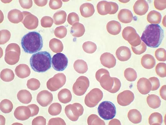
CHADS2 Variant Calculates Stroke Risk in Heart Failure Patients
NEW YORK - A variant of the CHADS2 score that's used to estimate ischemic stroke risk in patients with atrial fibrillation (AF) is also modestly accurate in heart failure patients, even in those without AF, researchers say. The variant, CHA2DS2-VASc, calculates stroke risk based on 10 possible points with higher scores indicating higher risk.
Line Melgaard from Aalborg University in Denmark and colleagues used three Danish nationwide registries to investigate whether the CHA2DS2-VASc score could predict ischemic stroke, thromboembolism, and death in patients with heart failure without AF as effectively as it does in patients with AF.
Patients with heart failure had a high risk of all three outcomes, whether or not AF was present, and the CHA2DS2-VASc score modestly predicted these endpoints at one-year and five-year follow-up (C statistics, 0.67 and 0.69, respectively).
Heart failure patients without AF whose CHA2DS2-VASc score was 4 or higher had increased risks of ischemic stroke, thromboembolism, and death in a manner comparable to patients with AF, according to the August 30 JAMA online report.
The negative predictive value (NPV) was around 90% at one-year follow-up for all three outcomes, although NPVs were strongly attenuated by the five-year follow-up.
"In our study, one of our principal findings was that the absolute risk of ischemic stroke among patients without AF was about 1.5% per year or higher with CHA2DS2-VASc scores of 2 or higher, with associated five-year absolute ischemic stroke risks in excess of 4% or more," the researchers noted. This risk level would be sufficient to prompt initiation of long-term anticoagulation in patients with AF, they say.
"The poor prognosis of atrial fibrillation for ischemic stroke and death in patients with heart failure was evident in our study and expected," Melgaard said. "But the observation that additional risk factors in patients with heart failure are particularly significant among those without atrial fibrillation is an important and (to some extent) unexpected result."
"I hope physicians will recognize that patients with heart failure and sinus rhythm have an increased risk of ischemic stroke, and that some subgroups within this population most likely need thromboprophylaxis," Melgaard concluded. "Especially patients with multiple comorbidities (high CHA2DS2-VASc score) need attention in the clinic."
Low-Dose Screening CT for High-Risk NSCLC/SCLC: The Providence VA’s Experience
Background: There are about 200,000 new cases of lung cancer each year in the U.S. and about 150,000 deaths. Lung cancer is the second most common malignancy but the most common cause of cancer deaths. In 2010, the National Lung Screening Trial described a 20.3% reduction in lung cancer mortality in high-risk patients who were screened by CT scan. In 2012, guidelines were set forth by multiple groups. The National Comprehensive Cancer Network encouraged low-dose CT screening for patients aged 55 to 75 years, ≥ 30 pack-years of smoking, or smoking cessation within 15 years.
Methods: In late 2013, screening was implemented at the Providence VA Medical Center (PVAMC); a rise in the incidence of early stage lung cancer was anticipated. We reviewed the number and stages of cases of non-small cell (NSCLC) and small cell (SCLC) lung cancer in 2012, 2013 (mostly pre-screening), 2014 (screening), and 2015 (4 months of screening).
Results: In 2012 there were 23 cases of NSCLC (5 stage I; 5 stage II; 8 stage III; 2 stage IV; 3 unknown) and 4 cases of SCLC (extensive). In 2013 there were 42 cases of NSCLC (9 stage I; 2 stage II; 10 stage III [1 screened]; 17 stage IV; 4 unknown [1 screened]) and 9 cases of SCLC (1 lim-ited, 8 extensive). In 2014, there were 54 cases of NSCLC (15 stage I [5 screened]; 10 stage II [2 screened]; 11 stage III [3 screened]; 14 stage IV [1 screened]; and 4 unknown) and 5 cases of SCLC (5 extensive). In the first 7 months of 2015, there were 36 cases of NSCLC (13 stage I [10 screened]; 5 stage II [3 screened]; 7 stage III [3 screened]; 8 stage IV [1 screened]; and 3 unknown [1 screened]) and 1 SCLC (extensive) not screened and 1 SCLC (limited) screened.
Discussion: The total number of lung cancer cases has increased with screening (36 in 2012; 51 in 2013; 59 in 2014; 38 in 7 months of 2015). With screening in 2014 there were more cases of late stage disease (NSCLC stage III/IV) than anticipated. In the first 7 months of 2015 72% of screened patients were stage I/II, 22% were stage III/IV, and 6% were unknown. As time progresses and the same patients are screened yearly, the percentage of early stage cases should increase and late stages decrease. It will be worthwhile to reevaluate these data over the next several years.
Gauging the Use of Single Fraction Radiotherapy for Painful Bone Metastases: The Experience of a Single Veterans Administration Radiation Oncology Center
Background: In addition to patient and/or family convenience, there is mounting category 1 evidence of the effectiveness of single fraction radiotherapy (SFRT) and its utility in the treatment of uncomplicated painful bone metastases. Surveys indicated that physicians in the U.S. still prefer multiple fractions radiotherapy (MFRT) for uncomplicated bone metastases compared with their Canadian or European colleagues. This report used code tracking to determine the scope of use of SFRT for painful malignant bone lesions compared with MFRT.
Methods: We reviewed encounters logged as international classification of disease (ICD) code 198.5 (secondary malignancy of bone and or bone marrow) and current procedural terminology (CPT) codes: 77427 (physician weekly visit for 3 to 5 treatments); and 77431 (physician weekly visit for 1 to 2 treatments) from 2002 to 2008 and 2009 to 2014.
Results: Data limitations/assumptions: The ICD 198.5 diagnosis code was pulled from either the primary or secondary position. By merging CPT/ICD codes to track use patterns, we noted a 43% increase in the use of SFRT for painful bone lesions at James J. Peters VA Medical Center. The CPT code 77431 was used on a yearly average of 2.45% of bone metastases cases from periods 2002 to 2008, and 5.67% (8.50% ex-cluding years 2009/2010) from 2009 to 2014. Of note, from the period years of 2003, 2008, and 2009 to 2010, CPT code 77431 was not used at all.
Conclusions: We saw an increased use of SFRT for bone metastases over the time period covered, and that tracking the encounters by ICD and CPT codes served, in part, as a useful tool in providing a snapshot view (if proper code is used) of SFRT usage. Physician education is a requisite for the proper use of CPT 77431 to capture the true rate of usage of SFRT in clinical practice.
Trabectedin superior for advanced liposarcoma, leiomyosarcoma
Trabectedin proved superior to standard dacarbazine therapy by numerous measures but not by overall survival in an industry-sponsored phase III clinical trial reported online Sept. 14 in Journal of Clinical Oncology.
Trabectedin, which has been used extensively in Europe for a decade but has not been approved in the U.S., has a complex mechanism of action that affects several critical cell biology processes within and surrounding tumor cells. It exhibited activity against metastatic soft tissue sarcomas in several phase II trials, said Dr. George D. Demetri of the Ludwig Center at Harvard Medical School and the Center for Sarcoma and Bone Oncology, Dana-Farber Cancer Institute, both in Boston, and his associates.
In this study, trabectedin was assessed in 518 patients aged 15 years and older who had heavily pretreated and rapidly progressing advanced or metastatic liposarcoma or leiomyosarcoma and were treated at 85 sites in four countries. These participants were randomly assigned to receive either trabectedin (345 patients) or dacarbazine (173 patients) via central intravenous infusion in 3-week cycles.
Compared with dacarbazine, trabectedin reduced the risk of disease progression by 45% (hazard ratio, 0.55), and superior disease control was discernible at the first patient assessment at 6 weeks. Median progression-free survival was significantly longer with trabectedin (4.2 months) than with dacarbazine (1.5 months), a benefit that was consistent across all 19 subgroups of patients assessed in sensitivity analyses, regardless of disease histology, previous therapies, or any clinical characteristics. Trabectedin also bested dacarbazine with regard to objective response rate (9.9% vs. 6.9%), median duration of response (6.5 months vs. 4.2 months), achievement of stable disease (51% vs. 35%), and achievement of durable stable disease (34% vs. 19%).
Trabectedin showed only a nonsignificant 13% reduction in overall survival, which was the primary endpoint of this study. However, several previous studies have demonstrated that it can be extremely difficult to prolong overall survival despite robust improvements in progression-free survival in patients with advanced sarcomas. Given this “historical difficulty in demonstrating overall survival improvement,” the documentation of disease control such as that achieved in this study may be considered a measure of clinically relevant efficacy in this setting, Dr. Demetri and his associates wrote (J. Clin. Oncol. 2015 Sep 14 [doi:10.1200/JCO.2015.62.4734]).
Adverse events in this study population “were consistent with the well-characterized safety and toxicity profiles of both study drugs.” Toxicity was more common with trabectedin, and deaths considered to be treatment-related occurred only in the trabectedin group: three cases of sepsis/septic shock and one each of rhabdomyolysis/sepsis, renal failure, cardiac arrest, and multiorgan failure, for a treatment-related mortality of 2.1%.
This study confirms what we originally heard a decade ago from European investigators: Trabectedin shows clinical activity against liposarcomas and leiomyosarcomas. But the benefits demonstrated here still seem small, and it is not yet clear whether the data are now sufficient to make trabectedin the standard of care for all patients who have these sarcomas.
The primary end point – improving overall survival to a greater degree than dacarbazine – was not met. However, in the investigators’ defense, this was partly because the dacarbazine group survived much longer than expected. This, in turn, may be because more patients who had disease progression with dacarbazine (56%) than with trabectedin (47%) crossed over to other anticancer drugs, including the receptor tyrosine kinase inhibitor pazopanib, which was approved during the course of this study.
Dr. Gary K. Schwartz is at the Herbert Irving Comprehensive Cancer Center and Columbia University, New York. He reported ties to Novartis, AstraZeneca, and Boehringer Ingelheim. Dr. Schwartz made these remarks in an editorial accompanying Dr. Demetri’s report (J. Clin. Oncol. 2015 Sep 14 [doi:10.1200/JCO.2015.63.5938]).
Trabectedin proved superior to standard dacarbazine therapy by numerous measures but not by overall survival in an industry-sponsored phase III clinical trial reported online Sept. 14 in Journal of Clinical Oncology.
Trabectedin, which has been used extensively in Europe for a decade but has not been approved in the U.S., has a complex mechanism of action that affects several critical cell biology processes within and surrounding tumor cells. It exhibited activity against metastatic soft tissue sarcomas in several phase II trials, said Dr. George D. Demetri of the Ludwig Center at Harvard Medical School and the Center for Sarcoma and Bone Oncology, Dana-Farber Cancer Institute, both in Boston, and his associates.
In this study, trabectedin was assessed in 518 patients aged 15 years and older who had heavily pretreated and rapidly progressing advanced or metastatic liposarcoma or leiomyosarcoma and were treated at 85 sites in four countries. These participants were randomly assigned to receive either trabectedin (345 patients) or dacarbazine (173 patients) via central intravenous infusion in 3-week cycles.
Compared with dacarbazine, trabectedin reduced the risk of disease progression by 45% (hazard ratio, 0.55), and superior disease control was discernible at the first patient assessment at 6 weeks. Median progression-free survival was significantly longer with trabectedin (4.2 months) than with dacarbazine (1.5 months), a benefit that was consistent across all 19 subgroups of patients assessed in sensitivity analyses, regardless of disease histology, previous therapies, or any clinical characteristics. Trabectedin also bested dacarbazine with regard to objective response rate (9.9% vs. 6.9%), median duration of response (6.5 months vs. 4.2 months), achievement of stable disease (51% vs. 35%), and achievement of durable stable disease (34% vs. 19%).
Trabectedin showed only a nonsignificant 13% reduction in overall survival, which was the primary endpoint of this study. However, several previous studies have demonstrated that it can be extremely difficult to prolong overall survival despite robust improvements in progression-free survival in patients with advanced sarcomas. Given this “historical difficulty in demonstrating overall survival improvement,” the documentation of disease control such as that achieved in this study may be considered a measure of clinically relevant efficacy in this setting, Dr. Demetri and his associates wrote (J. Clin. Oncol. 2015 Sep 14 [doi:10.1200/JCO.2015.62.4734]).
Adverse events in this study population “were consistent with the well-characterized safety and toxicity profiles of both study drugs.” Toxicity was more common with trabectedin, and deaths considered to be treatment-related occurred only in the trabectedin group: three cases of sepsis/septic shock and one each of rhabdomyolysis/sepsis, renal failure, cardiac arrest, and multiorgan failure, for a treatment-related mortality of 2.1%.
FROM JOURNAL OF CLINICAL ONCOLOGY
Key clinical point: Trabectedin was superior to standard dacarbazine by most measures in advanced, refractory liposarcoma and leiomyosarcoma, though not for overall survival.
Major finding: Trabectedin reduced the risk of disease progression by 45% (HR, 0.55).
Data source: An international industry-sponsored open-label randomized phase III clinical trial involving 518 patients.
Disclosures: This study was supported by Janssen Pharmaceuticals and the Adelson Medical Research Foundation. Dr. Demetri and his associates reported ties to numerous industry sources.

Smoother orthopedic implants may minimize bacterial adherence
SAN DIEGO – Rough materials used for orthopedic implants, such as cobalt chromium and titanium, increased bacterial adherence, while smoother materials such as stainless steel did not, results from an image analysis demonstrated.
“In light of these results, it is important to question why we utilize the types of materials we use for various orthopedic procedures,” Dioscaris R. Garcia, Ph.D., said in an interview in advance of the annual Interscience Conference on Antimicrobial Agents and Chemotherapy. “There was no one-size-fits-all material to minimize adherence per the findings of this study, but the findings may suggest that having a smoother surface may minimize the ability of bacterial pathogens to adhere to the surface.”

He characterized the topic of bacterial adherence to orthopedic implants as “an issue of great interest due to how little is known about the biological implications of the materials utilized. The most researched of these materials is titanium, which is touted for its biocompatibility. This study aims to provide a base and a glimpse into how the most commonly utilized orthopedic-relevant materials interact with some of the most commonly encountered pathogens.”
For the study, Dr. Garcia, a molecular pharmacologist at Rhode Island Hospital in Providence, Dr. Alan H. Daniels, an orthopedic surgeon at the hospital, and their associates used scanning electron microscopy and confocal laser scanning microscopy to evaluate the adherence pattern, density, and propagation of six commonly encountered bacterial pathogens (methicillin-sensitive Staphylococcus aureus, methicillin-resistant S. aureus, coagulase-negative Staphylococcus epidermidis, multidrug-resistant Acinetobacter baumannii, Propionibacterium acnes, and vancomycin-resistant Enterococcus faecalis) on five commonly used spinal implant materials (titanium, titanium alloy, stainless steel, cobalt chromium, and polyetherether ketone). The samples were fixed and dehydrated via ethanol dehydration gradient and critical point drying.
The researchers found that some pathogens, such as vancomycin-resistant E. faecalis and multidrug-resistant A. baumannii, were more likely to adhere to more textured materials such as cobalt chromium and titanium, compared with smoother materials such as stainless steel. “Additionally, the findings suggest that the microtopography of these materials may be the driving force behind the adherence of pathogens on the materials themselves,” Dr. Garcia said. Compared with smoother, polished materials, he explained, the rougher materials were more likely to harbor dense proliferation of bacterial pathogens, which could be characterized as biofilms.
“This study has been successful in providing a platform for future studies to build upon and expand to study additional parameters and statistical tools to give further insight into additional driving forces behind adherence and means for improvement of material design,” Dr. Garcia concluded.
Dr. Christopher T. Born, head of the Diane N. Weiss Center for Orthopedic Trauma Research at Rhode Island Hospital, was the study’s principal investigator. The study was supported by Stryker Corp.*
*Correction, 9/19/2015: Dr. Born is a consultant for Stryker. He also has stock ownership in Biointraface, does consulting for the company, and is a member of its board.
SAN DIEGO – Rough materials used for orthopedic implants, such as cobalt chromium and titanium, increased bacterial adherence, while smoother materials such as stainless steel did not, results from an image analysis demonstrated.
“In light of these results, it is important to question why we utilize the types of materials we use for various orthopedic procedures,” Dioscaris R. Garcia, Ph.D., said in an interview in advance of the annual Interscience Conference on Antimicrobial Agents and Chemotherapy. “There was no one-size-fits-all material to minimize adherence per the findings of this study, but the findings may suggest that having a smoother surface may minimize the ability of bacterial pathogens to adhere to the surface.”
He characterized the topic of bacterial adherence to orthopedic implants as “an issue of great interest due to how little is known about the biological implications of the materials utilized. The most researched of these materials is titanium, which is touted for its biocompatibility. This study aims to provide a base and a glimpse into how the most commonly utilized orthopedic-relevant materials interact with some of the most commonly encountered pathogens.”
For the study, Dr. Garcia, a molecular pharmacologist at Rhode Island Hospital in Providence, Dr. Alan H. Daniels, an orthopedic surgeon at the hospital, and their associates used scanning electron microscopy and confocal laser scanning microscopy to evaluate the adherence pattern, density, and propagation of six commonly encountered bacterial pathogens (methicillin-sensitive Staphylococcus aureus, methicillin-resistant S. aureus, coagulase-negative Staphylococcus epidermidis, multidrug-resistant Acinetobacter baumannii, Propionibacterium acnes, and vancomycin-resistant Enterococcus faecalis) on five commonly used spinal implant materials (titanium, titanium alloy, stainless steel, cobalt chromium, and polyetherether ketone). The samples were fixed and dehydrated via ethanol dehydration gradient and critical point drying.
The researchers found that some pathogens, such as vancomycin-resistant E. faecalis and multidrug-resistant A. baumannii, were more likely to adhere to more textured materials such as cobalt chromium and titanium, compared with smoother materials such as stainless steel. “Additionally, the findings suggest that the microtopography of these materials may be the driving force behind the adherence of pathogens on the materials themselves,” Dr. Garcia said. Compared with smoother, polished materials, he explained, the rougher materials were more likely to harbor dense proliferation of bacterial pathogens, which could be characterized as biofilms.
“This study has been successful in providing a platform for future studies to build upon and expand to study additional parameters and statistical tools to give further insight into additional driving forces behind adherence and means for improvement of material design,” Dr. Garcia concluded.
Dr. Christopher T. Born, head of the Diane N. Weiss Center for Orthopedic Trauma Research at Rhode Island Hospital, was the study’s principal investigator. The study was supported by Stryker Corp.*
*Correction, 9/19/2015: Dr. Born is a consultant for Stryker. He also has stock ownership in Biointraface, does consulting for the company, and is a member of its board.
AT ICAAC 2015
Key clinical point: Orthopedic implants made from smoother material may minimize bacterial adherence.
Major finding: Some pathogens, such as vancomycin-resistant Enterococcus faecalis and multidrug-resistant Acinetobacter baumannii, were more likely to adhere to more textured materials like cobalt chromium and titanium, compared with smoother materials such as stainless steel.
Data source: A study that used scanning electron microscopy and confocal laser scanning microscopy to evaluate the adherence pattern, density, and propagation of six commonly encountered bacterial pathogens on five commonly used spinal implant materials.
Disclosures: The study was supported by Stryker Corp. Dr. Born is a consultant for Stryker. He also has stock ownership in Biointraface, does consulting for the company, and is a member of its board.*

EASD: SGLT2 inhibitor empagliflozin cuts CV risk in patients with type 2 diabetes
STOCKHOLM – The combined risk for cardiovascular death, nonfatal MI, and nonfatal stroke was reduced by 14% when empagliflozin was added to standard care for type 2 diabetes in patients at high cardiovascular risk, compared with standard care alone, in the EMPA-REG OUTCOME study.
This primary composite endpoint was reached by 10.5% of the 4,687 patients treated with the antidiabetic drug versus 12.1% of the 2,333 patients given a placebo, with a hazard ratio (HR) of 0.86 and 95% confidence interval (CI) of 0.74 to 0.99 (P = .04 for superiority and P less than .001 for noninferiority).
The results of the EMPA-REG OUTCOME study, which were reported for the first time at the annual meeting of the European Association for the Study of Diabetes, were published simultaneously Sept. 17 online in the New England Journal of Medicine (doi: 10.1056/NEJMoa1504720).

Dr. Silvio Inzucchi, professor of medicine at Yale University, New Haven, Conn., and the study investigator who presented the key outcome findings, said during the Q&A session that this was the first presentation of these data and that of course there had to be a little bit of caution before making any firm recommendations.
“It is really important that the diabetes and cardiology communities digest [these data], to try to understand them, and that we also have to be cautious here. This was a group of patients with 10 years’ duration of diabetes, all of whom had cardiovascular disease, so we can not necessarily ‘cut and paste’ these expectations to the entirety of the diabetes population.”
Putting the results into context ahead of their presentation in detail at the meeting, Dr. Naveed Sattar of the University of Glasgow, Scotland, said in an interview that there had been four other cardiovascular outcomes trials in this high-risk population of patients with type 2 diabetes and cardiovascular disease – three with DPP-4 inhibitors, namely the SAVOR-TIMI 53 trial, the EXAMINEtrial, and the recent TECOS. The fourth such trial involved a GLP-1 antagonist (ELIXA). All of these had shown modest or small additional reductions in blood glucose levels with the tested drugs versus standard of care or placebo control but had “shown unequivocally no benefit in [reducing] cardiovascular events.” Cardiovascular effects had simply been neutral or no cause for concern.
So to now have a trial with a drug that is potentially more potent at lowering blood glucose than the others and is showing a cardiovascular benefit is potentially game changing, said Dr. Sattar, professor of metabolic medicine at the Institute of Cardiovascular and Medical Sciences at the University of Glasgow.
Evidence suggests that empagliflozin does more than just lower blood glucose, said Dr. Sattar, who was not involved in the EMPA-REG trial. “Empagliflozin lowers blood pressure, it reduces weight, so is it these added effects that have conferred this benefit or is it something else?” He added: “Most people’s perception is that it is possibly a class effect, but the data need to be seen.”
Dr. Lars Rydén, professor of cardiology at the Karolinska Institute in Stockholm, singled out the EMPA-REG study as one to watch. Speaking in an interview at the annual meeting of the European Society of Cardiology in London, he said that if the cardiovascular risk reduction touted were true it would make empagliflozin “the only modern glucose-lowering drug to have shown such a capacity.”
EMPA-REG OUTCOME was a phase III, international, multicenter, randomized, parallel group, double-blind cardiovascular safety study of empagliflozin, given at an oral dose of 10 mg/day or 25 mg/day compared to the best usual care in patients with type 2 diabetes who were at increased cardiovascular risk due to the presence of established cardiovascular disease. The study was done at 590 sites in 42 countries across six continents and involved more than 7,000 patients observed over a median of 3.1 years.
Looking at the individual components of the primary composite endpoint, there was no significant difference in terms of the relative risk reduction for MI or stroke. However, the unexpected results were that cardiovascular death, hospitalization for heart failure, and all-cause mortality were all reduced by more than one-third, with respective relative risk reductions of 38% (HR, 0.62; 95% CI, 0.49-0.77; P less than .001), 35% (HR, 0.68; 95% CI, 0.57-0.82; P less than .001), and 32% (HR, 0.65; 95% CI, 0.50-0.85; P = .002).
Even when the results were shown separating out the two doses of empagliflozin, the HR remained highly significant for these and the primary composite endpoint.
The key secondary outcome of the trial was a composite of the primary outcome plus hospitalization for unstable angina. However, no significant difference for superiority was seen between the groups (P = .008, P less than .001 for noninferiority).
“I am here because of the importance of this,” said Dr. Andrew Boulton outgoing president of the EASD and professor of medicine at the University of Manchester, England, who introduced the EASD-sponsored symposium where the results were revealed.
In an interview, Dr. Boulton said it “looks promising.”
The results were presented by the senior authors of the study and independently commented upon afterward by the EASD-invited discussant Dr. Hertzel Gerstein of McMaster University and Hamilton (Ont.) Health Sciences. “Without question, the EMPA-REG investigators have designed a good protocol, and they have answered a very important question,” he said. “This was a well done and very important study,” he noted.
Dr. Gerstein observed that empagliflozin “clearly reduces CV death and heart failure hospitalization,” an effect that starts to appear after just 3 months. This early effect is unlikely to be down to just glucose-lowering, antihypertensive effects of the background medications used, or weight loss because of this speed of onset. It could be due to the diuretic properties of the SLGT2 inhibitor or, maybe more likely, a combination of beneficial effects. “It is probably a class effect,” he said, “but you can never be certain.”
Based on the findings, he suggested that empagliflozin “may become first-line treatment for middle aged people with type 2 diabetes at risk for CV outcomes” and that the trial had “identified a treatment that could save many lives and reduce much suffering.” The results were unexpected and will open up new avenues for research.
Dr. Bernard Zinman of Mount Sinai Hospital, Toronto, lead author for the trial, said during the session that the number of patients needed to be treated with empagliflozin for 3 years to prevent one cardiovascular death was 39. By comparison, he said, the numbers needed to treat with simvastatin or ramipril for 5 years were 30 and 59, respectively.
Good safety was observed in the trial, reported Dr. David Fitchett, fellow author. Dr. Fitchett, a cardiologist at St Michael’s Hospital and associate professor at the University of Toronto, noted that overall rates of both any adverse events and serious adverse events were similar between the groups, with the exception of genital infections that were mainly mycoses and mostly in women.
Rates of confirmed hypoglycemic events were similar, he said, and there was no increase in diabetic ketoacidosis or bone fracture, which have been the focus of recent warnings issued by the Food and Drug Administration with regard to SLGT2 inhibitors.
Boehringer Ingelheim and Eli Lilly funded the study. Dr. Sattar disclosed ties with Amgen, Sanofi, and Merck Sharp & Dohme. Dr. Sattar and Dr. Rydén were on the panel of experts invited to talk about the study in a satellite symposium sponsored by Boehringer Ingelheim and Eli Lilly. Dr. Boulton did not report having disclosures. Dr. Gerstein reported ties with Sanofi, Eli Lilly, Boehringer Ingelheim, AstraZeneca, Merck Sharp & Dohme, Novo Nordisk, Abbot, Bayer, Berlin Chemie, Roche, GSK, Amgen, and Kaneq Bioscience. Dr. Zinman disclosed ties with Boehringer Ingelheim. Dr. Fitchett disclosed consulting for Boehringer Ingelheim, Novo Nordisk, AstraZeneca, and Merck Sharp & Dohme. Dr. Inzucchi disclosed ties with Boehringer Ingelheim, Merck, Janssen, Novo Nordisk, Sanofi/Regeron, Intarcia, Lexicon, Paxel, Takeda, and Eli Lilly. He acknowledged CME-funding to Yale University from Boehringer Ingelheim, Eli Lilly, Novo Nordisk, Abbot, Merck Sharp & Dohme, and Sanofi.
The EMPA-REG OUTCOME study results are spectacular in terms of the hard endpoints that were measured and the explicit reductions seen. It is exciting news because we have had quite a few negative trials with DPP4-inhibtiors where nothing much has happened and we don’t have cardiovascular outcomes. So that’s the upside, and that’s very exciting.
The downside has to be that the population studied included people who were post MI or were at higher cardiovascular risk than the general population with type 2 diabetes. So the study participants were people who are way into their diabetes pathology and are already having cardiovascular events. While it is of course good that these patients greatly benefit, one must not start to generalize and say that this shows that everyone with type 2 diabetes ought to be on an SGLT2 inhibitor.
In the future, I think one has to be very careful about the way that trials are set up and about who has gone into those trials. We still do not know very much about people who are newly onset with type 2 diabetes and if the SGLT2 inhibitors will be useful for them generally, but it is very nice to have positive outcomes in the group of patients studied.
These are big effects, which, as clinicians, we all like to see, and these are unlikely to be by chance because they are so big. The EMPA-REG OUTCOME findings clearly did not occur by chance. This is a really positive and exciting outcome for empagliflozin and it will affect the way people treat their patients. But the warning is that we must not generalize from the specifics of this trial in terms of who the patients treated were: they tended to be male, they were all people who had major events.
These remarks come from an interview with Dr. David Matthews, conducted onsite at EASD 2015 and exclusive to this news organization. Dr. Matthews is professor of diabetes and emeritus founding chairman, Oxford Centre for Diabetes, Endocrinology & Metabolism, Oxford, England. He is the coinvestigator for a Janssen-sponsored canagliflozin trial and has received honoraria and support from Janssen.
STOCKHOLM – The combined risk for cardiovascular death, nonfatal MI, and nonfatal stroke was reduced by 14% when empagliflozin was added to standard care for type 2 diabetes in patients at high cardiovascular risk, compared with standard care alone, in the EMPA-REG OUTCOME study.
This primary composite endpoint was reached by 10.5% of the 4,687 patients treated with the antidiabetic drug versus 12.1% of the 2,333 patients given a placebo, with a hazard ratio (HR) of 0.86 and 95% confidence interval (CI) of 0.74 to 0.99 (P = .04 for superiority and P less than .001 for noninferiority).
The results of the EMPA-REG OUTCOME study, which were reported for the first time at the annual meeting of the European Association for the Study of Diabetes, were published simultaneously Sept. 17 online in the New England Journal of Medicine (doi: 10.1056/NEJMoa1504720).
Dr. Silvio Inzucchi, professor of medicine at Yale University, New Haven, Conn., and the study investigator who presented the key outcome findings, said during the Q&A session that this was the first presentation of these data and that of course there had to be a little bit of caution before making any firm recommendations.
“It is really important that the diabetes and cardiology communities digest [these data], to try to understand them, and that we also have to be cautious here. This was a group of patients with 10 years’ duration of diabetes, all of whom had cardiovascular disease, so we can not necessarily ‘cut and paste’ these expectations to the entirety of the diabetes population.”
Putting the results into context ahead of their presentation in detail at the meeting, Dr. Naveed Sattar of the University of Glasgow, Scotland, said in an interview that there had been four other cardiovascular outcomes trials in this high-risk population of patients with type 2 diabetes and cardiovascular disease – three with DPP-4 inhibitors, namely the SAVOR-TIMI 53 trial, the EXAMINEtrial, and the recent TECOS. The fourth such trial involved a GLP-1 antagonist (ELIXA). All of these had shown modest or small additional reductions in blood glucose levels with the tested drugs versus standard of care or placebo control but had “shown unequivocally no benefit in [reducing] cardiovascular events.” Cardiovascular effects had simply been neutral or no cause for concern.
So to now have a trial with a drug that is potentially more potent at lowering blood glucose than the others and is showing a cardiovascular benefit is potentially game changing, said Dr. Sattar, professor of metabolic medicine at the Institute of Cardiovascular and Medical Sciences at the University of Glasgow.
Evidence suggests that empagliflozin does more than just lower blood glucose, said Dr. Sattar, who was not involved in the EMPA-REG trial. “Empagliflozin lowers blood pressure, it reduces weight, so is it these added effects that have conferred this benefit or is it something else?” He added: “Most people’s perception is that it is possibly a class effect, but the data need to be seen.”
Dr. Lars Rydén, professor of cardiology at the Karolinska Institute in Stockholm, singled out the EMPA-REG study as one to watch. Speaking in an interview at the annual meeting of the European Society of Cardiology in London, he said that if the cardiovascular risk reduction touted were true it would make empagliflozin “the only modern glucose-lowering drug to have shown such a capacity.”
EMPA-REG OUTCOME was a phase III, international, multicenter, randomized, parallel group, double-blind cardiovascular safety study of empagliflozin, given at an oral dose of 10 mg/day or 25 mg/day compared to the best usual care in patients with type 2 diabetes who were at increased cardiovascular risk due to the presence of established cardiovascular disease. The study was done at 590 sites in 42 countries across six continents and involved more than 7,000 patients observed over a median of 3.1 years.
Looking at the individual components of the primary composite endpoint, there was no significant difference in terms of the relative risk reduction for MI or stroke. However, the unexpected results were that cardiovascular death, hospitalization for heart failure, and all-cause mortality were all reduced by more than one-third, with respective relative risk reductions of 38% (HR, 0.62; 95% CI, 0.49-0.77; P less than .001), 35% (HR, 0.68; 95% CI, 0.57-0.82; P less than .001), and 32% (HR, 0.65; 95% CI, 0.50-0.85; P = .002).
Even when the results were shown separating out the two doses of empagliflozin, the HR remained highly significant for these and the primary composite endpoint.
The key secondary outcome of the trial was a composite of the primary outcome plus hospitalization for unstable angina. However, no significant difference for superiority was seen between the groups (P = .008, P less than .001 for noninferiority).
“I am here because of the importance of this,” said Dr. Andrew Boulton outgoing president of the EASD and professor of medicine at the University of Manchester, England, who introduced the EASD-sponsored symposium where the results were revealed.
In an interview, Dr. Boulton said it “looks promising.”
The results were presented by the senior authors of the study and independently commented upon afterward by the EASD-invited discussant Dr. Hertzel Gerstein of McMaster University and Hamilton (Ont.) Health Sciences. “Without question, the EMPA-REG investigators have designed a good protocol, and they have answered a very important question,” he said. “This was a well done and very important study,” he noted.
Dr. Gerstein observed that empagliflozin “clearly reduces CV death and heart failure hospitalization,” an effect that starts to appear after just 3 months. This early effect is unlikely to be down to just glucose-lowering, antihypertensive effects of the background medications used, or weight loss because of this speed of onset. It could be due to the diuretic properties of the SLGT2 inhibitor or, maybe more likely, a combination of beneficial effects. “It is probably a class effect,” he said, “but you can never be certain.”
Based on the findings, he suggested that empagliflozin “may become first-line treatment for middle aged people with type 2 diabetes at risk for CV outcomes” and that the trial had “identified a treatment that could save many lives and reduce much suffering.” The results were unexpected and will open up new avenues for research.
Dr. Bernard Zinman of Mount Sinai Hospital, Toronto, lead author for the trial, said during the session that the number of patients needed to be treated with empagliflozin for 3 years to prevent one cardiovascular death was 39. By comparison, he said, the numbers needed to treat with simvastatin or ramipril for 5 years were 30 and 59, respectively.
Good safety was observed in the trial, reported Dr. David Fitchett, fellow author. Dr. Fitchett, a cardiologist at St Michael’s Hospital and associate professor at the University of Toronto, noted that overall rates of both any adverse events and serious adverse events were similar between the groups, with the exception of genital infections that were mainly mycoses and mostly in women.
Rates of confirmed hypoglycemic events were similar, he said, and there was no increase in diabetic ketoacidosis or bone fracture, which have been the focus of recent warnings issued by the Food and Drug Administration with regard to SLGT2 inhibitors.
Boehringer Ingelheim and Eli Lilly funded the study. Dr. Sattar disclosed ties with Amgen, Sanofi, and Merck Sharp & Dohme. Dr. Sattar and Dr. Rydén were on the panel of experts invited to talk about the study in a satellite symposium sponsored by Boehringer Ingelheim and Eli Lilly. Dr. Boulton did not report having disclosures. Dr. Gerstein reported ties with Sanofi, Eli Lilly, Boehringer Ingelheim, AstraZeneca, Merck Sharp & Dohme, Novo Nordisk, Abbot, Bayer, Berlin Chemie, Roche, GSK, Amgen, and Kaneq Bioscience. Dr. Zinman disclosed ties with Boehringer Ingelheim. Dr. Fitchett disclosed consulting for Boehringer Ingelheim, Novo Nordisk, AstraZeneca, and Merck Sharp & Dohme. Dr. Inzucchi disclosed ties with Boehringer Ingelheim, Merck, Janssen, Novo Nordisk, Sanofi/Regeron, Intarcia, Lexicon, Paxel, Takeda, and Eli Lilly. He acknowledged CME-funding to Yale University from Boehringer Ingelheim, Eli Lilly, Novo Nordisk, Abbot, Merck Sharp & Dohme, and Sanofi.
AT EASD 2015
Key clinical point: Empagliflozin reduced cardiovascular risk in patients with type 2 diabetes who were at high cardiovascular risk, making it the first antidiabetic drug to have been shown to have such an effect.
Major finding: This primary composite endpoint of cardiovascular death, nonfatal MI, and nonfatal stroke was reached by 10.5% (N = 4,687) of patients treated with empagliflozin versus 12.1% (N = 2,333) of those given placebo.
Data source: EMPA-REG OUTCOME: a phase III, international, multicenter, randomized, parallel group, double-blind cardiovascular safety study of empagliflozin added to usual care versus usual care alone in more than 7,000 patients with type 2 diabetes at high cardiovascular risk.
Disclosures: Boehringer Ingelheim and Eli Lilly funded the study. Dr. Sattar disclosed ties with Amgen, Sanofi, and Merck Sharp & Dohme. Dr. Sattar and Dr. Rydén were on the panel of experts invited to talk about the study in a satellite symposium sponsored by Boehringer Ingelheim and Eli Lilly. Dr. Boulton did not report having disclosures. Dr. Gerstein reported ties with Sanofi, Eli Lilly, Boehringer Ingelheim, AstraZeneca, Merck Sharp & Dohme, Novo Nordisk, Abbot, Bayer, Berlin Chemie, Roche, GSK, Amgen, and Kaneq Bioscience. Dr. Zinman disclosed ties with Boehringer Ingelheim. Dr. Fitchett disclosed consulting for Boehringer Ingelheim, Novo Nordisk, AstraZeneca, and Merck Sharp & Dohme. Dr. Inzucchi disclosed ties with Boehringer Ingelheim, Merck, Janssen, Novo Nordisk, Sanofi/Regeron, Intarcia, Lexicon, Paxel, Takeda, and Eli Lilly. He acknowledged CME-funding to Yale University from Boehringer Ingelheim, Eli Lilly, Novo Nordisk, Abbot, Merck Sharp & Dohme, and Sanofi.
The registered letter runaround
Registered letters are a pain in the butt.
In the grand scheme of things, they’re a minor nuisance, albeit necessary. Documentation is everything is this job.
So here and there, I stop by the post office on the way home. It’s almost $7 a letter now, so maybe $28 a month, $336 a year. Not a huge sum.
But it’s annoying. It’s money that could be used for other expenses, and 80% of the time, the reason I have to do it is someone isn’t returning a call.
I suspect the scenario is similar in your practice. Patients are due for a follow-up MRI, MR angiography, labs, or whatever. So your staff calls them a few times. If they don’t return the call, you mail them letters. If they don’t respond, you send them registered letters. That way, even if they never respond, you can document that someone at that address got the letter.
Patients have several chances to answer the phone or call my office to schedule or refuse the test before I resort to the letter. But, for whatever reasons, sometimes they ignore them, leaving me with a trip to the post office and $7 down.
About one-third of the time we still never hear back, which is fine if that’s what they want. As long as I get the signed card, I don’t mind. Another third of the time they call to schedule, often wondering why I had to resort to a registered letter. “I got your call and other letter, so why did you send that?” And the others? They call us, usually to say they don’t want the test, or just angry that we sent them a registered letter, or (my favorite) to accuse us of harassing them. Because, you know, I enjoy making the extra trip and cost just to do that.
Unfortunately, the consequences of not doing this are (potentially) worse. In most cases, nothing would happen. But, sooner or later you risk having a medical disaster possibly occur because of whatever you were following. And then, correctly or not, they may blame you and call a lawyer. While it’s not guaranteed protection, it’s helpful to be able to prove, with a signed card, that they got your letter and chose to ignore it.
Is that worth the $7 and extra stop? Absolutely. But still, I wish people would be kind enough to just answer the phone or return a call. If they don’t want to do the study, I’m not offended. I just don’t like wasting time and money because someone won’t pick up a phone.
Dr. Block has a solo neurology practice in Scottsdale, Ariz.
PROPPR finds resuscitation strategy had no effect on laparotomy outcomes
LAS VEGAS – Choice of damage control resuscitation – plasma-platelet-red blood cell ratio of either 1:1:1 or 1:1:2 – did not affect whether severely injured patients required an emergency laparotomy, nor did it affect time to laparotomy or survival following laparotomy, according to findings from the Pragmatic Randomized Optimal Platelet and Plasma Ratios (PROPPR) trial.
“We were unable to detect significant effects of damage control resuscitation on the frequency and time to emergency laparotomy, outcomes, disposition at 30 days, or main endpoint survival,” said Dr. Vicente J. Undurraga Perl of the Oregon Health and Science University, Portland. The lack of a difference between the treatment groups with respect to emergency laparotomy and 30-day survival may be a result of the low overall mortality of 23% and to the study being underpowered to detect a difference between the groups.
The PROPPR trial demonstrated that damage control resuscitation, defined as “a massive transfusion strategy targeting a balanced delivery of plasma-platelet-RBC in a ratio of 1:1:1,” allows earlier achievement of hemostasis in a greater number of severely injured patients than does a 1:1:2 ratio. A corresponding reduction in deaths because of exsanguination was observed in the study subjects, who were enrolled from 12 level-1 trauma centers in North America, where they presented with severe injuries.
Of 680 patients who had severe injuries and were predicted to require massive transfusions, 613 underwent a surgical procedure and 397 underwent a laparotomy. Of the latter, 346 were emergency laparotomies. Of those who received damage control resuscitation using the 1:1:1 ratio, 52% underwent emergency laparotomy (defined as laparotomy within 90 minutes of arrival at a trauma center). Of those who received the 1:1:2 ratio, 50% underwent emergency laparotomy. The difference between the groups was not statistically significant, Dr. Perl reported at the annual meeting of the American Association for the Surgery of Trauma.
The median time to laparotomy was 28 minutes in both groups, and the proportions of patients who survived to 3 hours, 6 hours, 24 hours, and 30 days also were similar in the two groups. For example, 88% and 85% of those in the 1:1:1 and 1:1:2 groups, respectively, survived to 24 hours; 82% and 77%, respectively, survived to 30 days, he said.
There was no overall difference in mortality between the groups (hazard ratio, 0.78), nor was there a difference in survival by study site, he noted.
Dr. Perl reported having no disclosures.
AT THE AAST ANNUAL MEETING
Key clinical point: Choice of damage control resuscitation – plasma-platelet-red blood cell ratio of either 1:1:1 or 1:1:2 – does not affect whether severely injured patients require an emergency laparotomy, time to laparotomy, or survival following laparotomy.
Major finding: 52% of patients in the 1:1:1-ratio emergency resuscitation group and 50% in the 1:1:2-ratio group underwent emergency laparotomy, and 30-day survival was 82% and 77%, respectively.
Data source: An analysis of data for 680 patients from the PROPPR trial.
Disclosures: Dr. Perl reported having no disclosures.
Say it with a smile
“Have a nice time!”
I looked around, trying to tell where the piping, childish voice was coming from. I’d just swiped the barcode of my pass to the local lake I swim in every summer. There it was – the voice of one of the high school kids who works at the lake. She wore a wan smile.
I’ve been swimming at this lake for 35 years. No kid ever said a word to me before. “Have a good swim!” said a young man standing at the desk where, “All Children Under 12 Must Check In!”
Goodness me, I thought. The management consultants have made it to the lake.
You know who I mean. The ones who see to it that front-desk personnel always flash a bright smile and recite the corporate script. At the hardware store, the fast-food chain, the airline counter. Even the people in the auto dealer’s service department smile and murmur sweet nothings. They used to glare and growl, and make you feel like an idiot. “Whatsamattter, Bud? Dontcha know anything about cars?” Now it’s all politeness and smiles and “How may we help you, kind sir?”
And of course there’s the pharmacy. When I fill my prescription, the tech flashes a bright grin of welcome. Either that, or she has tetanus.
“Welcome to DrugTown!” she says. “May I have your name?”
I tell her. She retrieves the prescription. “Verify your address?” I do.
“Do you have a DrugTown Rewards Card?” she asks. I enter in my cellphone number, swipe my card, turn to leave.
“Be sound!” she says, still grinning. The DrugTown motto is: “Where Safe Meets Sound!”
It is easy to mock this sort of thing as formulaic and false. Insincere or not, smiling makes a difference. Some say you can actually get happier by making yourself smile. Whether that’s true or not, watching other people smile and make eye contact makes you feel good. Seeing them scowl and look away does the reverse.
This is true in doctors’ offices too. I learned this recently by being a patient.
I approached the front desk at my first visit. The lone receptionist was looking at some papers. I tried to get her attention. “Hello,” I said, “My name is ... ”
Still looking down, she shoved a clipboard across the counter. “Sign in,” she said. “And fill this out.” She handed me a sheaf of forms. “Leave it here when you’re done.” She was still looking away.
I sat in one of the waiting room chairs to work on the forms. I felt bad. As I watched the clerk ignore a succession of other patients, I asked myself why I felt so bad. First of all, it wasn’t personal; she was churlish to everyone. Second, what did this have to do with my visit? I was there to see the doctor, not his receptionist. Weren’t his skill and expertise what mattered?
True enough, but I still felt lousy. At later visits I took on the personal challenge of trying to force the clerk to make eye contact. I failed. In truth, her behavior colored my impression of the medical experience – mixed anyway – more than the medical outcome.
Sometimes I force myself to look at my own online reviews. The bad ones often focus on the alleged rudeness of my staff. It can be hard to tell from cranky patients whether their complaints are justified. But sometimes they are.
Management consultants know this. They teach employers that the customer experience has to do with more than the quality of the good or service provided. Even if the quarter-pounder is delicious, it may not taste that way if the burger-flipper is having a bad day and doesn’t know how to hide it.
So my office manager now trains our front-desk staff to be insistently cheery. This can be hard when patients are stacked three-deep, each with a form to scan, a credit card to swipe, a follow-up to book. But smile we have them do.
We don’t, however, have them recite a script when smiling. (“Make the scene! Wear sunscreen!”) We’re not up to that chapter in the customer-service handbook.
Who do you think we are? The town lake?
Dr. Rockoff practices dermatology in Brookline, Mass., and is a longtime contributor to Dermatology News. He serves on the clinical faculty at Tufts University, Boston, and has taught senior medical students and other trainees for 30 years. Write to him at [email protected].
CLL exosomes promote stromal cell transition into cancer-associated fibroblasts
Exosomes released by chronic lymphocytic leukemia (CLL) cells induce stromal cells to adopt a cancer-associated fibroblast phenotype, thereby creating a microenvironment conducive to CLL cell adhesion, survival, and growth.
Although the role of exosomes in other cancers has been well studied, their role in hematologic malignancies has not been well characterized. Also, this study confirmed that exosomes are present in CLL lymph nodes and promote tumor growth in vivo.

“Our in vitro and in vivo data show that CLL exosomes harbor an oncogenic potential by stimulating stromal cells to induce an inflammatory and protumorigenic milieu, including increased angiogenesis, thus supporting the survival and outgrowth of CLL cells,” wrote Jerome Paggetti, Ph.D., of the laboratory of experimental hemato-oncology, Luxembourg Institute of Health (Blood 2015 Aug 27. doi:10.1182/blood-2014-12-618025).
Cells were obtained from 21 CLL patients; all patients had an absolute lymphocyte count of more than 30,000/mcL and were untreated for 3 months. The researchers established 30-day cocultures of bone marrow mesenchymal stem cells with primary CLL cells in culture inserts or they treated bone marrow mesenchymal stem cells weekly with exosomes. Similar experiments were performed with the Burkitt lymphoma cell line Namalwa to investigate whether the impact on stromal cells is CLL specific.
Based on gene expression analysis, CLL exosomes and CLL cells cocultured in inserts induced similar gene expression changes in bone marrow mesenchymal stem cells, highlighting the relevance of exosomes for microenvironment changes. “Importantly, lymphoma cells induced a distinct gene expression pattern in bone marrow mesenchymal stem cells, suggesting a specific response to CLL exosomes,” wrote Dr. Paggetti and coauthors.
The impact of CLL exosomes on tumor growth was studied in vivo by subcutaneously injecting cells with and without CLL exosomes into immunocompromised mice. Cells supplemented with exosomes resulted in an increased tumor size compared with tumor cells injected without additional exosomes. Also, the cells supplemented with exosomes accumulated in mice kidneys, confirming the renal involvement observed in CLL patients. “Our data demonstrate a protumorigenic effect of CLL-derived exosomes in vivo and their importance in the early onset of the disease when tumor cells impact the microenvironment to proliferate and promote angiogenesis,” the researchers concluded.
Chronic lymphocytic leukemia results in clonal expansion and invasive migration of cells that infiltrate the lymph nodes and bone marrow. Understanding the tumor microenvironment and the communication that occurs between malignant cells and their surroundings is imperative to improving cancer therapies.
Alongside well-studied signaling mechanisms involving cytokines, growth factors, and receptors, exosome shedding has emerged recently as a key player in cancer signaling. Paggetti et al. comprehensively analyzed CLL-derived exosomes and provided functional data illustrating the impact of exosomes on the tumor microenvironment by reprogramming healthy stromal cells into cancer-associated fibroblasts.
The RNA and proteins delivered by exosomes to stromal cells induce an inflammatory phenotype characteristic of cancer-associated fibroblasts.
The work supports the theory that tumor cell induction of cancer-associated fibroblasts is a universal feature of progression in both solid and blood cancers. Continued research may identify novel therapies that reconfigure the tumor microenvironment for antitumorigenic effect.
Dr. Benedetta Apollonio is a researcher and Dr. Alan Ramsey is a senior lecturer in lymphoma biology at King’s College, London. Their remarks were part of an editorial accompanying the report (Blood 2015 Aug 27. doi:10.1182/blood-2015-07-655233). The authors had no disclosures to report.
Exosomes released by chronic lymphocytic leukemia (CLL) cells induce stromal cells to adopt a cancer-associated fibroblast phenotype, thereby creating a microenvironment conducive to CLL cell adhesion, survival, and growth.
Although the role of exosomes in other cancers has been well studied, their role in hematologic malignancies has not been well characterized. Also, this study confirmed that exosomes are present in CLL lymph nodes and promote tumor growth in vivo.
“Our in vitro and in vivo data show that CLL exosomes harbor an oncogenic potential by stimulating stromal cells to induce an inflammatory and protumorigenic milieu, including increased angiogenesis, thus supporting the survival and outgrowth of CLL cells,” wrote Jerome Paggetti, Ph.D., of the laboratory of experimental hemato-oncology, Luxembourg Institute of Health (Blood 2015 Aug 27. doi:10.1182/blood-2014-12-618025).
Cells were obtained from 21 CLL patients; all patients had an absolute lymphocyte count of more than 30,000/mcL and were untreated for 3 months. The researchers established 30-day cocultures of bone marrow mesenchymal stem cells with primary CLL cells in culture inserts or they treated bone marrow mesenchymal stem cells weekly with exosomes. Similar experiments were performed with the Burkitt lymphoma cell line Namalwa to investigate whether the impact on stromal cells is CLL specific.
Based on gene expression analysis, CLL exosomes and CLL cells cocultured in inserts induced similar gene expression changes in bone marrow mesenchymal stem cells, highlighting the relevance of exosomes for microenvironment changes. “Importantly, lymphoma cells induced a distinct gene expression pattern in bone marrow mesenchymal stem cells, suggesting a specific response to CLL exosomes,” wrote Dr. Paggetti and coauthors.
The impact of CLL exosomes on tumor growth was studied in vivo by subcutaneously injecting cells with and without CLL exosomes into immunocompromised mice. Cells supplemented with exosomes resulted in an increased tumor size compared with tumor cells injected without additional exosomes. Also, the cells supplemented with exosomes accumulated in mice kidneys, confirming the renal involvement observed in CLL patients. “Our data demonstrate a protumorigenic effect of CLL-derived exosomes in vivo and their importance in the early onset of the disease when tumor cells impact the microenvironment to proliferate and promote angiogenesis,” the researchers concluded.
FROM BLOOD
Key clinical point: Exosomes derived from chronic lymphocytic leukemia (CLL) cells induce stromal cell transition to cancer-associated fibroblasts.
Major finding: CLL exosomes and CLL cells cocultured in inserts induced similar gene expression changes in bone marrow mesenchymal stem cells.
Data source: In vitro and in vivo studies that used cells obtained from 21 CLL patients.
Disclosures: Jerome Paggetti, Ph.D., and coauthors reported having no disclosures.