User login
Nemolizumab improved most common symptoms in moderate, severe atopic dermatitis
WASHINGTON – Nemolizumab, an anti-interleukin-31 receptor A monoclonal antibody, was shown to rapidly and consistently improve pruritus, dermatitis, and sleep disturbance in patients with previously uncontrolled moderate-to-severe atopic dermatitis in a phase II, randomized, double-blind, placebo-controlled trial.
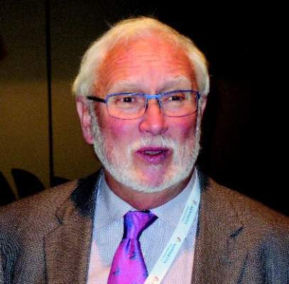
“The patients notice that the itch is gone very quickly, sometimes within days, even before their dermatitis starts to heal,” said Dr. Jon M. Hanifin, one of the treatment’s investigators. “It’s very exciting.”

Dr. Hanifin, a researcher at Oregon Health & Science University, Portland, made his comments while presenting the data during the late-breaking clinical research session at this year’s annual meeting of the American Academy of Dermatology.
The novel treatment, currently known as CIM331 (Chugai) targets elevated levels of interleukin-31, a cytokine that has been implicated in the pathophysiology of atopic dermatitis (AD) and pruritus.
The multicenter, multi-dose study assigned 264 patients, primarily in their mid-30s, with moderate-to-severe AD, uncontrolled by topical treatments, to either placebo or 0.1 mg/kg, 0.5 mg/kg, or 2.0 mg/kg CIM331 every 4 weeks. The study completion rate was 82%.
Using a visual analog scale of 0 to 10, with 10 characterized as “the worst imaginable,” at week 12, patients in the three study groups reported −41.5%, −61.2%, and −60.5% reductions in pruritus vs. −20.1% for placebo (P less than .01 for all).
The “significant reductions” in itch began as early as week 1, particularly in the group treated with the 2-mg/kg dose. Dr. Hanifin said that the patients had a very high mean pruritis score, and some had as much as half of their body surface area affected by AD.
Rescue medications such as topical corticosteroids were not permitted until after at least 1 month of treatment. Patients given rescue therapies were required to have both itch and dermatitis. “This was kind of a tough thing to get through, but [patients] did okay,” Dr. Hanifin said.
Patients treated with the 0.5-mg/kg dose showed the most improvement from baseline in Eczema Area and Severity Index scores at week 12 at −44.6%, compared with −20.9% for placebo. Across the study arms, the proportion of static Investigator’s Global Assessment of 1 or less was 20.9% vs. 4.7% for placebo. Additionally, Dr. Hanifin said sleep latency in the study groups was improved by half and there was an increase in total overall sleep time.
CIM331 was well tolerated, with the most common adverse events being exacerbation of AD and nasopharyngitis. “The risk of immune suppression seemed to be low,” Dr. Hanifin said.
On Twitter @whitneymcknight
WASHINGTON – Nemolizumab, an anti-interleukin-31 receptor A monoclonal antibody, was shown to rapidly and consistently improve pruritus, dermatitis, and sleep disturbance in patients with previously uncontrolled moderate-to-severe atopic dermatitis in a phase II, randomized, double-blind, placebo-controlled trial.
“The patients notice that the itch is gone very quickly, sometimes within days, even before their dermatitis starts to heal,” said Dr. Jon M. Hanifin, one of the treatment’s investigators. “It’s very exciting.”
Dr. Hanifin, a researcher at Oregon Health & Science University, Portland, made his comments while presenting the data during the late-breaking clinical research session at this year’s annual meeting of the American Academy of Dermatology.
The novel treatment, currently known as CIM331 (Chugai) targets elevated levels of interleukin-31, a cytokine that has been implicated in the pathophysiology of atopic dermatitis (AD) and pruritus.
The multicenter, multi-dose study assigned 264 patients, primarily in their mid-30s, with moderate-to-severe AD, uncontrolled by topical treatments, to either placebo or 0.1 mg/kg, 0.5 mg/kg, or 2.0 mg/kg CIM331 every 4 weeks. The study completion rate was 82%.
Using a visual analog scale of 0 to 10, with 10 characterized as “the worst imaginable,” at week 12, patients in the three study groups reported −41.5%, −61.2%, and −60.5% reductions in pruritus vs. −20.1% for placebo (P less than .01 for all).
The “significant reductions” in itch began as early as week 1, particularly in the group treated with the 2-mg/kg dose. Dr. Hanifin said that the patients had a very high mean pruritis score, and some had as much as half of their body surface area affected by AD.
Rescue medications such as topical corticosteroids were not permitted until after at least 1 month of treatment. Patients given rescue therapies were required to have both itch and dermatitis. “This was kind of a tough thing to get through, but [patients] did okay,” Dr. Hanifin said.
Patients treated with the 0.5-mg/kg dose showed the most improvement from baseline in Eczema Area and Severity Index scores at week 12 at −44.6%, compared with −20.9% for placebo. Across the study arms, the proportion of static Investigator’s Global Assessment of 1 or less was 20.9% vs. 4.7% for placebo. Additionally, Dr. Hanifin said sleep latency in the study groups was improved by half and there was an increase in total overall sleep time.
CIM331 was well tolerated, with the most common adverse events being exacerbation of AD and nasopharyngitis. “The risk of immune suppression seemed to be low,” Dr. Hanifin said.
On Twitter @whitneymcknight
AT AAD 2016
Key clinical point: Nemolizumab rapidly and consistently improved pruritus, dermatitis and sleep disturbance in patients with previously uncontrolled moderate-to-severe atopic dermatitis.
Major finding: At week 12, patients treated with nemolizumab reported reductions in pruritus of −41.5% for 0.1 mg/kg, −61.2% for 0.5 mg/kg, and −60.5% 2.0 mg/kg vs. −20.1% for placebo (P less than .01 for all).
Data source: Randomized, double-blind, placebo-controlled, multi-center, multi-dose phase II study of 264 patients with moderate-to-severe atopic dermatitis.
Disclosures: This trial was sponsored by Chugai Pharmaceuticals.
Outcomes in major surgery unchanged by continuing clopidogrel
MONTREAL – Patients who stayed on antiplatelet therapy close to – or even up until – major surgery fared just as well as those who stopped their medication earlier in a retrospective, single-center study.
The study found no difference in blood product administration, adverse perioperative events, or all-cause 30-day mortality regardless of whether patients stopped clopidogrel (Plavix) the recommended 5 days before surgery.
“We believe that continuing clopidogrel in elective and emergent surgical situations appears to be safe, and may challenge the current recommendations,” said presenter Dr. David Strosberg.
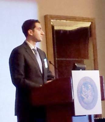
The study addressed a thorny question for surgeons, Dr. Strosberg said. “As surgeons, we face a dilemma: Do we take the risk of thrombotic complications in stopping the antiplatelet drugs, or do we take the risk of increased surgical bleeding with continuing therapy?”
The package insert for clopidogrel advises discontinuation 5 days prior to surgery. However, manufacturer labeling also states that discontinuation of clopidogrel can lead to adverse cardiac events, said Dr. Strosberg, a general surgery resident at Ohio State University in Columbus.
The aim of the study, presented at the annual meeting of the Central Surgical Association, was to ascertain whether continuing antiplatelet therapy increased the rate of adverse surgical outcomes in those undergoing major emergent or elective surgery.
Dr. Strosberg and his colleagues retrospectively reviewed the record of patients over a 4-year period at a single institution and included those undergoing major general, thoracic, or vascular surgery who were taking clopidogrel at the time of presentation.
Data collected included patient characteristics, including demographic data and comorbidities, as well as transfusion requirements and perioperative events.
A total of 200 patients who had 205 qualifying procedures and were taking clopidogrel were included in the study. Of these, 116 patients (Group A) had their clopidogrel held for at least 5 days preoperatively. The remaining 89 patients (Group B) had their clopidogrel held for less than 5 days, or not at all.
Patient demographics were similar between the two groups. Patients in Group A were more likely to have emergency surgery, to have peripheral stents placed, to have COPD or peripheral vascular disease, to have a malignancy, and to have received aspirin within five days of surgery (P less than .01 for all).
Blood product administration rates and volumes did not differ significantly between the two groups, and there was no difference between the groups in the incidence of myocardial infarctions, cerebrovascular events, or acute visceral or lower extremity ischemia.
Three patients in each group died within 30 days of the procedure, a nonsignificant difference. However, in the group that had clopidogrel held, three patients had perioperative myocardial infarctions, and two of these patients died. In discussing the study, Dr. Michael Dalsing said, “I think a lot of us would accept bleeding more over myocardial infarction.”
A subgroup analysis of the group who had clopidogrel held for fewer than 5 days compared outcomes for emergent vs. non-emergent surgery. The emergent surgery subgroup had a significantly higher rate of preoperative platelet transfusions, although numbers overall were small (2/17, 11.8%, vs. 0/72; P = .03).
Dr. Strosberg noted study limitations that included the retrospective, single-center nature of the study, and the fact that one variable, estimated blood loss, is notoriously subjective and inaccurate.
Dr. Dalsing, chief of vascular surgery at Indiana University, Indianapolis, said that he “was surprised that not even one patient went back for postoperative bleeding in this high-risk group of patients,” and raised the question of potential selection bias. Dr. Strosberg replied that comorbidities were ascertained by the physician at the time of surgery planning; since no differences were seen between study groups, investigators didn’t go back and parse out details about comorbid conditions.
In discussion following the presentation, surgeons spoke to the real-world challenges of performing surgery on a patient with antiplatelet therapy on board.
“Overall, I think your data support kind of a bias I have. Since I’m a vascular surgeon, we almost always operate on clopidogrel, and I don’t know if our bleeding risk is worse or better. But it’s something we almost have to do to keep our grafts going,” Dr. Dalsing said.
Dr. Peter Henke, professor of vascular surgery at the University of Michigan, Ann Arbor, said, “I’d be a little bit cautious with this. If you’ve ever done a big aortic procedure on someone on Plavix, I’ve seen them lose up to a couple of liters of blood just with oozing.”
“Those of us who do open aortic surgery know that very few things bleed like the back wall of an aortic anastomosis of a patient on Plavix,” echoed Dr. Peter Rossi, associate professor of vascular surgery at the Medical College of Wisconsin, Milwaukee.
The study authors reported no relevant disclosures.
On Twitter @karioakes
MONTREAL – Patients who stayed on antiplatelet therapy close to – or even up until – major surgery fared just as well as those who stopped their medication earlier in a retrospective, single-center study.
The study found no difference in blood product administration, adverse perioperative events, or all-cause 30-day mortality regardless of whether patients stopped clopidogrel (Plavix) the recommended 5 days before surgery.
“We believe that continuing clopidogrel in elective and emergent surgical situations appears to be safe, and may challenge the current recommendations,” said presenter Dr. David Strosberg.
The study addressed a thorny question for surgeons, Dr. Strosberg said. “As surgeons, we face a dilemma: Do we take the risk of thrombotic complications in stopping the antiplatelet drugs, or do we take the risk of increased surgical bleeding with continuing therapy?”
The package insert for clopidogrel advises discontinuation 5 days prior to surgery. However, manufacturer labeling also states that discontinuation of clopidogrel can lead to adverse cardiac events, said Dr. Strosberg, a general surgery resident at Ohio State University in Columbus.
The aim of the study, presented at the annual meeting of the Central Surgical Association, was to ascertain whether continuing antiplatelet therapy increased the rate of adverse surgical outcomes in those undergoing major emergent or elective surgery.
Dr. Strosberg and his colleagues retrospectively reviewed the record of patients over a 4-year period at a single institution and included those undergoing major general, thoracic, or vascular surgery who were taking clopidogrel at the time of presentation.
Data collected included patient characteristics, including demographic data and comorbidities, as well as transfusion requirements and perioperative events.
A total of 200 patients who had 205 qualifying procedures and were taking clopidogrel were included in the study. Of these, 116 patients (Group A) had their clopidogrel held for at least 5 days preoperatively. The remaining 89 patients (Group B) had their clopidogrel held for less than 5 days, or not at all.
Patient demographics were similar between the two groups. Patients in Group A were more likely to have emergency surgery, to have peripheral stents placed, to have COPD or peripheral vascular disease, to have a malignancy, and to have received aspirin within five days of surgery (P less than .01 for all).
Blood product administration rates and volumes did not differ significantly between the two groups, and there was no difference between the groups in the incidence of myocardial infarctions, cerebrovascular events, or acute visceral or lower extremity ischemia.
Three patients in each group died within 30 days of the procedure, a nonsignificant difference. However, in the group that had clopidogrel held, three patients had perioperative myocardial infarctions, and two of these patients died. In discussing the study, Dr. Michael Dalsing said, “I think a lot of us would accept bleeding more over myocardial infarction.”
A subgroup analysis of the group who had clopidogrel held for fewer than 5 days compared outcomes for emergent vs. non-emergent surgery. The emergent surgery subgroup had a significantly higher rate of preoperative platelet transfusions, although numbers overall were small (2/17, 11.8%, vs. 0/72; P = .03).
Dr. Strosberg noted study limitations that included the retrospective, single-center nature of the study, and the fact that one variable, estimated blood loss, is notoriously subjective and inaccurate.
Dr. Dalsing, chief of vascular surgery at Indiana University, Indianapolis, said that he “was surprised that not even one patient went back for postoperative bleeding in this high-risk group of patients,” and raised the question of potential selection bias. Dr. Strosberg replied that comorbidities were ascertained by the physician at the time of surgery planning; since no differences were seen between study groups, investigators didn’t go back and parse out details about comorbid conditions.
In discussion following the presentation, surgeons spoke to the real-world challenges of performing surgery on a patient with antiplatelet therapy on board.
“Overall, I think your data support kind of a bias I have. Since I’m a vascular surgeon, we almost always operate on clopidogrel, and I don’t know if our bleeding risk is worse or better. But it’s something we almost have to do to keep our grafts going,” Dr. Dalsing said.
Dr. Peter Henke, professor of vascular surgery at the University of Michigan, Ann Arbor, said, “I’d be a little bit cautious with this. If you’ve ever done a big aortic procedure on someone on Plavix, I’ve seen them lose up to a couple of liters of blood just with oozing.”
“Those of us who do open aortic surgery know that very few things bleed like the back wall of an aortic anastomosis of a patient on Plavix,” echoed Dr. Peter Rossi, associate professor of vascular surgery at the Medical College of Wisconsin, Milwaukee.
The study authors reported no relevant disclosures.
On Twitter @karioakes
AT THE ANNUAL MEETING OF THE CENTRAL SURGICAL ASSOCIATION
Key clinical point: Outcomes were similar whether patients did or didn’t stop clopidogrel before major surgery.
Major finding: No significant differences in blood product use, adverse events, or death were seen with continuing clopidogrel.
Data source: Retrospective, single-center review of 200 patients undergoing major elective or emergent surgery and taking clopidogrel.
Disclosures: The study authors reported no relevant disclosures.

SHM Announces 2016 Awards of Excellence Winners
The Society of Hospital Medicine (SHM) created the Awards of Excellence Program to honor its members whose exemplary contributions to the hospital medicine movement merit acknowledgment and celebration. In honor of their achievements, recipients of each Award of Excellence receive an all-expense paid trip to SHM’s annual meeting.
Award recipients also receive recognition on stage in front of friends, family, and colleagues at SHM’s annual meeting, in The Hospitalist, and on www.hospitalmedicine.org.
Congratulations to this year’s winners:
Clinical Excellence
Mark Thoelke, MD, SFHM
Dr. Thoelke became the first hospitalist at Barnes-Jewish Hospital in 1998 and helped form the Hospital Medicine Division of the Washington University School of Medicine in St. Louis in 2000, one of the first divisions in the U.S. The division is now composed of 70 physicians and eight nurse practitioners and consistently turns in superior performances on clinical outcomes as measured by UnitedHealthcare. The division has led the way with innovations in care models and teaching models and was one of the first to offer a sub-internship experience on the non-teaching service and one of the first to offer co-management with their oncology service in 2002. Dr. Thoelke still spends two-thirds of his time on clinical services and states that his job satisfaction comes largely from patient care and teaching.
Humanitarian Services
Bijay Acharya, MBBS, MD
Dr. Acharya works as a hospitalist at Massachusetts General Hospital in Boston and is currently completing the Harvard Medical School/CRICO Fellowship in Patient Safety and Quality. His humanitarian work started when he was in medical school, where he led many health camps in extremely poor villages, ran blood-donation drives, and established the poor-patient fund. After graduation, Dr. Acharya, with his friends, worked to establish a nonprofit clinic named NyayaHealth (now Possible) to serve the healthcare needs of a very remote district in rural Nepal. Prior to the clinic, there was no physician for more than a quarter million people. Recently, after the massive earthquake in Nepal, Dr. Acharya led the relief efforts for the earthquake victims. Dr. Acharya strongly believes in the capacity of hospitalists to be strong advocates for their patients, peers, and communities, both locally and globally.
Non-Physician
Tiffani M. Panek, MA, SFHM, CLHM
Panek is the hospitalist administrator for the Division of Hospital Medicine at the Johns Hopkins Bayview Medical Center in Baltimore. She is a Senior Fellow in Hospital Medicine and has also received her Certificate of Leadership in Hospital Medicine (CLHM) from SHM. She has been at Johns Hopkins for more than 12 years and has been instrumental in the significant growth and success of the Division of Hospital Medicine. Within SHM, she has been a member of the Practice Administrators Committee for three years and was recently elected to a two-year term as vice president of SHM’s Maryland Chapter. She is the first administrator to be elected to chapter leadership, to receive the CLHM, and to have an abstract accepted at an SHM annual meeting.
Outstanding Service
Thomas McIlraith, MD, SFHM, CLHM
Dr. McIlraith is the chairman of the Hospital Medicine Department at Mercy Medical Group in Sacramento, Calif. He improved patient flow between admissions and rounding with a novel operational system called Central Coordination, and it is now the standard for the Dignity Health facilities in Sacramento. The system markedly improved ED response, on-call hospitalist stress, and patient continuity. He has led many other quality and operational improvements, including unit-based rounding, rapid-response team development, and staff restructuring to improve physician coverage. Most recently, he became a leader in the Patient Experience Movement by developing the “Cognitive/Emotional Disconnect” model for understanding patient experience in hospital medicine. He is a member of the SHM Practice Management Committee.
Research
Vineet Chopra, MD, MSc, FHM
Dr. Chopra is an assistant professor of medicine and research scientist in the Patient Safety Enhancement Program at the University of Michigan and Ann Arbor VA Medical Center. Dr. Chopra’s research efforts are centered on improving the safety of hospitalized patients by preventing hospital-acquired complications. Using peripherally inserted central catheters (PICCs) as a model for this inquiry, his work has focused on quantifying current use of PICCs in hospitalized patients, estimating the risk of complications, and defining innovative ways to improve decision making for these devices. His research has been cited 1,962 times (1,580 times since 2010). He is an associate editor of The American Journal of Medicine and the Journal of Hospital Medicine and will serve as chair of SHM’s Research Committee in 2016.
Teaching
Alberto Puig, MD, PhD, SFHM
Dr. Puig has spent his career fully devoted to medical and clinical education. He is an associate professor of medicine at Harvard Medical School in Boston and director of the core educator faculty in the Department of Medicine at Massachusetts General Hospital, where he leads a unique group of physician-teachers fully devoted to clinical education. He is a regular discussant on educational programs for the academy at Harvard Medical School, and his contributions to medical education and clinical hospital teaching have made him a celebrated teacher and educator. Dr. Puig has played an important role at SHM and in the field of hospital medicine through his efforts as a medical educator; he is an avid student of the history of medicine and has been a frequent presenter at SHM’s annual meeting on this topic.
Teamwork
WellSpan Health, Active Bed Management

With the launch of ABM, Dr. Pfeiffer and Dr. Landis hoped to decrease ED length of stay by standardizing the hospitalist processes surrounding admission orders in computerized physician order entry. Ultimately, ABM at WellSpan has maintained the fastest time-to-admission order entry for any service at York Hospital—a decrease to 10 minutes from 80—with less variation for two years. ABM has also sustained national benchmark ED length of stay when the hospital is functioning at general capacity.
ABM also became instrumental in process and outcome objectives from a number of other hospital-wide initiatives. With ABM, more than 90% of a physician’s patient load is on one medical unit (up from 40%), which allowed the hospitalists to implement structured interdisciplinary bedside rounds (SIBR) on all medical units in York and Gettysburg hospitals. The success of ABM and SIBR allowed a transition-of-care project to focus on efficient discharges. Furthermore, Dr. Pfeiffer led a direct admission task force to improve direct admission referrals, safety, and acceptance, the number of which has since doubled. Without hospitalists’ ongoing leadership and effective teamwork, these significant improvements would not have been possible or sustained. TH
The Society of Hospital Medicine (SHM) created the Awards of Excellence Program to honor its members whose exemplary contributions to the hospital medicine movement merit acknowledgment and celebration. In honor of their achievements, recipients of each Award of Excellence receive an all-expense paid trip to SHM’s annual meeting.
Award recipients also receive recognition on stage in front of friends, family, and colleagues at SHM’s annual meeting, in The Hospitalist, and on www.hospitalmedicine.org.
Congratulations to this year’s winners:
Clinical Excellence
Mark Thoelke, MD, SFHM
Dr. Thoelke became the first hospitalist at Barnes-Jewish Hospital in 1998 and helped form the Hospital Medicine Division of the Washington University School of Medicine in St. Louis in 2000, one of the first divisions in the U.S. The division is now composed of 70 physicians and eight nurse practitioners and consistently turns in superior performances on clinical outcomes as measured by UnitedHealthcare. The division has led the way with innovations in care models and teaching models and was one of the first to offer a sub-internship experience on the non-teaching service and one of the first to offer co-management with their oncology service in 2002. Dr. Thoelke still spends two-thirds of his time on clinical services and states that his job satisfaction comes largely from patient care and teaching.
Humanitarian Services
Bijay Acharya, MBBS, MD
Dr. Acharya works as a hospitalist at Massachusetts General Hospital in Boston and is currently completing the Harvard Medical School/CRICO Fellowship in Patient Safety and Quality. His humanitarian work started when he was in medical school, where he led many health camps in extremely poor villages, ran blood-donation drives, and established the poor-patient fund. After graduation, Dr. Acharya, with his friends, worked to establish a nonprofit clinic named NyayaHealth (now Possible) to serve the healthcare needs of a very remote district in rural Nepal. Prior to the clinic, there was no physician for more than a quarter million people. Recently, after the massive earthquake in Nepal, Dr. Acharya led the relief efforts for the earthquake victims. Dr. Acharya strongly believes in the capacity of hospitalists to be strong advocates for their patients, peers, and communities, both locally and globally.
Non-Physician
Tiffani M. Panek, MA, SFHM, CLHM
Panek is the hospitalist administrator for the Division of Hospital Medicine at the Johns Hopkins Bayview Medical Center in Baltimore. She is a Senior Fellow in Hospital Medicine and has also received her Certificate of Leadership in Hospital Medicine (CLHM) from SHM. She has been at Johns Hopkins for more than 12 years and has been instrumental in the significant growth and success of the Division of Hospital Medicine. Within SHM, she has been a member of the Practice Administrators Committee for three years and was recently elected to a two-year term as vice president of SHM’s Maryland Chapter. She is the first administrator to be elected to chapter leadership, to receive the CLHM, and to have an abstract accepted at an SHM annual meeting.
Outstanding Service
Thomas McIlraith, MD, SFHM, CLHM
Dr. McIlraith is the chairman of the Hospital Medicine Department at Mercy Medical Group in Sacramento, Calif. He improved patient flow between admissions and rounding with a novel operational system called Central Coordination, and it is now the standard for the Dignity Health facilities in Sacramento. The system markedly improved ED response, on-call hospitalist stress, and patient continuity. He has led many other quality and operational improvements, including unit-based rounding, rapid-response team development, and staff restructuring to improve physician coverage. Most recently, he became a leader in the Patient Experience Movement by developing the “Cognitive/Emotional Disconnect” model for understanding patient experience in hospital medicine. He is a member of the SHM Practice Management Committee.
Research
Vineet Chopra, MD, MSc, FHM
Dr. Chopra is an assistant professor of medicine and research scientist in the Patient Safety Enhancement Program at the University of Michigan and Ann Arbor VA Medical Center. Dr. Chopra’s research efforts are centered on improving the safety of hospitalized patients by preventing hospital-acquired complications. Using peripherally inserted central catheters (PICCs) as a model for this inquiry, his work has focused on quantifying current use of PICCs in hospitalized patients, estimating the risk of complications, and defining innovative ways to improve decision making for these devices. His research has been cited 1,962 times (1,580 times since 2010). He is an associate editor of The American Journal of Medicine and the Journal of Hospital Medicine and will serve as chair of SHM’s Research Committee in 2016.
Teaching
Alberto Puig, MD, PhD, SFHM
Dr. Puig has spent his career fully devoted to medical and clinical education. He is an associate professor of medicine at Harvard Medical School in Boston and director of the core educator faculty in the Department of Medicine at Massachusetts General Hospital, where he leads a unique group of physician-teachers fully devoted to clinical education. He is a regular discussant on educational programs for the academy at Harvard Medical School, and his contributions to medical education and clinical hospital teaching have made him a celebrated teacher and educator. Dr. Puig has played an important role at SHM and in the field of hospital medicine through his efforts as a medical educator; he is an avid student of the history of medicine and has been a frequent presenter at SHM’s annual meeting on this topic.
Teamwork
WellSpan Health, Active Bed Management
With the launch of ABM, Dr. Pfeiffer and Dr. Landis hoped to decrease ED length of stay by standardizing the hospitalist processes surrounding admission orders in computerized physician order entry. Ultimately, ABM at WellSpan has maintained the fastest time-to-admission order entry for any service at York Hospital—a decrease to 10 minutes from 80—with less variation for two years. ABM has also sustained national benchmark ED length of stay when the hospital is functioning at general capacity.
ABM also became instrumental in process and outcome objectives from a number of other hospital-wide initiatives. With ABM, more than 90% of a physician’s patient load is on one medical unit (up from 40%), which allowed the hospitalists to implement structured interdisciplinary bedside rounds (SIBR) on all medical units in York and Gettysburg hospitals. The success of ABM and SIBR allowed a transition-of-care project to focus on efficient discharges. Furthermore, Dr. Pfeiffer led a direct admission task force to improve direct admission referrals, safety, and acceptance, the number of which has since doubled. Without hospitalists’ ongoing leadership and effective teamwork, these significant improvements would not have been possible or sustained. TH
AEs prompt EMA review of idelalisib

Photo courtesy of
Gilead Sciences, Inc.
The European Medicines Agency (EMA) is reviewing the safety of idelalisib (Zydelig), a drug approved to treat chronic lymphocytic leukemia (CLL) and follicular lymphoma in the European Union (EU).
The European Commission (EC) requested the review because of serious adverse events (AEs), including deaths, reported in 3 clinical trials investigating idelalisib in combination with other drugs.
The AEs were mostly infection-related.
The EMA is reviewing data from these studies to assess whether the findings have any consequences for the authorized uses of idelalisib.
In the meantime, the EMA advises that patients starting or already on treatment with idelalisib be carefully monitored for signs of infections. If the drug is well tolerated, treatment should not be stopped.
The EMA is considering whether any other immediate measures are necessary during the review period. The agency said it will communicate further and keep doctors and patients informed as appropriate.
About idelalisib
In the EU, idelalisib is approved for use in combination with rituximab to treat adults with CLL who have received at least 1 prior therapy or as first-line treatment in the presence of 17p deletion or TP53 mutation in CLL patients unsuitable for chemo-immunotherapy.
Idelalisib is also approved as monotherapy for adults with follicular lymphoma that is refractory to 2 prior lines of treatment.
About the trials
The trials in which patients have experienced serious AEs involve patients with CLL and indolent non-Hodgkin lymphoma (NHL).
In one trial (NCT01732926), researchers are evaluating idelalisib in combination with bendamustine and rituximab for previously treated indolent NHL.
In another (NCT01732913), researchers are testing idelalisib in combination with rituximab for previously treated indolent NHL.
And in the third (NCT01980888), researchers are evaluating idelalisib in combination with bendamustine and rituximab in patients with previously untreated CLL.
The EMA noted that these studies are investigating combinations of drugs that are currently not approved in the EU and include patients with disease characteristics different from those covered by the approved indications for idelalisib.
About the review
The EMA has begun the review of idelalisib at the request of the EC, under Article 20 of Directive 2001/83/EC.
The review is being carried out by the EMA’s Pharmacovigilance Risk Assessment Committee, the committee responsible for the evaluation of safety issues for human medicines, which will make a set of recommendations.
Those recommendations will then be forwarded to the Committee for Medicinal Products for Human Use, which is responsible for questions concerning medicines for human use and will adopt a final opinion on the safety of idelalisib.
The final stage of the review procedure is the EC’s adoption of a legally binding decision that is applicable in all EU member states. ![]()
Global Surgery: ‘Partnership Among Friends’
Surgery volunteerism has been on the rise for several decades. The American College of Surgeons is increasing its role in organizing and facilitating these programs via Operation Giving Back (OGB). And many ACS members are prominent participants in this endeavor.
A leader in global surgery is Michael L. Bentz, M.D., FAAP, FACS, professor of surgery, pediatrics, and neurosurgery, and chairman of the Division of Plastic and Reconstructive Surgery at the University of Wisconsin School of Medicine and Public Health. Dr. Bentz has led international missions in many countries of the world over nearly 20 years and has helped a team develop a long-term program of clinical care and training in Nicaragua. We talked with him about his experiences.
Q: You have been involved in international surgical missions for many years. Can you tell us something about your early projects?
I was first exposed to international work at the University of Pittsburgh. My mentor J. William Futrell, M.D., FACS, was a veteran of over 30 international surgical trips. I went on the first trip with him to Vietnam in the 1997 and have been going ever since. For that initial trip, we worked with a nonprofit organization called Interplast. I went with a large group of 20 people from the University that included plastic surgery attendings, plastic surgery residents, pediatric attendings, pediatric residents, and nursing and anesthesia staff.
In those days, many trips were based predominantly on clinical care – adult care and pediatric care. Teams would do a certain number of operations and then go home. We did cleft lip repairs, cleft palate repairs, burn reconstruction, congenital hand deformity surgery, and tumor management.
That would result in good outcomes for those who actually had a procedure done. But in any place I have ever worked overseas – Vietnam, China, Russia, Nicaragua – the need is overwhelming. The need far outstripped what surgical missions can provide in isolated, single trips back and forth.
Q: The years have brought changes to these missions. What are the most significant changes over the years in how these missions are conducted?
The scope and direction of global health is moving toward sustainable, long-term, and longitudinal education. In those earlier trips where there was an emphasis on doing as many operations as possible, people meant well – we meant well! But the real impact comes with the longitudinal education investment.
I have never been anywhere around the world where there weren’t interested, very capable, excellent surgeons committed to taking care of their patients who only need some support and facilitation.
If you compare the cases we are able to do on a trip with our partners with the cases they are able to do independently, it’s a logarithmic curve – they are far more productive than we could ever be on any number of trips. There is a multiplier effect that allows many more patients to be taken care of.
Q: Your institution has a long-term relationship with a hospital in Nicaragua. How does this work and what is the role of your team in the program?
The University of Wisconsin Division of Plastic Surgery and the Eduplast Foundation has a team of about 10 that goes to Nicaragua twice a year. Most importantly, we support a residency program in there. We move residents through a 3-year modular program much like programs in the U.S. and then examine them. We facilitate this educational process with trips there and we bring them to our institution in the U.S.
Over the past 10 years, we have been doing a weekly live webcast of our Plastic Surgery Grand Rounds which is received on several continents. This creates a very valuable bidirectional, and even tridirectional conversation. This webcast is simple, incredibly inexpensive, and has provided hundreds of hours of education over the years in addition to the on-site work we do.
There can be a language barrier in some cases, but we broadcast in English, with occasional translation support. In addition to Nicaragua, our webcast has been received in institutions in Thailand, China, Ecuador and across the United States. We keep records of cases performed. Our plastic surgery residents can get credit for the cases they do under faculty supervision at our international sites if we meet specific criteria set by our Resident Review Committee.
It is important to note that we take care of the patients in our partner institutions in Nicaragua exactly as we would care for patients in our institution in Wisconsin. There is no “practicing” as all operations are done by surgeons appropriately credentialed and trained for the task.
Q: Do you find that there is a cultural gap that you must bridge in working with colleagues and patients in Nicaragua?
Our program has an orientation session for team participants in advance of each trip, where we talk about the mechanics of the trip – safety, medical issues. We also talk about cultural considerations of each site. It is very important that the residents embed in the culture in which we are working. They also need to know the cultural norms of how to communicate with patients, parents, and children. Some of it is simply good manners – acting like your mother taught you!
The team can reside in a local hotel, but often stays in the homes of local hosts, and this can be a beautiful opportunity to learn about local norms and communication.
Q: What is your favorite part of these missions?
I have so many favorite parts! I like caring for people who otherwise might not receive medical care. This is “giving back” and I think all of the participants would agree that we come home feeling like we received much more than we gave. These experiences remind you of why you went to medical school. It is an opportunity to provide something in return for all the investment that has been made in us for our education. In working with colleagues from other countries, I learn as much as I teach. I come back a better surgeon.
The benefits to residents from our institution are many. They learn how to operate in a resource-limited setting, and they return with a greater appreciation for the equipment and supplies we have available at our institution in Madison. The cultural competence and awareness they also learn is an invaluable life skill.
I want to stress that the friendships with our fellow surgeons are what makes this work. We achieve a degree of continuity and even watch our pediatric patients grow up over the years because of our long-term relationship with the hospital in León and our dedicated colleagues there. This is a truly a partnership among friends.
Q: Do you have some advice for a surgeon interested in participating in an international program?
For those surgeons who were not exposed to these programs during residency, finding a mentor or mentoring organization is the way to begin. A beginner should consider making the first couple of trips with someone who knows the ropes in terms of understanding cultural competency, practical issues of safety, and relevant clinical issues. Almost every surgery discipline has an organization with the capability of identifying volunteer surgery groups in their specialty. ACS’ Operation Giving Back is a particularly important resource for helping Fellows find the right international program.
If you would like to learn more about global surgery programs, contact Operation Giving Back at [email protected]. Or if you would like to share your experiences as an international surgical volunteer, please email this publication at [email protected].
Apply now for 2016 international scholarships for surgical education
The American College of Surgeons (ACS) Division of Education and the International Relations Committee have announced the availability of two international scholarships focused on surgical education for 2016. These awards will offer faculty members from countries other than the U.S. and Canada the opportunity to participate in a variety of faculty development activities that will result in acquisition of new knowledge and skills in surgical education and training. The intent of the program is to help scholars improve surgical education and training in their home institutions and countries. All application materials and supporting documents are due no later than May 2 for attendance at the ACS Clinical Congress 2016, October 16−20 in Washington, DC.
About the program
The two scholars will participate in the annual Clinical Congress, including the Surgical Education: Principles and Practice course, as well as other plenary sessions and Postgraduate Courses that address surgical education and training needs across the continuum of professional development. This continuum may include the needs of practicing surgeons through their entire careers, as well as the needs of surgery residents, medical students, and other members of the surgical team.
After the Clinical Congress, each scholar will visit two Level I ACS Accredited Education Institutes (ACS-AEIs) selected in advance based on the scholars’ interest areas in surgical education and training. At the conclusion of the Clinical Congress and the scholars’ visits to the ACS-AEIs, each recipient will send to the International Relations Committee and the Division of Education a brief report outlining the outcomes that have been achieved as a result of their scholarship; this report should focus specifically on the objectives outlined in their application for the scholarship. The scholarships will facilitate the scholars’ involvement in subsequent collaborative ventures in education and training under the aegis of the ACS Division of Education.
Each scholarship provides a stipend of $10,000, supporting travel and per diem in North America and the cost of Postgraduate Courses undertaken at the Clinical Congress and at the ACS-AEIs to be visited. Clinical Congress registration and fees for attendance at the Surgical Education: Principles and Practices course will be provided. Assistance will be offered to reserve affordable housing in Washington, DC, during the Clinical Congress.
Application requirements
Applicants must document prior experience in surgical education and training, such as involvement in the development and evaluation of education modules, use of novel teaching and assessment strategies, or curriculum design. In addition, applicants must submit a one-paragraph description of their education philosophies, a list of specific educational goals and objectives for their visits, and evidence of support of these goals and objectives from the leadership at their home institutions. These documents will be reviewed by the Division of Education as part of the selection process. At least 5 years of experience beyond completion of all training and fellowships is required. Scholarships must be used in the year awarded; they may not be postponed.
Full scholarship requirements for this program may be reviewed at facs.org/member-services/scholarships/international/issurged. The application for the scholarship may be accessed at the bottom of the requirements page. Questions should be directed to Kate Early, ACS International Liaison, at [email protected].
International Guest Scholarships available for 2017
The American College of Surgeons (ACS) offers International Guest Scholarships to young surgeons from countries other than the U.S. or Canada who have demonstrated a strong interest in teaching and research. Twelve scholarships are available for 2017, in the amount of $10,000 each, and will provide the scholars with an opportunity to engage in clinical, teaching, and research activities in the U.S. and Canada and to attend and participate fully in the educational opportunities and activities of the ACS Clinical Congress. For consideration by the Selection Committee, completed applications for the 2017 International Guest Scholarships and all supporting documentation must be received at the office of the International Liaison Section by June 30, 2016.
Paul R. Hawley, MD, FACS(Hon), Past-ACS Executive Director (1950−1961), left a legacy to the College for the scholarship endowment. More recently, gifts to the International Guest Scholarship endowment from the family of Abdol Islami, MD, FACS, and Joan Islami; the Stavros Niarchos Foundation; and others have enabled the College to increase the number of these scholarship awards.
The scholarship requirements are as follows:
• Applicants must be medical school graduates who have completed their surgical training.
• Applicants must be between 35 and 50 years old on the date that the completed application is filed.
• Applicants must submit their applications from their intended permanent location. Applications will be accepted for processing only if the applicants have been in surgical practice, teaching, or research for a minimum of 1 year at their intended permanent location, following completion of all formal training (including fellowships and scholarships).
• Applicants must have demonstrated a commitment to teaching and/or research in accordance with the standards of the applicant’s country.
• Applicants early in their careers are deemed more suitable than those surgeons who are in senior academic positions.
• Applicants must submit a fully completed application form available on the ACS website at facs.org/member-services/scholarships/international/igs. The application and accompanying materials must be submitted in English. Submission of a curriculum vitae without a completed application is unacceptable.
• Applicants must provide a list of all of their publication credits and must submit three complete reprints or manuscripts from that list.
• Preference may be given to applicants who have not already experienced training or surgical fellowships in the U.S. or Canada.
• Applicants must submit independently prepared letters of recommendation from three of their colleagues. One letter must be from the chair of the department in which the applicant holds an academic appointment or an ACS Fellow residing in the applicant’s country. This letter must include a statement regarding the nature and extent of the teaching and other academic involvement of the applicant. Letters of recommendation should be submitted by the person making the recommendation.
• The online application form is structured to assist the Scholarship Selection Subcommittee and assists the applicant in submitting a structured curriculum vitae.
• The International Guest Scholarships must be used in the year for which they are designated. They may not be postponed.
• Applicants who are awarded scholarships will provide a full written report of the experiences provided through the scholarships upon completion of their tours.
• An unsuccessful applicant may reapply only twice and only by completing and submitting a new application and new supporting documentation.
The scholarships will provide successful applicants with public recognition of their presence. Assistance will be provided in arranging visits, following the Clinical Congress, to various clinics and universities of their choice.
For consideration by the Selection Committee, applicants must fulfill all of the requirements. Applicants are urged to submit their completed applications and supporting documents as early as possible to provide sufficient time for processing.
Send supporting materials to International Liaison Section, American College of Surgeons, 633 N. Saint Clair St., Chicago, IL, 60611-3211; or via fax to 312-202-5021. Questions should be directed to Kate Early, International Liaison, at [email protected].
Community Surgeon Travel Awards available for 2017
Applications and supporting documentation for two 2017 Community Surgeon Travel Awards, sponsored by the International Relations Committee of the American College of Surgeons (ACS), are due July 1, 2016. The travel awards, $4,000 each and available to surgeons ages 30–50 years, allow international surgeons to attend and participate in the educational activities of the annual ACS Clinical Congress. The awards are intended specifically to assist surgeons who work in community or regional hospitals or clinics in countries other than the United States and Canada, or who are from under-resourced academic departments of surgery in under-resourced countries.
The College will cover each awardee’s registration fees for Clinical Congress 2017, October 22−26, in San Diego, CA, as well as the cost of one Postgraduate Course at the meeting. The ACS also will assist the recipients in finding preferential housing in an economical hotel. All applicants will be notified of the Selection Committee’s decision in November 2016.
Application requirements are as follows:
• Applicants must be medical school graduates who have completed their surgical training.
• Applicants must be between 30 and 50 years old on the date that the application is filed.
• Applicants must submit their applications from their intended permanent location. Applications will be accepted for processing only when the applicants have been in surgical practice, teaching, or research for at least 1 year at their intended permanent location and following completion of all formal training (including fellowships and scholarships).
• Applicants must show evidence of commitment to quality care, surgical teaching, and improving access to surgical care in their community.
• Applicants must submit a fully completed application form provided on the ACS website at facs.org/member-services/scholarships/international/communitytravel. The application and accompanying materials must be submitted in English. Submission of a curriculum vitae without a completed application is unacceptable.
• Applicants who have not already experienced training or surgical fellowships in the U.S. or Canada will receive preference for the awards.
• Applicants must submit independently prepared letters of recommendation from three colleagues. One letter must be from the chair of the department in which the applicant holds a clinical or academic appointment or from an ACS Fellow residing in their country. The recommendation letter must directly address the applicant’s commitment to quality care, surgical teaching, and improving access to surgical care locally. Letters of recommendation should be submitted by the individuals making the recommendations.
• The Community Surgeon Travel Awards must be used in the year for which they are designated. They may not be postponed.
• Awardees are expected to provide a written report upon their return home, specifically focusing on the value of the visit to the awardee and the potentially beneficial effect for patients in the country of origin.
• Unsuccessful applicants may reapply only twice and only by completing and submitting a new application together with new supporting documentation.
To qualify for consideration by the Selection Committee, all of the requirements must be fulfilled.
Supporting materials and questions should be directed to Kate Early, International Liaison, at [email protected] or faxed to 312-202-5021.
Save the date for the ACS Surgeons as Leaders Course in June
Save the date for the American College of Surgeons (ACS) Surgeons as Leaders: From Operating Room to Boardroom course, June 5–8 in Durham, NC. Surgeons who aspire to meet the challenges of exemplary leadership across all settings are encouraged to join senior surgical leaders in the three-day course.
Faculty will include the following:
• Course Chair Andrew L. Warshaw, MD, FACS, FRCSEd(Hon), senior consultant, international and regional clinical relations, Massachusetts General Hospital and Partners HealthCare, Boston, MA, and Immediate Past-President of the ACS.
• Julie A. Freischlag, MD, FACS, vice-chancellor, human health sciences, and dean, school of medicine; University of California-Davis Health System, and Past-Chair of the ACS Board of Regents.
• Matthew M. Hutter, MD, MPH, FACS, director, Codman Center for Clinical Effectiveness in Surgery, Massachusetts General Hospital, and associate professor of surgery, Harvard Medical School, Boston.
• Larry R. Kaiser, MD, FACS, president and chief executive officer, Temple University Health System, and dean, Lewis Katz School of Medicine, Temple University, Philadelphia, PA.
• Fabrizio Michelassi, MD, FACS, Lewis Atterbury Stimson Professor and chairman, department of surgery, Weill Cornell Medical College; surgeon in chief, New York-Presbyterian/Weill Cornell Medical Center, New York, NY; and Chair, ACS Board of Governors.
• Carlos A. Pellegrini, MD, FACS, FRCSI(Hon), FRCS(Hon), FRCSEd(Hon), chief medical officer, UW Medicine; vice-president for medical affairs, University of Washington, Seattle; and ACS Past-President.
• Nathaniel J. Soper, MD, FACS, Loyal and Edith Davis Professor and chair, department of surgery, and surgeon in chief, Northwestern Medicine, Chicago, IL, and a Past-Governor of the ACS.
• Beth H. Sutton, MD, FACS, general surgeon, Wichita Falls, TX; clinical professor of surgery, University of Texas Southwestern Medical School, Dallas; and ACS Regent.
• Michael Useem, PhD, William and Jacalyn Egan Professor of Management and director, Center for Leadership and Change Management, Wharton School of University of Pennsylvania, Philadelphia.
• The keynote speaker will be David F. Torchiana, MD, FACS, president and chief executive officer, Partners HealthCare System, Boston
Organized by the ACS Division of Education, the course will help surgeons exhibit leadership attributes; use consensus development and vision to set, align, and achieve goals; build and maintain effective teams; identify factors that hamper the ability to lead; change culture, resolve conflict, and balance demands within the larger environment; and translate the principles of leadership into action. For additional information, e-mail [email protected], or call 312-202-5018. ♦
Register Now for 2016 Leadership & Advocacy Summit
Register today for the American College of Surgeons (ACS) 2016 Leadership & Advocacy Summit, April 9-12, at the J.W. Marriott, Washington, DC. This dual meeting offers volunteer leaders and advocates educational sessions focused on effective surgeon leadership, as well as interactive advocacy training with coordinated visits to congressional offices.
The 2016 Leadership Summit will commence Saturday, April 9, with an evening Welcome Reception, followed the next day by presentations on strategic thinking, the latest on social media for surgeons, building better team communication, improving emotional intelligence, and leading through team conflict, among other topics. Find more information about the Leadership Summit at https://www.facs.org/advocacy/participate/summit-2016. Summit attendees will also meet over lunch in small groups organized by state/region to identify areas for unified efforts in the upcoming year. The Leadership Summit preliminary agenda is available at https://www.facs.org/advocacy/participate/summit-2016/leadership-agenda.
The Advocacy Summit will kick off the evening of April 10 with a dinner featuring political pundit and MSNBC Hardball host and Today Show commentator Chris Matthews. The next day, a number of speakers will discuss the political environment in Washington, DC, and provide updates on important health care issues. Monday’s program will include a luncheon sponsored by the ACS Professional Association’s political action committee (ACSPA-SurgeonsPAC), featuring Larry J. Sabato, PhD, election analyst and author; professor of politics, University of Virginia Center for Politics, Charlottesville. Participants will use the lessons learned at the Advocacy Summit in meetings with their senators and representatives and/or congressional staff on Tuesday. Find more information on the Advocacy Summit at https://www.facs.org/advocacy/participate/summit-2016/advocacy-agenda.