User login
Tips for performing complex laparoscopic gyn surgery

Share your thoughts! Send your Letter to the Editor to [email protected]. Please include your name and the city and state in which you practice.
Share your thoughts! Send your Letter to the Editor to [email protected]. Please include your name and the city and state in which you practice.
Share your thoughts! Send your Letter to the Editor to [email protected]. Please include your name and the city and state in which you practice.

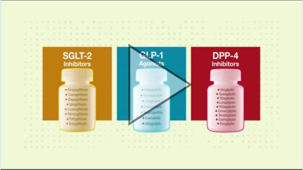
VIDEO: Meta-analysis: Mortality, safety data may favor SGLT2 inhibitors in T2DM
Sodium-glucose cotransporter 2 (SGLT2) inhibitors and glucagonlike peptide–1 (GLP-1) agonists were both associated with a lower mortality risk, compared with that seen with dipeptidyl peptidase–4 (DPP-4) inhibitors and in controls, in patients with type 2 diabetes, according to findings from a large network meta-analysis.
In addition, the GLP-1 agonists were associated with a higher risk of adverse events that led to study withdrawal, compared with SGLT2 inhibitors, according to the analysis conducted by Sean L. Zheng, BM BCh, of the department of endocrinology at the Imperial College Healthcare NHS Foundation Trust, London, and his coinvestigators.
“Of the 3 classes tested, SGLT2 inhibition may be preferred over the incretin-based therapies based on their association with lower mortality and their favorable adverse-event profile,” Dr. Zheng and his coinvestigators wrote in a report on the study published in JAMA.
The video associated with this article is no longer available on this site. Please view all of our videos on the MDedge YouTube channel
Source: JAMA
For patients with type 2 diabetes who don’t achieve target glycemic control on metformin, Dr. Zheng and his coauthors noted, international guidelines recommend SGLT2 inhibitors or incretin-based treatments as a next step.
However, there has been little exploration of the relative clinical effectiveness of these drug classes, which has led to uncertainty about what treatment approach is optimal. “When no head-to-head trial exists, network meta-analysis can be used to estimate the effect,” the authors wrote.
To compare the efficacy of the drug classes in reducing mortality and cardiovascular outcomes, Dr. Zheng and his colleagues conducted a systematic review and meta-analysis of 236 randomized clinical trials including 176,310 participants.
The primary outcome of the study was all-cause mortality.
Both SGLT2 inhibitors and GLP-1 agonists were associated with significantly lower all-cause mortality than that seen in controls (placebo or no treatment), while DPP-4 inhibitors were not, investigators found in the meta-analysis.
For that endpoint, SGLT2 inhibitors had an absolute risk difference of –1.0%, with a hazard ratio of 0.80, and GLP-1 agonists had an absolute RD of –0.6% and an HR of 0.88. By contrast, DPP-4 inhibitors had an absolute RD of 0.1% and an HR of 1.02, according to the published report.
Moreover, when compared with DPP-4 inhibitors, SGLT2 inhibitors and GLP-1 agonists were associated with reduced all-cause mortality, with an absolute risk difference of –0.9% and –0.5%, respectively, they found.
SGLT2 inhibitors and GLP-1 agonists also were significantly associated with lower cardiovascular mortality than controls were, while SGLT2 inhibitors were significantly associated with lower heart failure event rates versus those seen controls, they also found.
Safety outcomes analysis showed that GLP-1 agonists, compared with SGLT2 inhibitors and DPP-4 inhibitors, had a higher risk of adverse events that led patients to withdraw from the study.
The DPP-4 inhibitors were associated with increased acute pancreatitis risk, according to the safety analysis. “SGLT2 inhibitors were associated with increased risk of genital infections but not urinary tract infections. There was a high degree of heterogeneity for lower-limb amputations driven by the significant increase in events with canagliflozin but neutral effects of empagliflozin,” they said.
“Careful treatment selection may be necessary to minimize these outcomes in at-risk patients,” Dr. Zheng and his coauthors concluded.
One author reported potential conflicts of interest with Roche Diabetes, Dexcom, Medtronics Diabetes, and others. Another was supported by a grant from the British Heart Foundation. No other conflicts were reported.
SOURCE: Zheng SL et al. JAMA. 2018;319(15):1580-91.
FROM JAMA
Key clinical point: that were either placebo or no treatment.
Major finding: When compared with DPP-4 inhibitors, SGLT2 inhibitors and GLP-1 agonists were both associated with reduced all-cause mortality, with an absolute risk difference of –0.9% and –0.5%, respectively.
Study details: A systematic review and meta-analysis of 236 randomized clinical trials including 176,310 participants.
Disclosures: One author reported potential conflicts of interest with Roche Diabetes, Dexcom, Medtronics Diabetes, and others. Another was supported by a grant from the British Heart Foundation. No other conflicts were reported.
Source: Zheng SL et al. JAMA. 2018;319(15):1580-91.
Dyspareunia Associated with Vulvovaginal Atrophy: Innovations in Counseling, Diagnosis, and Management

In the United States, there are approximately 64 million women who are postmenopausal. Of these women, it is estimated that 50%—or more than 32 million— have symptoms of vulvovaginal atrophy (VVA) and/or dyspareunia (painful sexual intercourse). These two conditions, along with several others, are components of the Genitourinary Syndrome of Menopause (GSM).
This CME-designated journal supplement is comprised of three articles that provide information and strategies regarding best practices as to patient counseling, diagnosis, and treatment of VVA, and its associated dyspareunia. The goal is to provide women’s health clinicians the knowledge and tools they need to optimize the care they provide to their menopausal patients.
Click here to read the supplement.
https://omniaeducation.com/dyspareunia/
An early version of this supplement distributed at the 2018 American College of Obstetricians and Gynecologists Annual Clinical and Scientific Meetings incorrectly dated the second photo in Figure 9 on page S16 “8-4-2010”. This was later corrected to read “8-4-2011”.

Top AAN picks from Clinical Neurology News’ medical editor
Standout presentations at this year’s American Academy of Neurology annual meeting range from targeting tau in Alzheimer’s disease, to new treatments for spinal muscular atrophy, to the controversial topic of allowing your child to play contact sports, but all are sure to have an impact, according to Clinical Neurology News Medical Editor Richard J. Caselli, MD.
“There are a lot of good talks and papers being presented, and it is impossible without having seen and heard them all to accurately predict what will be the real standouts, but from a purely personal perspective, and with all due apologies to any others not mentioned below, these are some of the ones I think could have large and, in some cases, almost immediate impact or potential impact,” said Dr. Caselli, professor of neurology at the Mayo Clinic Arizona in Scottsdale and also associate director and clinical core director of the Arizona Alzheimer’s Disease Center.
Targeting tau in Alzheimer’s

The measuring of plasma tau to detect preclinical Alzheimer’s, as is described in the abstract from Pase and colleagues, is an “intriguing” approach, Dr. Caselli said. In that study, higher plasma tau levels were observed across correlates of preclinical Alzheimer’s: poorer cognitive function and smaller hippocampal volumes on MRI. Plasma tau level was also a strong predictor of future dementia. It will be presented Friday, April 27, 1:00-3:00 in S48, “Novel Biomarkers in Aging and Dementia.”
Focus continues on SMA
More advancements continue to be made in the treatment of various forms of spinal muscular trophy. In Monday morning’s Presidential Plenary Session, Richard Finkel’s presentation in receipt of the Sidney Carter Award in Child Neurology, should chart the development, current state, and future of antisense oligonucleotide therapy for SMA.
In the Emerging Science poster program on Wednesday, April 25, attendees will get an update on trial results for a different approach to the treatment of SMA using AVXS-101 gene replacement therapy for SMA type 1. John W. Day, MD, PhD, will provide longer-term outcomes after last year’s presentation of results in 15 patients.
Big news in stroke
Gregory Albers, MD, will describe in the Clinical Trials Plenary Session how new evidence from stroke thrombectomy trials such as DEFUSE 3 have led to new recommendations for extending the time window for thrombectomy. The results of DEFUSE 3 were first reported in January at the International Stroke Conference.
Other plenary presentations
In Wednesday’s Frontiers in Neuroscience Session, Dr. Caselli recommended Alan Evans’ discussion of the development and current and upcoming work to use and update the giant, freely accessible “BigBrain” High Resolution 3D Digital Human Brain Atlas.
In the always “fun and interesting” Controversies in Neurology on the morning of Thursday, April 26, the debate on “Should We Use Biomarkers Alone For Diagnosis of Alzheimer’s?” takes on greater interest now that the National Institute on Aging and the Alzheimer’s Association have defined Alzheimer’s disease as a diagnosis based on biomarkers. The separate debate of “Would You Let Your Child Play Contact Sports?” should also bring lots of interesting questions to the forefront of attendees’ minds.
Dr. Steven R. Messé’s talk, “Finally, Some Closure on PFO Closure,” at the Neurology Year in Review on Friday morning, April 27, is “of immediate relevance” as recent clinical trials have begun to determine patient groups for whom PFO closure appears worthwhile, Dr. Caselli said.
He has no relevant disclosures.
Standout presentations at this year’s American Academy of Neurology annual meeting range from targeting tau in Alzheimer’s disease, to new treatments for spinal muscular atrophy, to the controversial topic of allowing your child to play contact sports, but all are sure to have an impact, according to Clinical Neurology News Medical Editor Richard J. Caselli, MD.
“There are a lot of good talks and papers being presented, and it is impossible without having seen and heard them all to accurately predict what will be the real standouts, but from a purely personal perspective, and with all due apologies to any others not mentioned below, these are some of the ones I think could have large and, in some cases, almost immediate impact or potential impact,” said Dr. Caselli, professor of neurology at the Mayo Clinic Arizona in Scottsdale and also associate director and clinical core director of the Arizona Alzheimer’s Disease Center.
Targeting tau in Alzheimer’s
The measuring of plasma tau to detect preclinical Alzheimer’s, as is described in the abstract from Pase and colleagues, is an “intriguing” approach, Dr. Caselli said. In that study, higher plasma tau levels were observed across correlates of preclinical Alzheimer’s: poorer cognitive function and smaller hippocampal volumes on MRI. Plasma tau level was also a strong predictor of future dementia. It will be presented Friday, April 27, 1:00-3:00 in S48, “Novel Biomarkers in Aging and Dementia.”
Focus continues on SMA
More advancements continue to be made in the treatment of various forms of spinal muscular trophy. In Monday morning’s Presidential Plenary Session, Richard Finkel’s presentation in receipt of the Sidney Carter Award in Child Neurology, should chart the development, current state, and future of antisense oligonucleotide therapy for SMA.
In the Emerging Science poster program on Wednesday, April 25, attendees will get an update on trial results for a different approach to the treatment of SMA using AVXS-101 gene replacement therapy for SMA type 1. John W. Day, MD, PhD, will provide longer-term outcomes after last year’s presentation of results in 15 patients.
Big news in stroke
Gregory Albers, MD, will describe in the Clinical Trials Plenary Session how new evidence from stroke thrombectomy trials such as DEFUSE 3 have led to new recommendations for extending the time window for thrombectomy. The results of DEFUSE 3 were first reported in January at the International Stroke Conference.
Other plenary presentations
In Wednesday’s Frontiers in Neuroscience Session, Dr. Caselli recommended Alan Evans’ discussion of the development and current and upcoming work to use and update the giant, freely accessible “BigBrain” High Resolution 3D Digital Human Brain Atlas.
In the always “fun and interesting” Controversies in Neurology on the morning of Thursday, April 26, the debate on “Should We Use Biomarkers Alone For Diagnosis of Alzheimer’s?” takes on greater interest now that the National Institute on Aging and the Alzheimer’s Association have defined Alzheimer’s disease as a diagnosis based on biomarkers. The separate debate of “Would You Let Your Child Play Contact Sports?” should also bring lots of interesting questions to the forefront of attendees’ minds.
Dr. Steven R. Messé’s talk, “Finally, Some Closure on PFO Closure,” at the Neurology Year in Review on Friday morning, April 27, is “of immediate relevance” as recent clinical trials have begun to determine patient groups for whom PFO closure appears worthwhile, Dr. Caselli said.
He has no relevant disclosures.
Upadacitinib for RA shows encouraging results in phase 3 trial
In a recent phase 3 trial, the investigational oral JAK1 inhibitor upadacitinib met all primary and secondary endpoints in patients with moderate to severe rheumatoid arthritis – including clinical remission – compared with adalimumab and placebo, according to its manufacturer.
The ongoing SELECT-COMPARE trial randomized patients with a stable background on methotrexate but with a limited response to the drug to upadacitinib (n = 651), adalimumab (n = 327), or placebo (n = 651). In terms of primary endpoints, 28% of patients taking upadacitinib achieved remission based on 28-joint Disease Activity Score using C-reactive protein at week 12, compared with 18% of those taking adalimumab and 6% taking placebo. A total of 71% achieved an American College of Rheumatology 20% (ACR20) level of response, compared with 63% of those taking adalimumab and 36% of those taking placebo.
In addition, upadacitinib met the ranked secondary endpoints, showing superiority over adalimumab. It did so in terms of ACR50 and ACR70 at week 12, as well as reduction in patient pain scores and improvement in physical function at week 12. Furthermore, it significantly inhibited radiographic progression.
The safety profile was consistent with previous findings: By week 26, 3.7% of patients taking upadacitinib experienced serious adverse events, compared with 4.3% of patients taking adalimumab and 2.9% of those taking placebo.
Find out more in AbbVie’s press release.
Grind it out
“And five more, four more, three more, two more, one more, and done!” Just when you thought you could not stand the searing pain any longer, it ends. Your spin instructor is not only helping you be fit, she is also teaching you an important lesson for life: Sometimes you just need to grind it out.
. College basketball teams need to simply grind it out to advance in the NCAA championship tournament. How might Tiger Woods recover from a disastrous few holes at the Masters? “He’ll just have to grind it out on the back nine.” How will you finally finish your PhD thesis? You’ll have to grind it out this month. It’s how I’m writing this column, how I got my taxes in on time, and, sometimes, how I get through clinic.
The phrase is used to describe something which needs to be done that is tedious, laborious, or joyless. Although the outcome of grinding it out is always pleasant, the task is often considered arduous.
In my dermatology practice, patient demand came in like a lion this March, and to meet our awesome access goals, we needed to add clinics on Saturdays, early mornings, and even a few nights. We met our goal, with supply to spare, and felt proud of our accomplishments. Physician wellness gurus (this author not included) say that, to avoid burnout from such excess work, you must find meaning in your work. Be grateful to help that 24-year-old with acne at 8:15 p.m. Think about how lucky you are to serve that lawyer with hand dermatitis at 8:45 p.m. Celebrate the mom’s cancer-free skin screening at 9:00 p.m. By finding meaning in our work, we’re told, we can achieve clinic nirvana. Except it doesn’t always work, and sometimes it serves us badly.

For the long days that ended in night clinic last month, I found myself counting down those last few patients – “four more, three more, two more, and last one.” I love my work and care about my patients, but sometimes I just have to grind it out. I’m proud of what I’ve accomplished.
Now it’s on to spin class.
Dr. Benabio is director of Healthcare Transformation and chief of dermatology at Kaiser Permanente San Diego. The opinions expressed in this column are his own and do not represent those of Kaiser Permanente. Dr. Benabio is @Dermdoc on Twitter. Write to him at [email protected].
“And five more, four more, three more, two more, one more, and done!” Just when you thought you could not stand the searing pain any longer, it ends. Your spin instructor is not only helping you be fit, she is also teaching you an important lesson for life: Sometimes you just need to grind it out.
. College basketball teams need to simply grind it out to advance in the NCAA championship tournament. How might Tiger Woods recover from a disastrous few holes at the Masters? “He’ll just have to grind it out on the back nine.” How will you finally finish your PhD thesis? You’ll have to grind it out this month. It’s how I’m writing this column, how I got my taxes in on time, and, sometimes, how I get through clinic.
The phrase is used to describe something which needs to be done that is tedious, laborious, or joyless. Although the outcome of grinding it out is always pleasant, the task is often considered arduous.
In my dermatology practice, patient demand came in like a lion this March, and to meet our awesome access goals, we needed to add clinics on Saturdays, early mornings, and even a few nights. We met our goal, with supply to spare, and felt proud of our accomplishments. Physician wellness gurus (this author not included) say that, to avoid burnout from such excess work, you must find meaning in your work. Be grateful to help that 24-year-old with acne at 8:15 p.m. Think about how lucky you are to serve that lawyer with hand dermatitis at 8:45 p.m. Celebrate the mom’s cancer-free skin screening at 9:00 p.m. By finding meaning in our work, we’re told, we can achieve clinic nirvana. Except it doesn’t always work, and sometimes it serves us badly.
For the long days that ended in night clinic last month, I found myself counting down those last few patients – “four more, three more, two more, and last one.” I love my work and care about my patients, but sometimes I just have to grind it out. I’m proud of what I’ve accomplished.
Now it’s on to spin class.
Dr. Benabio is director of Healthcare Transformation and chief of dermatology at Kaiser Permanente San Diego. The opinions expressed in this column are his own and do not represent those of Kaiser Permanente. Dr. Benabio is @Dermdoc on Twitter. Write to him at [email protected].

FDA approves immunotherapy combo for advanced RCC
The Food and Drug Administration has granted approvals to
The approvals were based on statistically significant improvements in overall survival (OS) and objective response rate (ORR) for patients receiving the combination of nivolumab and ipilimumab (n = 425), compared with those receiving sunitinib (n = 422) in CheckMate 214, the FDA said in a press statement.
Median OS was not yet reached in the combination arm at follow-up of 32 months, compared with 25.9 months in the sunitinib arm (hazard ratio, 0.63; 95% confidence interval, 0.44-0.89; P less than .0001). The ORR was 41.6% (95% CI, 36.9-46.5) for the combination versus 26.5% (95% CI, 22.4-31) in the sunitinib arm (P less than .0001).
Efficacy of the combination was not established for patients with favorable-risk disease.
The most common adverse reactions were fatigue, rash, diarrhea, musculoskeletal pain, pruritus, nausea, cough, pyrexia, arthralgia, and decreased appetite.
The recommended schedule and dose is 3 mg/kg nivolumab, followed by 1 mg/kg ipilimumab, on the same day every 3 weeks for four doses, then 240 mg nivolumab every 2 weeks or 480 mg every 4 weeks, the FDA said.
Nivolumab is marketed as Opdivo and ipilimumab as Yervoy by Bristol-Myers Squibb.
MDedge Daily News: Can a nasal spray reverse suicidality?
Smoking boosts heart failure risk in black patients. Respiratory infections increase risk of heart attack and stroke. And pain relievers and inflammatory bowel disease? It’s complicated.
Listen to the MDedge Daily News podcast for all the details on today’s top news.
Smoking boosts heart failure risk in black patients. Respiratory infections increase risk of heart attack and stroke. And pain relievers and inflammatory bowel disease? It’s complicated.
Listen to the MDedge Daily News podcast for all the details on today’s top news.
Smoking boosts heart failure risk in black patients. Respiratory infections increase risk of heart attack and stroke. And pain relievers and inflammatory bowel disease? It’s complicated.
Listen to the MDedge Daily News podcast for all the details on today’s top news.

Is “Runner’s Kidney” a Thing?
Q) Many of my patients are athletes. I recall reading something about kidney disease in marathon runners. Am I remembering correctly?
Although data on acute kidney injury (AKI) in marathon runners are limited, two recent studies have added to our knowledge. In 2017, Mansour et al studied 22 marathon runners, collecting urine and blood samples 24 hours before, immediately after, and 24 hours after a race. The results showed that in 82% of the subjects, serum creatinine increased to a level correlated with stage 1 or 2 AKI (as defined by the Acute Kidney Injury Network criteria).1
Based on urine microscopy results, as well as serum creatinine and novel biomarker levels, the researchers concluded that the runners’ AKI was caused by acute tubular injury—likely induced by ischemia. However, the subjects did not show any evidence of chronic kidney disease (CKD), despite years of running and intensive training. One theory: Habitual running might condition the kidneys to transient ischemic conditions—in other words, they build tolerance to repetitive injury over time.1
Continue to: The other recent study
The other recent study examined use of NSAIDs by ultramarathon
In summary: While marathon runners are prone to AKI, the injury seems to be transient and does not progress to CKD. Furthermore, use of NSAIDs during endurance running may contribute to AKI development, so patients should be advised to use caution with these analgesics. Finally, remind your endurance runners to stay hydrated, since it may help to limit kidney damage. As for the casual runner? The impact on the kidney remains unclear and needs further investigation. —DSW
Danielle S. Wentworth, MSN, FNP-BC
Division of Nephrology, University of Viriginia Health System, Charlottesville
1. Mansour SG, Verma G, Pata RW, et al. Kidney injury and repair biomarkers in marathon runners. Am J Kidney Dis. 2017;70(2):252-261.
2. Lipman GS, Shea K, Christensen M, et al. Ibuprofen versus placebo effect on acute kidney injury in ultramarathons: a randomised controlled trial. Emerg Med J. 2017;34(10):637-642.
Clinician Reviews in partnership with
Renal Consult is edited by Jane S. Davis, CRNP, DNP, a member of the Clinician Reviews editorial board, who is a nurse practitioner in the Division of Nephrology at the University of Alabama at Birmingham and is the communications chairperson for the National Kidney Foundation's Council of Advanced Practitioners (NKF-CAP); and Kim Zuber, PA-C, MSPS, DFAAPA, a semi-retired PA who works with the American Academy of Nephrology PAs and is a past chair of the NKF-CAP. This month's responses were authored by Danielle S. Wentworth, MSN, FNP-BC, who practices in the Division of Nephrology at the University of Virginia Health System in Charlottesville, and Barbara Weis Malone, DNP, FNP-C, FNKF, who is an Assistant Professor in the Adult/Gerontology NP Program in the College of Nursing, and a Nurse Practitioner in the School of Medicine, at the University of Colorado Anschutz Medical Campus.
Q) Many of my patients are athletes. I recall reading something about kidney disease in marathon runners. Am I remembering correctly?
Although data on acute kidney injury (AKI) in marathon runners are limited, two recent studies have added to our knowledge. In 2017, Mansour et al studied 22 marathon runners, collecting urine and blood samples 24 hours before, immediately after, and 24 hours after a race. The results showed that in 82% of the subjects, serum creatinine increased to a level correlated with stage 1 or 2 AKI (as defined by the Acute Kidney Injury Network criteria).1
Based on urine microscopy results, as well as serum creatinine and novel biomarker levels, the researchers concluded that the runners’ AKI was caused by acute tubular injury—likely induced by ischemia. However, the subjects did not show any evidence of chronic kidney disease (CKD), despite years of running and intensive training. One theory: Habitual running might condition the kidneys to transient ischemic conditions—in other words, they build tolerance to repetitive injury over time.1
Continue to: The other recent study
The other recent study examined use of NSAIDs by ultramarathon
In summary: While marathon runners are prone to AKI, the injury seems to be transient and does not progress to CKD. Furthermore, use of NSAIDs during endurance running may contribute to AKI development, so patients should be advised to use caution with these analgesics. Finally, remind your endurance runners to stay hydrated, since it may help to limit kidney damage. As for the casual runner? The impact on the kidney remains unclear and needs further investigation. —DSW
Danielle S. Wentworth, MSN, FNP-BC
Division of Nephrology, University of Viriginia Health System, Charlottesville
1. Mansour SG, Verma G, Pata RW, et al. Kidney injury and repair biomarkers in marathon runners. Am J Kidney Dis. 2017;70(2):252-261.
2. Lipman GS, Shea K, Christensen M, et al. Ibuprofen versus placebo effect on acute kidney injury in ultramarathons: a randomised controlled trial. Emerg Med J. 2017;34(10):637-642.

Dr. Pellegrini receives Seattle Business Leaders in Health Care Lifetime Achievement Award
Carlos A. Pellegrini, MD, FACS, FRCSI(Hon), FRCS(Hon), FRCSEd(Hon), a Past-President of the American College of Surgeons, has received Seattle Business magazine’s 2018 Leaders in Health Care Lifetime Achievement Award for his committed service to improving the quality of patient care in the Seattle, WA, area.
Dr. Pellegrini has worked in the University of Washington (UW), Seattle, department of surgery since 1993, first as chair of the department and then in 1996 as the Henry N. Harkins Professor and Chair, until 2015, when he was appointed to serve as UW Medicine’s first chief medical officer (CMO).
According to the Seattle Business article on Dr. Pellegrini’s achievement, as CMO, Dr. Pellegrini oversees thousands of health care providers and has led a program that has visibly improved patient care quality, reduced costs, and “ensured that all of the health care system’s 270,000 patients have an assigned primary care provider across its primary care clinics.” He also integrated clinical services for key programs and created a training program to prepare young clinicians for leadership roles.
Dr. Pellegrini said that his motivation has always been to help people, as a surgeon, a mentor, or, as he notes about his role as CMO, by “advancing social issues and the care that we provide our patients.”
Read more about Dr. Pellegrini’s life and career in the Seattle Business article at http://seattlebusinessmag.com:8080/health-care/2018-leaders-health-care-lifetime-achievement-award-carlos-pellegrini-uw-medicine.