User login
NAFLD patients with abnormal liver tests may not get statins when indicated
WASHINGTON – Though the liver safety of statins in patients with low-level liver enzyme elevations has long been established, some providers still hesitate to prescribe them to the patients with the conditions for which they are indicated.
Nonalcoholic fatty liver disease (NAFLD), hyperlipidemia, metabolic syndrome, and diabetes, which often co-occur, are also involved in cardiovascular disease. Cardiovascular disease is the most common cause of mortality in NAFLD, before liver disease.
Sonal Kumar, MD, MPH, of New York–Presbyterian Hospital described in a video interview at the annual Digestive Disease Week® a study she and her colleagues conducted to evaluate statin use in patients with hyperlipidemia by using data from the National Health and Nutrition Examination Survey during 2005-2014 (NHANES). Adult patients aged over 18 years were included if they did not have viral hepatitis, did not excessively consume alcohol, were not pregnant, and did not have transaminase levels over 500 IU/L.
Statin use was assessed in 136,833,627 participants by NHANES interviewers. Of these participants, 74.6% had hyperlipidemia (defined as LDL cholesterol greater than 130 mg/dL) and 93.5% were taking a statin. Patients with hyperlipidemia with abnormal alanine aminotransferase values were significantly less likely to be taking a statin (86.3% vs. 89.1%, P = .001). With multivariate analysis, abnormal ALT significantly decreased the odds of patients receiving a statin if they had diabetes (odds ratio, 0.75; 95% confidence interval, 0.57-0.99) when sex and age were controlled for.
Statins are underutilized in patients with NAFLD and diabetes, patient groups in whom they could help control cardiovascular disease risk factors, said Dr. Kumar. Providers need to be educated on the safety of statins in these patients to improve cardiovascular outcomes.
Dr. Kumar reported receiving support from Gilead and AbbVie.
REPORTING FROM DDW 2018
Key clinical point: Patients diagnosed with hyperlipidemia who had abnormal ALT levels were less likely to take a statin (86.3% vs. 89.1%, P = .001).
Major finding: Abnormal ALT significantly decreased the odds of patients receiving a statin if they had diabetes (odds ratio, 0.75; 95% confidence interval, 0.57-0.99) when sex and age were controlled for.
Data source: Data from 136,833,627 adult patients from the National Health and Nutrition Examination Survey collected during 2005-2014.
Disclosures: Dr. Kumar reported receiving support from Gilead and AbbVie.

Check SVS Website for New Research Opportunities
Looking for a research opportunity? Check our updated website for current programs in your area. If your institution has an opportunity to promote, let us know at [email protected].
Looking for a research opportunity? Check our updated website for current programs in your area. If your institution has an opportunity to promote, let us know at [email protected].
Looking for a research opportunity? Check our updated website for current programs in your area. If your institution has an opportunity to promote, let us know at [email protected].
Percutaneous procedure gives alternative to anticoagulation for portal vein thrombosis
WASHINGTON – Catheter-directed clot lysis and thrombectomy with creation of a bypass shunt is a reasonable alternative to prolonged anticoagulation for treating patients with portal vein thrombosis (PVT) based on the accumulated reported experience since 1993 using this percutaneous treatment.
” Nelson Valentin, MD, said at the annual Digestive Disease Week.® “TIPS should be considered a viable treatment option for patients with PVT,” said Dr. Valentin, a gastroenterology fellow at Mount Sinai Beth Israel hospital in New York.
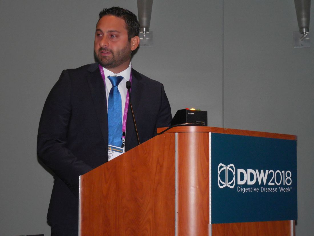
“There is sufficient evidence from these reports to at least consider TIPS as an adjunct to anticoagulation or perhaps as primary therapy,” especially for patients with PVT who have a contraindication for anticoagulation, Dr. Valentin said in an interview. Standard anticoagulation for PVT would today involve acute treatment with a low-molecular-weight heparin followed by oral anticoagulation for a total treatment time of at least 6 months and continued for a year or longer in some patients. A recently published review of reported experience using anticoagulation to treat PVT found a complete recanalization rate of 41% and a complete or partial rate of 66%, which suggests that TIPS is at least as effective, although Dr. Valentin cautioned that no reported study has directly compared the two alternative approaches. A study designed to make this direct comparison is warranted by the reported results using TIPS, Dr. Valentin said. And the experience with TIPS positions it as an option for patients who do not respond to anticoagulation or would prefer an alternative to prolonged anticoagulation.
One factor currently limiting use of TIPS, which is usually performed by an interventional radiologist, is that the procedure is technically demanding, with a limited number of operators with the expertise to perform it. If TIPS became more widely accepted as an option for treating PVT, then the pool of interventionalists experienced with performing the procedure would grow, Dr. Valentin noted.
[email protected]
On Twitter @mitchelzoler
SOURCE: Valentin N et al. Digestive Disease Week, Presentation 361.
WASHINGTON – Catheter-directed clot lysis and thrombectomy with creation of a bypass shunt is a reasonable alternative to prolonged anticoagulation for treating patients with portal vein thrombosis (PVT) based on the accumulated reported experience since 1993 using this percutaneous treatment.
” Nelson Valentin, MD, said at the annual Digestive Disease Week.® “TIPS should be considered a viable treatment option for patients with PVT,” said Dr. Valentin, a gastroenterology fellow at Mount Sinai Beth Israel hospital in New York.
“There is sufficient evidence from these reports to at least consider TIPS as an adjunct to anticoagulation or perhaps as primary therapy,” especially for patients with PVT who have a contraindication for anticoagulation, Dr. Valentin said in an interview. Standard anticoagulation for PVT would today involve acute treatment with a low-molecular-weight heparin followed by oral anticoagulation for a total treatment time of at least 6 months and continued for a year or longer in some patients. A recently published review of reported experience using anticoagulation to treat PVT found a complete recanalization rate of 41% and a complete or partial rate of 66%, which suggests that TIPS is at least as effective, although Dr. Valentin cautioned that no reported study has directly compared the two alternative approaches. A study designed to make this direct comparison is warranted by the reported results using TIPS, Dr. Valentin said. And the experience with TIPS positions it as an option for patients who do not respond to anticoagulation or would prefer an alternative to prolonged anticoagulation.
One factor currently limiting use of TIPS, which is usually performed by an interventional radiologist, is that the procedure is technically demanding, with a limited number of operators with the expertise to perform it. If TIPS became more widely accepted as an option for treating PVT, then the pool of interventionalists experienced with performing the procedure would grow, Dr. Valentin noted.
[email protected]
On Twitter @mitchelzoler
SOURCE: Valentin N et al. Digestive Disease Week, Presentation 361.
REPORTING FROM DDW 2018
Key clinical point: Reported worldwide experience with TIPS in 439 patients shows it works and is relatively safe.
Major finding: TIPS was technically successful in 87% of reported patients and achieved complete portal recanalization in 74% of patients.
Study details: Systematic review of 18 published case series from 1993 to 2016 with 439 total patients.
Disclosures: Dr. Valentin had no disclosures.
Source: Valentin N et al. Digestive Disease Week, Presentation 361.
Volunteers Sought for ‘Task Force on Future of Vascular Surgery’
The SVS is recruiting volunteers to serve on a new Task Force on the Future of Vascular Surgery, which will examine a number of critical trends shaping the specialty. President-elect Michael Makaroun, MD, will chair the new group. Learn more, including how to volunteer, here.
The SVS is recruiting volunteers to serve on a new Task Force on the Future of Vascular Surgery, which will examine a number of critical trends shaping the specialty. President-elect Michael Makaroun, MD, will chair the new group. Learn more, including how to volunteer, here.
The SVS is recruiting volunteers to serve on a new Task Force on the Future of Vascular Surgery, which will examine a number of critical trends shaping the specialty. President-elect Michael Makaroun, MD, will chair the new group. Learn more, including how to volunteer, here.
Submit a case to VAM's “Ask the Experts"
Help build the Vascular Annual Meeting’ new “Ask the Experts” sessions. The four small-group sessions will focus on common issues in vascular disease — coding, aortic care for occlusive disease, hemodialysis and PAD — with an expert reviewing a member's prior case. Members are invited to submit a case on these topics, briefly describing the case and providing a brief history and physical exam of the patient, the results and any additional issues they want to discuss during the program. Sessions will be held daily, Wednesday through Saturday. Submit a case here.
Help build the Vascular Annual Meeting’ new “Ask the Experts” sessions. The four small-group sessions will focus on common issues in vascular disease — coding, aortic care for occlusive disease, hemodialysis and PAD — with an expert reviewing a member's prior case. Members are invited to submit a case on these topics, briefly describing the case and providing a brief history and physical exam of the patient, the results and any additional issues they want to discuss during the program. Sessions will be held daily, Wednesday through Saturday. Submit a case here.
Help build the Vascular Annual Meeting’ new “Ask the Experts” sessions. The four small-group sessions will focus on common issues in vascular disease — coding, aortic care for occlusive disease, hemodialysis and PAD — with an expert reviewing a member's prior case. Members are invited to submit a case on these topics, briefly describing the case and providing a brief history and physical exam of the patient, the results and any additional issues they want to discuss during the program. Sessions will be held daily, Wednesday through Saturday. Submit a case here.
Don’t Miss the Upcoming Vascular Annual Meeting; Register Today
Don’t miss the vascular surgery world’s headline event! Join colleagues and friends in Boston for this year’s Vascular Annual Meeting, June 20 to 23. Scientific sessions are June 21-23 and the Exhibit Hall is open June 21 to 22. Click here to register. To get a full schedule and begin creating your own personal agenda, complete with marking sessions as favorites, see the VAM Planner.
Don’t miss the vascular surgery world’s headline event! Join colleagues and friends in Boston for this year’s Vascular Annual Meeting, June 20 to 23. Scientific sessions are June 21-23 and the Exhibit Hall is open June 21 to 22. Click here to register. To get a full schedule and begin creating your own personal agenda, complete with marking sessions as favorites, see the VAM Planner.
Don’t miss the vascular surgery world’s headline event! Join colleagues and friends in Boston for this year’s Vascular Annual Meeting, June 20 to 23. Scientific sessions are June 21-23 and the Exhibit Hall is open June 21 to 22. Click here to register. To get a full schedule and begin creating your own personal agenda, complete with marking sessions as favorites, see the VAM Planner.
When is denosumab an option in myeloma?
CHICAGO – , G. David Roodman, MD, PhD, reported at the annual meeting of the American Society of Clinical Oncology.
“We use denosumab in patients with compromised renal function,” said Dr. Roodman, director of the Division of Hematology-Oncology at Indiana University, Indianapolis, noting one such scenario. That use of denosumab echoes recently published ASCO guidelines on bone-modifying therapy.
The second scenario for denosumab use is in patients who aren’t tolerating bisphosphonates: “We switch them from zoledronic acid to pamidronate, and they still have terrible acute phase reactions,” Dr. Roodman said.
Dr. Roodman’s comments on use of denosumab were in response to an audience question about when he would use denosumab, given the considerable cost difference between the RANK ligand inhibitor and bisphosphonates.
The recent ASCO guidelines, of which Dr. Roodman is a coauthor, state that denosumab “is more expensive than zoledronic acid or pamidronate and must be considered in treatment decisions.”
Previously, ASCO guidelines recommended use of intravenous bisphosphonates for patients with myeloma and evidence of bone disease. Based on consideration of new evidence, the guideline authors eliminated the requirement for evidence of bone disease and added denosumab as an alternative treatment choice.
The addition of denosumab was based in part on results of a recent randomized phase 3 trial that comprised 1,718 myeloma patients who were treated with either denosumab or zoledronic acid.
The primary endpoint, time to first on-study skeletal-related event, was evaluated after 676 skeletal-related events had accrued on study. The investigators found no difference in time to first event (hazard ratio [HR], 0.98; 95% confidence interval, 0.85-1.14; P = 0.82).
Likewise, the secondary endpoint of overall survival showed no difference between arms (HR, 0.90; 95% CI, 0.70-1.16), though an exploratory analysis did suggest denosumab was superior on the endpoint of progression-free survival (HR, 0.82; 95% CI, 0.68-0.99).
The ASCO guidelines also recommend that clinicians consider less-frequent dosing in patients with responsive or stable disease. That recommendation is based on results of two studies of less-frequent dosing prompted by concerns over the risk of osteonecrosis of the jaw, an uncommon but potentially serious complication associated with bone-modifying agents.
Both studies suggested every-3-months dosing of zoledronic acid could be effective. However, Dr. Roodman noted that both studies had limitations that need to be considered, including small numbers of myeloma patients, limited duration of therapy studied, and a high dropout rate in the case of one study. Due to those limitations, “it’s very difficult to draw conclusions about this today,” Dr. Roodman said.
Dr. Roodman reported that he had a consulting or advisory role with Amgen.
EXPERT ANALYSIS FROM ASCO 2018
What do the genes GDF15 and IGFBP7 mean for the future of hyperemesis gravidarum treatment?
The video associated with this article is no longer available on this site. Please view all of our videos on the MDedge YouTube channel
The video associated with this article is no longer available on this site. Please view all of our videos on the MDedge YouTube channel
The video associated with this article is no longer available on this site. Please view all of our videos on the MDedge YouTube channel
WHAT DOES THIS MEAN FOR PRACTICE?
Genes GDF15 and IGFBP7 have been associated with hyperemesis gravidarum
The association may allow for future techniques in the prediction, prevention, and treatment of hyperemesis gravidarum

IMPACT study: Matched targeted therapy improves survival in advanced cancer
CHICAGO – according to findings from a retrospective analysis of molecularly profiled patients.
Of 3,743 patients tested as part of IMPACT (Initiative for Molecular Profiling and Advanced Cancer Therapy), 1,307 (34.9%) had at least one targetable molecular alteration. Of those, 711 (54.4%) received either matched targeted therapy that was being tested in a clinical trial or – in a small number of cases – therapy with an approved treatment used off label, and 596 (45.6%) received nonmatched therapy, Apostolia-Maria Tsimberidou, MD, reported during a press briefing at the annual meeting of the American Society of Clinical Oncology.
The objective response rates in 697 evaluable matched therapy patients was 16.2% versus 5.4% in 571 evaluable nonmatched patients, and stable disease for at least 6 months occurred in 18.7% and 14.7% of patients, respectively, for an overall disease control rate of 34.9% versus 20.1%, said Dr. Tsimberidou, a professor at the University of Texas MD Anderson Cancer Center, Houston.
Median progression-free survival in those who received matched versus nonmatched therapy was 4.0 months and 2.8 months, respectively (hazard ratio, 0.67), and median overall survival was 9.3 and 7.3 months, respectively (HR, 0.72), she said.
The 3-year overall survival rate was 15% versus 7%, respectively, and 10-year survival was 6% and 1%, respectively.
Patients included in IMPACT had a mean age of 57 years, and 39% were men. They were heavily pretreated (mean number of prior therapies was 4); only 2.8% of patients had no prior treatment. Cancers included gastrointestinal (24.2%), gynecologic (19.4%), breast (13.5%), melanoma (11.9%) and lung (8.7%).
In this video interview, Dr. Tsimberidou describes the rationale, methodology, and findings of IMPACT, including the use of a prognostic scoring system developed as part of the study to predict overall survival based on baseline characteristics, such as baseline p13K/AKT/mTOR pathway molecular alterations, which were shown on multivariate analysis in IMPACT to predict shorter overall survival versus other alterations. Other predictors of shorter survival included liver metastases, elevated lactate dehydrogenase levels, poor functional status, low albumin levels, elevated platelet counts, and age of 60 years or older.
“We [also] wanted to see if adding the intervention ... would hold significance in this multivariate model, and we found that ... nonmatched therapy was associated with adverse survival; it was an independent factor associated with worse survival,” she said. “Therefore, matched targeted therapy is associated with longer survival.”
In the randomized, phase 2 trial IMPACT 2, progression-free survival will be compared in patients with and without matched targeted therapy, and the prognostic scoring system developed as part of IMPACT to predict overall survival based on baseline characteristics will be further evaluated, she said.
During a discussion of the findings during the press briefing, ASCO Expert Catherine M. Diefenbach, MD, said the type of precision medicine studied in IMPACT is “the wave of the future.
“Large scale efforts such as ASCO’s TAPUR or the NCI-MATCH trial will bring these efforts to many, many more patients, and hopefully usher in a new way of treating advanced cancer patients that will improve overall survival for many more patients,” said Dr. Diefenbach, of New York University.
Dr. Tsimberidou reported a consulting or advisory role with Roche, as well as research funding to her institution from EMD Serono, Baxter, Foundation Medicine, ONYX, Bayer, Boston Biomedical, Placon, IMMATICS, Karus Therapeutics, and StemCells.
SOURCE: Tsimberidou AM et al. ASCO 2018, Abstract LBA 2553.
REPORTING FROM ASCO 2018
Key clinical point: Matched targeted therapy improved survival in patients with advanced cancer.
Major finding: The 3-yearoverall survival rate with matched versus nonmatched therapy was 15% and 7%, respectively.
Study details: A retrospective analysis (IMPACT) of 3,743 molecularly profiled advanced cancer patients.
Disclosures: Dr. Tsimberidou reported a consulting or advisory role with Roche, as well as research funding to her institution from EMD Serono, Baxter, Foundation Medicine, ONYX Medical, Bayer, Boston Biomedical, Placon, IMMATICS, Karus Therapeutics, and StemCells.
Source: Tsimberidou AM et al. ASCO 2018, Abstract LBA 2553.
Frontline immunotherapy boosts survival in NSCLC patients
CHICAGO – Pembrolizumab (Keytruda) as first-line treatment of advanced non–small-cell lung cancer (NSCLC) offered longer overall survival with better tolerability compared with chemotherapy, results of the Keynote-042 phase 3 randomized trial show.
Among 1,274 patients with advanced, previously untreated NSCLC with expression of the PD-L1 on 1% or more of tumor cells, median overall survival after a median follow-up of 12.8 months was 16.7 months for patients treated with pembrolizumab monotherapy, compared with 12.1 months for patients treated with either paclitaxel or pemetrexed plus carboplatin, reported lead author Gilberto Lopes, MD, of the Sylvester Comprehensive Cancer Center at the University of Miami.
For all three PD-L1 expression groups, the median duration of response was 20.2 months, compared with 10.8-8.3 months for patients in the chemotherapy arm.
“These are responses that are unlike anything that we have seen with chemotherapy in the past for non–small-cell lung cancer,” Dr. Lopes said at a briefing prior to his presentation of the data in a plenary session.
“In addition to that, and probably more importantly, patients had fewer adverse events [with pembrolizumab]. Overall, about 60% had any treatment-related adverse event with pembrolizumab, vs. 90% with chemotherapy,” he added.
‘A true milestone’
ASCO expert John Heymach, MD, PhD, of the University of Texas MD Anderson Cancer Center in Houston, said at the briefing that “this study represents a true milestone for the field, because now, for the first time, we can say that among non–small-cell lung cancer patients receiving first-line therapy, the vast majority can receive immunotherapy with pembrolizumab instead of chemotherapy.”
He noted that an earlier study, Keynote-024, showed that pembrolizumab significantly improved progression-free survival in patients with tumors expressing PD-L1 on at least 50% of cells compared with standard platinum-based chemotherapy (10.3 vs. 6 months).

The Keynote-042 investigators enrolled 1,274 patients with locally advanced or metastatic NSCLC, and randomly assigned them to receive either a maximum of 35 cycles of pembrolizumab 200 mg every 3 weeks, or the investigators’ choice of not more than 6 cycles of either paclitaxel/carboplatin or pemetrexed/carboplatin, with optional pemetrexed maintenance for patients with nonsquamous histologies only.
The randomization was stratified by region (Asia vs. non–East Asia), Eastern Cooperative Oncology Group performance status 0 or 1, squamous vs. nonsquamous histology, and PD-L1 expression, or TPS (tumor proportion score) greater than 50% vs. 1%-49%.
As noted before, the primary endpoint of overall survival among all patients with a TPS of 1% or greater was met, with respective median overall survival in the pembrolizumab vs. chemotherapy groups of 16.7 vs. 12.1 months, translating into a hazard ratio favoring pembrolizumab of 0.81 (P = .0018). Respective hazard ratios for the TPS 20% or greater and TPS 50% or greater groups were 0.77 (P = .0020), and 0.69 (P = .0003).
At 12.8 months of median follow-up, 13% of patients assigned to pembrolizumab were still on the drug, and 4.3% of patients were receiving maintenance pemetrexed.
Treatment-related adverse events of any grade occurred in 399 of 636 patients assigned to pembrolizumab (62.7%), vs. 553 of 615 patients assigned to chemotherapy (89.9%).
Grade 3 or greater events occurred in 17.8% vs. 41% of patients, respectively, There were 13 deaths related to therapy in the pembrolizumab arm (2.0%), and 14 in the chemotherapy arm (2.3%).
Adverse events leading to discontinuation were similar between the groups, at 9% and 9.4%, respectively.
There were more immune-mediated adverse events in the pembrolizumab arm (27.8% vs. 7.2%), and of these, grade 3 or greater events occurred in 8% vs. 1.5% of patients, respectively.
There was one immune-mediated death, from pneumonitis, in the immunotherapy arm; there were no deaths related to immune-mediated side effects in the chemotherapy arm.
“I really view this as a ‘double whammy’ for patients,” Dr. Heymach said at the briefing. “Often advances in survival for our lung cancer patients come at the cost of significant toxicities. Here, by contrast, not only are patients living longer and having a much higher likelihood of prolonged survival in years, often instead of months, but they’re also receiving a treatment that has substantially less toxicity across virtually all measures, and this really impacts the day-to-day life of these patients.”
Leena Gandhi, MD, PhD, of the Perlmutter Cancer Center at New York University, the invited discussant at the plenary, agreed that pembrolizumab improves survival, compared with chemotherapy patients with PD-L1 expression levels greater than 1%, but noted that most of the benefit – as also seen in Keynote-024 – was in those patients whose tumors had high levels of PD-L1 expression.
She emphasized that although PD-L1 is an imperfect biomarker, it should still be used to help select patients for therapy, and may be complementary with tumor mutational burden for more precise treatment selection.
“What we know, and what this study adds to, is that PD-L1 really does define a patient population that could receive benefit from pembrolizumab over chemotherapy. Patients with low or no PD-L1 expression likely should get some type of combination therapy,” she said.
“I do think this study extends what we’ve seen from other recent studies, which is that chemotherapy alone is no longer a first-line standard of care in non–small-cell lung cancer,” she added.
Merck funded the study. Dr. Lopes disclosed institutional research funding from Merck Sharp & Dohme, EMD Serono, and AstraZeneca. Dr. Heymach disclosed stock/ownership in Bio-Tree and Cardinal Spine, a consulting or advisory role for Abbvie, ARIAD, AstraZeneca, Boehringer Ingelheim, Bristol-Myers Squibb, Calithera Biosciences, Genentech, Medivation, Novartis, Oncomed, and Synta, and institutional research funding from AstraZeneca. Dr. Gandhi reported having no relevant disclosures.
SOURCE: Lobes G et al. ASCO 2018, abstract LBA4.
CHICAGO – Pembrolizumab (Keytruda) as first-line treatment of advanced non–small-cell lung cancer (NSCLC) offered longer overall survival with better tolerability compared with chemotherapy, results of the Keynote-042 phase 3 randomized trial show.
Among 1,274 patients with advanced, previously untreated NSCLC with expression of the PD-L1 on 1% or more of tumor cells, median overall survival after a median follow-up of 12.8 months was 16.7 months for patients treated with pembrolizumab monotherapy, compared with 12.1 months for patients treated with either paclitaxel or pemetrexed plus carboplatin, reported lead author Gilberto Lopes, MD, of the Sylvester Comprehensive Cancer Center at the University of Miami.
For all three PD-L1 expression groups, the median duration of response was 20.2 months, compared with 10.8-8.3 months for patients in the chemotherapy arm.
“These are responses that are unlike anything that we have seen with chemotherapy in the past for non–small-cell lung cancer,” Dr. Lopes said at a briefing prior to his presentation of the data in a plenary session.
“In addition to that, and probably more importantly, patients had fewer adverse events [with pembrolizumab]. Overall, about 60% had any treatment-related adverse event with pembrolizumab, vs. 90% with chemotherapy,” he added.
‘A true milestone’
ASCO expert John Heymach, MD, PhD, of the University of Texas MD Anderson Cancer Center in Houston, said at the briefing that “this study represents a true milestone for the field, because now, for the first time, we can say that among non–small-cell lung cancer patients receiving first-line therapy, the vast majority can receive immunotherapy with pembrolizumab instead of chemotherapy.”
He noted that an earlier study, Keynote-024, showed that pembrolizumab significantly improved progression-free survival in patients with tumors expressing PD-L1 on at least 50% of cells compared with standard platinum-based chemotherapy (10.3 vs. 6 months).
The Keynote-042 investigators enrolled 1,274 patients with locally advanced or metastatic NSCLC, and randomly assigned them to receive either a maximum of 35 cycles of pembrolizumab 200 mg every 3 weeks, or the investigators’ choice of not more than 6 cycles of either paclitaxel/carboplatin or pemetrexed/carboplatin, with optional pemetrexed maintenance for patients with nonsquamous histologies only.
The randomization was stratified by region (Asia vs. non–East Asia), Eastern Cooperative Oncology Group performance status 0 or 1, squamous vs. nonsquamous histology, and PD-L1 expression, or TPS (tumor proportion score) greater than 50% vs. 1%-49%.
As noted before, the primary endpoint of overall survival among all patients with a TPS of 1% or greater was met, with respective median overall survival in the pembrolizumab vs. chemotherapy groups of 16.7 vs. 12.1 months, translating into a hazard ratio favoring pembrolizumab of 0.81 (P = .0018). Respective hazard ratios for the TPS 20% or greater and TPS 50% or greater groups were 0.77 (P = .0020), and 0.69 (P = .0003).
At 12.8 months of median follow-up, 13% of patients assigned to pembrolizumab were still on the drug, and 4.3% of patients were receiving maintenance pemetrexed.
Treatment-related adverse events of any grade occurred in 399 of 636 patients assigned to pembrolizumab (62.7%), vs. 553 of 615 patients assigned to chemotherapy (89.9%).
Grade 3 or greater events occurred in 17.8% vs. 41% of patients, respectively, There were 13 deaths related to therapy in the pembrolizumab arm (2.0%), and 14 in the chemotherapy arm (2.3%).
Adverse events leading to discontinuation were similar between the groups, at 9% and 9.4%, respectively.
There were more immune-mediated adverse events in the pembrolizumab arm (27.8% vs. 7.2%), and of these, grade 3 or greater events occurred in 8% vs. 1.5% of patients, respectively.
There was one immune-mediated death, from pneumonitis, in the immunotherapy arm; there were no deaths related to immune-mediated side effects in the chemotherapy arm.
“I really view this as a ‘double whammy’ for patients,” Dr. Heymach said at the briefing. “Often advances in survival for our lung cancer patients come at the cost of significant toxicities. Here, by contrast, not only are patients living longer and having a much higher likelihood of prolonged survival in years, often instead of months, but they’re also receiving a treatment that has substantially less toxicity across virtually all measures, and this really impacts the day-to-day life of these patients.”
Leena Gandhi, MD, PhD, of the Perlmutter Cancer Center at New York University, the invited discussant at the plenary, agreed that pembrolizumab improves survival, compared with chemotherapy patients with PD-L1 expression levels greater than 1%, but noted that most of the benefit – as also seen in Keynote-024 – was in those patients whose tumors had high levels of PD-L1 expression.
She emphasized that although PD-L1 is an imperfect biomarker, it should still be used to help select patients for therapy, and may be complementary with tumor mutational burden for more precise treatment selection.
“What we know, and what this study adds to, is that PD-L1 really does define a patient population that could receive benefit from pembrolizumab over chemotherapy. Patients with low or no PD-L1 expression likely should get some type of combination therapy,” she said.
“I do think this study extends what we’ve seen from other recent studies, which is that chemotherapy alone is no longer a first-line standard of care in non–small-cell lung cancer,” she added.
Merck funded the study. Dr. Lopes disclosed institutional research funding from Merck Sharp & Dohme, EMD Serono, and AstraZeneca. Dr. Heymach disclosed stock/ownership in Bio-Tree and Cardinal Spine, a consulting or advisory role for Abbvie, ARIAD, AstraZeneca, Boehringer Ingelheim, Bristol-Myers Squibb, Calithera Biosciences, Genentech, Medivation, Novartis, Oncomed, and Synta, and institutional research funding from AstraZeneca. Dr. Gandhi reported having no relevant disclosures.
SOURCE: Lobes G et al. ASCO 2018, abstract LBA4.
REPORTING FROM ASCO 2018
Key clinical point: Many patients with previously untreated non–small-cell lung cancer could benefit from first-line therapy with the checkpoint inhibitor pembrolizumab.
Major finding: Among all patients with expression of PD-L1 on 1% or more of tumor, overall survival was 16.7 months with pembrolizumab, vs. 12.1 months for chemotherapy.
Study details: Randomized phase 3 trial of 1,274 patients with advanced or metastatic non–small-cell lung cancer.
Disclosures: Merck funded the study. Dr. Lopes disclosed institutional research funding from Merck Sharp & Dohme, EMD Serono, and AstraZeneca. Dr. Heymach disclosed stock/ownership in Bio-Tree and Cardinal Spine, a consulting or advisory role for Abbvie, ARIAD, AstraZeneca, Boehringer Ingelheim, Bristol-Myers Squibb, Calithera Biosciences, Genentech, Medivation, Novartis, Oncomed, and Synta, and institutional research funding from AstraZeneca. Dr. Gandhi reported having no relevant disclosures.
Source: Lobes G et al. ASCO 2018, abstract LBA4.