User login
Acute Kwashiorkor in the Setting of Cerebral Palsy and Pancreatic Insufficiency
To the Editor:
Kwashiorkor, or protein-calorie malnutrition, is a common issue in developing countries subject to starvation. In economically advanced nations, however, kwashiorkor is extremely rare and may appear in children placed on restrictive diets instituted by well-meaning guardians. Kwashiorkor also may occur because of gastrointestinal malabsorption. We present a unique case of kwashiorkor that revealed an underlying diagnosis of pancreatic insufficiency.
A 12-year-old girl presented to the hospital with 4 days of watery nonbloody diarrhea occurring with every feeding as well as new onset of presumed diaper dermatitis that had not responded to nystatin cream. Facial swelling also was noted the day prior to admission. Her medical history was notable for cerebral palsy secondary to nonaccidental trauma, leaving the patient nonverbal and quadriplegic. She had numerous prior admissions for sepsis with marked hypotension and more recently was diagnosed with insulin-dependent type 2 diabetes mellitus. She had never lived outside the United States and resided at home with her adoptive parents.
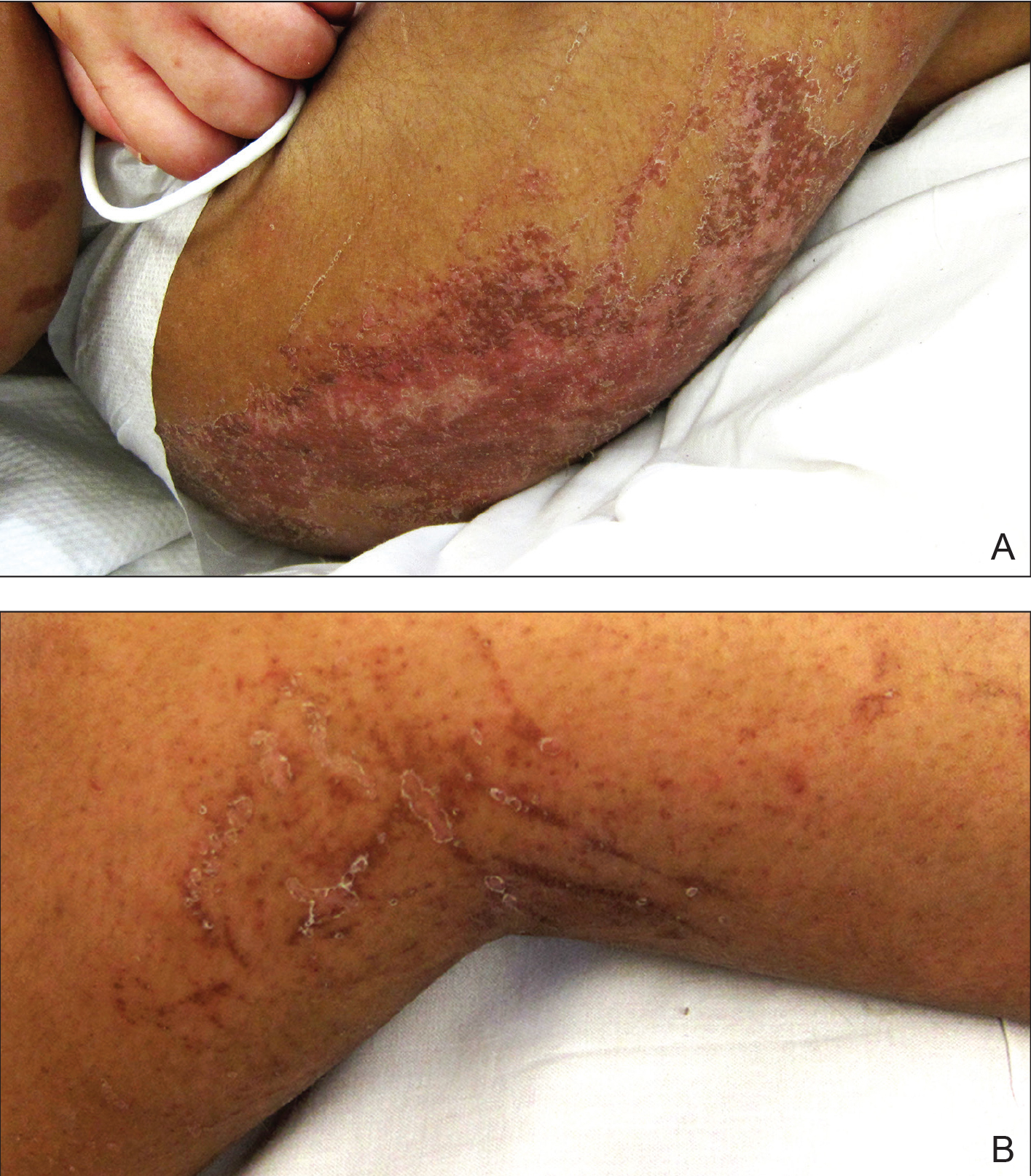
Physical examination revealed a nonverbal underweight girl (weight, 25 kg). Large areas of denudation with surrounding desquamated skin resembling flaking enamel paint covered the buttocks and posterior legs bilaterally (Figure). She had linear hyperpigmented patches on the dorsal hands with one superficial erosion on the left wrist. Marked periorbital edema as well as nonpitting edema of the face, arms, and legs were present.
Upon additional questioning, the patient’s adoptive parent reported a diet of formula containing 1.0 cal/mL with 200-mL feedings 3 times daily through a Geiger-Müller tube, providing a daily protein intake of approximately 17.7 g per day (0.7 g/kg per day). On the day of admission, abnormal laboratory findings included low protein and albumin levels at 4.6 g/dL (reference range, 5.7–8.2 g/dL) and 2.1 g/dL (reference range, 3.2–4.8 g/dL), respectively; an elevated aspartate aminotransferase level of 73 U/L (reference range, 10–34 U/L); and an elevated alanine aminotransferase level of 80 U/L (reference range, 10–40 U/L). Based on the patient’s characteristic clinical findings and abnormal laboratory values, a diagnosis of acute kwashiorkor was made. Although the zinc level was low at 0.29 µg/mL (reference range, 0.66–1.10 µg/mL), the patient did not have any periorificial involvement to support a diagnosis of acrodermatitis enteropathica.
Upon further workup, stool elastase was measured at less than 50 µg per gram of stool (reference range, >200 µg pancreatic elastase per gram of stool), confirming a diagnosis of severe pancreatic insufficiency. Pancreatic enzyme supplementation was initiated along with an increase in protein intake to 1.5 g/kg per day. The patient’s hospital course was complicated by respiratory distress and sepsis, leading to a prolonged hospital stay. A component of refeeding syndrome may have contributed to the patient’s respiratory distress.
Kwashiorkor, a form of protein malnutrition, is caused by inadequate protein intake and usually is seen in developing countries when children are weaned from breastmilk to a diet high in starch and low in protein. It is characterized by edema, growth retardation, a characteristic dermatosis, depigmentation of hair, lethargy, and irritability.1 If left untreated, kwashiorkor can be fatal. Skin changes associated with kwashiorkor first occur in areas of friction or pressure. The skin develops patches of hyperpigmentation that subsequently desquamate in a pattern likened to flaky paint. In the current case of a nonmobile child with diarrhea, prominent involvement of the buttocks and thighs would be expected. This dermatosis does not appear in marasmus and is pathognomonic for kwashiorkor when seen in a child with edema.2
Children in the United States developing kwashiorkor secondary to severely restrictive diets has been reported.3 However, kwashiorkor also may occur due to underlying chronic malabsorptive disease. There have been rare reports of children with cystic fibrosis presenting with kwashiorkor,4 as well as a case of kwashiorkor secondary to underlying infantile Crohn disease.5
Cerebral palsy is associated with multiple different risk factors for malnutrition. Musculoskeletal deformities, oral-motor difficulties, medication side effects, limited communication skills, compromised pulmonary status, and poor muscle tone can all contribute to energy and nutrient deprivation.6 A 2018 study including 728 children registered into the Bangladesh Cerebral Palsy Register between January 2015 and December 2016 demonstrated that more than two-thirds were underweight (70.0%) and stunted (73.1%) and that children with tri/quadriplegic cerebral palsy presented with the highest proportion of severe malnutrition.7 In another report (N=142), up to 85% of children with spastic quadriplegia had severe feeding problems,8 making this population particularly high risk for poor nutritional status.
Pancreatic exocrine insufficiency is characterized by reduced secretion of amylase, lipase, and protease, and it may result in diarrhea, weight loss, malabsorption of essential nutrients, and malnutrition. Pancreatic exocrine insufficiency may occur in the setting of chronic pancreatitis, pancreatic surgery, and cystic fibrosis. Our patient had numerous hospitalizations for sepsis marked by hypotension, and in the absence of more typical causes, we postulate that both endocrine and exocrine pancreatic damage resulted from prolonged hypotension. A sweat chloride test was not performed, as the patient had not experienced frequent pulmonary infections or other signs of cystic fibrosis.
According to a report from the Food and Agriculture Organization of the United Nations/World Health Organization/United Nations University (FAO/WHO/UNU), protein should provide at least 10% of the total caloric intake in a child.9 Although the adoptive parent approximated that our patient received 12% of her daily calories in the form of protein, the amount that she absorbed in the context of pancreatic insufficiency was undoubtedly much lower.
In this case, the diagnosis of kwashiorkor led to the discovery of underlying pancreatic exocrine insufficiency. Low stool elastase confirmed the diagnosis. Because kwashiorkor is rare in developed countries, the classic signs and symptoms may go unrecognized, which can lead to delayed diagnosis and notable morbidity and mortality. New-onset edema and desquamative rash in a child, especially a child with cerebral palsy, should alert physicians to the possibility of acute kwashiorkor and prompt investigation into underlying medical issues that may have contributed to its development.
1. Trowell HC, Davies JN, Dean RF. Kwashiorkor. II. clinical picture, pathology, and differential diagnosis. Br Med J. 1952;2:798-801.
2. Latham MC. The dermatosis of kwashiorkor in young children. Semin Dermatol. 1991;10:270-272.
3. Liu T, Howard RM, Mancini AJ, et al. Kwashiorkor in the United States: fad diets, perceived and true milk allergy, and nutritional ignorance. Arch Dermatol. 2001;137:630-636.
4. Phillips RJ, Crock CM, Dillon MJ, et al. Cystic fibrosis presenting as kwashiorkor with florid skin rash. Arch Dis Childhood. 1993;69:446-448.
5. Al-Mubarak L, Al-Khenaizan S, Al Goufi T. Cutaneous presentation of kwashiorkor due to infantile Crohn’s disease. Eur J Pediatr. 2010;169:117-119.
6. Wittenbrook W. Nutritional assessment and intervention in cerebral palsy. Practical Gastroenterol. Feb 2011;92:16-32. http://www.practicalgastro.com/pdf/February11/WittenbrookArticle.pdf.
7. Jahan I, Muhit M, Karim T, et al. What makes children with cerebral palsy vulnerable to malnutrition? findings from the Bangladesh cerebral palsy register (BCPR)[published online April 16, 2018]. Disabil Rehabil. doi:10.1080/09638288.2018.1461260.
8. Stallings VA, Charney EB, Davies JC, et al. Nutrition-related growth failure of children with quadriplegic cerebral palsy. Dev Med Child Neurol. 1993;35:126-138.
9. World Health Organization. Energy and Protein Requirements: Report of a Joint FAO/WHO/UNU Expert Consultation. Geneva, Switzerland: World Health Organization; 1985. Technical Report Series 724.
To the Editor:
Kwashiorkor, or protein-calorie malnutrition, is a common issue in developing countries subject to starvation. In economically advanced nations, however, kwashiorkor is extremely rare and may appear in children placed on restrictive diets instituted by well-meaning guardians. Kwashiorkor also may occur because of gastrointestinal malabsorption. We present a unique case of kwashiorkor that revealed an underlying diagnosis of pancreatic insufficiency.
A 12-year-old girl presented to the hospital with 4 days of watery nonbloody diarrhea occurring with every feeding as well as new onset of presumed diaper dermatitis that had not responded to nystatin cream. Facial swelling also was noted the day prior to admission. Her medical history was notable for cerebral palsy secondary to nonaccidental trauma, leaving the patient nonverbal and quadriplegic. She had numerous prior admissions for sepsis with marked hypotension and more recently was diagnosed with insulin-dependent type 2 diabetes mellitus. She had never lived outside the United States and resided at home with her adoptive parents.
Physical examination revealed a nonverbal underweight girl (weight, 25 kg). Large areas of denudation with surrounding desquamated skin resembling flaking enamel paint covered the buttocks and posterior legs bilaterally (Figure). She had linear hyperpigmented patches on the dorsal hands with one superficial erosion on the left wrist. Marked periorbital edema as well as nonpitting edema of the face, arms, and legs were present.
Upon additional questioning, the patient’s adoptive parent reported a diet of formula containing 1.0 cal/mL with 200-mL feedings 3 times daily through a Geiger-Müller tube, providing a daily protein intake of approximately 17.7 g per day (0.7 g/kg per day). On the day of admission, abnormal laboratory findings included low protein and albumin levels at 4.6 g/dL (reference range, 5.7–8.2 g/dL) and 2.1 g/dL (reference range, 3.2–4.8 g/dL), respectively; an elevated aspartate aminotransferase level of 73 U/L (reference range, 10–34 U/L); and an elevated alanine aminotransferase level of 80 U/L (reference range, 10–40 U/L). Based on the patient’s characteristic clinical findings and abnormal laboratory values, a diagnosis of acute kwashiorkor was made. Although the zinc level was low at 0.29 µg/mL (reference range, 0.66–1.10 µg/mL), the patient did not have any periorificial involvement to support a diagnosis of acrodermatitis enteropathica.
Upon further workup, stool elastase was measured at less than 50 µg per gram of stool (reference range, >200 µg pancreatic elastase per gram of stool), confirming a diagnosis of severe pancreatic insufficiency. Pancreatic enzyme supplementation was initiated along with an increase in protein intake to 1.5 g/kg per day. The patient’s hospital course was complicated by respiratory distress and sepsis, leading to a prolonged hospital stay. A component of refeeding syndrome may have contributed to the patient’s respiratory distress.
Kwashiorkor, a form of protein malnutrition, is caused by inadequate protein intake and usually is seen in developing countries when children are weaned from breastmilk to a diet high in starch and low in protein. It is characterized by edema, growth retardation, a characteristic dermatosis, depigmentation of hair, lethargy, and irritability.1 If left untreated, kwashiorkor can be fatal. Skin changes associated with kwashiorkor first occur in areas of friction or pressure. The skin develops patches of hyperpigmentation that subsequently desquamate in a pattern likened to flaky paint. In the current case of a nonmobile child with diarrhea, prominent involvement of the buttocks and thighs would be expected. This dermatosis does not appear in marasmus and is pathognomonic for kwashiorkor when seen in a child with edema.2
Children in the United States developing kwashiorkor secondary to severely restrictive diets has been reported.3 However, kwashiorkor also may occur due to underlying chronic malabsorptive disease. There have been rare reports of children with cystic fibrosis presenting with kwashiorkor,4 as well as a case of kwashiorkor secondary to underlying infantile Crohn disease.5
Cerebral palsy is associated with multiple different risk factors for malnutrition. Musculoskeletal deformities, oral-motor difficulties, medication side effects, limited communication skills, compromised pulmonary status, and poor muscle tone can all contribute to energy and nutrient deprivation.6 A 2018 study including 728 children registered into the Bangladesh Cerebral Palsy Register between January 2015 and December 2016 demonstrated that more than two-thirds were underweight (70.0%) and stunted (73.1%) and that children with tri/quadriplegic cerebral palsy presented with the highest proportion of severe malnutrition.7 In another report (N=142), up to 85% of children with spastic quadriplegia had severe feeding problems,8 making this population particularly high risk for poor nutritional status.
Pancreatic exocrine insufficiency is characterized by reduced secretion of amylase, lipase, and protease, and it may result in diarrhea, weight loss, malabsorption of essential nutrients, and malnutrition. Pancreatic exocrine insufficiency may occur in the setting of chronic pancreatitis, pancreatic surgery, and cystic fibrosis. Our patient had numerous hospitalizations for sepsis marked by hypotension, and in the absence of more typical causes, we postulate that both endocrine and exocrine pancreatic damage resulted from prolonged hypotension. A sweat chloride test was not performed, as the patient had not experienced frequent pulmonary infections or other signs of cystic fibrosis.
According to a report from the Food and Agriculture Organization of the United Nations/World Health Organization/United Nations University (FAO/WHO/UNU), protein should provide at least 10% of the total caloric intake in a child.9 Although the adoptive parent approximated that our patient received 12% of her daily calories in the form of protein, the amount that she absorbed in the context of pancreatic insufficiency was undoubtedly much lower.
In this case, the diagnosis of kwashiorkor led to the discovery of underlying pancreatic exocrine insufficiency. Low stool elastase confirmed the diagnosis. Because kwashiorkor is rare in developed countries, the classic signs and symptoms may go unrecognized, which can lead to delayed diagnosis and notable morbidity and mortality. New-onset edema and desquamative rash in a child, especially a child with cerebral palsy, should alert physicians to the possibility of acute kwashiorkor and prompt investigation into underlying medical issues that may have contributed to its development.
1. Trowell HC, Davies JN, Dean RF. Kwashiorkor. II. clinical picture, pathology, and differential diagnosis. Br Med J. 1952;2:798-801.
2. Latham MC. The dermatosis of kwashiorkor in young children. Semin Dermatol. 1991;10:270-272.
3. Liu T, Howard RM, Mancini AJ, et al. Kwashiorkor in the United States: fad diets, perceived and true milk allergy, and nutritional ignorance. Arch Dermatol. 2001;137:630-636.
4. Phillips RJ, Crock CM, Dillon MJ, et al. Cystic fibrosis presenting as kwashiorkor with florid skin rash. Arch Dis Childhood. 1993;69:446-448.
5. Al-Mubarak L, Al-Khenaizan S, Al Goufi T. Cutaneous presentation of kwashiorkor due to infantile Crohn’s disease. Eur J Pediatr. 2010;169:117-119.
6. Wittenbrook W. Nutritional assessment and intervention in cerebral palsy. Practical Gastroenterol. Feb 2011;92:16-32. http://www.practicalgastro.com/pdf/February11/WittenbrookArticle.pdf.
7. Jahan I, Muhit M, Karim T, et al. What makes children with cerebral palsy vulnerable to malnutrition? findings from the Bangladesh cerebral palsy register (BCPR)[published online April 16, 2018]. Disabil Rehabil. doi:10.1080/09638288.2018.1461260.
8. Stallings VA, Charney EB, Davies JC, et al. Nutrition-related growth failure of children with quadriplegic cerebral palsy. Dev Med Child Neurol. 1993;35:126-138.
9. World Health Organization. Energy and Protein Requirements: Report of a Joint FAO/WHO/UNU Expert Consultation. Geneva, Switzerland: World Health Organization; 1985. Technical Report Series 724.
Practice Points
• Pancreatic exocrine deficiency, confirmed by low stool elastase, can lead to kwashiorkor and requires a high index of suspicion for diagnosis.
• Kwashiorkor is not only seen in developing countries but also in certain at-risk populations in economically advantaged countries.
• For multiple reasons, patients with cerebral palsy are at particular risk for nutritional deficiencies including kwashiorkor.
External Trigeminal Neurostimulation Offers Relief
One-hour treatment with external trigeminal nerve stimulation resulted in significant headache pain relief compared to sham stimulation and was well tolerated, according to a recent double-blind, randomized, sham-controlled study conducted across 3 headache centers in the United States. Adult patients who were experiencing an acute migraine attack, with or without aura, were recruited on site and randomly assigned 1:1 to receive either verum or sham external trigeminal nerve stimulation treatment for 1 hour. Pain intensity was scored using a visual analog scale. Researchers found:
- One hundred and six patients were randomized and included in the intention-to-treat analysis (verum: n=52; sham: n=54).
- The primary outcome measure was significantly more reduced in the verum group than in the sham group: −3.46 ± 2.32 vs −1.78 ± 1.89, or −59% vs −30%.
- With regards to migraine subgroups, there was a significant difference in pain reduction between verum and sham for “migraine without aura” attacks.
- For “migraine with aura” attacks, pain reduction was numerically greater for verum vs sham, but did not reach significance: mean visual analog scale reduction at 1 hour was −4.3 ± 1.8 for the verum group vs −2.6 ± 1.9 for the sham group.
Chou DE, Yugrakh MS, Winegarner D, Rowe V, Kuruvilla D, Schoenen. Acute migraine therapy with external trigeminal neurostimulation (ACME): A randomized controlled trial. [Published online ahead of print November 17, 2018]. Cephalalgia. doi:10.1177%2F0333102418811573.
Alcoholic Beverages Recognized as Migraine Trigger
Alcoholic beverages, especially red wine, are recognized as a migraine trigger factor by patients with migraine and have a substantial effect on alcohol consumption behavior, according to a recent study. Researchers conducted a cross‐sectional, web‐based, questionnaire study among 2197 patients with migraine from the well‐defined Leiden University MIgraine Neuro‐Analysis (LUMINA) study population. They assessed alcoholic beverage consumption and self‐reported trigger potential, reasons behind alcohol abstinence, and time between alcohol consumption and migraine attack onset. They found:
- Alcoholic beverages were reported as a trigger by 35.6% of participants with migraine.
- In addition, more than 25% of patients with migraine who had stopped consuming or never consumed alcoholic beverages did so because of presumed trigger effects.
- Wine, especially red wine (77.8% of participants), was recognized as the most common trigger among the alcoholic beverages.
- However, red wine consistently led to an attack in only 8.8% of participants.
- Time of onset was rapid (<3 hours) in one‐third of patients and almost 90% had an onset of less than 10 hours independent of beverage type.
Onderwater GLC, van Oosterhaut WPJ, Schoonman GG, Ferrari MD, Terwindt GM. Alcoholic beverages as trigger factor and the effect on alcohol consumption behavior in patients with migraine. [Published online ahead of print December 18, 2018]. Eur J Neurol. doi:10.1111/ene.13861.
Inhibitory Pain Modulation in Adolescents Assessed
Adolescents with migraine and healthy adolescents have similar inhibitory pain modulation capability despite having marked differences in pain sensitivity, according to a recent study. Although participants with a family history of migraine (Fam-His) were asymptomatic, they demonstrated alterations in pain processing, which may serve as markers for prediction of migraine development. In order to determine if inhibitory pain modulation occurs in youth as it does in adults, researchers performed a quantitative sensory testing investigation in adolescents with migraine (n=19). These patients were compared to healthy adolescents with (n=20) or without (n=29) Fam-His of migraine (eg, first degree relative with migraine). They found:
- In response to graded heat stimuli, Fam-His participants reported higher pain intensity ratings compared to migraine patients, who in turn, reported higher pain intensity ratings than the healthy controls.
- For heat- and pressure- conditioned pain modulation (CPM), there was no significant group difference in the magnitude of CPM responses.
Nahman-Averbuch H, Leon E, Hunter BM, et al. Increased pain sensitivity but normal pain modulation in adolescents with migraine. [Published online ahead of print January 7, 2019]. Pain. doi:10.1097/j.pain.0000000000001477.
AAP infantile hemangioma guideline should empower primary care clinicians
given the dramatic increase in information available over the past decade. 
The aim in providing an evidence-based approach to evaluating, triaging, and managing IH cases is to arm primary care providers with the confidence needed to successfully treat high-risk cases, reported Daniel P. Krowchuk, MD, of the department of pediatrics and dermatology, Wake Forest University, Winston-Salem, N.C., and his associates who are members of the AAP subcommittee on the management of IHs.
With an occurrence rate of 4%-5%, IHs are the most common benign tumor presenting in childhood, especially occurring in girls, twins, preterm or low-birth-weight infants, and white neonates.
The AAP’s guideline “provides a framework for clinical decision-making” – it should not be considered a sole source of guidance. It also should not be used to replace clinical judgment or as a protocol for managing all patients with IHs, explained Dr. Krowchuk and his associates.
Clinicians, especially, are encouraged to consult promptly with a hemangioma specialist if they are not experienced in managing IHs.
According to one study cited by the authors, the mean age of examination by a dermatologist is 5 months, when most growth has already been completed. Lesions are first noticed, on average, at 2 weeks; 4 weeks has been recommended as the ideal time for professional consultation. It is important for clinicians to recognize the difficulty families are likely to face in obtaining an appointment, which makes caregiver and clinician advocacy on behalf of infants affected critical, urged Dr. Krowchuk and his colleagues. In cases or locations where hemangioma specialists are in short supply, telemedicine triage or photographic consultation is especially helpful.
Dr. Krowchuk and his associates noted several possible challenges in implementing this clinical practice guideline (CPG) published in Pediatrics. The growth of individual IHs is difficult to predict, especially in young infants, and there are no markers or imaging studies to correct this challenge. For this reason, they advised: “Prompt evaluation, either in-person or via photographs, is warranted for any infant reported by parents to have a changing birthmark during the first 2 months of life.”
Wide heterogeneity in terms of size, location, patterns, of distribution, and depth, when coupled with unpredictable growth, makes management of IHs unpredictable. Thus, there can be no one-size-fits-all treatment approach.
Further complicating implementation of the CPG is the long-held myth that IHs are benign and resolve spontaneously. While this may accurately describe the vast majority of outcomes, “ample evidence” demonstrates what can happen when family and/or caregivers yield to such “false reassurance.” According to Dr. Krowchuk and his associates, hemangioma specialists have seen their share of “examples of lost opportunities to intervene and prevent poor outcomes because of lack of or delayed referral.”
The paucity of data on high-risk cases in primary care and referral care settings should be the subject of future research, the authors noted. Scorings systems, such as the Hemangioma Severity Score, are growing in popularity as a triage tool, but more research is needed to demonstrate that primary care physicians are accurately interpreting findings and that high-risk cases are accurately identified to avoid over-referral to specialists.
Dr. Krowchuk and his colleagues did call attention to important evidence gaps that may be answered by research currently underway, or that may require further research in the future by asking the following questions: How safe is treatment with topical timolol in early infancy, and what proportion of patients can be observed without referral? For healthy infants 5 weeks or older, to what extent, if any, is cardiovascular monitoring for propranolol necessary? How should pediatricians be involved in beta-blocker management of infants and when should specialty reevaluation be made? What is the accuracy of primary care identification of high-risk IH cases using many of the parameters offered within this CPG? Are pediatric trainees being sufficiently trained in stratifying and managing IH risk?
One noteworthy barrier to improved management and outcomes noted by the authors is the “imprecision of current diagnostic codes.” At present, the existing coding in the International Classification of Diseases, 10th Revision does not include specific reference to IH but rather describes “hemangioma of the skin and subcutaneous tissues” and can include congenital as well as verrucous hemangioma. The codes also do not address the details characteristic of IHs or the higher risk aspects of IH, such as location or multifocality. Advocacy, in this instance, would be appropriate, advised Dr. Krowchuk and his associates.
In an interview, Dr. Krowchuk provided additional insight into what sets the AAP’s CPG apart from consensus statements published previously by European and Australasian expert groups. Although these might appear to be similar documents with analogous content at first glance, there are important differences, he said.
The consensus statements were based on expert opinion, while “the academy’s CPG was founded on an extensive review of the medical literature (1982-2017) regarding the potential benefits and harms of diagnostic modalities and pharmacologic and surgical treatments,” Dr. Krowchuk explained. The information that came out of this extensive review is what members of the subcommittee used to develop key action statements that pediatricians can use to evaluate and manage infants with IHs.
“The scope of the consensus statements was more limited, focusing primarily on the treatment of IH with propranolol. While the benefits of propranolol, its use and dosing, and potential adverse effects were addressed in depth in the academy’s CPG, the document went well beyond this,” he clarified.
The AAP also previously published a clinical report that provides a comprehensive evaluation of the pathogenesis, clinical features, and treatment of IH (Pediatrics. 2015 Oct. doi: 10.1542/peds.2015-2485).
There was no external funding for the CPG, and the authors said there were no potential conflicts of interest. Ilona J. Frieden, MD, is a member of the data monitoring safety board for Pfizer and the scientific advisory board for Venthera/Bridge Bio; Anthony J. Mancini, MD, said he has advisory board relationships with Verrica, Valeant, and Pfizer.
SOURCE: Krowchuk, DP et al. Pediatrics 2019;143(1):e20183475.
given the dramatic increase in information available over the past decade.
The aim in providing an evidence-based approach to evaluating, triaging, and managing IH cases is to arm primary care providers with the confidence needed to successfully treat high-risk cases, reported Daniel P. Krowchuk, MD, of the department of pediatrics and dermatology, Wake Forest University, Winston-Salem, N.C., and his associates who are members of the AAP subcommittee on the management of IHs.
With an occurrence rate of 4%-5%, IHs are the most common benign tumor presenting in childhood, especially occurring in girls, twins, preterm or low-birth-weight infants, and white neonates.
The AAP’s guideline “provides a framework for clinical decision-making” – it should not be considered a sole source of guidance. It also should not be used to replace clinical judgment or as a protocol for managing all patients with IHs, explained Dr. Krowchuk and his associates.
Clinicians, especially, are encouraged to consult promptly with a hemangioma specialist if they are not experienced in managing IHs.
According to one study cited by the authors, the mean age of examination by a dermatologist is 5 months, when most growth has already been completed. Lesions are first noticed, on average, at 2 weeks; 4 weeks has been recommended as the ideal time for professional consultation. It is important for clinicians to recognize the difficulty families are likely to face in obtaining an appointment, which makes caregiver and clinician advocacy on behalf of infants affected critical, urged Dr. Krowchuk and his colleagues. In cases or locations where hemangioma specialists are in short supply, telemedicine triage or photographic consultation is especially helpful.
Dr. Krowchuk and his associates noted several possible challenges in implementing this clinical practice guideline (CPG) published in Pediatrics. The growth of individual IHs is difficult to predict, especially in young infants, and there are no markers or imaging studies to correct this challenge. For this reason, they advised: “Prompt evaluation, either in-person or via photographs, is warranted for any infant reported by parents to have a changing birthmark during the first 2 months of life.”
Wide heterogeneity in terms of size, location, patterns, of distribution, and depth, when coupled with unpredictable growth, makes management of IHs unpredictable. Thus, there can be no one-size-fits-all treatment approach.
Further complicating implementation of the CPG is the long-held myth that IHs are benign and resolve spontaneously. While this may accurately describe the vast majority of outcomes, “ample evidence” demonstrates what can happen when family and/or caregivers yield to such “false reassurance.” According to Dr. Krowchuk and his associates, hemangioma specialists have seen their share of “examples of lost opportunities to intervene and prevent poor outcomes because of lack of or delayed referral.”
The paucity of data on high-risk cases in primary care and referral care settings should be the subject of future research, the authors noted. Scorings systems, such as the Hemangioma Severity Score, are growing in popularity as a triage tool, but more research is needed to demonstrate that primary care physicians are accurately interpreting findings and that high-risk cases are accurately identified to avoid over-referral to specialists.
Dr. Krowchuk and his colleagues did call attention to important evidence gaps that may be answered by research currently underway, or that may require further research in the future by asking the following questions: How safe is treatment with topical timolol in early infancy, and what proportion of patients can be observed without referral? For healthy infants 5 weeks or older, to what extent, if any, is cardiovascular monitoring for propranolol necessary? How should pediatricians be involved in beta-blocker management of infants and when should specialty reevaluation be made? What is the accuracy of primary care identification of high-risk IH cases using many of the parameters offered within this CPG? Are pediatric trainees being sufficiently trained in stratifying and managing IH risk?
One noteworthy barrier to improved management and outcomes noted by the authors is the “imprecision of current diagnostic codes.” At present, the existing coding in the International Classification of Diseases, 10th Revision does not include specific reference to IH but rather describes “hemangioma of the skin and subcutaneous tissues” and can include congenital as well as verrucous hemangioma. The codes also do not address the details characteristic of IHs or the higher risk aspects of IH, such as location or multifocality. Advocacy, in this instance, would be appropriate, advised Dr. Krowchuk and his associates.
In an interview, Dr. Krowchuk provided additional insight into what sets the AAP’s CPG apart from consensus statements published previously by European and Australasian expert groups. Although these might appear to be similar documents with analogous content at first glance, there are important differences, he said.
The consensus statements were based on expert opinion, while “the academy’s CPG was founded on an extensive review of the medical literature (1982-2017) regarding the potential benefits and harms of diagnostic modalities and pharmacologic and surgical treatments,” Dr. Krowchuk explained. The information that came out of this extensive review is what members of the subcommittee used to develop key action statements that pediatricians can use to evaluate and manage infants with IHs.
“The scope of the consensus statements was more limited, focusing primarily on the treatment of IH with propranolol. While the benefits of propranolol, its use and dosing, and potential adverse effects were addressed in depth in the academy’s CPG, the document went well beyond this,” he clarified.
The AAP also previously published a clinical report that provides a comprehensive evaluation of the pathogenesis, clinical features, and treatment of IH (Pediatrics. 2015 Oct. doi: 10.1542/peds.2015-2485).
There was no external funding for the CPG, and the authors said there were no potential conflicts of interest. Ilona J. Frieden, MD, is a member of the data monitoring safety board for Pfizer and the scientific advisory board for Venthera/Bridge Bio; Anthony J. Mancini, MD, said he has advisory board relationships with Verrica, Valeant, and Pfizer.
SOURCE: Krowchuk, DP et al. Pediatrics 2019;143(1):e20183475.
FROM PEDIATRICS
Dissuading parents from using corporal punishment
The American Academy of Pediatrics recently issued an updated policy statement on discipline,1 calling for us to teach parents not to use corporal punishment or verbally abuse their children. While a 2016 survey of 787 pediatricians found only 6% endorsed spanking as a positive, and, in a 2013 Harris Poll, fewer parents (72%) endorsed spanking, compared with 87% in 1995, we still have a lot of work to do given the even clearer adverse effects of painful discipline.

One of the difficult things about teaching parents to stop corporal punishment is that it works. A smack instantly stops many misbehaviors, but, when asked closely, parents admit that the pause is only about 10 minutes. Instant results are highly reinforcing, and smacking gives welcome emotional release for adults. Most parents who hit their children also were hit growing up. Hitting seems a natural and appropriate method of parenting because this is what their own beloved parents did. Hitting is not a logical decision but a reflex reinforced by early and current experiences.
Another barrier to stopping hitting is that, while some adverse effects appear immediately, most occur later. Immediate effects of the child screaming, telling the parent “I hate you,” throwing things, or stomping to their room may upset the parent, but also may be seen as signs that their action was effective, if retribution is their unconscious goal. Parenting comes at you like a fire hose, and our visits with families can be a special opportunity for reflection on their goals and how well their methods are working.
We can help parents see the later effects appearing hours or days after the hitting. Children feel degraded by spanking, and they may talk back; act sassy; refuse to follow directions or cooperate; and be mean to siblings, pets, or peers. Wait, you say, those were the behaviors the parent cited for hitting the child in the first place! This “hit, act up, hit” cycle perpetuating corporal punishment2 may be invisible to the parent.
Corporal punishment effects
“But he knows I love him,” parents will say, “and he respects me because of the way I have raised him.” Those things may be true, but the residual of loving combined with fearing has been shown to result in adulthood with increased aggression towards loved ones, including child abuse, partner violence, and sadistic sexual behaviors.
We can explain the much-later effects of corporal punishment: A child who experiences pain from the person they love and count on the most in life may develop very mixed feelings in future relationships. Especially if the pain was not countered by affection and admiration from the parent most of the time, the child may become aggressive; numb to others and to him/herself; and develop low self-esteem, learning difficulties, and depression or other mental health disorders. In some cases, the emotionally wounded child is driven to cause similar pain in others through mean acts, stealing things, hurting animals, and violence. “People hurt me so I am going to hurt them” is their unconscious path. As an adult, coping with old hurts may include numbing it with alcohol, drugs, overeating, smoking, or excessive sexual activities.
Do these sound like the familiar aftereffects of having adverse childhood experiences (ACE)? In fact, data from the original ACEs group who were recalling their childhoods showed that corporal punishment had a similar but independent impact as abuse, increasing suicide, and alcohol and substance use disorder.3 And the brain changes on MRIs of children with repeated corporal punishment had similar reductions of the prefrontal cortex and similar abnormalities of stress-related cortisol release.4
Parents commonly counter our advice not to hit their child by saying they were spanked and “came out okay.” But as for other medical problems, the effects of corporal punishment vary from child to child. Feelings are more easily and permanently damaged for some than for others, and we cannot predict who will have the worst outcomes. We do know that hitting is more harmful if not counteracted with affection, that more arbitrary hitting is worse than planned hitting for breaking prespecified rules, that more frequent hitting over time and to a later age has worse outcome, and that effects are smaller in studies of African Americans. Abuse, most often an acceleration of a disciplinary encounter, of course must be stopped and reported. Considered independently of parent factors, the children most likely to get hit are those with frequent impulsive misbehavior, such as ADHD, where our counseling to distinguish intentional from ADHD-related behaviors is most crucial. Anxious children likely take hitting to heart.
Specific strategies
We can’t just count on words and a handout to counter reflexes to hit, although these have some proven benefit. We have to convince parents to take action on other invisible health conditions such as high cholesterol or blood pressure, prescribing difficult changes in family diet and exercise. While these are also challenging they are not fraught with similar emotion. Parents resorting to hitting are more likely to be depressed, stressed, or have their own histories of ACEs. While we need to advise parents in practical strategies, we need to do this while attending to their strong feelings, family loyalty, frustration with the child’s misbehavior, and personal context, not just the facts about adverse outcomes.

Before it must come eliciting a specific example (What would s/he have to do to get hit? How did it work?), empathy with their pain (That sounds really [upsetting, frustrating, embarrassing]), problem solving (What have you tried so far? What has worked best?), and connecting to family opinions (What do your parents/partner say about this? How would your/his or her parents have handled this when you/he or she were growing up?).
Often advice for daily irrevocable special time and quick attention to desirable bits of behavior are first steps to breaking negative parent-child cycles. When a behavior requires intervention, eye contact at child level, acknowledgment of the child’s point of view, brief explanation of why a behavior is not okay, and an age- and offense-relevant consequence (removal of toy, time out, chore card, loss of privileges) have best evidence for reducing misbehavior over time. Letting them know that smaller consequences work better than larger ones is a relief for both child and parent!
The new AAP policy article has references for parenting programs, videos, and handouts – all good ideas. But parents are more likely to make the effort to use these resources when you develop understanding of their situation without judging them, explain reasons for choosing noncorporal discipline, provide evidence-based alternatives, and offer return visits to support them in changing their ways.
Dr. Howard is assistant professor of pediatrics at Johns Hopkins University, Baltimore, and creator of CHADIS (www.CHADIS.com). She reported no other relevant disclosures. Dr. Howard’s contribution to this publication was as a paid expert to MDedge News. Email her at [email protected].
References
1. Pediatrics. 2018 Dec 1;142[6]: e20183112.
2. J Youth Adolesc. 2015 Mar;44(3):658-69.
3. Child Abuse Negl. 2017 Sep;71:24-31.
4. Neuroimage. 2009 Aug;47 Suppl 2:T66-71.
The American Academy of Pediatrics recently issued an updated policy statement on discipline,1 calling for us to teach parents not to use corporal punishment or verbally abuse their children. While a 2016 survey of 787 pediatricians found only 6% endorsed spanking as a positive, and, in a 2013 Harris Poll, fewer parents (72%) endorsed spanking, compared with 87% in 1995, we still have a lot of work to do given the even clearer adverse effects of painful discipline.
One of the difficult things about teaching parents to stop corporal punishment is that it works. A smack instantly stops many misbehaviors, but, when asked closely, parents admit that the pause is only about 10 minutes. Instant results are highly reinforcing, and smacking gives welcome emotional release for adults. Most parents who hit their children also were hit growing up. Hitting seems a natural and appropriate method of parenting because this is what their own beloved parents did. Hitting is not a logical decision but a reflex reinforced by early and current experiences.
Another barrier to stopping hitting is that, while some adverse effects appear immediately, most occur later. Immediate effects of the child screaming, telling the parent “I hate you,” throwing things, or stomping to their room may upset the parent, but also may be seen as signs that their action was effective, if retribution is their unconscious goal. Parenting comes at you like a fire hose, and our visits with families can be a special opportunity for reflection on their goals and how well their methods are working.
We can help parents see the later effects appearing hours or days after the hitting. Children feel degraded by spanking, and they may talk back; act sassy; refuse to follow directions or cooperate; and be mean to siblings, pets, or peers. Wait, you say, those were the behaviors the parent cited for hitting the child in the first place! This “hit, act up, hit” cycle perpetuating corporal punishment2 may be invisible to the parent.
Corporal punishment effects
“But he knows I love him,” parents will say, “and he respects me because of the way I have raised him.” Those things may be true, but the residual of loving combined with fearing has been shown to result in adulthood with increased aggression towards loved ones, including child abuse, partner violence, and sadistic sexual behaviors.
We can explain the much-later effects of corporal punishment: A child who experiences pain from the person they love and count on the most in life may develop very mixed feelings in future relationships. Especially if the pain was not countered by affection and admiration from the parent most of the time, the child may become aggressive; numb to others and to him/herself; and develop low self-esteem, learning difficulties, and depression or other mental health disorders. In some cases, the emotionally wounded child is driven to cause similar pain in others through mean acts, stealing things, hurting animals, and violence. “People hurt me so I am going to hurt them” is their unconscious path. As an adult, coping with old hurts may include numbing it with alcohol, drugs, overeating, smoking, or excessive sexual activities.
Do these sound like the familiar aftereffects of having adverse childhood experiences (ACE)? In fact, data from the original ACEs group who were recalling their childhoods showed that corporal punishment had a similar but independent impact as abuse, increasing suicide, and alcohol and substance use disorder.3 And the brain changes on MRIs of children with repeated corporal punishment had similar reductions of the prefrontal cortex and similar abnormalities of stress-related cortisol release.4
Parents commonly counter our advice not to hit their child by saying they were spanked and “came out okay.” But as for other medical problems, the effects of corporal punishment vary from child to child. Feelings are more easily and permanently damaged for some than for others, and we cannot predict who will have the worst outcomes. We do know that hitting is more harmful if not counteracted with affection, that more arbitrary hitting is worse than planned hitting for breaking prespecified rules, that more frequent hitting over time and to a later age has worse outcome, and that effects are smaller in studies of African Americans. Abuse, most often an acceleration of a disciplinary encounter, of course must be stopped and reported. Considered independently of parent factors, the children most likely to get hit are those with frequent impulsive misbehavior, such as ADHD, where our counseling to distinguish intentional from ADHD-related behaviors is most crucial. Anxious children likely take hitting to heart.
Specific strategies
We can’t just count on words and a handout to counter reflexes to hit, although these have some proven benefit. We have to convince parents to take action on other invisible health conditions such as high cholesterol or blood pressure, prescribing difficult changes in family diet and exercise. While these are also challenging they are not fraught with similar emotion. Parents resorting to hitting are more likely to be depressed, stressed, or have their own histories of ACEs. While we need to advise parents in practical strategies, we need to do this while attending to their strong feelings, family loyalty, frustration with the child’s misbehavior, and personal context, not just the facts about adverse outcomes.
Before it must come eliciting a specific example (What would s/he have to do to get hit? How did it work?), empathy with their pain (That sounds really [upsetting, frustrating, embarrassing]), problem solving (What have you tried so far? What has worked best?), and connecting to family opinions (What do your parents/partner say about this? How would your/his or her parents have handled this when you/he or she were growing up?).
Often advice for daily irrevocable special time and quick attention to desirable bits of behavior are first steps to breaking negative parent-child cycles. When a behavior requires intervention, eye contact at child level, acknowledgment of the child’s point of view, brief explanation of why a behavior is not okay, and an age- and offense-relevant consequence (removal of toy, time out, chore card, loss of privileges) have best evidence for reducing misbehavior over time. Letting them know that smaller consequences work better than larger ones is a relief for both child and parent!
The new AAP policy article has references for parenting programs, videos, and handouts – all good ideas. But parents are more likely to make the effort to use these resources when you develop understanding of their situation without judging them, explain reasons for choosing noncorporal discipline, provide evidence-based alternatives, and offer return visits to support them in changing their ways.
Dr. Howard is assistant professor of pediatrics at Johns Hopkins University, Baltimore, and creator of CHADIS (www.CHADIS.com). She reported no other relevant disclosures. Dr. Howard’s contribution to this publication was as a paid expert to MDedge News. Email her at [email protected].
References
1. Pediatrics. 2018 Dec 1;142[6]: e20183112.
2. J Youth Adolesc. 2015 Mar;44(3):658-69.
3. Child Abuse Negl. 2017 Sep;71:24-31.
4. Neuroimage. 2009 Aug;47 Suppl 2:T66-71.
A Quick, Effective Screen for Youth at Risk for Suicide
In 2016 > 6,000 young people in the US killed themselves, according to the CDC. Most visited a health care provider or medical setting in the month prior. That means health care settings are not ideal for suicide intervention efforts. Researchers from the National Institute of Mental Health (NIMH) offer advice on how hospitals can address the rising suicide rate in a way that “is flexible and mindful of limited resources”—that is, an easily administered screening survey that provides immediate actionable response.
The NIMH report presents a 3-tiered clinical pathway system, created by a subcommittee of the Pathways in Clinical Care workgroup of the Physically Ill Child committee of the American Academy of Child and Adolescent Psychiatry.
The first step is an initial screen of all patients aged 10 to 24 years (for those with mental health issues, aged > 10 years), using the Ask Suicide-Screening Questions (ASQ) tool.
The first screening tool developed specifically for pediatric patients, the ASQ is free and available in 14 languages. Four questions (for example, “In the past few weeks, have you wished you were dead?”) take about 20 seconds to administer. A “yes” to one or more identified 97% of youth at risk in an NIMH study.
The second step is the most critical, the researchers say: A brief suicide safety assessment, which takes about 10 to 15 minutes to administer. This classifies a patient’s risk of suicide based on survey responses and clinical judgment.
The third step, if deemed necessary, is a full comprehensive safety evaluation by a licensed mental health provider, with the goal of addressing safety issues and establishing an intervention plan.
One of the biggest barriers to screening, NIMH says, is how to effectively and efficiently manage the patients who screen positive. Therefore, NIMH recommends that each health care setting have a plan. The ASQ Toolkit is designed to help with such a plan and to provide tools for managing care. The toolkit is organized according to medical setting: emergency department, inpatient medical/surgical unit, and outpatient primary care and specialty clinics. The kit includes information sheets, the Brief Suicide Safety Assessment Guide, nursing scripts, and information for parents and guardians.
The ASQ Toolkit is available at https://www.nimh.nih.gov/labs-at-nimh/asq-toolkit-materials/index.shtml.
Prioritize oral route for inpatient opioids with subcutaneous route as alternative
Clinical question: Can adoption of a local opioid standard of practice for hospitalized patients reduce intravenous and overall opioid exposure while providing effective pain control?
Background: Inpatient use of intravenous opioids may be excessive, considering that oral opioids may provide more consistent pain control with less risk of adverse effects. If oral treatment is not possible, subcutaneous administration of opioids is an effective and possibly less addictive alternative to the intravenous route.
Study design: Historical control pilot study.
Setting: Single adult general medicine unit in an urban academic medical center.

Synopsis: A 6-month historical period with 287 patients was compared with a 3-month intervention period with 127 patients. The intervention consisted of a clinical practice standard that was presented to medical and nursing staff via didactic sessions and email. The standard recommended the oral route for opioids in patients tolerating oral intake and endorsed subcutaneous over intravenous administration.
Intravenous doses decreased by 84% (0.06 vs. 0.39 doses/patient-day; P less than .001), the daily rate of patients receiving any parenteral opioid decreased by 57% (6% vs. 14%; P less than .001), and the mean daily overall morphine-milligram equivalents decreased by 31% (6.30 vs. 9.11). Pain scores were unchanged for hospital days 1 through 3 but were significantly improved on day 4 (P = .004) and day 5 (P = .009).
Limitations of this study include the small number of patients on one unit, in one institution, with one clinician group. Attractive features of the intervention include its scalability and potential for augmentation via additional processes such as EHR changes, prescribing restrictions, and pharmacy monitoring.
Bottom line: A standard of practice intervention with peer-to-peer education was associated with decreased intravenous opioid exposure, decreased total opioid exposure, and effective pain control.
Citation: Ackerman AL et al. Association of an opioid standard of practice intervention with intravenous opioid exposure in hospitalized patients. JAMA Int Med. 2018;178(6):759-63.
Dr. Wanner is director, hospital medicine section, and associate chief, division of general internal medicine, University of Utah, Salt Lake City.
Clinical question: Can adoption of a local opioid standard of practice for hospitalized patients reduce intravenous and overall opioid exposure while providing effective pain control?
Background: Inpatient use of intravenous opioids may be excessive, considering that oral opioids may provide more consistent pain control with less risk of adverse effects. If oral treatment is not possible, subcutaneous administration of opioids is an effective and possibly less addictive alternative to the intravenous route.
Study design: Historical control pilot study.
Setting: Single adult general medicine unit in an urban academic medical center.
Synopsis: A 6-month historical period with 287 patients was compared with a 3-month intervention period with 127 patients. The intervention consisted of a clinical practice standard that was presented to medical and nursing staff via didactic sessions and email. The standard recommended the oral route for opioids in patients tolerating oral intake and endorsed subcutaneous over intravenous administration.
Intravenous doses decreased by 84% (0.06 vs. 0.39 doses/patient-day; P less than .001), the daily rate of patients receiving any parenteral opioid decreased by 57% (6% vs. 14%; P less than .001), and the mean daily overall morphine-milligram equivalents decreased by 31% (6.30 vs. 9.11). Pain scores were unchanged for hospital days 1 through 3 but were significantly improved on day 4 (P = .004) and day 5 (P = .009).
Limitations of this study include the small number of patients on one unit, in one institution, with one clinician group. Attractive features of the intervention include its scalability and potential for augmentation via additional processes such as EHR changes, prescribing restrictions, and pharmacy monitoring.
Bottom line: A standard of practice intervention with peer-to-peer education was associated with decreased intravenous opioid exposure, decreased total opioid exposure, and effective pain control.
Citation: Ackerman AL et al. Association of an opioid standard of practice intervention with intravenous opioid exposure in hospitalized patients. JAMA Int Med. 2018;178(6):759-63.
Dr. Wanner is director, hospital medicine section, and associate chief, division of general internal medicine, University of Utah, Salt Lake City.
Rural suicidality and resilience
Overall U.S. life expectancy decreased from 78.7 years to 78.6 years from 2016 to 2017. Researchers from the Centers for Disease Control and Prevention noted that, along with drug overdose deaths, suicide also drove the decrease in average lifespan over that time period. Addressing suicide in rural communities presents unique challenges.
Dr. Bonham is vice chair of community behavioral health in the department of psychiatry and behavioral sciences at the University of New Mexico, Albuquerque. Dr. Kriechman is a child, adolescent and family psychiatrist at the university, where he serves as principal investigator on ASPYR – Alliance-building for Suicide Prevention & Youth Resilience.
You can hear more on resilience and suicide from the MDedge Psychcast in these past episodes:
- Find the MDedge Psychcast where ever you listen:

Courts stop contraceptive mandate
Too much sleep and too little sleep are linked to atherosclerosis, there is no drop in gout prevalence, but there isn’t an increase either, and back pain persists in one in five patients.
Amazon Alexa
Apple Podcasts
Google Podcasts
Spotify
Too much sleep and too little sleep are linked to atherosclerosis, there is no drop in gout prevalence, but there isn’t an increase either, and back pain persists in one in five patients.
Amazon Alexa
Apple Podcasts
Google Podcasts
Spotify
Too much sleep and too little sleep are linked to atherosclerosis, there is no drop in gout prevalence, but there isn’t an increase either, and back pain persists in one in five patients.
Amazon Alexa
Apple Podcasts
Google Podcasts
Spotify