User login
Advocacy strategies: Leveraging patient testimonials, physician expertise, and Google
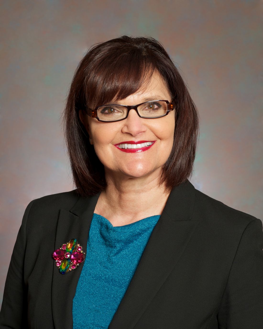
When an insurance coverage snafu threatened to take away a vital infusion drug for one of her patients, Julie Baak discovered that writing a letter wasn’t enough. Simponi Aria (golimumab) is the least expensive of all rheumatoid arthritis drugs for infusion, and at only six infusions a year offers a better experience for patients when compared with more expensive drugs like Humira (adalimumab), said Ms. Baak, practice manager at the Arthritis Center in St. Louis.

United Healthcare had drafted, then retracted, policy changes affecting and delaying access to RA drugs like Simponi Aria. Ms. Baak’s patient thrived on this drug after failing with others. UHC “kept pointing the finger at the employer, a self-funded plan,” she explained. Once correspondence efforts between the employer and payer fell through, she called a local news reporter, arranging an interview between Steven Baak, MD, the office’s medical director, and the patient, who was willing to go on the news. Through a 3-minute news segment, “we got that insurance company to allow us to get coverage for Simponi Aria on the medical side.”
Ms. Baak joined Karen Ferguson, Nilsa Cruz, and Madelaine A. Feldman, MD, at the Coalition of State Rheumatology Organizations 2020 State Society Advocacy Conference Virtual Meeting to discuss the power of advocacy in rheumatology, and impart strategies for enabling change. The Simponi Aria example underscores the importance of media and social media, Ms. Baak said. “When people do the wrong thing, you can bring light to others.”
The news spot on Dr. Baak and his patient mobilized Ms. Cruz to share it with others. “I took that interview and blasted it all over Wisconsin and to my colleagues because they needed to know what the options were for us in advocacy,” stressed Ms. Cruz, practice administrator of Milwaukee Rheumatology Center.

Rheumatologists are master problem solvers – the Sherlock Holmes’ of the medical industry, she continued. However, not many insurance companies understand the cognitive value of what they do. “There’s a lack of communication and education,” Ms. Cruz noted. Any advocacy measures calls for research on the topic, the panel stressed. It involves looking through medical and administrative insurance companies’ policies and using Google and social media, such as Twitter, to identify topics and key decision makers, a practice Ms. Cruz favors in particular.
Physicians as reliable sources
You need good documentation to support why you’re doing what you’re doing, advised Ms. Ferguson, practice administrator of Arthritis Northwest, Spokane, Wash. When an issue comes up, she always consults her doctors and her billing staff.
She recalled when biosimilars first came out, and one of them for Rituxan (rituximab) had not been indicated by the Food and Drug Administration for RA. “And yet, an insurance company was mandating that we use this nonindicated biosimilar,” said Ms. Ferguson, who learned about this from her billing staff. “I went immediately to the doctors and asked how they felt about this, and they said they were uncomfortable,” she continued. Ms. Ferguson found out that 45 states had legislation that prohibited interchangeability with non–FDA-indicated drugs. She was able to show this literature to the insurance company and get the mandate reversed. “One thing that’s so important is to be able to use your physicians’ knowledge and how they really feel and advocate based off of that,” using sound statutes to support your argument, she added.
“Ensuring that patients get the right medications that their physicians deem is important is critical for improved safety and efficacy,” Ms. Ferguson said. Yet, she acknowledged that facing off against an insurance company or a pharmacy benefit manager is often hard to do.
Many practices don’t have a Julie Baak or Karen Ferguson to fight on their behalf, Ms. Cruz noted. In this case, they should look within their state for high-profile advocates. “I guarantee you, every state has one. Practices should be able to reach out to those individuals, or respective state societies, or CSRO,” Ms. Cruz said. Best results are obtained when working through coalitions.
Patient testimony can make an impact
CSRO recently enacted a public relations campaign to shine a light on certain health plan policies that are harmful to patients. “We have been able to get the real impact of the policy on patients and their stories into the public eye,” Dr. Feldman, a rheumatologist in New Orleans and CSRO president, said in an interview. The group has spearheaded the effort to keep Stelara off of the Medicare self-administered drug list. CSRO has also built coalitions and provided testimony on several state step therapy and nonmedical switching bills. “When asked if these are pharma-backed bills, we say: ‘No, these are patient-backed bills,’ ” she added.

Patients act as powerful witnesses at hearings. “When legislation is being considered, it is important for both physicians and patients to be heard and to tell their stories,” Dr. Feldman said.
Ms. Cruz makes a point of getting involved with the payers themselves. As an example, she serves as a member on UHC’s National Steering Committee. “They know me to be very outspoken. Sometimes they listen, sometimes they don’t. Nevertheless, I consider it a compliment when they say they like that I hold them accountable. Every little thing that I can do to bring issues to the table,” filtering into their other divisions, is an accomplishment, she said.
When an insurance coverage snafu threatened to take away a vital infusion drug for one of her patients, Julie Baak discovered that writing a letter wasn’t enough. Simponi Aria (golimumab) is the least expensive of all rheumatoid arthritis drugs for infusion, and at only six infusions a year offers a better experience for patients when compared with more expensive drugs like Humira (adalimumab), said Ms. Baak, practice manager at the Arthritis Center in St. Louis.
United Healthcare had drafted, then retracted, policy changes affecting and delaying access to RA drugs like Simponi Aria. Ms. Baak’s patient thrived on this drug after failing with others. UHC “kept pointing the finger at the employer, a self-funded plan,” she explained. Once correspondence efforts between the employer and payer fell through, she called a local news reporter, arranging an interview between Steven Baak, MD, the office’s medical director, and the patient, who was willing to go on the news. Through a 3-minute news segment, “we got that insurance company to allow us to get coverage for Simponi Aria on the medical side.”
Ms. Baak joined Karen Ferguson, Nilsa Cruz, and Madelaine A. Feldman, MD, at the Coalition of State Rheumatology Organizations 2020 State Society Advocacy Conference Virtual Meeting to discuss the power of advocacy in rheumatology, and impart strategies for enabling change. The Simponi Aria example underscores the importance of media and social media, Ms. Baak said. “When people do the wrong thing, you can bring light to others.”
The news spot on Dr. Baak and his patient mobilized Ms. Cruz to share it with others. “I took that interview and blasted it all over Wisconsin and to my colleagues because they needed to know what the options were for us in advocacy,” stressed Ms. Cruz, practice administrator of Milwaukee Rheumatology Center.
Rheumatologists are master problem solvers – the Sherlock Holmes’ of the medical industry, she continued. However, not many insurance companies understand the cognitive value of what they do. “There’s a lack of communication and education,” Ms. Cruz noted. Any advocacy measures calls for research on the topic, the panel stressed. It involves looking through medical and administrative insurance companies’ policies and using Google and social media, such as Twitter, to identify topics and key decision makers, a practice Ms. Cruz favors in particular.
Physicians as reliable sources
You need good documentation to support why you’re doing what you’re doing, advised Ms. Ferguson, practice administrator of Arthritis Northwest, Spokane, Wash. When an issue comes up, she always consults her doctors and her billing staff.
She recalled when biosimilars first came out, and one of them for Rituxan (rituximab) had not been indicated by the Food and Drug Administration for RA. “And yet, an insurance company was mandating that we use this nonindicated biosimilar,” said Ms. Ferguson, who learned about this from her billing staff. “I went immediately to the doctors and asked how they felt about this, and they said they were uncomfortable,” she continued. Ms. Ferguson found out that 45 states had legislation that prohibited interchangeability with non–FDA-indicated drugs. She was able to show this literature to the insurance company and get the mandate reversed. “One thing that’s so important is to be able to use your physicians’ knowledge and how they really feel and advocate based off of that,” using sound statutes to support your argument, she added.
“Ensuring that patients get the right medications that their physicians deem is important is critical for improved safety and efficacy,” Ms. Ferguson said. Yet, she acknowledged that facing off against an insurance company or a pharmacy benefit manager is often hard to do.
Many practices don’t have a Julie Baak or Karen Ferguson to fight on their behalf, Ms. Cruz noted. In this case, they should look within their state for high-profile advocates. “I guarantee you, every state has one. Practices should be able to reach out to those individuals, or respective state societies, or CSRO,” Ms. Cruz said. Best results are obtained when working through coalitions.
Patient testimony can make an impact
CSRO recently enacted a public relations campaign to shine a light on certain health plan policies that are harmful to patients. “We have been able to get the real impact of the policy on patients and their stories into the public eye,” Dr. Feldman, a rheumatologist in New Orleans and CSRO president, said in an interview. The group has spearheaded the effort to keep Stelara off of the Medicare self-administered drug list. CSRO has also built coalitions and provided testimony on several state step therapy and nonmedical switching bills. “When asked if these are pharma-backed bills, we say: ‘No, these are patient-backed bills,’ ” she added.
Patients act as powerful witnesses at hearings. “When legislation is being considered, it is important for both physicians and patients to be heard and to tell their stories,” Dr. Feldman said.
Ms. Cruz makes a point of getting involved with the payers themselves. As an example, she serves as a member on UHC’s National Steering Committee. “They know me to be very outspoken. Sometimes they listen, sometimes they don’t. Nevertheless, I consider it a compliment when they say they like that I hold them accountable. Every little thing that I can do to bring issues to the table,” filtering into their other divisions, is an accomplishment, she said.
FROM CSRO 2020
Expanding Contraceptive Choices for Women: The Vaginal pH Modulator

Vaginal pH modulators (VPMs) add a new class of contraception that is now available in the United States. This method is nonhormonal, woman-controlled, and coitally dependent—and has the potential to increase overall contraceptive use and potentially reduce unintended pregnancy rates.
This CME supplement to OBG Management focuses on VPMs, their attributes, and the methodology surrounding the determination of contraceptive effectiveness.
The path to leadership
It was 6 a.m. on a rainy, cold Pacific Northwest morning as I walked from my apartment to the hospital, dodging puddles and dreaming of the mediocre-yet-hot physician-lounge coffee. Another long day full of clinical and administrative tasks awaited me.

I was 6 months’ pregnant with our first child and working my sixth 12-hour shift in a row. We had recently lost our medical director, and the C-suite had offered me the role. The day ahead seemed like an enormous mountain to climb.
I felt tired and more than a little overwhelmed. But I whispered to myself: “Today is going to be a fantastic day. I will not fail my team. I will not fail my patients!”
Physician leadership starts with a decision
The timing of this call to leadership had not been ideal. There’s probably never a perfect time to step into a medical director role. And my situation was no exception.
In addition to the baby on the way, my husband was traveling a lot for work. Also, the job of a medical director seemed a little daunting – especially to a young physician leading a team for the first time.
But I knew that leadership was my calling. While I didn’t yet have decades of experience, I had been selected as the chief resident in internal medicine, completed a nephrology fellowship, and mentored several medical students and residents along my career path.
I also knew that I was passionate about supporting my patients and hospitalist team. I’d previously served as associate medical director in charge of quality, readmission reduction, and patient experience. Having achieved the highest patient satisfaction scores on the team for 2 consecutive years, I was specially tasked to improve our team’s HCAHPS (Hospital Consumer Assessment of Healthcare Providers and Systems) scores.
These experiences taught me that coaching with positive reinforcement was in my blood. This gave me the courage to face my tallest mountain yet.
No one climbs a mountain alone
I also stepped into my new physician leadership role with amazing support. Our outgoing medical director had recommended me, and my entire team was rooting for me. My spouse was 100% behind the idea.

What’s more, I had received amazing feedback from patients throughout my 3 years at the hospital. I had papered an entire office wall with their thank-you notes. I even had a quilt that an 85-year-old patient’s wife made to thank me for my compassionate care.
As I weighed my decision, I realized that I had a higher calling to be a true advocate for my patients. I loved what I did. Each day, I resolved to bring my best and most authentic self for them – no matter how drained I felt.
My team and patients needed me now, not at some more convenient time down the road. A medical director job was the natural next step for me. And so, I resolved to climb the mountain.
Climbing through storms
Stepping into a medical director job forced me to grow into a completely new person. So maybe starting that role during pregnancy was a great metaphor!
Each day, there was immense pressure to perform, to deliver quality outcomes, and to simultaneously meet expectations of the C-suite as well as my hospitalist team. There was no room for failure, because too much was at stake.
Looking back today, I wouldn’t trade the experience for anything. The medical director role was one of the most gratifying experiences in my life, and I am truly thankful for it.
A leader’s role truly boils down to working tirelessly to collaborate with different care teams. It’s important to care not only about our patients but also about our fellow hospitalists. We can do this by truly leading by example – be it picking up extra shifts, covering holidays so team members can be with family, or coming in at 10 p.m. to round with your night team.
I was also able to bring a unique perspective to the hospital C-suite meetings as a woman, an immigrant, and a true “mama bear” – not only of my infant son but also of my team.

My first year as a medical director required more commitment and heart than I could have imagined. But all this hard work paid off when our hospitalist group received the coveted Best Team Award for most improved quality outcomes, financial performance, and patient experience.
The summit is the beginning
My first medical director job fueled my passion for patient satisfaction even further. I now serve as the director of patient experience for the more than 4,200 clinicians at Vituity. Together we care for more than 6.5 million lives a year across the country.
In 2019, I coached 300 physicians and hospital leaders on communication, collaboration, and service recovery skills, leading to significant improvement in their HCAHPS scores. I was delighted to receive the Vituity Distinguished Service Award for my contributions. It’s such an honor to be instrumental in impacting patient care at a larger scale.
This year, I was invited to serve as vice chair of the Society for Hospital Medicine’s patient experience committee and to join the executive board of the SHM San Francisco chapter. Together, we have created a COVID-19 patient communication resource and reached out to our hospitalists to provide them with a space to share their stories during this pandemic. I am so excited to share my knowledge and passion with clinicians across the country given the wide reach of Vituity & SHM!
Many hospitalists shy away from leadership roles. The mountain is tough to scale, but the view from the top is worth it. The key is to start, even if you don’t feel ready. I am here to tell you it can be done!
Dr. Mehta is a hospitalist and director of quality & performance and patient experience at Vituity in Emeryville, Calif. She is vice chair of the SHM patient experience committee and executive board member of the SHM San Francisco Bay Area chapter.
It was 6 a.m. on a rainy, cold Pacific Northwest morning as I walked from my apartment to the hospital, dodging puddles and dreaming of the mediocre-yet-hot physician-lounge coffee. Another long day full of clinical and administrative tasks awaited me.
I was 6 months’ pregnant with our first child and working my sixth 12-hour shift in a row. We had recently lost our medical director, and the C-suite had offered me the role. The day ahead seemed like an enormous mountain to climb.
I felt tired and more than a little overwhelmed. But I whispered to myself: “Today is going to be a fantastic day. I will not fail my team. I will not fail my patients!”
Physician leadership starts with a decision
The timing of this call to leadership had not been ideal. There’s probably never a perfect time to step into a medical director role. And my situation was no exception.
In addition to the baby on the way, my husband was traveling a lot for work. Also, the job of a medical director seemed a little daunting – especially to a young physician leading a team for the first time.
But I knew that leadership was my calling. While I didn’t yet have decades of experience, I had been selected as the chief resident in internal medicine, completed a nephrology fellowship, and mentored several medical students and residents along my career path.
I also knew that I was passionate about supporting my patients and hospitalist team. I’d previously served as associate medical director in charge of quality, readmission reduction, and patient experience. Having achieved the highest patient satisfaction scores on the team for 2 consecutive years, I was specially tasked to improve our team’s HCAHPS (Hospital Consumer Assessment of Healthcare Providers and Systems) scores.
These experiences taught me that coaching with positive reinforcement was in my blood. This gave me the courage to face my tallest mountain yet.
No one climbs a mountain alone
I also stepped into my new physician leadership role with amazing support. Our outgoing medical director had recommended me, and my entire team was rooting for me. My spouse was 100% behind the idea.
What’s more, I had received amazing feedback from patients throughout my 3 years at the hospital. I had papered an entire office wall with their thank-you notes. I even had a quilt that an 85-year-old patient’s wife made to thank me for my compassionate care.
As I weighed my decision, I realized that I had a higher calling to be a true advocate for my patients. I loved what I did. Each day, I resolved to bring my best and most authentic self for them – no matter how drained I felt.
My team and patients needed me now, not at some more convenient time down the road. A medical director job was the natural next step for me. And so, I resolved to climb the mountain.
Climbing through storms
Stepping into a medical director job forced me to grow into a completely new person. So maybe starting that role during pregnancy was a great metaphor!
Each day, there was immense pressure to perform, to deliver quality outcomes, and to simultaneously meet expectations of the C-suite as well as my hospitalist team. There was no room for failure, because too much was at stake.
Looking back today, I wouldn’t trade the experience for anything. The medical director role was one of the most gratifying experiences in my life, and I am truly thankful for it.
A leader’s role truly boils down to working tirelessly to collaborate with different care teams. It’s important to care not only about our patients but also about our fellow hospitalists. We can do this by truly leading by example – be it picking up extra shifts, covering holidays so team members can be with family, or coming in at 10 p.m. to round with your night team.
I was also able to bring a unique perspective to the hospital C-suite meetings as a woman, an immigrant, and a true “mama bear” – not only of my infant son but also of my team.
My first year as a medical director required more commitment and heart than I could have imagined. But all this hard work paid off when our hospitalist group received the coveted Best Team Award for most improved quality outcomes, financial performance, and patient experience.
The summit is the beginning
My first medical director job fueled my passion for patient satisfaction even further. I now serve as the director of patient experience for the more than 4,200 clinicians at Vituity. Together we care for more than 6.5 million lives a year across the country.
In 2019, I coached 300 physicians and hospital leaders on communication, collaboration, and service recovery skills, leading to significant improvement in their HCAHPS scores. I was delighted to receive the Vituity Distinguished Service Award for my contributions. It’s such an honor to be instrumental in impacting patient care at a larger scale.
This year, I was invited to serve as vice chair of the Society for Hospital Medicine’s patient experience committee and to join the executive board of the SHM San Francisco chapter. Together, we have created a COVID-19 patient communication resource and reached out to our hospitalists to provide them with a space to share their stories during this pandemic. I am so excited to share my knowledge and passion with clinicians across the country given the wide reach of Vituity & SHM!
Many hospitalists shy away from leadership roles. The mountain is tough to scale, but the view from the top is worth it. The key is to start, even if you don’t feel ready. I am here to tell you it can be done!
Dr. Mehta is a hospitalist and director of quality & performance and patient experience at Vituity in Emeryville, Calif. She is vice chair of the SHM patient experience committee and executive board member of the SHM San Francisco Bay Area chapter.
Sotorasib is a ‘triumph of drug discovery’ in cancer
KRAS, one of the most frequently mutated oncogenes in human cancer, has long been thought to be “undruggable,” but early results from a clinical trial of the experimental KRAS inhibitor sotorasib (Amgen) suggest that at least one KRAS mutation common in non–small cell lung cancers (NSCLC) has a soft underbelly.
In the phase 1 CodeBreaK 100 trial, sotorasib, an investigational first-in-class inhibitor of the KRAS p.G12C mutation, showed encouraging activity against advanced NSCLC and other solid tumors.
Among patients with NSCLC, 19 (32.2%) of 59 had a confirmed objective response to sotorasib monotherapy, and 52 (88.1%) had disease control, reported David S. Hong, MD, from the University of Texas MD Anderson Cancer Center, Houston.
“Sotorasib also demonstrated durable disease control in heavily pretreated patients with non–small cell lung cancer,” said Dr. Hong.
He presented secondary efficacy endpoint results from the trial in an online presentation during the European Society of Medical Oncology Virtual Congress 2020. The study was also published simultaneously online in the New England Journal of Medicine.
The trial met its primary endpoint of safety of sotorasib, with no dose-limiting toxicities or treatment-related fatal adverse events, and treatment-emergent grade 3 or higher adverse events occurring in less than 20% of patients.
“The safety profile is more favorable than that of other targeted agents, and I think the reason why you have a quite safe compound here is that sotorasib is very specific in its binding to KRAS G12C, and KRAS G12C is only present in the tumor,” coinvestigator Marwan G. Fakih, MD, a medical oncologist at City of Hope Comprehensive Cancer Center in Duarte, Calif., said in an interview. Fakih was co–lead author of the report in the New England Journal of Medicine.
A real “triumph”
Sotorasib is “a triumph of drug discovery,” commented Colin Lindsay, MD, from the University of Manchester (England), the invited discussant.
“We know that KRAS, over many years, over 3 decades, has been very difficult to target,” he said.
“The early development of KRAS G12C–targeted agents is just the beginning, lending hope that the ability to target not only other KRAS mutations but also other targets previously thought to be undruggable may be within reach,” write Patricia M. LoRusso, DO, from the Yale Cancer Center in New Haven, Conn., and Judith S. Sebolt-Leopold, PhD, from the University of Michigan Rogel Cancer Center, Ann Arbor, in an accompanying editorial.
The KRAS, which stands for Kristen rat sarcoma viral oncogene homologue, p.G12C mutation is a glycine-to-cysteine substitution that results in the oncogene being switched on in its active form. The mutation has been identified in approximately 13% of NSCLC tumors, in 1% to 7% of colorectal cancers, and in other solid tumors.
But the mutation has been considered too difficult to target because of KRAS’ strong binding affinity for guanosine triphosphate (GTP), an essential building block of RNA synthesis, and by a lack of accessible drug binding sites.
Sotorasib is a small-molecule, specific, and irreversible inhibitor of KRAS that interacts with a “pocket” on the gene’s surface that is present only in an inactive conformation of KRAS. The drug inhibits oncogenic signaling and tumorigenesis by preventing cycling of the oncogene into its active form, Dr. Fakih explained.
Study details
The CodeBreaK 100 investigators enrolled patients with 13 different locally advanced or metastatic solid tumor types, all bearing the KRAS p.G12C mutation.
The trial began with a dose-escalation phase, with two to four patients per cohort assigned to receive daily oral sotorasib at doses of 180, 360, 720, or 960 mg. The 960 mg dose was selected for expansion cohorts and for planned phase 2 studies, based on the safety profile and the lack of dose-limiting toxicities.
Hong and colleagues reported results for 129 patients treated in the dose-escalation and expansion cohorts, including 59 with NSCLC, 42 with colorectal cancer and 28 with other tumor types, but focused primarily on patients with NSCLC.
After a median follow-up of 11.7 months, 59 patients with NSCLC had been treated, 3 at the 180 mg dose, 16 at 360 mg, 6 at 720 mg, and 34 at 960 mg. At the time of data cutoff in June of this year, 14 patients were still on treatment and 45 had discontinued, either from disease progression (35 patients), death (5), patient request (4) or adverse events (1).
As noted, there were no dose-limiting toxicities or treatment-related fatalities reported.
Grade 3-4 treatment-related adverse events were reported in 18.6% of patients. The only grade 4 treatment-related event was diarrhea, in one patient. Grade 3 events included elevated liver transaminases in nine patients, increased alkaline phosphatase in two, anemia in one, and increased gamma-glutamyl transferase levels, decreased lymphocyte count, hepatitis, and hyponatremia in one patient each.
Dr. Fakih said that, given sotorasib’s high degree of specificity, it’s unclear what might be causing the observed adverse events.
Responses at all dose levels
The confirmed partial response rate was 32.2% for patients with NSCLC treated at all dose levels, and 35.3% for patients who received the 960 mg dose.
Among all NSCLC patients, and all treated at the highest 960-mg dose level, the stable disease rates were 55.9%. The respective disease control rates were 88.1% and 91.2%.
Tumor reductions occurred across all dose levels in patients with NSCLC. The median progression-free survival was 6.3 months.
Hong reported results for one patient, a 59-year-old man with the mutation who had experienced disease progression on five prior therapies including targeted agents, chemotherapy, and a checkpoint inhibitor, and had gamma-knife surgery for brain lesions.
This patient had a complete response in target lesions and a partial response overall, which included shrinkage of central nervous system metastases. He recently had progression in non-target lesions, after 1.5 years in response, Dr. Hong said.
The median duration of response was 10.9 months for patients with partial responses and 4 months for patients with stable disease.
Hong noted that response to sotorasib was seen across a range of co-mutational profiles, including several patients with four mutations in addition to KRAS p.G12C.
Other tumors, possible combinations
Among 42 patients with colorectal cancers bearing the KRAS p.G12C mutation, 3 (7.1%) had a partial response. There were also partial responses seen in one patient each with melanoma and with appendiceal, endometrial, and pancreatic tumors.
“Overall, the results of this trial are very encouraging, showing the first step in ‘drugging the undruggable,’ ” Dr. LoRusso and Dr. Sebolt-Leopold wrote in their editorial.
They suggested that therapy with sotorasib may be improved by combining it with other agents that could target resistance to KRAS inhibition.
“A recent study showed that KRAS G12C colorectal cancer cells have higher basal epidermal growth factor receptor (EGFR) activity than NSCLC cells, leading to a rapid rebound in mitogen-activated protein (MAP) kinase signaling and resistance to KRAS G12C inhibition,” the editorialists wrote. “This observation is consistent with the weaker observed clinical activity of sotorasib in patients with colorectal cancer, a problem that may be addressed by combining it with an EGFR inhibitor [e.g., cetuximab], as seen preclinically.”
“I think this drug is being positioned not only in refractory disease, but we’re hoping to see it move upfront in non–small cell lung cancer, and we’re hoping to improve its efficacy in colorectal cancer,” Dr. Fakih said in an interview.
The study was sponsored by Amgen and by grants from the National Institutes of Health. Dr. Hong disclosed research/grant funding and an advisory/consulting role with Amgen and others. Dr. Fakih disclosed a speaking engagement for Amgen and consulting for and grant support from others. Dr. Lindsay disclosed consulting for Amgen and institutional research funding from the company and others. Dr. LoRusso disclosed fees from multiple companies, not including Amgen. Dr. Sebolt-Leopold disclosed no relevant financial relationships.
This article first appeared on Medscape.com.
FROM ESMO 2020
Nivo-cabo combo joins advanced RCC treatment ranks
Promoting to the front line two drugs normally used in rearguard action to treat advanced renal cell carcinoma (RCC) – nivolumab (Opdivo) and cabozantinib (Cabometyx) – doubled overall response rates and progression-free survival (PFS) and significantly improved overall survival (OS), compared with first-line sunitinib (Sutent), investigators in the Checkmate 9ER trial reported.
Median PFS among patients with advanced RCC, which was the trial’s primary endpoint, was 16.6 months with nivolumab plus cabozantinib, compared with 8.3 months with sunitinib, translating into a hazard ratio of 0.51 for the combination (P < .0001). The median follow-up was 18.1 months.
Median OS had not been reached in either arm at the time of data cutoff, but the survival curves at the time of the analysis clearly favored nivolumab-cabozatinib, with an HR for death of .060 (P = .0010), said Tony K. Choueri, MD, from the Dana-Farber Cancer Institute in Boston.
“With expanding options in our patients with advanced RCC, the overall efficacy, safety, and quality-of-life benefit, as well as individual patient characteristics, are very important considerations when you select appropriate therapy,” he said in a press briefing prior to his presentation of the data in a presidential symposium at the European Society of Medical Oncology Virtual Congress 2020.
Although the nivolumab-cabozantinib combination therapy looks good, it’s late to the game, commented Dominik Berthold, MD, from the Centre Hospitalier Universitaire Vaudois, Lausanne, Switzerland, the invited discussant for the briefing.
“The question is, what’s the only drawback of this trial? It’s probably the fact that it’s not first in class in this situation,” he said.
Dr. Berthold noted that nivolumab-cabozantinib, if approved for the frontline setting, will join the combination of the tyrosine kinase inhibitor (TKI) axitinib (Inlyta) plus pembrolizumab (Keytruda), which, as previously reported, was associated with a nearly 50% reduction in the risk for death in the KEYNOTE-426 trial. This combination was approved by the Food and Drug Administration for the frontline setting in April 2019.
As shown in the CheckMate-214 study, the combination of the programmed cell death protein–1 (PD-1) inhibitor nivolumab with the CTLA-4 inhibitor ipilimumab (Yervoy) was associated with significantly higher objective response rates and OS rates compared with sunitinib. This combination was approved by the FDA in April 2018 as first-line therapy for patients with advanced intermediate- or poor-risk RCC.
CheckMate 9ER details
A total of 651 patients with previously untreated advanced or metastatic RCC that had a clear cell component in all International Metastatic RCC Database Consortium risk groups were enrolled and randomly assigned to receive either intravenous nivolumab at 240 mg every 2 weeks plus oral cabozantinib at 40 mg daily or oral sunitinib at 50 mg daily in cycles of 4 weeks on therapy/2 weeks off therapy. Patients were treated until disease progression or unacceptable toxicities occurred.
The primary PFS endpoint and the secondary OS endpoint both favored the combination, as did the objective response rate, which was 55.7% with nivolumab-cabozantinib versus 27.1% with sunitinib (P < .0001).
Complete responses were seen in 8% of patients who received the combination versus 4.6% with the patients who received sunitinib. Partial responses were seen in 47.7% and 22.6%, respectively.
Patients generally tolerated the combination. The incidence of the most common high-grade treatment-emergent adverse events and other adverse events of any grade was similar to that seen with sunitinib, Dr. Choueri said.
The rates of treatment-related events that led to discontinuation was 3.1% among patients who received the combination, 5.6% among patients who received the nivolumab component only, and 6.6% among patients who received cabozantinib only. It was 8.8% among patients who received sunitinib. More than 50% of patients in the combination arm needed a dose reduction of nivolumab-cabozantinib because of adverse events, however.
“Overall, it seems that the combination has a somewhat manageable safety profile in patients with advanced RCC,” Dr. Choueri said.
Patient-reported quality of life, as measured by the National Comprehensive Cancer Network/Functional Assessment of Cancer Therapy–Kidney Symptom Index 19 total score, was an exploratory endpoint. It was maintained over time with the combination but deteriorated over time with sunitinib, with statistically significant differences between the study arms at most time points to 91 weeks, he reported.
Making choices
Dr. Berthold acknowledged the benefit that having an additional therapy offers clinicians and patients.
“What we still need to learn here is, ‘Are there any patient populations who may benefit more on this combination compared with other combinations?’ ” he said. “Cabozantinib is quite a unique TKI which may target better bone metastases, for example, so I think there are things we need to learn from further data and longer follow-up.”
Camillo Porta, MD, from the University of Bari (Italy), the invited discussant for the presidential symposium, urged caution in comparing the three regimens, owing to differences in the drug used, study endpoints, baseline patient characteristics, and the distribution of patients among different prognostic groups.
When it comes to deciding between frontline regimens, “the only possible, though highly empiric, driver of our therapeutical choice should be the biological aggressiveness of the tumor,” he said.
For patients with highly aggressive disease, the use of an immune checkpoint inhibitor plus a vascular endothelial growth factor receptor (VEGFR)–directed TKI may help control disease long enough to give the checkpoint inhibitor time to work.
“Otherwise, one could head for the long-term benefit of the immune combo as well as for complete responses, trying to spare [patients] the additional toxicities deriving from the continuous use of the VEGFR TKI,” he added.
Dr. Porta noted that when considering the trade-off between efficacy and safety in the first-line setting, many patients are willing to accept more toxicities in exchange for clinical benefit.
The study was sponsored by Bristol-Myers Squibb. Dr. Choueri disclosed consultancy fees, advisory board activity, manuscript preparation, travel/lodging, honoraria, and grants for clinical trials from BMS and others. Dr. Berthold disclosed an advisory role for Ipsen, BMS, Merck, and Pfizer. Dr. Porta disclosed advisory/consulting activities and speakers bureau participation for BMS and others.
This article first appeared on Medscape.com.
FROM ESMO 2020
Lorlatinib: Another first-line option for ALK-positive NSCLC?
Lorlatinib also produced a higher overall and intracranial response rate, prolonging progression to CNS disease.
These findings “support the use of lorlatinib as a highly effective first-line therapy for patients with advanced ALK-positive NSCLC,” said Benjamin Solomon, MBBS, PhD, of the Peter MacCallum Cancer Centre in Melbourne.
“The CROWN study clearly establishes lorlatinib as another option” among other first-line ALK inhibitors, Dr. Solomon said when presenting the findings at the European Society for Medical Oncology Virtual Congress 2020.
“We now have multiple options for first-line treatment of patients with ALK-positive lung cancer,” noted study discussant Christine Lovly, MD, PhD, a medical oncologist and associate professor at Vanderbilt University Medical Center in Nashville, Tenn.
The question now, she said, is how to choose among these options. The drugs have all bested crizotinib in trials but haven’t gone head to head against one another.
Lorlatinib and CROWN
Lorlatinib is currently approved in the United States to treat ALK-positive metastatic NSCLC that has progressed on crizotinib and at least one other ALK inhibitor. Lorlatinib was granted accelerated approval for this indication based on response rate and duration.
The CROWN study was intended to support the conversion to full approval, according to Pfizer, which is developing both lorlatinib and crizotinib. Pfizer also plans to use the results of CROWN to seek a first-line indication for lorlatinib in NSCLC.
CROWN enrolled 296 patients with stage IIIB/IV ALK-positive NSCLC who had received no prior systemic treatment. Patients with asymptomatic treated or untreated CNS metastases were eligible.
There were 149 patients randomized to lorlatinib at 100 mg daily and 147 randomized to crizotinib at 250 mg twice daily. Five patients in the crizotinib arm were included in the analysis but were not treated, Dr. Solomon said.
The median age was 61 years in the lorlatinib arm and 56 years in the crizotinib arm. Nearly all patients were White (48% in the lorlatinib arm and 49% in the crizotinib arm) or Asian (44% in both arms). A majority of patients were women (56% in the lorlatinib arm and 62% in the crizotinib arm), and more than half said they never smoked (54% in the lorlatinib arm and 64% in the crizotinib arm).
Response and PFS
According to blinded independent central review, the objective response rate was 76% with lorlatinib (113/149) and 58% with crizotinib (85/147). There were four complete responses with lorlatinib and none with crizotinib.
Among patients who had measurable or nonmeasurable brain metastases at baseline, the intracranial ORR was 66% with lorlatinib (25/38) and 20% (8/40) with crizotinib. In patients with only measurable brain metastases at baseline, the intracranial ORR was 82% with lorlatinib (14/17) and 23% with crizotinib (3/13).
The 12-month PFS rate was 78% in the lorlatinib arm and 39% in the crizotinib arm. The median PFS was 9.3 months in the crizotinib arm but was not reached in the lorlatinib arm, which “corresponded to a 72% reduction in the risk of death or progression [hazard ratio, 0.21; P < .001],” Dr. Solomon said.
“[T]he PFS for alectinib in the first line is approximately 3 years,” Dr. Lovly noted. “We anxiously await additional data for lorlatinib to see how long the PFS will be.”
The median time to intracranial progression was 16.6 months in the crizotinib arm but was not reached in the lorlatinib arm (HR, 0.07; P < .001).
“These data indicate the ability of lorlatinib not only to delay the progression of existing brain metastases, but also to prevent the development of new brain metastases,” Dr. Solomon said.
Dr. Lovly noted that the efficacy of lorlatinib in the brain is “quite compelling,” but other ALK inhibitors have demonstrated similar results.
As for overall survival, the data are still immature. The median overall survival was not reached with lorlatinib or crizotinib (HR, 0.72).
Toxicity
Dr. Solomon noted that lorlatinib “does have a different toxicity profile, compared to other ALK inhibitors.” Specifically, lorlatinib is associated with hypercholesterolemia and hypertriglyceridemia, which have not been seen with other ALK inhibitors.
Lorlatinib is also associated with neurocognitive problems, including inattention, memory impairment, and mild confusion. Mood effects include emotional lability – “someone watching a movie might burst into tears when they wouldn’t have otherwise,” Dr. Solomon said – as well as anxiety and depression.
“So it’s important to tell not just the patient but their family about these things so that they identify when [the events] happen,” Dr. Solomon said. “That’s key because [the events are] completely reversible when you stop the drug. With dose interruption, those effects will resolve.”
Other adverse events that were more common with lorlatinib (a 10% or greater difference in frequency from crizotinib) included edema, weight gain, and peripheral neuropathy. Diarrhea, nausea, fatigue, vision disorders, constipation, and increased liver enzymes were more common with crizotinib. Grade 3-4 adverse events led to discontinuation in fewer than 10% of patients in each arm.
The study was funded by Pfizer, and the investigators included employees. Dr. Solomon is an adviser for Pfizer and other companies, and Dr. Lovly’s industry ties included being both an advisor and speaker for Pfizer.
SOURCE: Solomon B et al. ESMO 2020, Abstract LBA2.
FROM ESMO 2020
A young physician hopes to buck the status quo in Congress
On March 3 of this year, Bryant Cameron Webb, MD, JD, won two-thirds of the vote in Virginia’s Democratic primary race. In November, he’ll compete against Republican Bob Good to represent the state’s 5th Congressional District. If he succeeds, he will become the first Black physician ever elected to a seat in Congress.

The political and social unrest across the United States in recent months has resulted in millions of people becoming more proactive: from sports arenas to the halls of Congress, the rally cry of Black Lives Matter has echoed like never before after the killing of George Floyd and Breonna Taylor at the hands of law enforcement. Dr. Webb, a practicing internist and professor at the University of Virginia, Charlottesville, is among many physicians joining the cause. If elected, he hopes to bring a unique perspective to Washington and advocate for racial equity to help combat systemic racist policies that result in health disparities.
“For me as a professor at UVA in both public health sciences and in medicine, I have a lot to bring to this moment,” he commented, “real expertise on issues that are critical to the nation. Beyond my passion for health and wellness, I have a passion for justice.”
Dr. Webb also believes that serving in Congress is a way to help his patients. “I balance the work of direct patient care and patient advocacy in different spaces,” said the Spotsylvania County native. “Working in Congress is patient advocacy to me. It’s where I can be at my highest use to the people I take care of. It is different from direct patient care. I think this [unique] background that I have is needed in Congress.”
Dr. Webb has never held an elected office before, and he’s looking to get elected in a district that voted for President Trump in the past election. He knows challenges lie ahead.
A calling
The field of medicine called for Dr. Webb at an early age. He credits his family doctor, a Black man, for inspiring him. “With six kids in our family, we saw the doctor frequently. Dr. Yarboro was a young Black man just a few years out of residency. My mom had supreme confidence in him, and he made us feel at ease. So I wanted to be a doctor ever since I was 5 or 6 years old.”
Dr. Webb earned a bachelor’s degree from the University of Virginia in 2005. He entered medical school at Wake Forest University, Winston-Salem, N.C., the following year. Following his third year of medical training, he heeded another calling: He took time off to attend law school. He enrolled in Loyola University of Chicago School of Law and earned his juris doctorate in 2012.
The move may seem an unexpected turn. But Dr. Webb feels his law degree enhances his work. “I think that it’s because I’m so steeped in the legal resources that folks need to navigate. I think I am able to provide better care. ... It’s a complement and helpful to me professionally, whether it’s fighting with an insurance company or with a prescription drug company.”
After law school, Dr. Webb finished his medical training at Wake Forest and moved north, where he completed an internal medicine residency at New York–Presbyterian Hospital. Then came yet another twist in Dr. Webb’s unconventional career path: in 2016, he was selected by President Obama as a White House fellow. He spent the next 2 years in Washington, where he worked on Mr. Obama’s My Brother’s Keeper Task Force, an initiative that addresses opportunity gaps faced by boys and young men of color.
Adeze Enekwechi, MD, president of Impaq and associate professor at the George Washington University, Washington, worked with Dr. Webb at the White House. “This is the place where he will have the most impact. We’ve been talking and writing about health equity ever since our time [there]. Not everybody can speak that language.
Why here? Why now?
Dr. Webb sees patients 2-3 days a week on alternating weeks and knows well the concerns of people who struggle with health. Now he’s ready to have those conversations on a larger platform. “As a Black physician, it’s about bringing that healer mindset to these problems. It’s not about just going there to brow beat people or add to that divisive nature in Congress. You acknowledge that the problems exist, and then bridge,” he said, hoping that bridging party divides can be a catalyst for healing.
Carla Boutin-Foster, MD, associate dean, office of diversity education and research at the State University of New York, Brooklyn, has mentored Dr. Webb since 2013. With his credentials, confidence, and persistence, she believes, he will be a great representative of the medical community in D.C. “You need someone who respects the Constitution. When policy needs to be developed, you need a healer, someone who understands the science of vaccines. This is something Cam has been groomed for. It’s something he has been living and practicing for years.”
The killing of George Floyd and the uprising that ensued has opened the dialogue about racial inequality in America. Health care is not immune to racial bias, and the effects are palatable. One survey conducted by the Larry A. Green Center, in collaboration with the Primary Care Collaborative and 3rd Conversation, found that more than 40% of clinicians say Mr. Floyd’s demise has become a topic of concern among patients of all demographics.
When it comes to racism, Dr. Webb understands that he plays a critical role in moving America forward. “We have so many voices that are powerful and important in the highest level of legislation. We have to use those voices to root out the injustices in our society, like in the Breonna Taylor case. We have to do so because that is how you achieve the American dream,” he said.
The social determinants of health – or “ZIP-code risk” – has been proven to influence health outcomes, yet few physicians screen for them during patient visits. For Dr. Webb, discussing things like housing security and interpersonal violence are critical to providing care.
One of Dr. Webb’s biggest supporters is his wife of 11 years, Leigh Ann Webb, MD, MBA, an emergency medicine physician and assistant professor of emergency medicine at UVA. “He is an effective leader and a consensus builder,” she said of her husband, with whom she has two children. “There has always been something very unique and special about him and the way he engages the world. We need more thoughtful, intelligent people like him to help our country move forward.”
In addition to being the director of health policy and equity at UVA this fall, Dr. Webb plans to teach a course at UVA centered around the social determinants of health called Place Matters. “The focus is on understanding how education and housing and food insecurity all come together to cause illness,” he said. “Health doesn’t happen in hospitals and clinics. It happens in the community.”
A version of this article originally appeared on Medscape.com.
On March 3 of this year, Bryant Cameron Webb, MD, JD, won two-thirds of the vote in Virginia’s Democratic primary race. In November, he’ll compete against Republican Bob Good to represent the state’s 5th Congressional District. If he succeeds, he will become the first Black physician ever elected to a seat in Congress.
The political and social unrest across the United States in recent months has resulted in millions of people becoming more proactive: from sports arenas to the halls of Congress, the rally cry of Black Lives Matter has echoed like never before after the killing of George Floyd and Breonna Taylor at the hands of law enforcement. Dr. Webb, a practicing internist and professor at the University of Virginia, Charlottesville, is among many physicians joining the cause. If elected, he hopes to bring a unique perspective to Washington and advocate for racial equity to help combat systemic racist policies that result in health disparities.
“For me as a professor at UVA in both public health sciences and in medicine, I have a lot to bring to this moment,” he commented, “real expertise on issues that are critical to the nation. Beyond my passion for health and wellness, I have a passion for justice.”
Dr. Webb also believes that serving in Congress is a way to help his patients. “I balance the work of direct patient care and patient advocacy in different spaces,” said the Spotsylvania County native. “Working in Congress is patient advocacy to me. It’s where I can be at my highest use to the people I take care of. It is different from direct patient care. I think this [unique] background that I have is needed in Congress.”
Dr. Webb has never held an elected office before, and he’s looking to get elected in a district that voted for President Trump in the past election. He knows challenges lie ahead.
A calling
The field of medicine called for Dr. Webb at an early age. He credits his family doctor, a Black man, for inspiring him. “With six kids in our family, we saw the doctor frequently. Dr. Yarboro was a young Black man just a few years out of residency. My mom had supreme confidence in him, and he made us feel at ease. So I wanted to be a doctor ever since I was 5 or 6 years old.”
Dr. Webb earned a bachelor’s degree from the University of Virginia in 2005. He entered medical school at Wake Forest University, Winston-Salem, N.C., the following year. Following his third year of medical training, he heeded another calling: He took time off to attend law school. He enrolled in Loyola University of Chicago School of Law and earned his juris doctorate in 2012.
The move may seem an unexpected turn. But Dr. Webb feels his law degree enhances his work. “I think that it’s because I’m so steeped in the legal resources that folks need to navigate. I think I am able to provide better care. ... It’s a complement and helpful to me professionally, whether it’s fighting with an insurance company or with a prescription drug company.”
After law school, Dr. Webb finished his medical training at Wake Forest and moved north, where he completed an internal medicine residency at New York–Presbyterian Hospital. Then came yet another twist in Dr. Webb’s unconventional career path: in 2016, he was selected by President Obama as a White House fellow. He spent the next 2 years in Washington, where he worked on Mr. Obama’s My Brother’s Keeper Task Force, an initiative that addresses opportunity gaps faced by boys and young men of color.
Adeze Enekwechi, MD, president of Impaq and associate professor at the George Washington University, Washington, worked with Dr. Webb at the White House. “This is the place where he will have the most impact. We’ve been talking and writing about health equity ever since our time [there]. Not everybody can speak that language.
Why here? Why now?
Dr. Webb sees patients 2-3 days a week on alternating weeks and knows well the concerns of people who struggle with health. Now he’s ready to have those conversations on a larger platform. “As a Black physician, it’s about bringing that healer mindset to these problems. It’s not about just going there to brow beat people or add to that divisive nature in Congress. You acknowledge that the problems exist, and then bridge,” he said, hoping that bridging party divides can be a catalyst for healing.
Carla Boutin-Foster, MD, associate dean, office of diversity education and research at the State University of New York, Brooklyn, has mentored Dr. Webb since 2013. With his credentials, confidence, and persistence, she believes, he will be a great representative of the medical community in D.C. “You need someone who respects the Constitution. When policy needs to be developed, you need a healer, someone who understands the science of vaccines. This is something Cam has been groomed for. It’s something he has been living and practicing for years.”
The killing of George Floyd and the uprising that ensued has opened the dialogue about racial inequality in America. Health care is not immune to racial bias, and the effects are palatable. One survey conducted by the Larry A. Green Center, in collaboration with the Primary Care Collaborative and 3rd Conversation, found that more than 40% of clinicians say Mr. Floyd’s demise has become a topic of concern among patients of all demographics.
When it comes to racism, Dr. Webb understands that he plays a critical role in moving America forward. “We have so many voices that are powerful and important in the highest level of legislation. We have to use those voices to root out the injustices in our society, like in the Breonna Taylor case. We have to do so because that is how you achieve the American dream,” he said.
The social determinants of health – or “ZIP-code risk” – has been proven to influence health outcomes, yet few physicians screen for them during patient visits. For Dr. Webb, discussing things like housing security and interpersonal violence are critical to providing care.
One of Dr. Webb’s biggest supporters is his wife of 11 years, Leigh Ann Webb, MD, MBA, an emergency medicine physician and assistant professor of emergency medicine at UVA. “He is an effective leader and a consensus builder,” she said of her husband, with whom she has two children. “There has always been something very unique and special about him and the way he engages the world. We need more thoughtful, intelligent people like him to help our country move forward.”
In addition to being the director of health policy and equity at UVA this fall, Dr. Webb plans to teach a course at UVA centered around the social determinants of health called Place Matters. “The focus is on understanding how education and housing and food insecurity all come together to cause illness,” he said. “Health doesn’t happen in hospitals and clinics. It happens in the community.”
A version of this article originally appeared on Medscape.com.
Longer bisphosphonate use ups AFF risk, but not all is tied to drug
In a national study of older Danes who had previously had a fracture and were taking bisphosphonates, the risk of having a serious though rare atypical femoral fracture (AFF) was greater after 3-5 years of bisphosphonate use.
The risk quickly dropped after patients stopped taking a bisphosphonate, which suggests that bisphosphonate “holidays” may be useful for some patients, the researchers said. These findings support previous work.
But the study also found that 34% of the AFFs occurred in patients who had not been taking a bisphosphonate. That rate is higher than the 6%-22% that has been reported by others.
Doug Bauer, MD, from the University of California, San Francisco, presented the new study findings during the virtual American Society of Bone and Mineral Research 2020 annual meeting.
“We found no clear risk factor that accounts for this increased risk [for AFFs] among those not exposed to bisphosphonates,” he said, “but we believe this was a real finding, as our study protocol ensured that the study radiologists were completely blinded to treatments received.”
Suzanne N. Morin, MD, who was not involved in this research, pointed out that the reported AFF risks related to bisphosphonate dose and cessation are in keeping with findings of other studies, including a recent large study by Dennis M. Black, MD, and colleagues that was published in the New England Journal of Medicine.
That study found that Asians are at higher risk for AFFs than White persons. Others have reported that specific femur geometry or physique and use of glucocorticoids increase AFF risk, Dr. Morin, from the Research Institute of the McGill University Health Center, Montreal, said in an interview.
The current study suggests that rheumatoid arthritis may be a risk factor, she added.
The fact that the rate of AFFs among patients who had not been exposed to bisphosphonates was higher than previously reported “may be due to differences in the method they used to ascertain the fractures or in medication use,” she speculated.
The clinical implications of research to date are that “the risk of AFF should not dissuade patients and providers from short-term use of bisphosphonates [3-5 years],” Dr. Bauer said. He noted that most patients should not take a bisphosphonate for longer than this unless they have a very high fracture risk.
Similarly, Dr. Morin said that clinicians “should consider initiating bisphosphonate in those at high risk for fractures and reevaluate their use after 3-6 years, depending on individual’s risk profile.”
AFF is serious but rare complication of bisphosphonate use
“Since first reported over 10 years ago, it has become clear that AFFs are a rare but serious complication of bisphosphonate therapy,” Dr. Bauer explained. However, there is still uncertainty about the magnitude of this risk, including the absolute risk for AFFs among adults who take bisphosphonates and those who do not.
To study this, the researchers analyzed data from national health care and pharmacy records and a radiology image database in Denmark. They identified almost 5,000 adults who were aged 50 years or older and who experienced a subtrochanteric and femoral shaft fracture during the period from 2010 to 2015. Two expert radiologists who were blinded to the patients’ clinical history or treatment identified AFF on the basis of ASBMR 2014 criteria.
The researchers compared three patient groups: 189 patients with AFF, 2,397 patients with typical subtrochanteric and femoral shaft fractures (no AFF), and35,946 adults aged older than 50 years (control persons).
Compared with patients with typical fractures, patients with AFF were younger (aged 71 vs. 77), more likely to be women (79% vs. 69%), and more likely to have RA (12% vs. 2.5%).
Compared with patients in the other two groups, those with AFF were more likely to use corticosteroids, proton pump inhibitors, statins, and hormone replacement therapy.
They were also more likely to use bisphosphonates (58%) than patients with typical subtrochanteric and femoral shaft fractures (19%) or control patients (10%).
The bisphosphonates used in Denmark at the time were mostly alendronate (85%) and rarely ibandronate (6%), intravenous zoledronic acid (5%), etidronate (3%), or risedronate (1%).
One-third of patients with AFFs had no bisphosphonate exposure
In this national cohort of adults aged older than 50 years, the absolute rates of AFF per 10,000 person-years were as follows: 0.07 in nonusers of bisphosphonates, 1.84 in those with 3-5 years of bisphosphonate use, and 4.63 in those with >7 years of bisphosphonate use. As a comparison, the rate of classic hip fracture was 43.8 per 10,000 person-years.
Compared with no bisphosphonate use, the relative risk for AFF was close to 40 times higher with more than 7 years of use, after adjusting for multiple confounders. The risk for AFF was also significantly higher among patients with RA or hypertension and for those who used proton pump inhibitors.
“Note that age, gender, and previous fracture were not associated with the risk of AFF” after controlling for multiple confounders, Dr. Bauer stressed.
The relative risk for AFF fell significantly after it had been withheld from use for more than 1 year.
Among the 189 patients with confirmed AFF, 64 patients (34%) had never taken a bisphosphonate.
Preliminary analysis showed that, among patients with AFF, those who had not been exposed to bisphosphonates were younger, more likely to be male, and less likely to have had a previous fracture, RA, or to have used corticosteroids, proton pump inhibitors, statins, or hormone-replacement therapy.
The study was funded by the National Institute of Arthritis and Musculoskeletal and Skin Diseases. Dr. Bauer and Dr. Morin disclosed no relevant financial relationships.
A version of this article originally appeared on Medscape.com.
FROM ASBMR 2020
First-in-class ADC ups survival in mTNBC
The first-in-class drug is directed at trophoblast cell surface antigen 2 (Trop-2), which is highly expressed in breast cancer. Research on the drug was presented at the European Society for Medical Oncology Virtual Congress 2020.
ASCENT randomly assigned more than 500 patients who had metastatic TNBC and who had experienced disease progression after a median of four lines of therapy to receive either SG or physician’s choice of chemotherapy. SG significantly improved median progression-free survival (5.6 vs 1.7 months; hazard ratio [HR], 0.41; P < .0001) and median overall survival (12.1 vs 6.7 months; HR, 0.48; P < .0001).
The response rate was 35% for SG vs 5% for chemotherapy (P < .0001).
The study was presented by Aditya Bardia, MD, MPH, a medical oncologist at Massachusetts General Hospital, Boston, Massachusetts.
He said that because the safety profile of SG is consistent with previous reports and the treatment discontinuation rate was low, the clinical benefit “confirms the use of sacituzumab govitecan” in metastatic TNBC. This was a reference to the fact that the drug previously received accelerated approval on the basis of early data.
Bardia added that ongoing studies are evaluating the use of SG in earlier lines of therapy, including neoadjuvant settings in combination with other targeted agents, as well as in hormone receptor–positive metastatic breast cancer. One such study is the phase 3 TROPiCS-02 study, which is actively accruing patients.
Invited discussant Fatima Cardoso, MD, director of the Breast Unit at Champalimaud Clinical Center, Lisbon, Portugal, said the treatment algorithm for the management of TNBC will need to be updated in light of these results.
“In my opinion, we should now add sacituzumab govitecan as a new treatment option for patients treated with two or more lines of therapy,” she said.
She noted that the study design raised some questions over the way such trials should be conducted and the future sequencing of treatments.
Objections to study design and execution
Discussant Cardoso said the choice of progression-free survival as the primary endpoint for the trial was not ideal.
“In the metastatic setting, and particularly for triple-negative breast cancer, where the median survival is quite low and where each line of treatment, particularly after the second line, has a short duration, the primary endpoint should have been overall survival,” she commented.
“Luckily, we saw some results, but we could have missed it,” Cardoso said. She made “a plea to make sure that overall survival is at least the co–primary endpoint” in the future.
Cardoso also said it was not clear to her why the trial had to be stopped. “For the current patients, if there is no crossover, there is no benefit in stopping the trial,” she said.
She went on: “For future patients, it’s better to have the final results sufficiently powered.” She noted that the benefit seen in ASCENT was “moderate” and “so not a substantial breakthrough.” She added that it was “important not to stop trials too early.”
On the positive side, Cardoso said the median number of previous lines of therapy was “quite remarkable for this subtype, and it is important then for us to discuss sequencing,” particularly given that so many patients received checkpoint inhibitors before entering the trial.
Safety and a focus on diarrhea
Investigator Bardia explained that SG is an antibody-drug conjugate directed at Trop-2, which is highly expressed in breast cancer.
The antibody is linked to SN-38, the active metabolite of irinotecan, via a hydrolyzable linker that makes internalization and enzymatic cleavage by tumor cells unnecessary.
Hydrolysis of the linker releases SN-38 both within the tumor cell and extracellularly to induce a so-called bystander effect, in which neighboring tumors cells may be killed even if they do not express Trop-2.
On the basis of positive results from phase 1/2 trial data, SG was granted accelerated approval by the US Food and Drug Administration for patients with metastatic TNBC who experience disease progression after at least two prior therapies.
ASCENT was therefore a phase 3 confirmatory study. Patients with metastatic TNBC who had received at least two prior chemotherapy regimens were randomly assigned in a 1:1 ratio to receive intravenous SG on days 1 and 8 of a 21-day cycle or to receive physician’s choice of treatment.
Physicians could choose eribulin, vinorelbine, gemcitabine, or capecitabine.
The patients continued receiving treatment until disease progression or unacceptable toxicity occurred. On the unanimous recommendation of the data safety monitoring committee, the trial was ended early because of “compelling evidence of efficacy.”
In all, 267 patients were randomly assigned to receive SG. Of those patients, 15 remain on treatment. Treatment was discontinued for 199 patients who experienced disease progression. In the control arm, 262 patients were included, none of whom are still on treatment; 166 discontinued because of disease progression.
The current analysis is limited to 235 patients in the SG group and, in the control arm, 233 patients who did not have brain metastases. Patients with brain metastases will be the subject of a later analysis.
All but one patient in both treatment groups were women. The median age was approximately 54 years. The median number of prior treatment regimens was four. All patients had previously received chemotherapy, and between 26% and 29% have taken checkpoint inhibitors.
By the data cutoff of March 11, 2020, patients had received a median of seven treatment cycles with SG. Progression-free survival was adjudicated by blind, independent central review. The median duration of response was of borderline significance, at 6.3 months vs 3.6 months (P = .057).
Bardia showed that the results were consistent among all subgroups, including subgroups determined on the basis of age, number of prior therapies, whether patients had received prior immune checkpoint therapy, and the presence of liver metastases.
With respect to safety, the important grade 3 or higher treatment-related adverse events were neutropenia (seen in 51% of SG patients vs 33% of patients in the control arm), diarrhea (10% vs <1%), leukopenia (10% vs 5%), anemia (8% vs 5%), and febrile neutropenia (6% vs 2%).
Despite the fact that the adverse event rate was higher with SG than with physician’s choice of chemotherapy, the percentage of such events that led to treatment discontinuation was numerically lower, at 4.7% vs 5.4%.
Cardoso highlighted the “substantial percentage” of patients with diarrhea and nausea in the trial and noted that “all grades” of these adverse events “affect quality of life.”
The focus therefore should be on patient education, prophylaxis, and “the early management of side effects,” she said.
This point was taken up in the postpresentation debate. Bardia said the high rate of diarrhea “likely relates to the toxic payload, which is SN-38, which is known to cause diarrhea.
“Loperamide or immodium prophylaxis can be used in patients who receive this drug, and in general, our experience with the use of sacituzumab govitecan is you can control the diarrhea,” Bardia said.
He added: “There is also a different side effect that occurs during the infusion of SG, which is abdominal cramping and diarrhea, and that’s more of a cholinergic reaction. For that, atropine is the best medication to use.”
The study was funded by Immunomedics Inc. Bardia has disclosed financial ties with Immunomedics and multiple other pharmaceutical companies. Cardoso has disclosed financial ties to multiple drug companies.
This article first appeared on Medscape.com.
FROM ESMO 2020
Study offers reassurance to postmenopausal women taking hormone therapy
References
- Chlebowski RT, Anderson GL, Aragaki AK, et al. Association of menopausal hormone therapy with breast cancer incidence and mortality during long-term follow-up of the Women’s Health Initiative randomized clinical trials. JAMA. 2020;32:369-380.
- The North American Menopause Society. Menopause Guidebook. 8th ed. www.menopause.org/publications/consumer-publications/-em-menopause-guidebook-em-8th-edition. Accessed September 25, 2020.
- US Preventive Services Task Force; Grossman DC, Curry SJ, Owens DK, et al. Hormone therapy for the primary prevention of chronic conditions in postmenopausal women: US Preventive Services Task Force Recommendation Statement. JAMA. 2017;318:2224-2233.
