User login
Early BCR-ABL1 kinetics predicts subsequent TFR achievement in CML-CP
Key clinical point: Initial rate of BCR-ABL1 decline, measured as halving time, was a strong predictor of sustained treatment-free remission (TFR) post-tyrosine kinase inhibitor (TKI) cessation in patients with chronic-phase chronic myeloid leukemia (CML-CP) treated with firstline TKI therapies.
Major finding: Patients with a sustained TFR had a shorter BCR-ABL1 halving time vs. those with molecular relapse (10.1 vs. 21.7 days; P less than .001). The probability of sustained TFR was 80% vs. 4% among patients with halving time less than 9.35 days vs. more than 21.85 days (P less than .001).
Study details: Findings are from a retrospective analysis of 115 adult patients with CML-CP who attempted TFR and were followed up for at least 12 months post-TKI discontinuation.
Disclosures: The authors did not declare any source of funding. Some of the investigators including the lead author reported receiving honoraria and travel and accommodation expenses; being on the advisory board; and receiving research funding from various pharmaceutical companies.
Source: Shanmuganathan N et al. Blood. 2021 Mar 4. doi: 10.1182/blood.2020005514.
CML-CP: High red blood cell distribution width predicts poor outcomes in TKI-treated patients
Key clinical point: High red blood cell distribution width (RDW) at diagnosis was associated with poor prognosis and treatment response in patients with chronic-phase chronic myeloid leukemia (CML-CP) treated with tyrosine kinase inhibitors (TKIs).
Major finding: High RDW was a significant predictor of poor overall survival (hazard ratio, [HR], 9.741; P = .005) and progression-free survival (HR, 16.74; P = .009). Patients with high RDW had worse treatment responses at 3 months (P = .03) and 6 months (P = .02).
Study details: Findings are from a retrospective analysis of 93 patients with newly diagnosed CML-CP and treated with TKIs. Patients were categorized into low (18.65% or lesser; n=58) and high (more than 18.65%; n=35) RDW groups.
Disclosures: No funding source was reported. The authors declared no conflicts of interest.
Source: Mao XL et al. Medicine. 2021 Mar 12. doi: 10.1097/MD.0000000000024003.
Reflections on George Floyd, Derek Chauvin, and racism in America
Exhaustion, numbness, dissociation, and most notably, anger are my emotional response when viewing the video of George Floyd’s death. The homicide trial of former Minneapolis police officer Derek Chauvin activates the shared stress of those who experienced intergenerational trauma and the legacy of racism in the United States of America.

On May 25, 2020, Mr. Floyd died after Derek Chauvin used a lethal maneuver and placed his knee on Mr. Floyd’s neck for 9 minutes and 29 seconds. Mr. Floyd has died physically, but his death is replayed through high-definition social media daily, if not hourly, as I write this article and think of the generational legacy of trauma that African Americans must cope with on an everyday basis. I struggle daily to explain this legacy to my daughters, students, residents, and colleagues. I hope to share with you some of my perspectives on the current trial and give you some insight as to how my training and personal life experience have affected my views on police brutality and the use of lethal force toward African American men.
My earliest recollection of public video-recorded images of police brutality occurred when Rodney King was beaten and assaulted by the Los Angeles Police Department on March 3, 1991. At that time, I was a senior in high school, and the world was different. My clear expectation was that any attempt to resist police arrest would be met with overwhelming and potentially lethal force. This was simply a matter of my daily reality, so, while witnessing the assault of Mr. King, the 17-year-old child didn’t expect much, if any, real change to come about in regard to police brutality. At that time, my mother kept me focused on one singular goal – becoming a physician – and protected me as best she could from the effects of intergenerational trauma woven into the African American experience.
The issue of police brutality and police-involved deaths has been recognized as a significant public health concern for some time. Over the 3 decades since the assault on Mr. King, several researchers have examined these issues. A review of all the research is beyond the scope of this opinion piece. Still, I will highlight a study that I believe illustrates some conclusions scholars have come to regarding police use of lethal force and subsequent mortality in African American men. A recent study by Frank Edwards, PhD, and colleagues, published in the Proceedings of the National Academy of Sciences, showed that Black men were 2.5 times more likely to be killed by police over their life course than White men.
The researchers also developed predictive models that about 1 in 1,000 Black men and boys will be killed by police over their life course, and that among all age groups Black men and boys face the highest lifetime risk. The authors concluded that “Our analysis shows that the risk of being killed by police is jointly patterned by one’s race, gender, and age. Police violence is a leading cause of death for young men, and young men of color face an exceptionally high risk of being killed by police. Inequalities in risk are pronounced throughout the life course. This study reinforces calls to treat police violence as a public health issue.”
Research such as this helps validate on a visceral level what I already was taught: “As a Black male, encounters with police can quickly become deadly, and you must remain calm, or you could die.” This thought process informed much of my thinking whenever I heard about a Black male being fatally shot by police. My first response was to ask, “Was he resisting arrest?” At this time, my naive impression was that “if you don’t resist or conflict, you’ll live.” It wasn’t until my training in psychiatry that I realized that the duty to calm, support, and most importantly, protect was the responsibility of the person who is given the trust of the public. As a psychiatrist, I am humbled by the trust the public places in physicians to restrain patients and take part in their involuntary hospitalizations. Over the years, I learned from my attending physicians, colleagues in security, social work, nursing, assertive community treatment (ACT) teams, and many other allied health professions that the responsibility to show restraint, calm, and compassion lies with those who have the power and trust of the public.
Mostly, I learned from my patients. They taught me to meet distress with compassion and humanity and not simply with force. With those lessons in mind, I now fast forward to July 17, 2014, and the death of Eric Garner. On July 17, New York Police Department officers approached Mr. Garner on the suspicion that he was selling loose cigarettes. Amid this encounter, Mr. Garner was subjected to a chokehold, and his face was pinned to the ground while he can be heard saying, “I can’t breathe.” At this time in my professional career, I had just become a dean of student affairs at the George Washington School of Medicine and Health Sciences. I can still remember the response of my minority students, and the sense of pain and anguish they felt watching the video of a chokehold being used on a man stating, “I can’t breathe.” At this point, my training would not allow me to see this as anything other than an unnecessary use of lethal force that would subsequently be ruled a homicide. I hoped that we as a nation had reached a “reckoning “ because of Mr. Garner’s death and Michael Brown Jr.’s subsequent death in Ferguson, Mo., in St. Louis County, on Aug. 9, 2014. I hoped we were ready to finally address police brutality and excessive use of force that had disproportionately affected Black men. I was utterly wrong. Black men such as Alton Sterling, Jamar Clark, and many others would die in fatal police encounters. So would Tamir Rice, who was 12 years old when he was shot and killed by a police officer.
This brings me back to the death of Mr. Floyd. As I listened to the witnesses’ testimony, it triggered an emotional response from sadness, fear, shock, but mostly anger. Some would consider it progress that the Minneapolis Police Department’s top homicide detective testified that kneeling on Mr. Floyd’s neck after he had been restrained was “unnecessary.” The officer stated, “If your knee is on someone’s neck, that could kill him.” While I acknowledge this is a form of progress, we must ultimately address the other “substantial causal factor of death” for Mr. Floyd. Namely, the systemic racism present in a criminal justice system in the form of policies and procedures that allow for continued racial disparities and inequities.
There will be coverage of the court proceedings and a detailed dissection of the legal arguments. Questions regarding Mr. Floyd’s physical health and struggle with opiate use disorder will be raised by the defense. The debate about the substantial causal factor will be played out in the court and the media. Ultimately, we, as health professionals, need to ask ourselves, “Who has the power and the duty to do no harm?”
Dr. Norris is associate dean of student affairs and administration at George Washington University, Washington. He has no disclosures.
Exhaustion, numbness, dissociation, and most notably, anger are my emotional response when viewing the video of George Floyd’s death. The homicide trial of former Minneapolis police officer Derek Chauvin activates the shared stress of those who experienced intergenerational trauma and the legacy of racism in the United States of America.
On May 25, 2020, Mr. Floyd died after Derek Chauvin used a lethal maneuver and placed his knee on Mr. Floyd’s neck for 9 minutes and 29 seconds. Mr. Floyd has died physically, but his death is replayed through high-definition social media daily, if not hourly, as I write this article and think of the generational legacy of trauma that African Americans must cope with on an everyday basis. I struggle daily to explain this legacy to my daughters, students, residents, and colleagues. I hope to share with you some of my perspectives on the current trial and give you some insight as to how my training and personal life experience have affected my views on police brutality and the use of lethal force toward African American men.
My earliest recollection of public video-recorded images of police brutality occurred when Rodney King was beaten and assaulted by the Los Angeles Police Department on March 3, 1991. At that time, I was a senior in high school, and the world was different. My clear expectation was that any attempt to resist police arrest would be met with overwhelming and potentially lethal force. This was simply a matter of my daily reality, so, while witnessing the assault of Mr. King, the 17-year-old child didn’t expect much, if any, real change to come about in regard to police brutality. At that time, my mother kept me focused on one singular goal – becoming a physician – and protected me as best she could from the effects of intergenerational trauma woven into the African American experience.
The issue of police brutality and police-involved deaths has been recognized as a significant public health concern for some time. Over the 3 decades since the assault on Mr. King, several researchers have examined these issues. A review of all the research is beyond the scope of this opinion piece. Still, I will highlight a study that I believe illustrates some conclusions scholars have come to regarding police use of lethal force and subsequent mortality in African American men. A recent study by Frank Edwards, PhD, and colleagues, published in the Proceedings of the National Academy of Sciences, showed that Black men were 2.5 times more likely to be killed by police over their life course than White men.
The researchers also developed predictive models that about 1 in 1,000 Black men and boys will be killed by police over their life course, and that among all age groups Black men and boys face the highest lifetime risk. The authors concluded that “Our analysis shows that the risk of being killed by police is jointly patterned by one’s race, gender, and age. Police violence is a leading cause of death for young men, and young men of color face an exceptionally high risk of being killed by police. Inequalities in risk are pronounced throughout the life course. This study reinforces calls to treat police violence as a public health issue.”
Research such as this helps validate on a visceral level what I already was taught: “As a Black male, encounters with police can quickly become deadly, and you must remain calm, or you could die.” This thought process informed much of my thinking whenever I heard about a Black male being fatally shot by police. My first response was to ask, “Was he resisting arrest?” At this time, my naive impression was that “if you don’t resist or conflict, you’ll live.” It wasn’t until my training in psychiatry that I realized that the duty to calm, support, and most importantly, protect was the responsibility of the person who is given the trust of the public. As a psychiatrist, I am humbled by the trust the public places in physicians to restrain patients and take part in their involuntary hospitalizations. Over the years, I learned from my attending physicians, colleagues in security, social work, nursing, assertive community treatment (ACT) teams, and many other allied health professions that the responsibility to show restraint, calm, and compassion lies with those who have the power and trust of the public.
Mostly, I learned from my patients. They taught me to meet distress with compassion and humanity and not simply with force. With those lessons in mind, I now fast forward to July 17, 2014, and the death of Eric Garner. On July 17, New York Police Department officers approached Mr. Garner on the suspicion that he was selling loose cigarettes. Amid this encounter, Mr. Garner was subjected to a chokehold, and his face was pinned to the ground while he can be heard saying, “I can’t breathe.” At this time in my professional career, I had just become a dean of student affairs at the George Washington School of Medicine and Health Sciences. I can still remember the response of my minority students, and the sense of pain and anguish they felt watching the video of a chokehold being used on a man stating, “I can’t breathe.” At this point, my training would not allow me to see this as anything other than an unnecessary use of lethal force that would subsequently be ruled a homicide. I hoped that we as a nation had reached a “reckoning “ because of Mr. Garner’s death and Michael Brown Jr.’s subsequent death in Ferguson, Mo., in St. Louis County, on Aug. 9, 2014. I hoped we were ready to finally address police brutality and excessive use of force that had disproportionately affected Black men. I was utterly wrong. Black men such as Alton Sterling, Jamar Clark, and many others would die in fatal police encounters. So would Tamir Rice, who was 12 years old when he was shot and killed by a police officer.
This brings me back to the death of Mr. Floyd. As I listened to the witnesses’ testimony, it triggered an emotional response from sadness, fear, shock, but mostly anger. Some would consider it progress that the Minneapolis Police Department’s top homicide detective testified that kneeling on Mr. Floyd’s neck after he had been restrained was “unnecessary.” The officer stated, “If your knee is on someone’s neck, that could kill him.” While I acknowledge this is a form of progress, we must ultimately address the other “substantial causal factor of death” for Mr. Floyd. Namely, the systemic racism present in a criminal justice system in the form of policies and procedures that allow for continued racial disparities and inequities.
There will be coverage of the court proceedings and a detailed dissection of the legal arguments. Questions regarding Mr. Floyd’s physical health and struggle with opiate use disorder will be raised by the defense. The debate about the substantial causal factor will be played out in the court and the media. Ultimately, we, as health professionals, need to ask ourselves, “Who has the power and the duty to do no harm?”
Dr. Norris is associate dean of student affairs and administration at George Washington University, Washington. He has no disclosures.
Hospital medicine around the world
Similar needs, local adaptations
Hospital medicine has evolved rapidly and spread widely across the United States in the past 25 years in response to the health care system’s needs for patient safety, quality, efficiency, and effective coordination of care in the ever-more complex environment of the acute care hospital.
But hospital care can be just as complex in other countries, so it’s not surprising that there’s a lot of interest around the world in the U.S. model of hospital medicine. But adaptations of that model vary across – and within – countries, reflecting local culture, health care systems, payment models, and approaches to medical education.
Other countries have looked to U.S. experts for consultations, to U.S.-trained doctors who might be willing to relocate, and to the Society of Hospital Medicine as an internationally focused source of networking and other resources. Some U.S.-based institutions, led by the Cleveland Clinic, Johns Hopkins Medicine, and Weill-Cornell Medical School, have established teaching outposts in other countries, with opportunities for resident training that prepares future hospitalists on the ground.
SHM CEO Eric E. Howell, MD, MHM, said that he personally has interacted with developing hospital medicine programs in six countries, who called upon him in part because of his past research on managing length of hospital stays. Dr. Howell counts himself among a few dozen U.S. hospitalists who are regularly invited to come and consult or to give talks to established or developing hospitalist programs in other countries. Because of the COVID-19 epidemic, in-person visits to other countries have largely been curtailed, but that has introduced a more virtual world of online meetings.

“I think the interesting thing about the ‘international consultants’ for hospital medicine is that while they come from professionally diverse backgrounds, they are all working to solve remarkably similar problems: How to make health care more affordable and higher quality while staying abreast of up-to-date best practice for physicians,” he said.
“Hospital care is costly no matter where you go. Other countries are also trying to limit expense in ways that don’t compromise the quality of that care,” Dr. Howell said. Also, hospitalized patients are more complex than ever, with increasing severity of illness and comorbidities, which makes having a hospitalist available on site more important.
Dr. Howell hopes to encourage more dialogue with international colleagues. SHM has established collaborations with medical societies in other countries and makes time at its conferences for international hospitalist participants to meet and share their experiences. Hospitalists from 33 countries were represented at SHM’s 2017 conference, and the upcoming virtual SHM Converge, May 3-7, 2021, includes a dedicated international session. SHM chapters have formed in a number of other countries.
Flora Kisuule, MD, MPH, SFHM, director of the Division of Hospital Medicine at Johns Hopkins Bayview Medical Center in Baltimore, said her international hospital medicine work1 started 7 years ago when she was invited to the Middle East to help Aramco, the Saudi Arabian Oil Company, develop a hospital medicine program based on the U.S. model for its employees. This was a joint venture with Johns Hopkins Medicine. “We went there and looked at their processes and made recommendations such as duration of hospitalist shifts and how to expand the footprint of hospital medicine in the hospital,” she said.
Then Dr. Kisuule was asked to help develop a hospital medicine program in Panama, where the drivers for developing hospital medicine were improving quality of care and ensuring patient safety. The biggest barrier has been remuneration and how to pay salaries that will allow doctors to work at only one hospital. In Panama, doctors typically work at multiple hospitals or clinics so they can earn enough to make ends meet.
The need for professional identity
Arpana Vidyarthi, MD, “grew up” professionally in hospital medicine at the University of California, San Francisco, a pioneering institution for hospital medicine, and in SHM. “We used to say: If you’ve seen one hospital medicine group, you’ve seen one hospital medicine group,” she said.

Dr. Vidyarthi went to Singapore in 2011, taking a job as a hospitalist at Singapore General Hospital and the affiliated Duke–National University Medical School, eventually directing the Division of Advanced Internal Medicine (general and hospital medicine) at the National University Health System, before moving back to UCSF in 2020.
“Professional identity is one of the biggest benefits hospital medicine can bestow in Singapore and across Asia, where general medicine is underdeveloped. Just as it did 20 years ago in the U.S., that professional identity offers a road map to achieving competency in practicing medicine in the hospital setting,” Dr. Vidyarthi said.
At UCSF, the professional identity of a hospitalist is broad but defined. The research agenda, quality, safety, and educational competencies are specific, seen through a system lens, she added. “We take pride in that professional identify. This is an opportunity for countries where general medicine is underdeveloped and undervalued.”
But the term hospital medicine – or the American model – isn’t always welcomed by health care systems in other countries, Dr Vidyarthi said. “The label of 'hospital medicine' brings people together in professional identify, and that professional identity opens doors. But for it to have legs in other countries, those skills need to be of value to the local system. It needs to make sense, as it did in the United States, and to add value for the identified gaps that need to be filled.”
In Singapore, the health care system turned to the model of acute medical units (AMUs) and the acute medicine physician specialty developed in the United Kingdom, which created a new way of delivering care, a new geography of care, and new set of competencies around which to build training and certification.
AMUs manage the majority of acute medical patients who present to the emergency department and get admitted, with initial treatment for a maximum of 72 hours. Acute physicians, trained in the specialty of assessment, diagnosis, and treatment of adult patients with urgent medical needs, work in a unit situated between the emergency department entrance and the specialty care units. This specialty has been recognized since 2009.2
“Acute medicine is the standard care model in the UK and is now found in all government hospitals in Singapore. This model is being adapted across Europe, Asia, and the Pacific Islands,” Dr. Vidyarthi said. “Advantages include the specific geography of the unit, and outcomes that are value-added to these systems such as decreased use of hospital beds in areas with very high bed occupancy rates.”
In many locales, a variety of titles are used to describe doctors who are not hospitalists as we understand them but whose work is based in the hospital, including house officer, duty officer, junior officer, registrar, or general practitioner. Often these hospital-based doctors, who may in fact be residents or nongraduated trainees, lack the training and the scope of practice of a hospitalist. Because they typically need to consult the supervising physician before making inpatient management decisions, they aren’t able to provide the timely response to the patient’s changing medical condition that is needed to manage today’s acute patients.
Defining the fee schedule
In South Korea, a hospitalist model has emerged since 2015 in response to the insufficient number of hospital-based physicians needed to cover all admitted patients and to address related issues of patient safety, health care quality, and limitations on total hours per week medical residents are allowed to work.
South Korea in 1989 adopted a universal National Health Insurance System (NHIS), which took 12 years to implement. But inadequate coverage for medical work in the hospital has deterred physicians from choosing to work there. South Korea had longer lengths of hospital stay, fewer practicing physicians per 1,000 patients, and a much higher number of hospital patients per practicing physician than other countries in the Organization for Economic Cooperation and Development, according to a new study in the Journal of Hospital Medicine detailing hospitalist development in South Korea.3
A council representing leading medical associations was formed to develop a South Korean hospitalist system and charged by the Ministry of Health with designing an official proposal for implementing it. A pilot study focused on quality and on defining a fee schedule for hospital work was tested in four hospitals, and then a second phase in 31 of South Korea’s 344 general hospitals tested the proposed fee schedule, said Wonjeong Chae, MPH, the first named author on the study, based in the Department of Public Health in the College of Medicine at Yonsei University in Seoul. “But we’re still working on making the fee schedule better,” she said.
Ms. Chae estimates that there are about 250 working hospitalists in South Korea today, which leaves a lot of gaps in practice. “We did learn from America, but we have a different system, so the American concept had to be adapted. Hospital medicine is still growing in Korea despite the impact of the pandemic. We are at the beginning stages of development, but we expect it will grow more with government support.”
In Brazil, a handful of hospital medicine pioneers such as Guilherme Barcellos, MD, SFHM, in Porto Alegre have tried to grow the hospitalist model, networking with colleagues across Latin America through the Pan American Society of Hospitalists and the Brazilian chapter of SHM.
Individual hospitals have developed hospitalist programs, but there is no national model to lead the way. Frequent turnover for the Minister of Health position has made it harder to develop consistent national policy, and the country is largely still in the early stages of developing hospital medicine, depending on isolated initiatives, as Dr. Barcellos described it in a November 2015 article in The Hospitalist.4 Growth is slow but continuing, with new programs such as the one led by Reginaldo Filho, MD at Hospital São Vicente in Curitiba standing out in the confrontation against COVID-19, Dr. Barcellos said.
What can we learn from others?
India-born, U.S.-trained hospitalist Anand Kartha, MD, MS, SFHM, currently heads the Hospital Medicine Program at Hamad General Hospital in Doha, Qatar. He moved from Boston to this small nation on the Arabian Peninsula in 2014. Under the leadership of the hospital’s Department of Medicine, this program was developed to address difficulties such as scheduling, transitions of care, and networking with home care and other providers – the same issues seen in hospitals around the world.

These are not novel problems, Dr. Kartha said, but all of them have a common solution in evidence-based practice. “As hospitalists, our key is to collaborate with everyone in the hospital, using the multidisciplinary approach that is a unique feature of hospital medicine.”
The model has continued to spread across hospitals in Qatar, including academic and community programs. “We now have a full-fledged academic hospitalist system, which collaborates with community hospitals and community programs including a women’s hospital and an oncologic hospital,” he said. “Now the focus is on expanding resource capacity and the internal pipeline for hospitalists. I am getting graduates from Weill Cornell Medicine in Qatar.” Another key collaborator has been the Boston-based Institute for Healthcare Improvement, helping to develop best practices in Qatar and sponsoring the annual Middle East Forum on Quality and Safety in Health Care.
The residency training program at Hamad General is accredited by ACGME, with the same expected competencies as in the U.S. “We don’t use the term ‘hospitalist,’ ” Dr. Kartha said. “It’s better to focus on the model of care – which clearly was American. That model has encountered some resistance in some countries – on many of the same grounds U.S. hospitalists faced 20 years ago. You have to be sensitive to local culture. For hospitalists to succeed internationally, they have to possess a high degree of cultural intelligence.” There’s no shortage of issues such as language barriers, he said. “But that’s no different than at Boston Medical Center.”
SHM’s Middle East Chapter was off to a great start and then was slowed down by regional politics and COVID-19, but is looking forward to a great reboot in 2021, Dr. Kartha said. The pandemic also has been an opportunity to show how hospital medicine is the backbone of the hospital’s ability to respond, although of course many other professionals also pitched in.
Other countries around the world have learned a lot from the American model of hospital medicine. But sources for this article wonder if U.S. hospitalists, in turn, could learn from their adaptations and innovations.
“We can all learn better how to practice our field of medicine in the hospital with less resource utilization,” Dr. Vidyarthi concluded. “So many innovations are happening around us. If we open our eyes to our global colleagues and infuse some of their ideas, it could be wonderful for hospital professionals in the United States.”
References
1. Kisuule F, Howell E. Hospital medicine beyond the United States. Int J Gen Med. 2018;11:65-71. doi: 10.2147/IJGM.S151275.
2. Stosic J et al. The acute physician: The future of acute hospital care in the UK. Clin Med (Lond). 2010 Apr; 10(2):145-7. doi: 10.7861/clinmedicine.10-2-145.
3. Yan Y et al. Adoption of Hospitalist Care in Asia: Experiences From Singapore, Taiwan, Korea, and Japan. J Hosp Med. Published Online First 2021 June 11. doi: 10.12788/jhm.3621.
4. Beresford L. Hospital medicine flourishing around the world. The Hospitalist. Nov 2015.
Similar needs, local adaptations
Similar needs, local adaptations
Hospital medicine has evolved rapidly and spread widely across the United States in the past 25 years in response to the health care system’s needs for patient safety, quality, efficiency, and effective coordination of care in the ever-more complex environment of the acute care hospital.
But hospital care can be just as complex in other countries, so it’s not surprising that there’s a lot of interest around the world in the U.S. model of hospital medicine. But adaptations of that model vary across – and within – countries, reflecting local culture, health care systems, payment models, and approaches to medical education.
Other countries have looked to U.S. experts for consultations, to U.S.-trained doctors who might be willing to relocate, and to the Society of Hospital Medicine as an internationally focused source of networking and other resources. Some U.S.-based institutions, led by the Cleveland Clinic, Johns Hopkins Medicine, and Weill-Cornell Medical School, have established teaching outposts in other countries, with opportunities for resident training that prepares future hospitalists on the ground.
SHM CEO Eric E. Howell, MD, MHM, said that he personally has interacted with developing hospital medicine programs in six countries, who called upon him in part because of his past research on managing length of hospital stays. Dr. Howell counts himself among a few dozen U.S. hospitalists who are regularly invited to come and consult or to give talks to established or developing hospitalist programs in other countries. Because of the COVID-19 epidemic, in-person visits to other countries have largely been curtailed, but that has introduced a more virtual world of online meetings.
“I think the interesting thing about the ‘international consultants’ for hospital medicine is that while they come from professionally diverse backgrounds, they are all working to solve remarkably similar problems: How to make health care more affordable and higher quality while staying abreast of up-to-date best practice for physicians,” he said.
“Hospital care is costly no matter where you go. Other countries are also trying to limit expense in ways that don’t compromise the quality of that care,” Dr. Howell said. Also, hospitalized patients are more complex than ever, with increasing severity of illness and comorbidities, which makes having a hospitalist available on site more important.
Dr. Howell hopes to encourage more dialogue with international colleagues. SHM has established collaborations with medical societies in other countries and makes time at its conferences for international hospitalist participants to meet and share their experiences. Hospitalists from 33 countries were represented at SHM’s 2017 conference, and the upcoming virtual SHM Converge, May 3-7, 2021, includes a dedicated international session. SHM chapters have formed in a number of other countries.
Flora Kisuule, MD, MPH, SFHM, director of the Division of Hospital Medicine at Johns Hopkins Bayview Medical Center in Baltimore, said her international hospital medicine work1 started 7 years ago when she was invited to the Middle East to help Aramco, the Saudi Arabian Oil Company, develop a hospital medicine program based on the U.S. model for its employees. This was a joint venture with Johns Hopkins Medicine. “We went there and looked at their processes and made recommendations such as duration of hospitalist shifts and how to expand the footprint of hospital medicine in the hospital,” she said.
Then Dr. Kisuule was asked to help develop a hospital medicine program in Panama, where the drivers for developing hospital medicine were improving quality of care and ensuring patient safety. The biggest barrier has been remuneration and how to pay salaries that will allow doctors to work at only one hospital. In Panama, doctors typically work at multiple hospitals or clinics so they can earn enough to make ends meet.
The need for professional identity
Arpana Vidyarthi, MD, “grew up” professionally in hospital medicine at the University of California, San Francisco, a pioneering institution for hospital medicine, and in SHM. “We used to say: If you’ve seen one hospital medicine group, you’ve seen one hospital medicine group,” she said.
Dr. Vidyarthi went to Singapore in 2011, taking a job as a hospitalist at Singapore General Hospital and the affiliated Duke–National University Medical School, eventually directing the Division of Advanced Internal Medicine (general and hospital medicine) at the National University Health System, before moving back to UCSF in 2020.
“Professional identity is one of the biggest benefits hospital medicine can bestow in Singapore and across Asia, where general medicine is underdeveloped. Just as it did 20 years ago in the U.S., that professional identity offers a road map to achieving competency in practicing medicine in the hospital setting,” Dr. Vidyarthi said.
At UCSF, the professional identity of a hospitalist is broad but defined. The research agenda, quality, safety, and educational competencies are specific, seen through a system lens, she added. “We take pride in that professional identify. This is an opportunity for countries where general medicine is underdeveloped and undervalued.”
But the term hospital medicine – or the American model – isn’t always welcomed by health care systems in other countries, Dr Vidyarthi said. “The label of 'hospital medicine' brings people together in professional identify, and that professional identity opens doors. But for it to have legs in other countries, those skills need to be of value to the local system. It needs to make sense, as it did in the United States, and to add value for the identified gaps that need to be filled.”
In Singapore, the health care system turned to the model of acute medical units (AMUs) and the acute medicine physician specialty developed in the United Kingdom, which created a new way of delivering care, a new geography of care, and new set of competencies around which to build training and certification.
AMUs manage the majority of acute medical patients who present to the emergency department and get admitted, with initial treatment for a maximum of 72 hours. Acute physicians, trained in the specialty of assessment, diagnosis, and treatment of adult patients with urgent medical needs, work in a unit situated between the emergency department entrance and the specialty care units. This specialty has been recognized since 2009.2
“Acute medicine is the standard care model in the UK and is now found in all government hospitals in Singapore. This model is being adapted across Europe, Asia, and the Pacific Islands,” Dr. Vidyarthi said. “Advantages include the specific geography of the unit, and outcomes that are value-added to these systems such as decreased use of hospital beds in areas with very high bed occupancy rates.”
In many locales, a variety of titles are used to describe doctors who are not hospitalists as we understand them but whose work is based in the hospital, including house officer, duty officer, junior officer, registrar, or general practitioner. Often these hospital-based doctors, who may in fact be residents or nongraduated trainees, lack the training and the scope of practice of a hospitalist. Because they typically need to consult the supervising physician before making inpatient management decisions, they aren’t able to provide the timely response to the patient’s changing medical condition that is needed to manage today’s acute patients.
Defining the fee schedule
In South Korea, a hospitalist model has emerged since 2015 in response to the insufficient number of hospital-based physicians needed to cover all admitted patients and to address related issues of patient safety, health care quality, and limitations on total hours per week medical residents are allowed to work.
South Korea in 1989 adopted a universal National Health Insurance System (NHIS), which took 12 years to implement. But inadequate coverage for medical work in the hospital has deterred physicians from choosing to work there. South Korea had longer lengths of hospital stay, fewer practicing physicians per 1,000 patients, and a much higher number of hospital patients per practicing physician than other countries in the Organization for Economic Cooperation and Development, according to a new study in the Journal of Hospital Medicine detailing hospitalist development in South Korea.3
A council representing leading medical associations was formed to develop a South Korean hospitalist system and charged by the Ministry of Health with designing an official proposal for implementing it. A pilot study focused on quality and on defining a fee schedule for hospital work was tested in four hospitals, and then a second phase in 31 of South Korea’s 344 general hospitals tested the proposed fee schedule, said Wonjeong Chae, MPH, the first named author on the study, based in the Department of Public Health in the College of Medicine at Yonsei University in Seoul. “But we’re still working on making the fee schedule better,” she said.
Ms. Chae estimates that there are about 250 working hospitalists in South Korea today, which leaves a lot of gaps in practice. “We did learn from America, but we have a different system, so the American concept had to be adapted. Hospital medicine is still growing in Korea despite the impact of the pandemic. We are at the beginning stages of development, but we expect it will grow more with government support.”
In Brazil, a handful of hospital medicine pioneers such as Guilherme Barcellos, MD, SFHM, in Porto Alegre have tried to grow the hospitalist model, networking with colleagues across Latin America through the Pan American Society of Hospitalists and the Brazilian chapter of SHM.
Individual hospitals have developed hospitalist programs, but there is no national model to lead the way. Frequent turnover for the Minister of Health position has made it harder to develop consistent national policy, and the country is largely still in the early stages of developing hospital medicine, depending on isolated initiatives, as Dr. Barcellos described it in a November 2015 article in The Hospitalist.4 Growth is slow but continuing, with new programs such as the one led by Reginaldo Filho, MD at Hospital São Vicente in Curitiba standing out in the confrontation against COVID-19, Dr. Barcellos said.
What can we learn from others?
India-born, U.S.-trained hospitalist Anand Kartha, MD, MS, SFHM, currently heads the Hospital Medicine Program at Hamad General Hospital in Doha, Qatar. He moved from Boston to this small nation on the Arabian Peninsula in 2014. Under the leadership of the hospital’s Department of Medicine, this program was developed to address difficulties such as scheduling, transitions of care, and networking with home care and other providers – the same issues seen in hospitals around the world.
These are not novel problems, Dr. Kartha said, but all of them have a common solution in evidence-based practice. “As hospitalists, our key is to collaborate with everyone in the hospital, using the multidisciplinary approach that is a unique feature of hospital medicine.”
The model has continued to spread across hospitals in Qatar, including academic and community programs. “We now have a full-fledged academic hospitalist system, which collaborates with community hospitals and community programs including a women’s hospital and an oncologic hospital,” he said. “Now the focus is on expanding resource capacity and the internal pipeline for hospitalists. I am getting graduates from Weill Cornell Medicine in Qatar.” Another key collaborator has been the Boston-based Institute for Healthcare Improvement, helping to develop best practices in Qatar and sponsoring the annual Middle East Forum on Quality and Safety in Health Care.
The residency training program at Hamad General is accredited by ACGME, with the same expected competencies as in the U.S. “We don’t use the term ‘hospitalist,’ ” Dr. Kartha said. “It’s better to focus on the model of care – which clearly was American. That model has encountered some resistance in some countries – on many of the same grounds U.S. hospitalists faced 20 years ago. You have to be sensitive to local culture. For hospitalists to succeed internationally, they have to possess a high degree of cultural intelligence.” There’s no shortage of issues such as language barriers, he said. “But that’s no different than at Boston Medical Center.”
SHM’s Middle East Chapter was off to a great start and then was slowed down by regional politics and COVID-19, but is looking forward to a great reboot in 2021, Dr. Kartha said. The pandemic also has been an opportunity to show how hospital medicine is the backbone of the hospital’s ability to respond, although of course many other professionals also pitched in.
Other countries around the world have learned a lot from the American model of hospital medicine. But sources for this article wonder if U.S. hospitalists, in turn, could learn from their adaptations and innovations.
“We can all learn better how to practice our field of medicine in the hospital with less resource utilization,” Dr. Vidyarthi concluded. “So many innovations are happening around us. If we open our eyes to our global colleagues and infuse some of their ideas, it could be wonderful for hospital professionals in the United States.”
References
1. Kisuule F, Howell E. Hospital medicine beyond the United States. Int J Gen Med. 2018;11:65-71. doi: 10.2147/IJGM.S151275.
2. Stosic J et al. The acute physician: The future of acute hospital care in the UK. Clin Med (Lond). 2010 Apr; 10(2):145-7. doi: 10.7861/clinmedicine.10-2-145.
3. Yan Y et al. Adoption of Hospitalist Care in Asia: Experiences From Singapore, Taiwan, Korea, and Japan. J Hosp Med. Published Online First 2021 June 11. doi: 10.12788/jhm.3621.
4. Beresford L. Hospital medicine flourishing around the world. The Hospitalist. Nov 2015.
In low-risk thyroid cancer, no advantage found for postsurgical iodine
After 3 years of follow-up in patients with low-risk differentiated thyroid cancer (DTC), there is no significant difference in the rate of tumor-related events in those treated with postsurgical radioactive iodine (RAI) relative to those who are not, according to a randomized phase 3 trial.
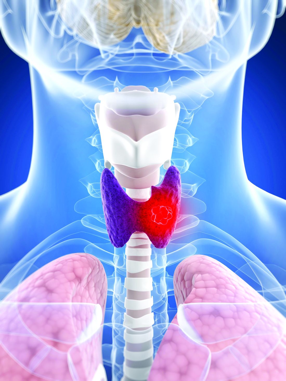
The controversy about whether postoperative RAI is beneficial or an overtreatment in low-risk DTC has persisted for years, but it can now be addressed with level one data, according to Sophie Leboulleux, MD, PhD, who heads the thyroid cancer division at Gustave Roussy Cancer Institute, Villejuif, France.
The trial, called ESTIMABL2, included 776 low-risk DTC patients managed at 35 participating treatment centers in France. Two to five months after surgery, patients were randomized to receive RAI or to follow-up without RAI if there were no suspicious findings, such as changes in lateral neck lymph nodes, on ultrasonography.
Three years after randomization in 729 evaluable patients, tumor-related effects occurred in 4.1% of those randomized to RAI and 4.9% of those randomized to follow-up without RAI.
“Thus, 95.1% of patients in the followed group and 95.9% in the RAI group had no event over 3 years of follow-up. The difference of 0.8% was noninferior by the prespecified definition,” Dr. Leboulleux reported at the annual meeting of the Endocrine Society.
The 0.8% difference in events is noninferior
The prespecified definition was a less than 5% difference in events at the end of 5 years. The 0.8% difference in this study was comfortably within the 95% confidence interval (–3.3%-1.8%).
On yearly evaluations under levothyroxine treatment, events were defined as an indication for treatment, whether surgery or RAI administration if there was abnormal RAI intake on the posttherapeutic whole-body scan or elevated thyroglobulin or thyroglobulin antibodies. In the latter case, this was an event even in the absence of abnormal neck ultrasonography.
“Elevated thyroglobulin levels on levothyroxine treatment were defined as greater than 2 ng/mL in the group that did not received RAI and greater than 1 ng/mL in the group that did,” Dr. Leboulleux reported.
Thyroglobulin antibodies were considered elevated with a greater than 50% increase over the upper limit of normal on two consecutive determinations 6 months apart, Dr. Leboulleux said. The same or similar definitions of an event have been used previously.
Of the study population, 83% were women and almost all (96%) had papillary DTC. The mean age was 52 years. Of the tumors, 81.1% were T1b, of which more than half were unevaluable for node status and about 40% were node negative. The remaining tumors were grade T1a, of which about two-thirds had unevaluable node status and the remainder were confirmed node negative.
Prior studies also question RAI benefit
Several retrospective studies have also failed to associated RAI with a significant protection against thyroid events in low-risk DTC. In the most recent guidelines, the American Thoracic Society recommended against routine use of RAI following surgery in low-risk DTC patients. However, these guidelines acknowledged this is a “weak recommendation” based on “low-quality evidence.”
Recurrence rates following surgery in low-risk DTC are not zero. According to a review article coauthored by Dr. Leboulleux, these may occur in up to 3% of patients. This measurable risk might explain why many published retrospective data suggest low-risk DTC patients are continuing to receive postsurgical RAI. In one, using data taken from the Surveillance, Epidemiology, and End Results (SEER) database, nearly 25% of 17,286 low-risk papillary thyroid cancers had received postsurgical RAI.
The ESTIMABL2 trial is one of two randomized studies launched over the last several years to address the controversy regarding postsurgical RAI in low-risk DTC with prospective randomized data. The ongoing IoN trial is the other. The researchers plan a much longer follow-up, with data not expected until 2031.
In fact, one criticism of the ESTIMABL2 is “the low event rate and relatively short follow-up,” reported Megan Haymart, MD, a professor in the division of metabolism, endocrinology, and diabetes at the University of Michigan, Ann Arbor.
“The strengths of this study include the involvement of 35 French centers, the use of a cohort that is representative of the distribution of low-risk thyroid cancer at large, and the use of biochemical markers during follow-up,” Dr. Haymart said in an interview.
Ultimately, despite the limited follow-up, “this is a clinically important study as it supports the recent paradigm shift towards less use of RAI for low-risk thyroid cancer,” she said.
Dr. Leboulleux and Dr. Haymart report no relevant conflicts of interest.
After 3 years of follow-up in patients with low-risk differentiated thyroid cancer (DTC), there is no significant difference in the rate of tumor-related events in those treated with postsurgical radioactive iodine (RAI) relative to those who are not, according to a randomized phase 3 trial.
The controversy about whether postoperative RAI is beneficial or an overtreatment in low-risk DTC has persisted for years, but it can now be addressed with level one data, according to Sophie Leboulleux, MD, PhD, who heads the thyroid cancer division at Gustave Roussy Cancer Institute, Villejuif, France.
The trial, called ESTIMABL2, included 776 low-risk DTC patients managed at 35 participating treatment centers in France. Two to five months after surgery, patients were randomized to receive RAI or to follow-up without RAI if there were no suspicious findings, such as changes in lateral neck lymph nodes, on ultrasonography.
Three years after randomization in 729 evaluable patients, tumor-related effects occurred in 4.1% of those randomized to RAI and 4.9% of those randomized to follow-up without RAI.
“Thus, 95.1% of patients in the followed group and 95.9% in the RAI group had no event over 3 years of follow-up. The difference of 0.8% was noninferior by the prespecified definition,” Dr. Leboulleux reported at the annual meeting of the Endocrine Society.
The 0.8% difference in events is noninferior
The prespecified definition was a less than 5% difference in events at the end of 5 years. The 0.8% difference in this study was comfortably within the 95% confidence interval (–3.3%-1.8%).
On yearly evaluations under levothyroxine treatment, events were defined as an indication for treatment, whether surgery or RAI administration if there was abnormal RAI intake on the posttherapeutic whole-body scan or elevated thyroglobulin or thyroglobulin antibodies. In the latter case, this was an event even in the absence of abnormal neck ultrasonography.
“Elevated thyroglobulin levels on levothyroxine treatment were defined as greater than 2 ng/mL in the group that did not received RAI and greater than 1 ng/mL in the group that did,” Dr. Leboulleux reported.
Thyroglobulin antibodies were considered elevated with a greater than 50% increase over the upper limit of normal on two consecutive determinations 6 months apart, Dr. Leboulleux said. The same or similar definitions of an event have been used previously.
Of the study population, 83% were women and almost all (96%) had papillary DTC. The mean age was 52 years. Of the tumors, 81.1% were T1b, of which more than half were unevaluable for node status and about 40% were node negative. The remaining tumors were grade T1a, of which about two-thirds had unevaluable node status and the remainder were confirmed node negative.
Prior studies also question RAI benefit
Several retrospective studies have also failed to associated RAI with a significant protection against thyroid events in low-risk DTC. In the most recent guidelines, the American Thoracic Society recommended against routine use of RAI following surgery in low-risk DTC patients. However, these guidelines acknowledged this is a “weak recommendation” based on “low-quality evidence.”
Recurrence rates following surgery in low-risk DTC are not zero. According to a review article coauthored by Dr. Leboulleux, these may occur in up to 3% of patients. This measurable risk might explain why many published retrospective data suggest low-risk DTC patients are continuing to receive postsurgical RAI. In one, using data taken from the Surveillance, Epidemiology, and End Results (SEER) database, nearly 25% of 17,286 low-risk papillary thyroid cancers had received postsurgical RAI.
The ESTIMABL2 trial is one of two randomized studies launched over the last several years to address the controversy regarding postsurgical RAI in low-risk DTC with prospective randomized data. The ongoing IoN trial is the other. The researchers plan a much longer follow-up, with data not expected until 2031.
In fact, one criticism of the ESTIMABL2 is “the low event rate and relatively short follow-up,” reported Megan Haymart, MD, a professor in the division of metabolism, endocrinology, and diabetes at the University of Michigan, Ann Arbor.
“The strengths of this study include the involvement of 35 French centers, the use of a cohort that is representative of the distribution of low-risk thyroid cancer at large, and the use of biochemical markers during follow-up,” Dr. Haymart said in an interview.
Ultimately, despite the limited follow-up, “this is a clinically important study as it supports the recent paradigm shift towards less use of RAI for low-risk thyroid cancer,” she said.
Dr. Leboulleux and Dr. Haymart report no relevant conflicts of interest.
FROM ENDO 2021
Clinical Edge Journal Scan Commentary: Breast Cancer April 2021

Adjuvant endocrine therapy (ET) has been shown to reduce risk of recurrence and improve survival in HR+ early breast cancer (EBC). A retrospective matched cohort analysis of 1,972 patients ≥70 years of age with HR+/HER2-negative, node-negative EBC and multiple medical co-morbidities, demonstrated higher median overall survival (OS) in patients who received ET versus those who did not (79.2 months compared to 67.7 months, p<0.0001). Older women with multiple medical co-morbidities may be at greater risk of toxicities related to ET, which can impact quality of life and adherence. The probability of initiating ET has been shown to be greater among patients who underwent radiation, thus highlighting the need to identify those who will adhere to ET when making decisions regarding radiation therapy to optimize care. ET should be discussed with all women who are candidates for this therapy, with a focus on individualized benefit and risk of these agents.
Studies of immunotherapy in metastatic triple-negative breast cancer have shown promising results, notably in patients with PD-L1 enriched tumors (IMpassion 130 and KEYNOTE-355). KEYNOTE-119 was a phase 3 study including 1,098 patients with metastatic TNBC who had received one or two previous systemic therapies for metastatic disease and progressed on most recent treatment, with randomization to pembrolizumab or physician’s choice chemotherapy . Median OS for pembrolizumab compared to chemotherapy was similar in the overall population (9.9 months versus 10.8 months, HR 0.97) and in patients with PD-L1 CPS score of ≥10 (12.7 months versus 11.6 months, HR 0.78, p=0.057). Although not statistically significant, greater PD-L1 expression was associated with longer median OS with pembrolizumab. Responses to immunotherapy may be durable in select patients, and these agents have an overall favorable toxicity profile. Novel immunotherapy combinations as well as biomarkers to predict response are certainly desired in this space.
Combination endocrine therapy plus a CDK 4/6 inhibitor is standard first-line treatment for metastatic HR+/HER2-negative breast cancer. Neutropenia is a common adverse event (AE) seen with the CDK 4/6 inhibitor palbociclib, and dosing guidelines provide a clear algorithm for drug monitoring and adjustment for cytopenias. Cutaneous toxicities, although not commonly seen, have been reported in the literature. Chawla and colleagues performed a retrospective analysis including 324 patients with advanced HR+/HER2-negative breast cancer who received palbociclib plus endocrine therapy. Cutaneous AEs were seen in 14.2%, with a significant proportion occurring early (41% occurred during or after the first cycle) and 50% resolved within 14 days (average 43 days). Of those who developed cutaneous AEs (n=46), only 15% and 4% required temporary hold and permanent cessation of therapy, respectively. These findings indicate a low overall incidence of cutaneous AEs associated with palbociclib, however highlight the importance of prompt recognition, management, and dermatology referral as appropriate, to help maintain patients on effective cancer-directed therapy.
References:
Yau C, van der Noordaa M, Wei J, et al. Residual cancer burden after neoadjuvant therapy and long-term survival outcomes in breast cancer: a multi-center pooled analysis. Presented at: 2019 San Antonio Breast Cancer Symposium; December 10-14, 2019; San Antonio, TX. Abstract GS5-01.
Wei M, Wang X, Zimmerman DN, Burt LM, Haaland B, Henry NL. Endocrine therapy and radiotherapy use among older women with hormone receptor-positive, clinically node-negative breast cancer. Breast Cancer Res Treat. 2021 Jan 9. doi: 10.1007/s10549-020-06071-w.Epub ahead of print.
Emens LA, Adams S, Barrios CH, et al. IMpassion130: Final OS analysis from the pivotal phase III study of atezolizumab plus nabpaclitaxel versus placebo plus nabpaclitaxel in previously untreated locally advanced or metastatic triple-negative breast cancer. Presented at: ESMO Virtual Congress 2020, Ann Oncol. 2020;31S:ESMO #LBA16.
Khan NAJ, Alsharedi M. Bullous Skin Rash: A Rare Case of Palbociclib-Induced Dermatological Toxicity. Cureus. 2020 Sep 3;12(9):e10229. doi: 10.7759/cureus.10229.
Liver transplant emerges for hepatic mets in colorectal cancer
Liver transplant might be the next big thing for extensive hepatic metastases in colorectal cancer, according to an editorial published in JAMA Surgery .
“Many centers around the world, including our own, [have] started offering liver transplant to patients with colorectal liver metastases,” write Ralph Quillin III, MD, and Shimul Shah, MD, liver transplant surgeons at the University of Cincinnati.
However, they admit that the concept is controversial. “One need only to go to a tumor board to hear critics of the concept of liver transplant for isolated colorectal liver metastases,” the pair write, adding that opponents might argue that transplant patients could have done as well with chemotherapy.
There’s also the problem of deceased donor organ scarcity, Dr. Quillin and Dr. Shah acknowledge.
Nevertheless, the editorial is effusive about a new comparative effectiveness study that it accompanies in JAMA Surgery from a team at the Oslo University Hospital, led by Svein Dueland, MD, PhD.
The new study shows that for select patients with liver metastases, the 5-year survival rate with transplant is higher than for patients who receive standard of care – portal vein embolization (PVE) and surgical resection.
“[Dr.] Dueland et al. have laid the groundwork for the world to follow and took a concept from being experimental to possibly standard of care: liver transplant for colorectal liver metastases with heavy tumor burden,” the editorialists declare.
Using data from their hospital, the Oslo team compared 50 patients who had colorectal cancer liver metastases from previous liver transplant studies (2006-2019) with a retrospective cohort of 53 patients (2006-2015) from studies with similar selection criteria who underwent PVE with the intent to perform liver resection.
Patients with a high liver-only metastatic load had nine or more tumors, or the largest of their tumors had a diameter of at least 5.5 cm. Among 29 such patients treated with liver transplant, the 5-year overall survival (OS) rate was 33.4%, and the median OS was 40.5 months. Among eight such patients treated with PVE and liver resection, the 5-year OS rate was 12.5%, and the median OS was 29.8 months.
The OS among patients with a high metastatic load who received a transplant may be unprecedented, according to the investigators.
The Oslo team “should be commended for a very aggressive surgical approach” for patients whom “many would deem unresectable and palliative from the start,” comment Dr. Quillin and Dr. Shah.
Refining patient selection
In the past, finding the ideal candidate with extensive colorectal liver metastases for liver transplant was like “discovering the proverbial needle in the haystack,” Dr. Quillin and Dr. Shah say. Critics have said the impressive outcomes reported in past studies reflect extreme patient selection.
The new study results make selection easier and may expand the transplant patient population.
The patients in the study had no extrahepatic metastases, and their primary colorectal tumors had been resected. Such patients may represent an expanded transplant population. They were younger than 72 years, and their performance status score was 0-1. They had undergone standard-of-care chemotherapy before their liver procedures.
The high-burden transplant patients were all judged to have nonresectable tumors by the university’s tumor board; 14 of 29 had 16 or more liver lesions.
The maximum number of tumors in the PVE group was 15, so these patients were expected to live longer, owing to the fact that survival is longer for colorectal cancer patients who have fewer liver metastases. However, as noted above, that’s not how it worked out.
Different story for low liver burdens
The Oslo investigators caution that, despite the findings, liver transplant for colorectal liver metastases “should still be considered a work in progress.”
The Oslo team says that to avoid “the futile use of liver grafts,” prospective research is needed to clearly identify colorectal liver metastases patients “who would benefit the most” from transplant.
Patients with extensive nonresectable metastases seem to be high on the list, but there might also be a role in resectable disease, say the study authors. One scenario, for instance, would be one in which PVE fails to expand the liver enough to allow resection; this was the case in 15 of the 53 PVE patients in the study.
As for patients with low liver tumor burden, transplant didn’t seem to offer much long-term survival benefit; 5-year OS was 72.4% among 21 low-burden transplant patients, vs. 69.3% among 23 who underwent PVE and resection.
Right-sided disease – having a primary tumor in the ascending colon, a known predictor of worse outcomes – might also prove to be a contraindication. The median OS after liver transplant was 12.2 months among patients with right-sided primaries and high-burden liver metastases. No such patients were alive at 5 years. On the other hand, the median OS was 59.9 months, and the 5-year OS was 45.3% among high-burden patients with left-sided primaries.
A call for a consortium
In a second editorial, Yuman Fong, MD, of the City of Hope Medical Center, Duarte, Calif., is less enthusiastic than the pair from Cincinnati. He calls the data “intriguing” but emphasizes that “we must recognize that cadaveric livers for transplant remain a finite resource,” and about 1,000 people die annually while on the waiting list in the United States.
The Cincinnati editorialists call for a multicenter consortium “for the cancer community to increase our understanding of this indication” and “pave the way for liver transplant as an option for colorectal liver metastases, just like we have for hepatocellular carcinoma, unresectable hilar cholangiocarcinoma, and metastatic neuroendocrine tumor.”
The investigators note that survival with transplant might be better because liver resection leaves behind microscopic disease that leads to liver recurrence. Most of the transplant patients in the study also experienced relapse, but recurrence after transplant tends to occur in the lungs with small, slow-growing lesions that are amenable to resection.
The study was funded by Oslo University Hospital, the Norwegian Cancer Society, and the South-Eastern Norway Regional Health Authority. The investigators and the editorialists in Cincinnati have disclosed no relevant financial relationships. Dr. Fong is a consultant for Medtronic and Johnson & Johnson and has received royalties from Merck and Imugene.
A version of this article first appeared on Medscape.com.
HER2+ BC: Recurrence risk remains even after achieving pCR with neoadjuvant pertuzumab+trastuzumab
Key clinical point: Patients with human epidermal growth factor receptor 2-positive (HER2+) early breast cancer (BC) who achieved a pathologic complete response (pCR) with neoadjuvant pertuzumab, trastuzumab, and chemotherapy (nPTC) and then received adjuvant trastuzumab remained at risk for recurrence, indicating the need for alternative therapies.
Major finding: At 4 years, the invasive disease-free survival rates for the overall, node-positive, and node-negative cohorts were 90.0% (95% confidence interval [CI], 84.6%-93.6%), 86.2% (95% CI, 78.1%-91.4%), and 96.0% (95% CI, 88.0%-98.7%), respectively.
Study details: Findings are from a real-world, retrospective analysis of 217 patients with HER2+ locally advanced, high-risk early-stage BC, including 135 patients with node-positive and 77 with node-negative disease. Patients received nPTC, had pCR, and then received adjuvant trastuzumab.
Disclosures: This study was funded by Genentech, Inc. Some investigators including the lead author reported ties with various pharmaceutical companies, including Genentech. J Sussell, A Cheng, and A Fung declared being employees of Genentech, Inc.
Source: O’Shaughnessy J et al. Breast Cancer Res Treat. 2021 Mar 1. doi: 10.1007/s10549-021-06137-3.
Excess deaths jump 23% in U.S. in 2020, mostly because of COVID-19
The United States saw nearly 23% more deaths than expected during the first 9 months of the pandemic, and almost three-quarters of those deaths involved COVID-19.
For comparison, the death rate increased by 2.5% or less annually in recent years.
At the same time, rates of deaths from heart disease, Alzheimer’s disease or dementia, and diabetes also increased from March 1, 2020, to Jan. 2, 2021, especially during COVID-19 surges.
“Excess deaths surged in the east in April, followed by extended summer and early winter surges concentrated in Southern and Western states, respectively. Many of these states weakly embraced, or discouraged, pandemic control measures and lifted restrictions earlier than other states,” lead author Steven H. Woolf, MD, MPH, from the Virginia Commonwealth University, Richmond, and colleagues wrote in a research letter published online April 2, 2021, in JAMA.
COVID-19 mortality included all deaths for which it was cited as an underlying or contributing cause in records from the District of Columbia and 49 states. North Carolina was excluded for insufficient data.
More than half a million excess deaths
Between March 1, 2020, and Jan. 2, 2021, the United States experienced 2,801,439 deaths, or 522,368 excess deaths. A total 72.4% of these events were attributed to COVID-19.
Not all racial and ethnic groups were equally represented. For example, the rate of excess deaths was higher among non-Hispanic Black populations, at 208.4 deaths per 100,000. Non-Hispanic White populations experienced 157 deaths per 100,000, and Hispanic populations experienced 139.8 deaths per 100,000.
Further, non-Hispanic Black individuals accounted for 16.9% of the excess deaths but only 12.5% of the U.S. population, which reflects “racial disparities in COVID-19 mortality,” the authors noted.
Not adjusting for population aging is a potential limitation, as was reliance on provisional data and the likelihood that some death certificates were inaccurate.
In February, Anthony S. Fauci, MD, chief medical adviser to President Joe Biden, stated that political divisions likely played a role in the 500,000-plus COVID-19–related deaths in the United States.
Then a report came out on March 26 indicating that a different U.S. response to the pandemic could have avoided almost 400,000 COVID-19 deaths. In addition, an April 1 study in the CDC’s Morbidity and Mortality Weekly Report revealed that COVID-19 is now the third leading cause of death in the United States, after heart disease and cancer.
‘Massive’ excessive mortality
“There is no more visible or alarming manifestation of the toll of the COVID-19 pandemic than the deaths it has caused. In this issue of JAMA, Dr. Woolf and colleagues provide updated analyses that demonstrate that the excess mortality in the U.S. between March 1, 2020, and Jan. 2, 2021, has been massive,” Alan Garber, MD, PhD, wrote in an accompanying editorial.
“It seems likely that COVID-19 will have contributed to nearly as many deaths in the U.S. as the great influenza pandemic of 1918, and more than in any influenza outbreak in the U.S. since then,” added Dr. Garber, provost of Harvard University in Cambridge, Mass.
This study of excess mortality illustrates what is at stake, he added. “Despite the scientific, medical and public health progress of recent decades, the loss of life attributable to the COVID-19 pandemic exceeds the mortality of major wars. No nation should squander this opportunity to do what it takes to prepare for the next one.”
Dr. Woolf and Dr. Garber disclosed no relevant financial relationships. The National Institutes of Health supported the research through its National Center for Advancing Translational Sciences and the National Institute on Aging.
A version of this article first appeared on Medscape.com.
Risk of 5-year local recurrence declines with event-free years in newly diagnosed breast cancer
Key clinical point: In patients with newly diagnosed breast cancer (BC), the risk of local recurrence (LR) as a first event within 5 years of diagnosis was low and declined further with the number of event-free years.
Major finding: Overall, the incidence of LR as a first event within 5 years of diagnosis was 3.0%, which decreased to 2.4%, 1.6%, 1.0%, and 0.6% after 1, 2, 3, and 4 event-free years, respectively.
Study details: Findings are from the nationwide Netherlands Cancer Registry database, including 34,453 patients with newly diagnosed breast cancer between 2005 and 2008.
Disclosures: This study was supported by CZ fonds 201400316. ML Smidt reported receiving grant from Servier Pharma. Other authors had no disclosures.
Source: Moossdorff M et al. Breast Cancer Res Treat. 2021 Mar 10. doi: 10.1007/s10549-020-06040-3.