User login
EoE Prevalence in US Reaches 1 in 700, Costs $1B Annually
, according to recent research.
Although EoE has been considered a rare disease, the chronic condition is becoming more common, and healthcare providers should expect to encounter EoE in clinical settings, the study authors wrote.
“Our last assessment of the prevalence and burden of EoE was more than 10 years ago, and we had a strong suspicion we would continue to see increased numbers of patients with EoE and an increasing cost burden related to the condition in the United States,” said senior author Evan S. Dellon, MD, MPH, AGAF, professor of gastroenterology and hepatology and director of the Center for Esophageal Diseases and Swallowing at the University of North Carolina School of Medicine, Chapel Hill, North Carolina.

“EoE is becoming more common,” Dellon said. “Healthcare providers should expect to see EoE in their practices, including in the primary care setting, emergency departments, allergy practices, GI [gastrointestinal] practices, ENT clinics, and endoscopy suites.”
The study was published in Clinical Gastroenterology and Hepatology.
Estimating EoE Prevalence
Dellon and colleagues analyzed the Merative MarketScan Commercial Claims and Encounters and Medicare Fee-for-Service databases to calculate the annual prevalence of EoE, as well as age- and sex-stratified estimates standardized to the US population. They also calculated healthcare utilization, including medications and endoscopic procedures, to estimate annual EoE-associated costs. Since the EoE billing code was introduced in 2008, the analysis included 2009-2022 MarketScan and 2009-2017 Medicare data.
In the MarketScan database, the research team identified 20,435 EoE cases in 2022, with a mean age of 38 years, 16% younger than 18 years, 62% men, and 41% with a comorbid allergic disease code. The most common symptoms and diagnoses were dysphagia (39%), abdominal pain or dyspepsia (24%), and esophageal stricture (19%). Over time, patients also had previous codes for comorbid allergic diseases (64%), dysphagia (62%), or esophageal stricture (32%).
In the Medicare database, the research team identified 1913 EoE cases in 2017, with a mean age of 73 years, 47% men, 90% non-Hispanic White, and 36% with a comorbid allergic disease. The most common symptoms and diagnoses were dysphagia (49%), abdominal pain or dyspepsia (35%), and esophageal stricture (30%). Over time, patients also had codes for comorbid allergic diseases (64%), dysphagia (65%), or esophageal stricture (42%).
The database numbers translated to EoE prevalences of about 163 cases per 100,000 people in MarketScan in 2022 and 64 cases per 100,000 people in Medicare in 2017. Since 2009, there has been a fivefold increase in prevalence in both databases.
In MarketScan, the prevalence was higher among men than among women, at 204 vs 122 cases per 100,000 people. For both sexes, peak prevalence occurred between ages 40 and 44.
In Medicare, prevalence was also higher among men than among women, at 79 vs 55 cases per 100,000 people. Peak prevalence occurred between ages 65 and 69.
Standardized to the US population, EoE prevalence was 142.5 cases per 100,000 people, extrapolating to 472,380 cases. The overall prevalence was approximately 1 in 700, with rates of 1 in 617 for those younger than 65 years and 1 in 1562 for those aged ≥ 65 years.
“The rapidly increasing prevalence year over year for the entire timeframe of the study was surprising, as were our estimates of the total number of EoE patients in the US, which suggests that EoE is no longer a rare disease and is now seen in about 1 in 700 people,” Dellon said. “This almost triples our prior estimates of 1 in 2000 from 10 years ago, with all trends suggesting that the prevalence will continue to increase.”
Calculating EoE Costs
In terms of procedures, endoscopy with dilation or biopsy was used in about 60%-70% of patients with EoE in both MarketScan and Medicare during the years analyzed. In addition, upper endoscopy with biopsy was coded in 80%-90% of patients, guidewire-based dilation in 11%-17% of patients, and balloon-based dilation in 13%-20% of patients.
In terms of prescription medications, proton pump inhibitors (41%) and topical steroids (26%) were the most common in MarketScan in 2022, as well as in Medicare in 2017, at 32% and 9%, respectively.
When looking at costs by age and sex, the male cohort with the highest costs was aged 10-14 years, estimated at $106.7 million. Among the female cohort, the highest costs were associated with ages 15-19, estimated at $46.5 million.
Overall, total EoE-associated healthcare costs were estimated to be $1.04 billion in 2017, and when adjusted for inflation, the costs were estimated at $1.32 billion in 2024. This is likely an underestimate, the authors wrote, given that EoE prevalence has likely increased for ages 65 or older since 2017 and for all ages since 2022.
“Researching the prevalence and costs is essential to improving patient care by highlighting the growing burden of this recently recognized and growing chronic disease, guiding policy and insurer decisions, and advocating for better access to effective treatments and support for patients,” said Joy Chang, MD, assistant professor of medicine in the Division of Gastroenterology, University of Michigan, Ann Arbor, Michigan.Chang, who wasn’t involved with this study, specializes in eosinophilic GI diseases and researches patient-physician preferences and decision-making in EoE care.
“Clinicians should remain vigilant for symptoms, utilize guideline-based diagnostic approaches, and consider both medical and dietary treatment strategies to optimize patient outcomes and reduce long-term costs,” she said. “Increased awareness and timely intervention can help mitigate the growing impact of this chronic condition.”
The study was supported by a National Institutes of Health grant and used resources from the University of North Carolina Center for Gastrointestinal Biology and Disease. Dellon reported receiving research funding from and having consultant roles with numerous pharmaceutical companies and organizations. Chang reported having no relevant disclosures.
A version of this article appeared on Medscape.com.
, according to recent research.
Although EoE has been considered a rare disease, the chronic condition is becoming more common, and healthcare providers should expect to encounter EoE in clinical settings, the study authors wrote.
“Our last assessment of the prevalence and burden of EoE was more than 10 years ago, and we had a strong suspicion we would continue to see increased numbers of patients with EoE and an increasing cost burden related to the condition in the United States,” said senior author Evan S. Dellon, MD, MPH, AGAF, professor of gastroenterology and hepatology and director of the Center for Esophageal Diseases and Swallowing at the University of North Carolina School of Medicine, Chapel Hill, North Carolina.
“EoE is becoming more common,” Dellon said. “Healthcare providers should expect to see EoE in their practices, including in the primary care setting, emergency departments, allergy practices, GI [gastrointestinal] practices, ENT clinics, and endoscopy suites.”
The study was published in Clinical Gastroenterology and Hepatology.
Estimating EoE Prevalence
Dellon and colleagues analyzed the Merative MarketScan Commercial Claims and Encounters and Medicare Fee-for-Service databases to calculate the annual prevalence of EoE, as well as age- and sex-stratified estimates standardized to the US population. They also calculated healthcare utilization, including medications and endoscopic procedures, to estimate annual EoE-associated costs. Since the EoE billing code was introduced in 2008, the analysis included 2009-2022 MarketScan and 2009-2017 Medicare data.
In the MarketScan database, the research team identified 20,435 EoE cases in 2022, with a mean age of 38 years, 16% younger than 18 years, 62% men, and 41% with a comorbid allergic disease code. The most common symptoms and diagnoses were dysphagia (39%), abdominal pain or dyspepsia (24%), and esophageal stricture (19%). Over time, patients also had previous codes for comorbid allergic diseases (64%), dysphagia (62%), or esophageal stricture (32%).
In the Medicare database, the research team identified 1913 EoE cases in 2017, with a mean age of 73 years, 47% men, 90% non-Hispanic White, and 36% with a comorbid allergic disease. The most common symptoms and diagnoses were dysphagia (49%), abdominal pain or dyspepsia (35%), and esophageal stricture (30%). Over time, patients also had codes for comorbid allergic diseases (64%), dysphagia (65%), or esophageal stricture (42%).
The database numbers translated to EoE prevalences of about 163 cases per 100,000 people in MarketScan in 2022 and 64 cases per 100,000 people in Medicare in 2017. Since 2009, there has been a fivefold increase in prevalence in both databases.
In MarketScan, the prevalence was higher among men than among women, at 204 vs 122 cases per 100,000 people. For both sexes, peak prevalence occurred between ages 40 and 44.
In Medicare, prevalence was also higher among men than among women, at 79 vs 55 cases per 100,000 people. Peak prevalence occurred between ages 65 and 69.
Standardized to the US population, EoE prevalence was 142.5 cases per 100,000 people, extrapolating to 472,380 cases. The overall prevalence was approximately 1 in 700, with rates of 1 in 617 for those younger than 65 years and 1 in 1562 for those aged ≥ 65 years.
“The rapidly increasing prevalence year over year for the entire timeframe of the study was surprising, as were our estimates of the total number of EoE patients in the US, which suggests that EoE is no longer a rare disease and is now seen in about 1 in 700 people,” Dellon said. “This almost triples our prior estimates of 1 in 2000 from 10 years ago, with all trends suggesting that the prevalence will continue to increase.”
Calculating EoE Costs
In terms of procedures, endoscopy with dilation or biopsy was used in about 60%-70% of patients with EoE in both MarketScan and Medicare during the years analyzed. In addition, upper endoscopy with biopsy was coded in 80%-90% of patients, guidewire-based dilation in 11%-17% of patients, and balloon-based dilation in 13%-20% of patients.
In terms of prescription medications, proton pump inhibitors (41%) and topical steroids (26%) were the most common in MarketScan in 2022, as well as in Medicare in 2017, at 32% and 9%, respectively.
When looking at costs by age and sex, the male cohort with the highest costs was aged 10-14 years, estimated at $106.7 million. Among the female cohort, the highest costs were associated with ages 15-19, estimated at $46.5 million.
Overall, total EoE-associated healthcare costs were estimated to be $1.04 billion in 2017, and when adjusted for inflation, the costs were estimated at $1.32 billion in 2024. This is likely an underestimate, the authors wrote, given that EoE prevalence has likely increased for ages 65 or older since 2017 and for all ages since 2022.
“Researching the prevalence and costs is essential to improving patient care by highlighting the growing burden of this recently recognized and growing chronic disease, guiding policy and insurer decisions, and advocating for better access to effective treatments and support for patients,” said Joy Chang, MD, assistant professor of medicine in the Division of Gastroenterology, University of Michigan, Ann Arbor, Michigan.Chang, who wasn’t involved with this study, specializes in eosinophilic GI diseases and researches patient-physician preferences and decision-making in EoE care.
“Clinicians should remain vigilant for symptoms, utilize guideline-based diagnostic approaches, and consider both medical and dietary treatment strategies to optimize patient outcomes and reduce long-term costs,” she said. “Increased awareness and timely intervention can help mitigate the growing impact of this chronic condition.”
The study was supported by a National Institutes of Health grant and used resources from the University of North Carolina Center for Gastrointestinal Biology and Disease. Dellon reported receiving research funding from and having consultant roles with numerous pharmaceutical companies and organizations. Chang reported having no relevant disclosures.
A version of this article appeared on Medscape.com.
FROM CLINICAL GASTROENTEROLOGY AND HEPATOLOGY
AI Algorithm Predicts Transfusion Need, Mortality Risk in Acute GI Bleeds
SAN DIEGO — , researchers reported at Digestive Disease Week® (DDW) 2025.
Acute GI bleeding is the most common cause of digestive disease–related hospitalization, with an estimated 500,000 hospital admissions annually. It’s known that predicting the need for red blood cell transfusion in the first 24 hours may improve resuscitation and decrease both morbidity and mortality.
However, an existing clinical score known as the Rockall Score does not perform well for predicting mortality, Xi (Nicole) Zhang, an MD-PhD student at McGill University, Montreal, Quebec, Canada, told attendees at DDW. With an area under the curve of 0.65-0.75, better prediction is needed, said Zhang, whose coresearchers included Dennis Shung, MD, MHS, PhD, director of Applied Artificial Intelligence at Yale University School of Medicine, New Haven, Connecticut.

“We’d like to predict multiple outcomes in addition to mortality,” said Zhang, who is also a student at the Mila-Quebec Artificial Intelligence Institute.
As a result, the researchers turned to the TFM approach, applying it to ICU patients with acute GI bleeding to predict both the need for transfusion and in-hospital mortality risk. The all-cause mortality rate is up to 11%, according to a 2020 study by James Y. W. Lau, MD, and colleagues. The rebleeding rate of nonvariceal upper GI bleeds is up to 10.4%. Zhang said the rebleeding rate for variceal upper gastrointestinal bleeding is up to 65%.
The AI method the researchers used outperformed a standard deep learning model at predicting the need for transfusion and estimating mortality risk.
Defining the AI Framework
“Probabilistic flow matching is a class of generative artificial intelligence that learns how a simple distribution becomes a more complex distribution with ordinary differential equations,” Zhang told GI & Hepatology News. “For example, if you had a few lines and shapes you could learn how it could become a detailed portrait of a face. In our case, we start with a few blood pressure and heart rate measurements and learn the pattern of blood pressures and heart rates over time, particularly if they reflect clinical deterioration with hemodynamic instability.”
Another way to think about the underlying algorithm, Zhang said, is to think about a river with boats where the river flow determines where the boats end up. “We are trying to direct the boat to the correct dock by adjusting the flow of water in the canal. In this case we are mapping the distribution with the first few data points to the distribution with the entire patient trajectory.”
The information gained, she said, could be helpful in timing endoscopic evaluation or allocating red blood cell products for emergent transfusion.
Study Details
The researchers evaluated a cohort of 2602 patients admitted to the ICU, identified from the publicly available MIMIC-III database. They divided the patients into a training set of 2342 patients and an internal validation set of 260 patients. Input variables were severe liver disease comorbidity, administration of vasopressor medications, mean arterial blood pressure, and heart rate over the first 24 hours.
Excluded was hemoglobin, since the point was to test the trajectory of hemodynamic parameters independent of hemoglobin thresholds used to guide red blood cell transfusion.
The outcome measures were administration of packed red blood cell transfusion within 24 hours and all-cause hospital mortality.
The TFM was more accurate than a standard deep learning model in predicting red blood cell transfusion, with an accuracy of 93.6% vs 43.2%; P ≤ .001. It was also more accurate at predicting all-cause in-hospital mortality, with an accuracy of 89.5% vs 42.5%, P = .01.
The researchers concluded that the TFM approach was able to predict the hemodynamic trajectories of patients with acute GI bleeding defined as deviation and outperformed the baseline from the measured mean arterial pressure and heart rate.
Expert Perspective
“This is an exciting proof-of-concept study that shows generative AI methods may be applied to complex datasets in order to improve on our current predictive models and improve patient care,” said Jeremy Glissen Brown, MD, MSc, an assistant professor of medicine and a practicing gastroenterologist at Duke University who has published research on the use of AI in clinical practice. He reviewed the study for GI & Hepatology News but was not involved in the research.
“Future work will likely look into the implementation of a version of this model on real-time data.” he said. “We are at an exciting inflection point in predictive models within GI and clinical medicine. Predictive models based on deep learning and generative AI hold the promise of improving how we predict and treat disease states, but the excitement being generated with studies such as this needs to be balanced with the trade-offs inherent to the current paradigm of deep learning and generative models compared to more traditional regression-based models. These include many of the same ‘black box’ explainability questions that have risen in the age of convolutional neural networks as well as some method-specific questions due to the continuous and implicit nature of TFM.”
Elaborating on that, Glissen Brown said: “TFM, like many deep learning techniques, raises concerns about explainability that we’ve long seen with convolutional neural networks — the ‘black box’ problem, where it’s difficult to interpret exactly how and why the model arrives at a particular decision. But TFM also introduces unique challenges due to its continuous and implicit formulation. Since it often learns flows without explicitly defining intermediate representations or steps, it can be harder to trace the logic or pathways it uses to connect inputs to outputs. This makes standard interpretability tools less effective and calls for new techniques tailored to these continuous architectures.”
“This approach could have a real clinical impact,” said Robert Hirten, MD, associate professor of medicine and artificial intelligence, Icahn School of Medicine at Mount Sinai, New York City, who also reviewed the study. “Accurately predicting transfusion needs and mortality risk in real time could support earlier, more targeted interventions for high-risk patients. While these findings still need to be validated in prospective studies, it could enhance ICU decision-making and resource allocation.”
“For the practicing gastroenterologist, we envision this system could help them figure out when to perform endoscopy in a patient admitted with acute gastrointestinal bleeding in the ICU at very high risk of exsanguination,” Zhang told GI & Hepatology News.
The approach, the researchers said, will be useful in identifying unique patient characteristics, make possible the identification of high-risk patients and lead to more personalized medicine.
Hirten, Zhang, and Shung had no disclosures. Glissen Brown reported consulting relationships with Medtronic, OdinVision, Doximity, and Olympus. The National Institutes of Health funded this study.
A version of this article appeared on Medscape.com.
SAN DIEGO — , researchers reported at Digestive Disease Week® (DDW) 2025.
Acute GI bleeding is the most common cause of digestive disease–related hospitalization, with an estimated 500,000 hospital admissions annually. It’s known that predicting the need for red blood cell transfusion in the first 24 hours may improve resuscitation and decrease both morbidity and mortality.
However, an existing clinical score known as the Rockall Score does not perform well for predicting mortality, Xi (Nicole) Zhang, an MD-PhD student at McGill University, Montreal, Quebec, Canada, told attendees at DDW. With an area under the curve of 0.65-0.75, better prediction is needed, said Zhang, whose coresearchers included Dennis Shung, MD, MHS, PhD, director of Applied Artificial Intelligence at Yale University School of Medicine, New Haven, Connecticut.
“We’d like to predict multiple outcomes in addition to mortality,” said Zhang, who is also a student at the Mila-Quebec Artificial Intelligence Institute.
As a result, the researchers turned to the TFM approach, applying it to ICU patients with acute GI bleeding to predict both the need for transfusion and in-hospital mortality risk. The all-cause mortality rate is up to 11%, according to a 2020 study by James Y. W. Lau, MD, and colleagues. The rebleeding rate of nonvariceal upper GI bleeds is up to 10.4%. Zhang said the rebleeding rate for variceal upper gastrointestinal bleeding is up to 65%.
The AI method the researchers used outperformed a standard deep learning model at predicting the need for transfusion and estimating mortality risk.
Defining the AI Framework
“Probabilistic flow matching is a class of generative artificial intelligence that learns how a simple distribution becomes a more complex distribution with ordinary differential equations,” Zhang told GI & Hepatology News. “For example, if you had a few lines and shapes you could learn how it could become a detailed portrait of a face. In our case, we start with a few blood pressure and heart rate measurements and learn the pattern of blood pressures and heart rates over time, particularly if they reflect clinical deterioration with hemodynamic instability.”
Another way to think about the underlying algorithm, Zhang said, is to think about a river with boats where the river flow determines where the boats end up. “We are trying to direct the boat to the correct dock by adjusting the flow of water in the canal. In this case we are mapping the distribution with the first few data points to the distribution with the entire patient trajectory.”
The information gained, she said, could be helpful in timing endoscopic evaluation or allocating red blood cell products for emergent transfusion.
Study Details
The researchers evaluated a cohort of 2602 patients admitted to the ICU, identified from the publicly available MIMIC-III database. They divided the patients into a training set of 2342 patients and an internal validation set of 260 patients. Input variables were severe liver disease comorbidity, administration of vasopressor medications, mean arterial blood pressure, and heart rate over the first 24 hours.
Excluded was hemoglobin, since the point was to test the trajectory of hemodynamic parameters independent of hemoglobin thresholds used to guide red blood cell transfusion.
The outcome measures were administration of packed red blood cell transfusion within 24 hours and all-cause hospital mortality.
The TFM was more accurate than a standard deep learning model in predicting red blood cell transfusion, with an accuracy of 93.6% vs 43.2%; P ≤ .001. It was also more accurate at predicting all-cause in-hospital mortality, with an accuracy of 89.5% vs 42.5%, P = .01.
The researchers concluded that the TFM approach was able to predict the hemodynamic trajectories of patients with acute GI bleeding defined as deviation and outperformed the baseline from the measured mean arterial pressure and heart rate.
Expert Perspective
“This is an exciting proof-of-concept study that shows generative AI methods may be applied to complex datasets in order to improve on our current predictive models and improve patient care,” said Jeremy Glissen Brown, MD, MSc, an assistant professor of medicine and a practicing gastroenterologist at Duke University who has published research on the use of AI in clinical practice. He reviewed the study for GI & Hepatology News but was not involved in the research.
“Future work will likely look into the implementation of a version of this model on real-time data.” he said. “We are at an exciting inflection point in predictive models within GI and clinical medicine. Predictive models based on deep learning and generative AI hold the promise of improving how we predict and treat disease states, but the excitement being generated with studies such as this needs to be balanced with the trade-offs inherent to the current paradigm of deep learning and generative models compared to more traditional regression-based models. These include many of the same ‘black box’ explainability questions that have risen in the age of convolutional neural networks as well as some method-specific questions due to the continuous and implicit nature of TFM.”
Elaborating on that, Glissen Brown said: “TFM, like many deep learning techniques, raises concerns about explainability that we’ve long seen with convolutional neural networks — the ‘black box’ problem, where it’s difficult to interpret exactly how and why the model arrives at a particular decision. But TFM also introduces unique challenges due to its continuous and implicit formulation. Since it often learns flows without explicitly defining intermediate representations or steps, it can be harder to trace the logic or pathways it uses to connect inputs to outputs. This makes standard interpretability tools less effective and calls for new techniques tailored to these continuous architectures.”
“This approach could have a real clinical impact,” said Robert Hirten, MD, associate professor of medicine and artificial intelligence, Icahn School of Medicine at Mount Sinai, New York City, who also reviewed the study. “Accurately predicting transfusion needs and mortality risk in real time could support earlier, more targeted interventions for high-risk patients. While these findings still need to be validated in prospective studies, it could enhance ICU decision-making and resource allocation.”
“For the practicing gastroenterologist, we envision this system could help them figure out when to perform endoscopy in a patient admitted with acute gastrointestinal bleeding in the ICU at very high risk of exsanguination,” Zhang told GI & Hepatology News.
The approach, the researchers said, will be useful in identifying unique patient characteristics, make possible the identification of high-risk patients and lead to more personalized medicine.
Hirten, Zhang, and Shung had no disclosures. Glissen Brown reported consulting relationships with Medtronic, OdinVision, Doximity, and Olympus. The National Institutes of Health funded this study.
A version of this article appeared on Medscape.com.
FROM DDW 2025
Ostomy Innovation Grabs ‘Shark Tank’ Win
The “Shark Tank” winning innovation at the American Gastroenterological Association (AGA) Tech Summit in Chicago this April has “life-altering” potential for ostomy patients, according to one of the judges, and eliminates the need for constant pouch wear.
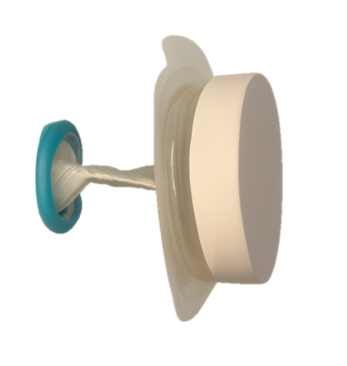
The innovation is called Twistomy and it is designed to replace current ostomy-pouch systems that can cause leaks, odor, skin irritation, embarrassment, and social and emotional distress. The AGA Committee for GI Innovation and Technology (CGIT) organizes the annual Tech Summit.

Twistomy’s winning design includes a flexible ring and sleeve, which are inserted into the stoma and secured on the outside with a set of rings that make up the housing unit attached to a standard wafer. The housing unit twists the sleeve closed, allowing the user to control fecal output. For evacuation, the user attaches a pouch, untwists the sleeve, evacuates cleanly and effectively, and then discards the pouch.
Twistomy cofounders Devon Horton, BS, senior bioengineer, and Lily Williams, BS, biomedical researcher and engineer, both work for the department of surgery at University of Colorado, Denver.

Horton said in an interview that when he was approached with the idea to create a better ostomy solution for a senior-year capstone project he was intrigued because the traditional ostomy system “has not changed in more than 70 years. It was crazy that no one had done anything to change that.”
The Twistomy team also won the Grand Prize this spring at the Emerging Medical Innovation Valuation Competition at the Design of Medical Devices Conference held at the University of Minnesota, Minneapolis.
Witnessing the Struggle as a CNA
Horton also works as a certified nursing assistant at an inpatient unit at University of Colorado Hospital and the ostomy patients he sees there every shift help drive his passion to find a better solution.
He hears the emotional stories of people who manage their ostomy daily.
“Many express feelings of depression and anxiety, feeling isolated with their severe inability to go out and do things because of the fear of the noise the stoma makes, or the crinkling of the plastic bag in a yoga class,” he said. “We want to help them regain that control of quality of life.”
They also hope to cut down on the ostomy management time. “Initial user testing [for Twistomy] was less than 75 seconds to insert and assemble,” he said. “I did an interview with a patient yesterday who said they probably spend an hour a day managing their ostomy,” including cleaning and replacing.
Horton and Williams have a patent on the device and currently use three-dimensional printing for the prototypes.
Williams said they are now conducting consumer discovery studies through the National Science Foundation and are interviewing 30 stakeholders — “anyone who has a relationship with an ostomy,” whether a colorectal surgeon, a gastrointestinal nurse, ostomy patients, or insurers.
Those interviews will help in refining the device so they can start consulting with manufacturers and work toward approval as a Class II medical device from the US Food and Drug Administration (FDA), Williams said.
Saving Healthcare Costs
Another potential benefit for Twistomy is its ability to cut healthcare costs, Horton said. Traditional ostomies are prone to leakage, which can lead to peristomal skin complications.
He pointed to a National Institutes of Health analysis that found that on average peristomal skin complications caused upwards of $80,000 more per ostomy patient in increased healthcare costs over a 3-month period than for those without the complications.
“With Twistomy, we are reducing leakage most likely to zero,” Horton said. “We set out to say if we could reduce [infections] by half or a little less than half, we can cut out those tens of thousands of dollars that insurance companies and payers are spending.”
Permanent and Temporary Ostomy Markets
He pointed out that not all ostomies are permanent ostomies, adding that the reversal rate “is about 65%.” Often those reversal surgeries cannot take place until peristomal skin complications have been healed.
“We’re not only hoping to market to the permanent stoma patients, but the patients with temporary stomas as well,” he said.
The team estimates it will need $4 million–$6 million in funding for manufacturing and consultation costs as well as costs involved in seeking FDA approval.
Horton and Williams project the housing unit cost will be $399 based on known out-of-pocket expenses for patients with ostomy care products and the unit would be replaced annually. Disposable elements would be an additional cost.
Assuming insurance acceptance of the product, he said, “With about an 80/20 insurance coverage, typical for many patients, it would be about $100 in out-of-pocket expenses per month to use our device, which is around the lower end of what a lot of patients are spending out of pocket.”
One of the Tech Summit judges, Somaya Albhaisi, MD, a gastroenterology/hepatology fellow at University of Southern California, Los Angeles, said in an interview that the Shark Tank results were unanimous among the five judges and Twistomy also took the fan favorite vote.
She said the teams were judged on quality of pitch, potential clinical impact, and feasibility of business plan. Teams got 5-7 minutes to pitch and answered questions afterward.
“Deep Understanding” of Patient Need
“They combined smart engineering with deep understanding of patient need, which is restoring control, dignity, and quality of life for ostomy users while also reducing healthcare costs. It is rare to see a solution this scalable and impactful. It was a deeply empathetic solution overall.” She noted that nearly 1 million people in the United States currently use an ostomy.
Ostomy users’ quality of life is compromised, and they often have mental health challenges, Albhaisi said. This innovation appears to offer easy use, more dignity and control.
The other four Shark Tank finalists were:
- AI Lumen, which developed a retroview camera system, which attaches to the colonoscope and enhances imaging to detect hidden polyps that may evade conventional endoscopes.
- Amplified Sciences, which developed an ultrasensitive diagnostic platform that detects biomarker activities in minute volumes of fluid from pancreatic cystic lesions, helping to stratify patients into low risk or potential malignancy, reducing unneeded surgeries, costs, and comorbidities.
- KITE Endoscopic Innovations, which designed the Dynaflex TruCut needle to offer a simpler endoscopic ultrasound (EUS)–guided biopsy procedure with fewer needle passes, deeper insights into tumor pathology, and more tissue for geonomic analysis.
- MicroSteer, which designed a device to facilitate semiautomated endoscopic submucosal dissection (ESD) by decoupling the dissecting knife from the endoscope, enhancing safety and effectiveness during the procedure.
The Twistomy Team “Surprised Everyone”
The competitors’ scores were “very close,” one of the judges, Kevin Berliner, said in an interview. “The Twistomy team surprised everyone — the judges and the crowd — with their succinct, informative, and impactful pitch. That presentation disparity was the tiebreaker for me,” said Berliner, who works for Medtronic, a sponsor of the competition, in Chicago.
He said Horton and Williams were the youngest presenters and had the earliest stage pitch they judged, but they “outpresented other competitors in clarity, simplification, and storytelling.”
Also impressive was their description of their “commercially viable path to success” and their plan for the challenges ahead, he said.
Those challenges to get Twistomy to market center “on the ongoing changing climate we have with research funds lately,” Horton said. “We’re giving it an estimate of 3-5 years.”
Horton, Williams, Albhaisi, and Berliner reported no relevant financial relationships.
The “Shark Tank” winning innovation at the American Gastroenterological Association (AGA) Tech Summit in Chicago this April has “life-altering” potential for ostomy patients, according to one of the judges, and eliminates the need for constant pouch wear.
The innovation is called Twistomy and it is designed to replace current ostomy-pouch systems that can cause leaks, odor, skin irritation, embarrassment, and social and emotional distress. The AGA Committee for GI Innovation and Technology (CGIT) organizes the annual Tech Summit.
Twistomy’s winning design includes a flexible ring and sleeve, which are inserted into the stoma and secured on the outside with a set of rings that make up the housing unit attached to a standard wafer. The housing unit twists the sleeve closed, allowing the user to control fecal output. For evacuation, the user attaches a pouch, untwists the sleeve, evacuates cleanly and effectively, and then discards the pouch.
Twistomy cofounders Devon Horton, BS, senior bioengineer, and Lily Williams, BS, biomedical researcher and engineer, both work for the department of surgery at University of Colorado, Denver.
Horton said in an interview that when he was approached with the idea to create a better ostomy solution for a senior-year capstone project he was intrigued because the traditional ostomy system “has not changed in more than 70 years. It was crazy that no one had done anything to change that.”
The Twistomy team also won the Grand Prize this spring at the Emerging Medical Innovation Valuation Competition at the Design of Medical Devices Conference held at the University of Minnesota, Minneapolis.
Witnessing the Struggle as a CNA
Horton also works as a certified nursing assistant at an inpatient unit at University of Colorado Hospital and the ostomy patients he sees there every shift help drive his passion to find a better solution.
He hears the emotional stories of people who manage their ostomy daily.
“Many express feelings of depression and anxiety, feeling isolated with their severe inability to go out and do things because of the fear of the noise the stoma makes, or the crinkling of the plastic bag in a yoga class,” he said. “We want to help them regain that control of quality of life.”
They also hope to cut down on the ostomy management time. “Initial user testing [for Twistomy] was less than 75 seconds to insert and assemble,” he said. “I did an interview with a patient yesterday who said they probably spend an hour a day managing their ostomy,” including cleaning and replacing.
Horton and Williams have a patent on the device and currently use three-dimensional printing for the prototypes.
Williams said they are now conducting consumer discovery studies through the National Science Foundation and are interviewing 30 stakeholders — “anyone who has a relationship with an ostomy,” whether a colorectal surgeon, a gastrointestinal nurse, ostomy patients, or insurers.
Those interviews will help in refining the device so they can start consulting with manufacturers and work toward approval as a Class II medical device from the US Food and Drug Administration (FDA), Williams said.
Saving Healthcare Costs
Another potential benefit for Twistomy is its ability to cut healthcare costs, Horton said. Traditional ostomies are prone to leakage, which can lead to peristomal skin complications.
He pointed to a National Institutes of Health analysis that found that on average peristomal skin complications caused upwards of $80,000 more per ostomy patient in increased healthcare costs over a 3-month period than for those without the complications.
“With Twistomy, we are reducing leakage most likely to zero,” Horton said. “We set out to say if we could reduce [infections] by half or a little less than half, we can cut out those tens of thousands of dollars that insurance companies and payers are spending.”
Permanent and Temporary Ostomy Markets
He pointed out that not all ostomies are permanent ostomies, adding that the reversal rate “is about 65%.” Often those reversal surgeries cannot take place until peristomal skin complications have been healed.
“We’re not only hoping to market to the permanent stoma patients, but the patients with temporary stomas as well,” he said.
The team estimates it will need $4 million–$6 million in funding for manufacturing and consultation costs as well as costs involved in seeking FDA approval.
Horton and Williams project the housing unit cost will be $399 based on known out-of-pocket expenses for patients with ostomy care products and the unit would be replaced annually. Disposable elements would be an additional cost.
Assuming insurance acceptance of the product, he said, “With about an 80/20 insurance coverage, typical for many patients, it would be about $100 in out-of-pocket expenses per month to use our device, which is around the lower end of what a lot of patients are spending out of pocket.”
One of the Tech Summit judges, Somaya Albhaisi, MD, a gastroenterology/hepatology fellow at University of Southern California, Los Angeles, said in an interview that the Shark Tank results were unanimous among the five judges and Twistomy also took the fan favorite vote.
She said the teams were judged on quality of pitch, potential clinical impact, and feasibility of business plan. Teams got 5-7 minutes to pitch and answered questions afterward.
“Deep Understanding” of Patient Need
“They combined smart engineering with deep understanding of patient need, which is restoring control, dignity, and quality of life for ostomy users while also reducing healthcare costs. It is rare to see a solution this scalable and impactful. It was a deeply empathetic solution overall.” She noted that nearly 1 million people in the United States currently use an ostomy.
Ostomy users’ quality of life is compromised, and they often have mental health challenges, Albhaisi said. This innovation appears to offer easy use, more dignity and control.
The other four Shark Tank finalists were:
- AI Lumen, which developed a retroview camera system, which attaches to the colonoscope and enhances imaging to detect hidden polyps that may evade conventional endoscopes.
- Amplified Sciences, which developed an ultrasensitive diagnostic platform that detects biomarker activities in minute volumes of fluid from pancreatic cystic lesions, helping to stratify patients into low risk or potential malignancy, reducing unneeded surgeries, costs, and comorbidities.
- KITE Endoscopic Innovations, which designed the Dynaflex TruCut needle to offer a simpler endoscopic ultrasound (EUS)–guided biopsy procedure with fewer needle passes, deeper insights into tumor pathology, and more tissue for geonomic analysis.
- MicroSteer, which designed a device to facilitate semiautomated endoscopic submucosal dissection (ESD) by decoupling the dissecting knife from the endoscope, enhancing safety and effectiveness during the procedure.
The Twistomy Team “Surprised Everyone”
The competitors’ scores were “very close,” one of the judges, Kevin Berliner, said in an interview. “The Twistomy team surprised everyone — the judges and the crowd — with their succinct, informative, and impactful pitch. That presentation disparity was the tiebreaker for me,” said Berliner, who works for Medtronic, a sponsor of the competition, in Chicago.
He said Horton and Williams were the youngest presenters and had the earliest stage pitch they judged, but they “outpresented other competitors in clarity, simplification, and storytelling.”
Also impressive was their description of their “commercially viable path to success” and their plan for the challenges ahead, he said.
Those challenges to get Twistomy to market center “on the ongoing changing climate we have with research funds lately,” Horton said. “We’re giving it an estimate of 3-5 years.”
Horton, Williams, Albhaisi, and Berliner reported no relevant financial relationships.
Chatbot Helps Users Adopt a Low FODMAP Diet
SAN DIEGO — Low fermentable oligo-, di-, and monosaccharides and polyols (FODMAPs) dietary advice has been shown to be effective in easing bloating and abdominal pain, especially in patients with irritable bowel syndrome (IBS), but limited availability of dietitians makes delivering this advice challenging. Researchers from Thailand have successfully enlisted a chatbot to help.
In a randomized controlled trial, they found that chatbot-assisted dietary advice with brief guidance effectively reduced high FODMAP intake, bloating severity, and improved dietary knowledge, particularly in patients with bothersome bloating.
“Chatbot-assisted dietary advice for FODMAPs restriction was feasible and applicable in patients with bloating symptoms that had baseline symptoms of moderate severity,” study chief Pochara Somvanapanich, with the Division of Gastroenterology, Chulalongkorn University and King Chulalongkorn Memorial Hospital, Bangkok, Thailand, told GI & Hepatology News.
Somvanapanich, who developed the chatbot algorithm, presented the study results at Digestive Disease Week (DDW) 2025.
More Knowledge, Less Bloating
The trial enrolled 86 adults with disorders of gut-brain interaction experiencing bloating symptoms for more than 6 months and consuming more than seven high-FODMAPs items per week. Half of them had IBS.
At baseline, gastrointestinal (GI) symptoms and the ability to identify FODMAPs were assessed. All participants received a 5-minute consultation on FODMAPs avoidance from a GI fellow and were randomly allocated (stratified by IBS diagnosis and education) into two groups.
The chatbot-assisted group received real-time dietary advice via a chatbot which helped them identify high, low, and non-FODMAP foods from a list of more than 300 ingredients/dishes of Thai and western cuisines.
The control group received only brief advice on high FODMAPs restriction. Both groups used a diary app to log food intake and postprandial symptoms. Baseline bloating, abdominal pain and global symptoms severity were similar between the two groups. Data on 64 participants (32 in each group) were analyzed.
After 4 weeks, significantly more people in the chatbot group than the control group responded — achieving a 30% or greater reduction in daily worst bloating, abdominal pain or global symptoms (19 [59%] vs 10 [31%], P < .05). Responder rates were similar in the IBS and non-IBS subgroups.
Subgroup analysis revealed significant differences between groups only for participants with bothersome bloating, not those with mild bloating severity.
In those with bothersome bloating severity, the chatbot group had a higher response rate (69.5% vs 36.3%) and fewer bloating symptoms (P < .05). They also had a greater reduction in high FODMAPs intake (10 vs 23 items/week) and demonstrated improved knowledge in identifying FODMAPs (P < .05).
“Responders in a chatbot group consistently engaged more with the app, performing significantly more weekly item searches than nonresponders (P < .05),” the authors noted in their conference abstract.
“Our next step is to develop the chatbot-assisted approach for the reintroduction and personalization phase based on messenger applications (including Facebook Messenger and other messaging platforms),” Somvanapanich told GI & Hepatology News.
“Once we’ve gathered enough data to confirm these are working effectively, we definitely plan to create a one-stop service application for FODMAPs dietary advice,” Somvanapanich added.
Lack of Robust Data on Digital GI Health Apps
Commenting on this research for GI & Hepatology News, Sidhartha R. Sinha, MD, Director of Digital Health and Innovation, Division of Gastroenterology and Hepatology, Stanford University in Stanford, California, noted that there is a “notable lack of robust data supporting digital health tools in gastroenterology. Despite hundreds of apps available, very few are supported by well-designed trials.”
“The study demonstrated that chatbot-assisted dietary advice significantly improved bloating symptoms, reduced intake of high-FODMAP foods, and enhanced patients’ dietary knowledge compared to brief dietary counseling alone, especially in those with bothersome symptoms,” said Sinha, who wasn’t involved in the study.
“Patients actively used the chatbot to manage their symptoms, achieving a higher response rate than those in the control arm who received brief counseling on avoiding high-FODMAP food,” he noted.
Sinha said in his practice at Stanford, “in the heart of Silicon Valley,” patients do use digital resources to manage their GI symptoms, including diseases like IBS and inflammatory bowel disease (IBD) — and he believes this is “increasingly common nationally.”
“However, the need for evidence-based tools is critical and the lack here often prevents many practitioners from regularly recommending them to patients. This study aligns well with clinical practice, and supports the use of this particular app to improve IBS symptoms, particularly when access to dietitians is limited. These results support chatbot-assisted dietary management as a feasible, effective, and scalable approach to patient care,” Sinha told GI & Hepatology News.
The study received no commercial funding. Somvanapanich and Sinha had no relevant disclosures.
A version of this article appeared on Medscape.com.
FROM DDW 2025
Blood-Based Test May Predict Crohn’s Disease 2 Years Before Onset
SAN DIEGO — Crohn’s disease (CD) has become more common in the United States, and an estimated 1 million Americans have the condition. Still, much is unknown about how to evaluate the individual risk for the disease.
“It’s pretty much accepted that Crohn’s disease does not begin at diagnosis,” said Ryan Ungaro, MD, associate professor of medicine at the Icahn School of Medicine at Mount Sinai, New York City, speaking at Digestive Disease Week (DDW)® 2025.
Although individual blood markers have been associated with the future risk for CD, what’s needed, he said, is to understand which combination of biomarkers are most predictive.
Now, .
It’s an early version that will likely be further improved and needs additional validation, Ungaro told GI & Hepatology News.
“Once we can accurately identify individuals at risk for developing Crohn’s disease, we can then imagine a number of potential interventions,” Ungaro said.
Approaches would vary depending on how far away the onset is estimated to be. For people who likely wouldn’t develop disease for many years, one intervention might be close monitoring to enable diagnosis in the earliest stages, when treatment works best, he said. Someone at a high risk of developing CD in the next 2 or 3 years, on the other hand, might be offered a pharmaceutical intervention.
Developing and Testing the Risk Score
To develop the risk score, Ungaro and colleagues analyzed data of 200 patients with CD and 100 healthy control participants from PREDICTS, a nested case-controlled study of active US military service members. The study is within the larger Department of Defense Serum Repository, which began in 1985 and has more than 62.5 million samples, all stored at −30 °C.
The researchers collected serum samples at four timepoints up to 6 or more years before the diagnosis. They assayed antimicrobial antibodies using the Prometheus Laboratories platform, proteomic markers using the Olink inflammation panel, and anti–granulocyte macrophage colony-stimulating factor autoantibodies using enzyme-linked immunosorbent assay.
Participants (median age, 33 years for both groups) were randomly divided into equally sized training and testing sets. In both the group, 83% of patients were White and about 90% were men.
Time-varying trajectories of marker abundance were estimated for each biomarker. Then, logistic regression modeled disease status as a function of each marker for different timepoints and multivariate modeling was performed via logistic LASSO regression.
A risk score to predict CD onset within 2 years was developed. Prediction models were fit on the testing set and predictive performance evaluated using receiver operating characteristic curves and area under the curve (AUC).
Blood proteins and antibodies have differing associations with CD depending on the time before diagnosis, the researchers found.
The integrative model to predict CD onset within 2 years incorporated 10 biomarkers associated significantly with CD onset.
The AUC for the model was 0.87 (considered good, with 1 indicating perfect discrimination). It produced a specificity of 99% and a positive predictive value of 84%.
The researchers stratified the model scores into quartiles and found the CD incidence within 2 years increased from 2% in the first quartile to 57.7% in the fourth. The relative risk of developing CD in the top quartile individuals vs lower quartile individuals was 10.4.
The serologic and proteomic markers show dynamic changes years before the diagnosis, Ungaro said.
A Strong Start
The research represents “an ambitious and exciting frontier for the future of IBD [inflammatory bowel disease] care,” said Victor G. Chedid, MD, MS, consultant and assistant professor of medicine at Mayo Clinic, Rochester, Minnesota, who reviewed the findings but was not involved in the study.

Currently, physicians treat IBD once it manifests, and it’s difficult to predict who will get CD, he said.
The integrative model’s AUC of 0.87 is impressive, and its specificity and positive predictive value levels show it is highly accurate in predicting the onset of CD within 2 years, Chedid added.
Further validation in larger and more diverse population is needed, Chedid said, but he sees the potential for the model to be practical in clinical practice.
“Additionally, the use of blood-based biomarkers makes the model relatively noninvasive and easy to implement in a clinical setting,” he said.
Now, the research goal is to understand the best biomarkers for characterizing the different preclinical phases of CD and to test different interventions in prevention trials, Ungaro told GI & Hepatology News.
A few trials are planned or ongoing, he noted. The trial PIONIR trial will look at the impact of a specific diet on the risk of developing CD, and the INTERCEPT trial aims to develop a blood-based risk score that can identify individuals with a high risk of developing CD within 5 years after initial evaluation.
Ungaro reported being on the advisory board of and/or receiving speaker or consulting fees from AbbVie, Bristol Myer Squibb, Celltrion, ECM Therapeutics, Genentech, Jansen, Eli Lilly, Pfizer, Roivant, Sanofi, and Takeda. Chedid reported having no relevant disclosures.
The PROMISE Consortium is funded by the Helmsley Charitable Trust.
A version of this article appeared on Medscape.com.
SAN DIEGO — Crohn’s disease (CD) has become more common in the United States, and an estimated 1 million Americans have the condition. Still, much is unknown about how to evaluate the individual risk for the disease.
“It’s pretty much accepted that Crohn’s disease does not begin at diagnosis,” said Ryan Ungaro, MD, associate professor of medicine at the Icahn School of Medicine at Mount Sinai, New York City, speaking at Digestive Disease Week (DDW)® 2025.
Although individual blood markers have been associated with the future risk for CD, what’s needed, he said, is to understand which combination of biomarkers are most predictive.
Now, .
It’s an early version that will likely be further improved and needs additional validation, Ungaro told GI & Hepatology News.
“Once we can accurately identify individuals at risk for developing Crohn’s disease, we can then imagine a number of potential interventions,” Ungaro said.
Approaches would vary depending on how far away the onset is estimated to be. For people who likely wouldn’t develop disease for many years, one intervention might be close monitoring to enable diagnosis in the earliest stages, when treatment works best, he said. Someone at a high risk of developing CD in the next 2 or 3 years, on the other hand, might be offered a pharmaceutical intervention.
Developing and Testing the Risk Score
To develop the risk score, Ungaro and colleagues analyzed data of 200 patients with CD and 100 healthy control participants from PREDICTS, a nested case-controlled study of active US military service members. The study is within the larger Department of Defense Serum Repository, which began in 1985 and has more than 62.5 million samples, all stored at −30 °C.
The researchers collected serum samples at four timepoints up to 6 or more years before the diagnosis. They assayed antimicrobial antibodies using the Prometheus Laboratories platform, proteomic markers using the Olink inflammation panel, and anti–granulocyte macrophage colony-stimulating factor autoantibodies using enzyme-linked immunosorbent assay.
Participants (median age, 33 years for both groups) were randomly divided into equally sized training and testing sets. In both the group, 83% of patients were White and about 90% were men.
Time-varying trajectories of marker abundance were estimated for each biomarker. Then, logistic regression modeled disease status as a function of each marker for different timepoints and multivariate modeling was performed via logistic LASSO regression.
A risk score to predict CD onset within 2 years was developed. Prediction models were fit on the testing set and predictive performance evaluated using receiver operating characteristic curves and area under the curve (AUC).
Blood proteins and antibodies have differing associations with CD depending on the time before diagnosis, the researchers found.
The integrative model to predict CD onset within 2 years incorporated 10 biomarkers associated significantly with CD onset.
The AUC for the model was 0.87 (considered good, with 1 indicating perfect discrimination). It produced a specificity of 99% and a positive predictive value of 84%.
The researchers stratified the model scores into quartiles and found the CD incidence within 2 years increased from 2% in the first quartile to 57.7% in the fourth. The relative risk of developing CD in the top quartile individuals vs lower quartile individuals was 10.4.
The serologic and proteomic markers show dynamic changes years before the diagnosis, Ungaro said.
A Strong Start
The research represents “an ambitious and exciting frontier for the future of IBD [inflammatory bowel disease] care,” said Victor G. Chedid, MD, MS, consultant and assistant professor of medicine at Mayo Clinic, Rochester, Minnesota, who reviewed the findings but was not involved in the study.
Currently, physicians treat IBD once it manifests, and it’s difficult to predict who will get CD, he said.
The integrative model’s AUC of 0.87 is impressive, and its specificity and positive predictive value levels show it is highly accurate in predicting the onset of CD within 2 years, Chedid added.
Further validation in larger and more diverse population is needed, Chedid said, but he sees the potential for the model to be practical in clinical practice.
“Additionally, the use of blood-based biomarkers makes the model relatively noninvasive and easy to implement in a clinical setting,” he said.
Now, the research goal is to understand the best biomarkers for characterizing the different preclinical phases of CD and to test different interventions in prevention trials, Ungaro told GI & Hepatology News.
A few trials are planned or ongoing, he noted. The trial PIONIR trial will look at the impact of a specific diet on the risk of developing CD, and the INTERCEPT trial aims to develop a blood-based risk score that can identify individuals with a high risk of developing CD within 5 years after initial evaluation.
Ungaro reported being on the advisory board of and/or receiving speaker or consulting fees from AbbVie, Bristol Myer Squibb, Celltrion, ECM Therapeutics, Genentech, Jansen, Eli Lilly, Pfizer, Roivant, Sanofi, and Takeda. Chedid reported having no relevant disclosures.
The PROMISE Consortium is funded by the Helmsley Charitable Trust.
A version of this article appeared on Medscape.com.
FROM DDW 2025
Winning Strategies to Retain Private Practice Gastroenterologists
SAN DIEGO — With the recently updated recommendations by the US Preventive Services Task Force lowering the age for colorectal cancer screening to 45 instead of 50, an additional 19 million patients now require screening, Asma Khapra, MD, AGAF, a gastroenterologist at Gastro Health in Fairfax, Virginia, told attendees at Digestive Disease Week® (DDW) 2025.

That change, coupled with the expected shortage of gastroenterologists, means one thing: The current workforce can’t meet patient demand, she said. , Khapra added.
The private practice model is already declining, she said. The fraction of US gastroenterologists in “fully independent” private practice was about 30% in 2019, Khapra noted. Then, “COVID really changed the landscape even more.” By 2022, “that number has shrunk to 13%.” Meanwhile, 67% are employed gastroenterologists (not in private practice), 7% work in large group practices, and 13% are private equity (PE) backed.
That makes effective retention strategies crucial for private practices, Khapra said. She first addressed the common attractions of private practices, then the challenges, and finally the winning strategies to retain and keep a viable private practice gastroenterology workforce.
The Attractions of Private Practice
The reasons for choosing private practice are many, Khapra said, including:
- Autonomy,
- Flexibility,
- Competitive compensation,
- Ownership mindset,
- Partnership paths, and
- Work-life balance including involvement in community and culture.
On the other hand, private practices have unique challenges, including:
- Administrative burdens such as EHR documentation, paperwork, prior authorizations, and staffing issues,
- Financial pressures, including competition with the employment packages offered by hospitals, as reimbursements continue to drop and staffing costs increase,
- Burnout,
- Variety of buy-ins and partnership tracks,
- Limited career development, and
- The strains of aging and endoscopy. “We used to joke in our practice that at any given time, three staff members are in physical therapy due to injuries and disabilities.”
Employing the Iceberg Model
One strategy, Khapra said, is to follow Edward T. Hall’s Iceberg Model of Culture , which focuses on the importance of both visible and invisible elements.
“The key to retention in private practice is to develop a value system where everyone is treated well and respected and compensated fairly,” she said. “That doesn’t mean you split the pie [equally].”
“Visible” elements of the model include the physical environment, policies and practices, symbols and behaviors, she said. While under the surface (“invisible” elements) are shared values, perceptions and attitudes, leadership style, conflict resolution, decision making and unwritten rules.
The key, she said, is to provide physicians an actual voice in decision making and to avoid favouritism, thus avoiding comments such as “Why do the same two people always get the prime scoping blocks?”
Financial transparency is also important, Khapra said. And people want flexibility without it being called special treatment. She provided several practical suggestions to accomplish the invisible Iceberg goals.
For instance, she suggested paying for activities outside the practice that physicians do, such as serving on committees. If the practice can’t afford that, she suggested asking the affiliated hospitals to do so, noting that such an initiative can often build community support.
Paying more attention to early associates than is typical can also benefit the practice, Khapra said. “So much effort is made to recruit them, and then once there, we’re on to the next [recruits].” Instead, she suggested, “pay attention to their needs.”
Providing support to physicians who are injured is also crucial and can foster a community culture, she said. For example, one Gastro Health physician was out for 4 weeks due to complications from surgery. “Everyone jumped in” to help fill the injured physician’s shifts, she said, reassuring the physician that the money would be figured out later. “That’s the culture you want to instill.”
To prevent burnout, another key to retaining physicians, “you have to provide support staff.” And offering good benefits, including parental and maternal leave and disability benefits, is also crucial, Khapra said. Consider practices such as having social dinners, another way to build a sense of community.
Finally, bring in national and local gastroenterologist organizations for discussions, including advocating for fair reimbursement for private practice. Consider working with the Digestive Health Physicians Alliance, which describes itself as the voice of independent gastroenterology, she suggested.
More Perspectives
Jami Kinnucan, MD, AGAF, a gastroenterologist and associate professor of medicine at Mayo Clinic, Jacksonville , Florida, spoke about optimizing recruitment of young gastroenterologists and provided perspective on Khapra’s talk.
“I think there’s a lot of overlap” with her topic and retaining private practice gastroenterologists, she said in an interview with GI & Hepatology News. Most important, she said, is having an efficient system in which the administrative flow is left to digital tools or other staff, not physicians. “That will also help to reduce burnout,” she said, and allow physicians to do what they most want to do, which is to focus on providing care to patients.
“People want to feel valued for their work,” she agreed. “People want opportunity for career development, opportunities for growth.”
As gastroenterologists age, flexibility is important, as it in in general for all physicians, Kinnucan said. She suggested schedule flexibility as one way. For instance, “if I tell 10 providers, ‘I need you to see 100 patients this week, but you can do it however you want,’ that promotes flexibility. They might want to see all of them on Monday and Tuesday, for instance. If you give people choice and autonomy, they are more likely to feel like they are part of the decision.”
How do you build a high-functioning team? “You do it by letting them operate autonomously,” and “you let people do the things they are really excited about.” And always, as Khapra said, focus on the invisible elements that are so crucial.
Khapra and Kinnucan had no relevant disclosures. Khapra received no funding for her presentation.
A version of this article appeared on Medscape.com.
SAN DIEGO — With the recently updated recommendations by the US Preventive Services Task Force lowering the age for colorectal cancer screening to 45 instead of 50, an additional 19 million patients now require screening, Asma Khapra, MD, AGAF, a gastroenterologist at Gastro Health in Fairfax, Virginia, told attendees at Digestive Disease Week® (DDW) 2025.
That change, coupled with the expected shortage of gastroenterologists, means one thing: The current workforce can’t meet patient demand, she said. , Khapra added.
The private practice model is already declining, she said. The fraction of US gastroenterologists in “fully independent” private practice was about 30% in 2019, Khapra noted. Then, “COVID really changed the landscape even more.” By 2022, “that number has shrunk to 13%.” Meanwhile, 67% are employed gastroenterologists (not in private practice), 7% work in large group practices, and 13% are private equity (PE) backed.
That makes effective retention strategies crucial for private practices, Khapra said. She first addressed the common attractions of private practices, then the challenges, and finally the winning strategies to retain and keep a viable private practice gastroenterology workforce.
The Attractions of Private Practice
The reasons for choosing private practice are many, Khapra said, including:
- Autonomy,
- Flexibility,
- Competitive compensation,
- Ownership mindset,
- Partnership paths, and
- Work-life balance including involvement in community and culture.
On the other hand, private practices have unique challenges, including:
- Administrative burdens such as EHR documentation, paperwork, prior authorizations, and staffing issues,
- Financial pressures, including competition with the employment packages offered by hospitals, as reimbursements continue to drop and staffing costs increase,
- Burnout,
- Variety of buy-ins and partnership tracks,
- Limited career development, and
- The strains of aging and endoscopy. “We used to joke in our practice that at any given time, three staff members are in physical therapy due to injuries and disabilities.”
Employing the Iceberg Model
One strategy, Khapra said, is to follow Edward T. Hall’s Iceberg Model of Culture , which focuses on the importance of both visible and invisible elements.
“The key to retention in private practice is to develop a value system where everyone is treated well and respected and compensated fairly,” she said. “That doesn’t mean you split the pie [equally].”
“Visible” elements of the model include the physical environment, policies and practices, symbols and behaviors, she said. While under the surface (“invisible” elements) are shared values, perceptions and attitudes, leadership style, conflict resolution, decision making and unwritten rules.
The key, she said, is to provide physicians an actual voice in decision making and to avoid favouritism, thus avoiding comments such as “Why do the same two people always get the prime scoping blocks?”
Financial transparency is also important, Khapra said. And people want flexibility without it being called special treatment. She provided several practical suggestions to accomplish the invisible Iceberg goals.
For instance, she suggested paying for activities outside the practice that physicians do, such as serving on committees. If the practice can’t afford that, she suggested asking the affiliated hospitals to do so, noting that such an initiative can often build community support.
Paying more attention to early associates than is typical can also benefit the practice, Khapra said. “So much effort is made to recruit them, and then once there, we’re on to the next [recruits].” Instead, she suggested, “pay attention to their needs.”
Providing support to physicians who are injured is also crucial and can foster a community culture, she said. For example, one Gastro Health physician was out for 4 weeks due to complications from surgery. “Everyone jumped in” to help fill the injured physician’s shifts, she said, reassuring the physician that the money would be figured out later. “That’s the culture you want to instill.”
To prevent burnout, another key to retaining physicians, “you have to provide support staff.” And offering good benefits, including parental and maternal leave and disability benefits, is also crucial, Khapra said. Consider practices such as having social dinners, another way to build a sense of community.
Finally, bring in national and local gastroenterologist organizations for discussions, including advocating for fair reimbursement for private practice. Consider working with the Digestive Health Physicians Alliance, which describes itself as the voice of independent gastroenterology, she suggested.
More Perspectives
Jami Kinnucan, MD, AGAF, a gastroenterologist and associate professor of medicine at Mayo Clinic, Jacksonville , Florida, spoke about optimizing recruitment of young gastroenterologists and provided perspective on Khapra’s talk.
“I think there’s a lot of overlap” with her topic and retaining private practice gastroenterologists, she said in an interview with GI & Hepatology News. Most important, she said, is having an efficient system in which the administrative flow is left to digital tools or other staff, not physicians. “That will also help to reduce burnout,” she said, and allow physicians to do what they most want to do, which is to focus on providing care to patients.
“People want to feel valued for their work,” she agreed. “People want opportunity for career development, opportunities for growth.”
As gastroenterologists age, flexibility is important, as it in in general for all physicians, Kinnucan said. She suggested schedule flexibility as one way. For instance, “if I tell 10 providers, ‘I need you to see 100 patients this week, but you can do it however you want,’ that promotes flexibility. They might want to see all of them on Monday and Tuesday, for instance. If you give people choice and autonomy, they are more likely to feel like they are part of the decision.”
How do you build a high-functioning team? “You do it by letting them operate autonomously,” and “you let people do the things they are really excited about.” And always, as Khapra said, focus on the invisible elements that are so crucial.
Khapra and Kinnucan had no relevant disclosures. Khapra received no funding for her presentation.
A version of this article appeared on Medscape.com.
FROM DDW 2025
Blood Detection Capsule Helpful in Suspected Upper GI Bleeding
SAN DIEGO — , a study found.
Notably, patients with negative capsule results had shorter hospital stays and lower acuity markers, and in more than one third of cases, an esophagogastroduodenoscopy (EGD) was avoided altogether without any observed adverse events or readmissions, the study team found.
“Our study shows that this novel capsule that detects blood in the upper GI tract (PillSense) was highly sensitive and specific (> 90%) for detecting recent or active upper GI blood, influenced clinical management in 80% of cases and allowed about one third of patients to be safely discharged from the emergency department, with close outpatient follow-up,” Linda Lee, MD, AGAF, medical director of endoscopy, Brigham and Women’s Hospital and associate professor of medicine, Harvard Medical School, Boston, told GI & Hepatology News.
The study was presented at Digestive Disease Week® (DDW) 2025.
Real-World Insights
EGD is the gold standard for diagnosing suspected upper GI bleeding, but limited access to timely EGD complicates diagnosis and resource allocation.
Approved by the US Food and Drug Administration, PillSense (EnteraSense) is an ingestible capsule with a reusable receiver that provides a rapid, noninvasive method for detecting upper GI bleeding. The capsule analyzes light absorption to identify blood and transmits the result within 10 minutes.
Lee and colleagues evaluated the real-world impact of this point-of-care device on clinical triage and resource allocation, while assessing its safety profile.
They analyzed data on 43 patients (mean age 60 years; 72% men) with clinical suspicion of upper GI bleeding in whom the device was used. The most common symptoms were symptomatic anemia (70%), melena (67%), and hematemesis (33%).
Sixteen PillSense studies (37%) were positive for blood detection, and 27 (63%) were negative.
Compared to patients with a positive capsule results, those without blood detected by the capsule had shorter hospital stays (mean, 3.8 vs 13.4 days, P = .02), lower GBS scores (mean, 7.93 vs 12.81; P = .005), and fewer units of blood transfused (mean, 1.19 vs 10.94; P = .01) and were less apt to be hemodynamically unstable (5 vs 8 patients; P = .03).
Capsule results influenced clinical management in 80% of cases, leading to avoidance of EGD in 37% and prioritization of urgent EGD in 18% (all had active bleeding on EGD).
Capsule use improved resource allocation in 51% of cases. This included 12 patients who were discharged from the ED, six who were assigned an inpatient bed early, and four who underwent expedited colonoscopy as upper GI bleeding was ruled out, they noted.
Among the eight patients who did not undergo EGD, there were no readmissions within 30 days and no adverse events. There were no capsule-related adverse events.
“Clinicians should consider using this novel capsule PillSense as another data point in the management of suspected upper GI bleed,” Lee told GI & Hepatology News.
“This could include in helping to triage patients for safe discharge from the ED or to more urgent endoscopy, to differentiate between upper vs lower GI bleed and to manage ICU patients with possible rebleeding,” Lee said.
Important Real-World Evidence
Reached for comment, Shahin Ayazi, MD, esophageal surgeon, Director, Allegheny Health Network Chevalier Jackson Esophageal Research Center, Pittsburgh, Pennsylvania, said this study is important for several reasons.
“Prior investigations have established that PillSense possesses a high negative predictive value for detecting upper GI bleeding and have speculated on its utility in triage, decision-making, and potentially avoiding unnecessary endoscopy. This study is important because it substantiates that speculation with clinical data,” Ayazi, who wasn’t involved in the study, told GI & Hepatology News.
“These findings support the capsule’s practical application in patient stratification and clinical workflow, particularly when diagnostic uncertainty is high and endoscopic resources are limited,” Ayazi noted.
In his experience, PillSense is “highly useful as a triage adjunct in the evaluation of suspected upper GI bleeding. It provides direct and objective evidence as to whether blood is currently present in the stomach,” he said.
“In patients whose presentation is ambiguous or whose clinical scores fall into an intermediate risk zone, this binary result can provide clarity that subjective assessment alone may not achieve. This is particularly relevant in settings where the goal is to perform endoscopy within 24 hours, but the volume of consults exceeds procedural capacity,” Ayazi explained.
“In such scenarios, PillSense enables physicians to stratify patients based on objective evidence of active bleeding, helping to prioritize those who require urgent endoscopy and defer or even avoid endoscopic evaluation in those who do not. The result is a more efficient allocation of endoscopic resources without compromising patient safety,” he added.
Ayazi cautioned that the PillSense capsule should not be used as a replacement for clinical evaluation or established risk stratification protocols.
“It is intended for hemodynamically stable patients and has not been validated in cases of active or massive bleeding. Its diagnostic yield depends on the presence of blood in the stomach at the time of capsule transit; intermittent or proximal bleeding that has ceased may not be detected, introducing the potential for false-negative results,” Ayazi told GI & Hepatology News.
“However, in prior studies, the negative predictive value was high, and in the present study, no adverse outcomes were observed in patients who did not undergo endoscopy following a negative PillSense result,” Ayazi noted.
“It must also be understood that PillSense does not localize the source of bleeding or replace endoscopy in patients with a high likelihood of active hemorrhage. It is not designed to detect bleeding from the lower GI tract or distal small bowel. Rather, it serves as an adjunct that can provide immediate clarity when the need for endoscopy is uncertain, and should be interpreted within the broader context of clinical findings, laboratory data, and established risk stratification tools,” he added.
The study had no specific funding. Lee and Ayazi had no relevant disclosures.
A version of this article appeared on Medscape.com.
FROM DDW 2025
Why Is Early-Onset CRC Rising? New Study Provides a Clue
The numbers don’t lie: Colorectal cancer (CRC) has been on the rise in younger people in the United States for over two decades.
While the data show a clear trend, researchers still face a glaring unanswered question: Why is this happening?
A recent report in Nature may offer an important clue to start unraveling this early-onset CRC mystery.
What the Study Found
The new analysis found that
The bacterial toxin, called colibactin, is produced by certain strains of Escherichia coli and other bacteria — more specifically, polyketide synthase (PKS)–positive strains. Previous research has found colibactin-related mutations can occur in up to 15% of CRC cases overall, but a link to early-onset disease has been less clear.
In this recent genetic analysis, investigators led by Marcos Díaz-Gay, PhD, analyzed CRC biopsies from 981 patients across 11 countries and 4 continents. The team tracked DNA damage from colibactin by identifying distinctive mutational signatures — called SBS88 and ID18 — left by the toxin.
Díaz-Gay and colleagues found that these mutational signatures were 3.3 times more common in patients diagnosed before 40 years of age than in those over 70 years.
Colibactin exposure was also linked to about a quarter of mutations that inactivate the colorectal tumor suppressor gene APC.
However, epidemiologic factors linked to CRC, such as body mass index, diet, and lifestyle, were not considered in the study, which the investigators noted is a key limitation.
“Our results show for the first time an association between the presence of colibactin-induced mutational signatures and early-onset colorectal cancer,” Díaz-Gay, a genomic researcher at the Spanish National Cancer Research Center, Madrid, Spain, and colleagues wrote.
“Prior studies have indicated that mutagenesis due to colibactin exposure can occur within the first decade of life and then ceases,” the investigators explained. But “this ‘head start’ could plausibly result in an increased risk of early-onset cancers.”
What the Study Means
Trevor Graham, PhD, a professor of genomics and evolution at The Institute of Cancer Research, London, England, helped put the study findings into context.
Others have proposed that colibactin “could have a role in causing early-onset disease,” Graham commented in a statement from the UK nonprofit Science Media Centre. “This work provides [the] strong[est] data yet that the hypothesis is correct.”
Plus, Graham added, “This is very good quality research. The authors have collected bowel cancers from countries around the world and performed whole genome sequencing on them.”
“Most importantly,” he said, the colibactin mutations were more common in people who got bowel cancer before 50 years of age, which “suggests the mutations caused by these bugs in the bowel could be a cause of early-onset bowel cancer, although further studies are needed to confirm this.”
Although the study doesn’t prove causation, “this is a very important finding,” Alan Venook, MD, a gastrointestinal medical oncologist and CRC specialist at the University of California, San Francisco, told GI & Hepatology News. “This gives us a hook to understand what’s going on.”

However, “it’s not at all likely that this single entity is entirely responsible for early-onset CRC,” Venook clarified.
But if childhood exposure to colibactin is responsible, at least in part, for the growing incidence of early-onset CRC, it would suggest that PKS-positive bacteria have become more common in the gut microbiome of younger people over the past few decades. PKS-positive E coli are common, in general — found in up to 20% of healthy people and about 67% of patients with CRC.
If these bacteria are becoming more common in younger people, the reason isn’t yet clear. “The working hypothesis is overuse of antibiotics in young kids,” said Venook, who is collaborating with colleagues to launch an additional multi-institution investigation into the issue.
There are also clinical implications if the findings pan out, Venook said.
This work could lead to a diagnostic test — perhaps one looking for circulating mutational DNA in the blood — that could “give us a leg up on who’s at risk for early-onset CRC,” Venook said. “That’s how this could really make a difference.”
The work was funded by the National Institutes of Health, Cancer Research UK, and others. Several investigators disclosed ties to io9, Inocras, Hologic, Quotient Therapeutics, and Microbiotica. Venook didn’t have any disclosures; disclosure information for Graham was unavailable.
A version of this article appeared on Medscape.com.
The numbers don’t lie: Colorectal cancer (CRC) has been on the rise in younger people in the United States for over two decades.
While the data show a clear trend, researchers still face a glaring unanswered question: Why is this happening?
A recent report in Nature may offer an important clue to start unraveling this early-onset CRC mystery.
What the Study Found
The new analysis found that
The bacterial toxin, called colibactin, is produced by certain strains of Escherichia coli and other bacteria — more specifically, polyketide synthase (PKS)–positive strains. Previous research has found colibactin-related mutations can occur in up to 15% of CRC cases overall, but a link to early-onset disease has been less clear.
In this recent genetic analysis, investigators led by Marcos Díaz-Gay, PhD, analyzed CRC biopsies from 981 patients across 11 countries and 4 continents. The team tracked DNA damage from colibactin by identifying distinctive mutational signatures — called SBS88 and ID18 — left by the toxin.
Díaz-Gay and colleagues found that these mutational signatures were 3.3 times more common in patients diagnosed before 40 years of age than in those over 70 years.
Colibactin exposure was also linked to about a quarter of mutations that inactivate the colorectal tumor suppressor gene APC.
However, epidemiologic factors linked to CRC, such as body mass index, diet, and lifestyle, were not considered in the study, which the investigators noted is a key limitation.
“Our results show for the first time an association between the presence of colibactin-induced mutational signatures and early-onset colorectal cancer,” Díaz-Gay, a genomic researcher at the Spanish National Cancer Research Center, Madrid, Spain, and colleagues wrote.
“Prior studies have indicated that mutagenesis due to colibactin exposure can occur within the first decade of life and then ceases,” the investigators explained. But “this ‘head start’ could plausibly result in an increased risk of early-onset cancers.”
What the Study Means
Trevor Graham, PhD, a professor of genomics and evolution at The Institute of Cancer Research, London, England, helped put the study findings into context.
Others have proposed that colibactin “could have a role in causing early-onset disease,” Graham commented in a statement from the UK nonprofit Science Media Centre. “This work provides [the] strong[est] data yet that the hypothesis is correct.”
Plus, Graham added, “This is very good quality research. The authors have collected bowel cancers from countries around the world and performed whole genome sequencing on them.”
“Most importantly,” he said, the colibactin mutations were more common in people who got bowel cancer before 50 years of age, which “suggests the mutations caused by these bugs in the bowel could be a cause of early-onset bowel cancer, although further studies are needed to confirm this.”
Although the study doesn’t prove causation, “this is a very important finding,” Alan Venook, MD, a gastrointestinal medical oncologist and CRC specialist at the University of California, San Francisco, told GI & Hepatology News. “This gives us a hook to understand what’s going on.”
However, “it’s not at all likely that this single entity is entirely responsible for early-onset CRC,” Venook clarified.
But if childhood exposure to colibactin is responsible, at least in part, for the growing incidence of early-onset CRC, it would suggest that PKS-positive bacteria have become more common in the gut microbiome of younger people over the past few decades. PKS-positive E coli are common, in general — found in up to 20% of healthy people and about 67% of patients with CRC.
If these bacteria are becoming more common in younger people, the reason isn’t yet clear. “The working hypothesis is overuse of antibiotics in young kids,” said Venook, who is collaborating with colleagues to launch an additional multi-institution investigation into the issue.
There are also clinical implications if the findings pan out, Venook said.
This work could lead to a diagnostic test — perhaps one looking for circulating mutational DNA in the blood — that could “give us a leg up on who’s at risk for early-onset CRC,” Venook said. “That’s how this could really make a difference.”
The work was funded by the National Institutes of Health, Cancer Research UK, and others. Several investigators disclosed ties to io9, Inocras, Hologic, Quotient Therapeutics, and Microbiotica. Venook didn’t have any disclosures; disclosure information for Graham was unavailable.
A version of this article appeared on Medscape.com.
Colorectal Cancer Screening Choices: Is Compliance Key?
SAN DIEGO — , and when it comes to potentially life-saving screening measures, picking the optimal screening tool is critical.
Regarding tests, “perfect is not possible,” said William M. Grady, MD, AGAF, of the Fred Hutchinson Cancer Center, University of Washington School of Medicine, in Seattle, who took part in a debate on the pros and cons of key screening options at Digestive Disease Week® (DDW) 2025.

“We have to remember that that’s the reality of colorectal cancer screening, and we need to meet our patients where they live,” said Grady, who argued on behalf of blood-based tests, including cell-free (cf) DNA (Shield, Guardant Health) and cfDNA plus protein biomarkers (Freenome).
A big point in their favor is their convenience and higher patient compliance — better tests that don’t get done do not work, he stressed.
He cited data that showed suboptimal compliance rates with standard colonoscopy: Rates range from about 70% among non-Hispanic White individuals to 67% among Black individuals, 51% among Hispanic individuals, and the low rate of just 26% among patients aged between 45 and 50 years.
With troubling increases in CRC incidence among younger patients, “that’s a group we’re particularly concerned about,” Grady said.
Meanwhile, studies show compliance rates with blood-based tests are ≥ 80%, with similar rates seen among those racial and ethnic groups, with lower rates for conventional colonoscopy, he noted.
Importantly, in terms of performance in detecting CRC, blood-based tests stand up to other modalities, as demonstrated in a real-world study conducted by Grady and his colleagues showing a sensitivity of 83% for the cfDNA test, 74% for the fecal immunochemical test (FIT) stool test, and 92% for a multitarget stool DNA test compared with 95% for colonoscopy.
“What we can see is that the sensitivity of blood-based tests looks favorable and comparable to other tests,” he said.
Among the four options, cfDNA had a highest patient adherence rate (85%-86%) compared with colonoscopy (28%-42%), FIT (43%-65%), and multitarget stool DNA (48%-60%).
“The bottom line is that these tests decrease CRC mortality and incidence, and we know there’s a potential to improve compliance with colorectal cancer screening if we offer blood-based tests for average-risk people who refuse colonoscopy,” Grady said.
Blood-Based Tests: Caveats, Harms?
Arguing against blood-based tests in the debate, Robert E. Schoen, MD, MPH, professor of medicine and epidemiology, Division of Gastroenterology, Hepatology and Nutrition, at the University of Pittsburgh, in Pittsburgh, Pennsylvania, checked off some of the key caveats.
While the overall sensitivity of blood-based tests may look favorable, these tests don’t detect early CRC well,” said Schoen. The sensitivity rates for stage 1 CRC are 64.7% with Guardant Health and 57.1% with Freenome.
Furthermore, their rates of detecting advanced adenomas are very low; the rate with Guardant Health is only about 13%, and with Freenome is even lower at 12.5%, he reported.
These rates are “similar to the false positive rate, with poor discrimination and accuracy for advanced adenomas,” Schoen said. “Without substantial detection of advanced adenomas, blood-based testing is inferior [to other options].”
Importantly, the low advanced adenoma rate translates to a lack of CRC prevention, which is key to reducing CRC mortality, he noted.
Essential to success with blood-based biopsies, as well as with stool tests, is the need for a follow-up colonoscopy if results are positive, but Schoen pointed out that this may or may not happen.
He cited research from FIT data showing that among 33,000 patients with abnormal stool tests, the rate of follow-up colonoscopy within a year, despite the concerning results, was a dismal 56%.
“We have a long way to go to make sure that people who get positive noninvasive tests get followed up,” he said.
In terms of the argument that blood-based screening is better than no screening at all, Schoen cited recent research that projected reductions in the risk for CRC incidence and mortality among 100,000 patients with each of the screening modalities.
Starting with standard colonoscopy performed every 10 years, the reductions in incidence and mortality would be 79% and 81%, respectively, followed by annual FIT, at 72% and 76%; multitarget DNA every 3 years, at 68% and 73%; and cfDNA (Shield), at 45% and 55%.
Based on those rates, if patients originally opting for FIT were to shift to blood-based tests, “the rate of CRC deaths would increase,” Schoen noted.
The findings underscore that “blood testing is unfavorable as a ‘substitution test,’” he added. “In fact, widespread adoption of blood testing could increase CRC morbidity.”
“Is it better than nothing?” he asked. “Yes, but only if performance of a colonoscopy after a positive test is accomplished.”
What About FIT?
Arguing that stool-based testing, or FIT, is the ideal choice as a first-line CRC test Jill Tinmouth, MD, PhD, a professor at the University of Toronto, Ontario, Canada, pointed to its prominent role in organized screening programs, including regions where resources may limit the widespread utilization of routine first-line colonoscopy screening. In addition, it narrows colonoscopies to those that are already prescreened as being at risk.
Data from one such program, reported by Kaiser Permanente of Northern California, showed that participation in CRC screening doubled from 40% to 80% over 10 years after initiating FIT screening. CRC mortality over the same period decreased by 50% from baseline, and incidence fell by as much as 75%.
In follow-up colonoscopies, Tinmouth noted that collective research from studies reflecting real-world participation and adherence to FIT in populations in the United Kingdom, the Netherlands, Taiwan, and California show follow-up colonoscopy rates of 88%, 85%, 70%, and 78%, respectively.
Meanwhile, a recent large comparison of biennial FIT (n = 26,719) vs one-time colonoscopy (n = 26,332) screening, the first study to directly compare the two, showed noninferiority, with nearly identical rates of CRC mortality at 10 years (0.22% colonoscopy vs 0.24% FIT) as well as CRC incidence (1.13% vs 1.22%, respectively).
“This study shows that in the context of organized screening, the benefits of FIT are the same as colonoscopy in the most important outcome of CRC — mortality,” Tinmouth said.
Furthermore, as noted with blood-based screening, the higher participation with FIT shows a much more even racial/ethnic participation than that observed with colonoscopy.
“FIT has clear and compelling advantages over colonoscopy,” she said. As well as better compliance among all groups, “it is less costly and also better for the environment [by using fewer resources],” she added.
Colonoscopy: ‘Best for First-Line Screening’
Making the case that standard colonoscopy should in fact be the first-line test, Swati G. Patel, MD, director of the Gastrointestinal Cancer Risk and Prevention Center at the University of Colorado Anschutz Medical Center, Aurora, Colorado, emphasized the robust, large population studies showing its benefits. Among them is a landmark national policy study showing a significant reduction in CRC incidence and mortality associated with first-line colonoscopy and adenoma removal.
A multitude of other studies in different settings have also shown similar benefits across large populations, Patel added.
In terms of its key advantages over FIT, the once-a-decade screening requirement for average-risk patients is seen as highly favorable by many, as evidenced in clinical trial data showing that individuals highly value tests that are accurate and do not need to be completed frequently, she said. Research from various other trials of organized screening programs further showed patients crossing over from FIT to colonoscopy, including one study of more than 3500 patients comparing colonoscopy and FIT, which had approximately 40% adherence with FIT vs nearly 90% with colonoscopy.
Notably, as many as 25% of the patients in the FIT arm in that study crossed over to colonoscopy, presumably due to preference for the once-a-decade regimen, Patel said.
“Colonoscopy had a substantial and impressive long-term protective benefit both in terms of developing colon cancer and dying from colon cancer,” she said.
Regarding the head-to-head FIT and colonoscopy comparison that Tinmouth described, Patel noted that a supplemental table in the study’s appendix of patients who completed screening does reveal increasing separation between the two approaches, favoring colonoscopy, in terms of longer-term CRC incidence and mortality.
The collective findings underscore that “colonoscopy as a standalone test is uniquely cost-effective,” in the face of costs related to colon cancer treatment.
Instead of relying on biennial tests with FIT, colonoscopy allows clinicians to immediately risk-stratify those individuals who can benefit from closer surveillance and really relax surveillance for those who are determined to be low risk, she said.
Grady had been on the scientific advisory boards for Guardant Health and Freenome and had consulted for Karius. Shoen reported relationships with Guardant Health and grant/research support from Exact Sciences, Freenome, and Immunovia. Tinmouth had no disclosures to report. Patel disclosed relationships with Olympus America and Exact Sciences.
A version of this article appeared on Medscape.com.
SAN DIEGO — , and when it comes to potentially life-saving screening measures, picking the optimal screening tool is critical.
Regarding tests, “perfect is not possible,” said William M. Grady, MD, AGAF, of the Fred Hutchinson Cancer Center, University of Washington School of Medicine, in Seattle, who took part in a debate on the pros and cons of key screening options at Digestive Disease Week® (DDW) 2025.
“We have to remember that that’s the reality of colorectal cancer screening, and we need to meet our patients where they live,” said Grady, who argued on behalf of blood-based tests, including cell-free (cf) DNA (Shield, Guardant Health) and cfDNA plus protein biomarkers (Freenome).
A big point in their favor is their convenience and higher patient compliance — better tests that don’t get done do not work, he stressed.
He cited data that showed suboptimal compliance rates with standard colonoscopy: Rates range from about 70% among non-Hispanic White individuals to 67% among Black individuals, 51% among Hispanic individuals, and the low rate of just 26% among patients aged between 45 and 50 years.
With troubling increases in CRC incidence among younger patients, “that’s a group we’re particularly concerned about,” Grady said.
Meanwhile, studies show compliance rates with blood-based tests are ≥ 80%, with similar rates seen among those racial and ethnic groups, with lower rates for conventional colonoscopy, he noted.
Importantly, in terms of performance in detecting CRC, blood-based tests stand up to other modalities, as demonstrated in a real-world study conducted by Grady and his colleagues showing a sensitivity of 83% for the cfDNA test, 74% for the fecal immunochemical test (FIT) stool test, and 92% for a multitarget stool DNA test compared with 95% for colonoscopy.
“What we can see is that the sensitivity of blood-based tests looks favorable and comparable to other tests,” he said.
Among the four options, cfDNA had a highest patient adherence rate (85%-86%) compared with colonoscopy (28%-42%), FIT (43%-65%), and multitarget stool DNA (48%-60%).
“The bottom line is that these tests decrease CRC mortality and incidence, and we know there’s a potential to improve compliance with colorectal cancer screening if we offer blood-based tests for average-risk people who refuse colonoscopy,” Grady said.
Blood-Based Tests: Caveats, Harms?
Arguing against blood-based tests in the debate, Robert E. Schoen, MD, MPH, professor of medicine and epidemiology, Division of Gastroenterology, Hepatology and Nutrition, at the University of Pittsburgh, in Pittsburgh, Pennsylvania, checked off some of the key caveats.
While the overall sensitivity of blood-based tests may look favorable, these tests don’t detect early CRC well,” said Schoen. The sensitivity rates for stage 1 CRC are 64.7% with Guardant Health and 57.1% with Freenome.
Furthermore, their rates of detecting advanced adenomas are very low; the rate with Guardant Health is only about 13%, and with Freenome is even lower at 12.5%, he reported.
These rates are “similar to the false positive rate, with poor discrimination and accuracy for advanced adenomas,” Schoen said. “Without substantial detection of advanced adenomas, blood-based testing is inferior [to other options].”
Importantly, the low advanced adenoma rate translates to a lack of CRC prevention, which is key to reducing CRC mortality, he noted.
Essential to success with blood-based biopsies, as well as with stool tests, is the need for a follow-up colonoscopy if results are positive, but Schoen pointed out that this may or may not happen.
He cited research from FIT data showing that among 33,000 patients with abnormal stool tests, the rate of follow-up colonoscopy within a year, despite the concerning results, was a dismal 56%.
“We have a long way to go to make sure that people who get positive noninvasive tests get followed up,” he said.
In terms of the argument that blood-based screening is better than no screening at all, Schoen cited recent research that projected reductions in the risk for CRC incidence and mortality among 100,000 patients with each of the screening modalities.
Starting with standard colonoscopy performed every 10 years, the reductions in incidence and mortality would be 79% and 81%, respectively, followed by annual FIT, at 72% and 76%; multitarget DNA every 3 years, at 68% and 73%; and cfDNA (Shield), at 45% and 55%.
Based on those rates, if patients originally opting for FIT were to shift to blood-based tests, “the rate of CRC deaths would increase,” Schoen noted.
The findings underscore that “blood testing is unfavorable as a ‘substitution test,’” he added. “In fact, widespread adoption of blood testing could increase CRC morbidity.”
“Is it better than nothing?” he asked. “Yes, but only if performance of a colonoscopy after a positive test is accomplished.”
What About FIT?
Arguing that stool-based testing, or FIT, is the ideal choice as a first-line CRC test Jill Tinmouth, MD, PhD, a professor at the University of Toronto, Ontario, Canada, pointed to its prominent role in organized screening programs, including regions where resources may limit the widespread utilization of routine first-line colonoscopy screening. In addition, it narrows colonoscopies to those that are already prescreened as being at risk.
Data from one such program, reported by Kaiser Permanente of Northern California, showed that participation in CRC screening doubled from 40% to 80% over 10 years after initiating FIT screening. CRC mortality over the same period decreased by 50% from baseline, and incidence fell by as much as 75%.
In follow-up colonoscopies, Tinmouth noted that collective research from studies reflecting real-world participation and adherence to FIT in populations in the United Kingdom, the Netherlands, Taiwan, and California show follow-up colonoscopy rates of 88%, 85%, 70%, and 78%, respectively.
Meanwhile, a recent large comparison of biennial FIT (n = 26,719) vs one-time colonoscopy (n = 26,332) screening, the first study to directly compare the two, showed noninferiority, with nearly identical rates of CRC mortality at 10 years (0.22% colonoscopy vs 0.24% FIT) as well as CRC incidence (1.13% vs 1.22%, respectively).
“This study shows that in the context of organized screening, the benefits of FIT are the same as colonoscopy in the most important outcome of CRC — mortality,” Tinmouth said.
Furthermore, as noted with blood-based screening, the higher participation with FIT shows a much more even racial/ethnic participation than that observed with colonoscopy.
“FIT has clear and compelling advantages over colonoscopy,” she said. As well as better compliance among all groups, “it is less costly and also better for the environment [by using fewer resources],” she added.
Colonoscopy: ‘Best for First-Line Screening’
Making the case that standard colonoscopy should in fact be the first-line test, Swati G. Patel, MD, director of the Gastrointestinal Cancer Risk and Prevention Center at the University of Colorado Anschutz Medical Center, Aurora, Colorado, emphasized the robust, large population studies showing its benefits. Among them is a landmark national policy study showing a significant reduction in CRC incidence and mortality associated with first-line colonoscopy and adenoma removal.
A multitude of other studies in different settings have also shown similar benefits across large populations, Patel added.
In terms of its key advantages over FIT, the once-a-decade screening requirement for average-risk patients is seen as highly favorable by many, as evidenced in clinical trial data showing that individuals highly value tests that are accurate and do not need to be completed frequently, she said. Research from various other trials of organized screening programs further showed patients crossing over from FIT to colonoscopy, including one study of more than 3500 patients comparing colonoscopy and FIT, which had approximately 40% adherence with FIT vs nearly 90% with colonoscopy.
Notably, as many as 25% of the patients in the FIT arm in that study crossed over to colonoscopy, presumably due to preference for the once-a-decade regimen, Patel said.
“Colonoscopy had a substantial and impressive long-term protective benefit both in terms of developing colon cancer and dying from colon cancer,” she said.
Regarding the head-to-head FIT and colonoscopy comparison that Tinmouth described, Patel noted that a supplemental table in the study’s appendix of patients who completed screening does reveal increasing separation between the two approaches, favoring colonoscopy, in terms of longer-term CRC incidence and mortality.
The collective findings underscore that “colonoscopy as a standalone test is uniquely cost-effective,” in the face of costs related to colon cancer treatment.
Instead of relying on biennial tests with FIT, colonoscopy allows clinicians to immediately risk-stratify those individuals who can benefit from closer surveillance and really relax surveillance for those who are determined to be low risk, she said.
Grady had been on the scientific advisory boards for Guardant Health and Freenome and had consulted for Karius. Shoen reported relationships with Guardant Health and grant/research support from Exact Sciences, Freenome, and Immunovia. Tinmouth had no disclosures to report. Patel disclosed relationships with Olympus America and Exact Sciences.
A version of this article appeared on Medscape.com.
FROM DDW 2025