User login
Complaints of foot pain
This patient's physical findings are consistent with a diagnosis of claw toe, which can be caused by diabetes-related peripheral neuropathy.
According to the International Diabetes Federation, diabetes currently affects approximately 537 million adults worldwide. The number of individuals living with diabetes is expected to exceed 640 million by 2030 and 780 million by 2045. In the United States, more than 37 million people are living with diabetes.
Foot complications related to diabetes represent a significant economic and social burden and can profoundly affect a patient's quality of life and medical outcomes. Common diabetes-related foot complications include foot deformity and peripheral neuropathy, both of which increase the risk for ulceration and amputation. The most common deformity is at the metatarsophalangeal joint (MTPJ). As many as 85% of patients with a history of ulcers and amputation have an MTPJ deformity such as claw toe or hammertoe.
Although they are often grouped together, claw toe and hammertoe have distinct features. Extended MTPJ, flexed proximal interphalangeal joint (PIPJ), and flexed distal interphalangeal joint (DIPJ) are characteristic of claw toe. While hammertoe also has extended MTPJ and flexed PIPJ, the DIPJ is extended rather than flexed. In both cases, the area of high pressure at risk for skin breakdown and ulceration is at the metatarsal head as a result of MTPJ hyperextension deformity.
Prompt detection and care of diabetes-related foot complications can minimize progression and negative consequences on patients' health and quality of life. According to the American Diabetes Association, all patients with diabetes should undergo a comprehensive foot evaluation at least annually to identify risk factors for ulceration and amputation, which include foot deformities, poor glycemic control, peripheral neuropathy, cigarette smoking, preulcerative callus or corn, peripheral artery disease, chronic kidney disease, visual impairment, and a history of ulceration or amputation. When patients present with a history of ulceration or amputation, a foot inspection should be conducted at each visit.
A comprehensive foot evaluation should include inspection of the skin, evaluation of any foot deformities, a neurologic assessment (10-g monofilament testing with at least one other assessment: pinprick, temperature, vibration), and a vascular assessment, including pulses in the legs and feet.
Patients should be educated on risk factors and appropriate management of foot-related complications, including the importance of effective glycemic control and daily monitoring of feet. Treatment may be medical, surgical, or both, as indicated by the individual patient's presentation. Conservative treatment approaches include footwear that is extra wide or deep, avoiding high-heeled and narrow-toed shoes, use of a metatarsal bar or pad, cushioning sleeves or stocking caps with silicon linings, and a longitudinal pad beneath the toes.
Complete recommendations on achieving glycemic control in T2D can be found in the 2022 American Diabetes Association Standards of Medical Care. Guidelines on foot care are also available.
Romesh K. Khardori, MD, PhD, Professor, Department of Internal Medicine, Division of Diabetes, Endocrine, and Metabolic Disorders, Eastern Virginia Medical School; EVMS Medical Group, Norfolk, Virginia
Romesh K. Khardori, MD, PhD, has disclosed no relevant financial relationships.
Image Quizzes are fictional or fictionalized clinical scenarios intended to provide evidence-based educational takeaways.

A 59-year-old woman newly diagnosed with type 2 diabetes (T2D) and hypercholesterolemia presents with complaints of foot pain, particularly while wearing shoes. Physical examination reveals an extended metatarsophalangeal joint, a flexed proximal interphalangeal joint, and flexed distal interphalangeal joint. Her toenails are discolored with a yellowish hue, and callus formation is noted over the metatarsal area. The patient reports pain at the tip of the toe from pressure against the point of the distal phalanx. She states that she has not experienced any numbness, tingling, or muscle weakness. Before her recent diabetes diagnosis, the patient had not been receiving regular medical care. The patient's current medications include metformin 500 mg/d, empagliflozin 10 mg/d, and rosuvastatin 10 mg/d.

Children with asymptomatic celiac disease may have severe disease histology
Some pediatric patients with celiac disease whose condition is diagnosed after screening because a first-degree relative has the disease may appear asymptomatic but have severe disease histology, according to a new report.
About half of these patients had no symptoms, but disease histology was as severe as among those screened for other reasons, such as having symptomatic disease or high-risk conditions.
“This data supports current recommendations to screen all first-degree relatives of patients with celiac disease, especially pediatric patients in whom the ramifications of untreated disease may be significant,” wrote Michelle Gould, MD, and colleagues at the University of Toronto and McMaster University, Hamilton, Ont.
The study was published online in the Journal of Pediatric Gastroenterology and Nutrition.
Clinical characteristics
The incidence of celiac disease is higher among first-degree relatives of patients with the disease than among the general population, yet the clinical characteristics aren’t well described, the study authors wrote. Determining the clinical, serologic, and histologic phenotype of these patients could help clinicians determine whether continued universal screening of first-degree relatives is appropriate.
Dr. Gould and colleagues conducted a retrospective review of 227 patients diagnosed with celiac disease at McMaster Children’s Hospital between 1996 and 2014. The patients were categorized as being screened for celiac disease because a first-degree relative had the disease or because of other reasons. The other reasons included symptoms consistent with celiac disease or the presence of a high-risk clinical condition for which screening is recommended, such as type 1 diabetes or Down syndrome.
All patients were screened via tissue transglutaminase (tTG-IgA) tests. Positive serology was defined as tTG-IgA greater than the upper limit of normal in the presence of normal IgA immunoglobulin level for age.
The patients who were included in the study had biopsy-proven celiac disease in accordance with the Marsh criteria, which included Marsh III histology, Marsh II histology with positive serology, or Marsh I histology with positive serology and clinical symptoms.
The average age of the patients (144 girls and 83 boys) was 8 years at diagnosis. Among the patients, 49 (21.6%) were screened because a first-degree relative had celiac disease. Of those 49 patients, 24 (49%) were symptomatic, and 25 (51%) were asymptomatic.
By contrast, among the 178 patients who were screened for other reasons, 149 (83.7%) were symptomatic, and 29 (16.3%) were asymptomatic.
There was no significant difference between the patient groups with respect to Marsh score at biopsy and tTG-IgA levels at screening. Among the children who were screened because of family history, Marsh scores were equally severe as among other patients, whether they were symptomatic or not.
In addition, no statistically significant differences were found for other clinical characteristics, including body mass index z-score, weight z-score, height z-score, the presence of anemia, or a low mean corpuscular volume for age.
When comparing the characteristics of those screened because of family history and those screened for other high-risk conditions (type 1 diabetes and Down syndrome), the researchers found that rates of asymptomatic presentation were statistically similar between the groups, as were tissue transglutaminase values, Marsh scores, BMI z-scores, and hemoglobin levels at diagnosis. Although there was a statistical difference between the groups with respect to the mean corpuscular volume values at diagnosis, it was unlikely to be of clinical significance, the authors noted.
At 6 months, 1 year, and 2 years after diagnosis, among patients with repeat tTG-IgA measurements, 93 of 143 patients (65%), 52 of 68 patients (76.5%), and 80 of 90 patients (88.9%) had normal serum tTG-IgA levels, respectively. In comparing the proportion of patients whose tTG-IgA levels were normal, there was no difference between those screened because of family history and those screened for other reasons at any time point after diagnosis.
“This may suggest that the natural history of celiac disease is similar in these two groups following initiation of a gluten-free diet and that there are similar rates of compliance with this therapy regardless of the initial indication for screening,” the study authors wrote.
Clinical implications
Dr. Gould and colleagues noted that celiac disease was considered histologically severe – with a Marsh III score or higher – among nearly all patients whose condition was diagnosed because of family history. Histology was equally severe regardless of whether the patients were symptomatic or asymptomatic at screening – 100% of symptomatic patients had a high score, and 96% of asymptomatic patients had a high score.
“This emphasizes the importance of celiac screening in all patients with first-degree relatives with celiac disease, as symptom status does not predict diagnosis or severity of disease,” they wrote.
Previous studies have indicated that the prevalence of celiac disease is highest among siblings of patients with celiac disease, compared with other types of first-degree relatives, the authors wrote. However, they lacked this information in their records, which would be valuable for analysis in future studies.
In addition, ongoing research should investigate the optimal frequency of screening for first-degree relatives, they noted.
“One study suggests that individuals screened before 10 years of age should have repeat screening in their second decade for a small increased pick-up of diagnoses,” they wrote.
No funding for the study has been reported. The authors have disclosed no relevant financial relationships.
A version of this article first appeared on Medscape.com.
FROM THE JOURNAL OF PEDIATRIC GASTROENTEROLOGY AND NUTRITION
Severe abdominal pain and vomiting
On the basis of the patient’s family history, personal history, and presentation, the likely diagnosis is Crohn disease. Although the disease may be diagnosed at any age, onset shows a bimodal distribution, with the first, more predominant wave occurring in adolescence and early adulthood. Peak global onset is between the ages of 15 and 30. Compared with adult-onset disease, pediatric Crohn disease is associated with a more serious disease course. Patients of Ashkenazi Jewish descent are at higher risk of developing this autoimmune disease than any other ethnic group.
Colonoscopy is the first-line approach for diagnosing and monitoring inflammatory bowel disease. Typical findings in patients with Crohn disease include histologic changes, such as focal crypt irregularity, transmural lymphoid aggregates, fissures and fistulas, and perianal disorders. In the differential diagnosis, ulcerative colitis (UC) must be carefully ruled out. UC involves only the large bowel, rarely causes fistulas, and is frequently seen with bleeding. Crohn disease is characteristically noncontiguous, with linear ulcerations of a cobblestone appearance. In addition, noncaseating granulomas are specific for Crohn disease. Micronutrient and vitamin levels are usually low, as seen in the present case. During workup, fecal calprotectin can help differentiate inflammatory bowel disease from irritable bowel syndrome.
The patient in this case may be a candidate for 5-aminosalicylic acid, together with a nutritional plan, used in mild or moderate cases of pediatric Crohn disease. Clinical improvement plus a decrease of fecal calprotectin would be an indication of positive treatment response. Being newly diagnosed, if the patient does not achieve remission after the induction period, he may be at risk for a more complicated disease course. Treatment for Crohn disease in the pediatric setting, as in the adult setting, should be implemented through a step-up approach. Other treatment options for pediatric disease include antibiotics; immunomodulators; and, in moderate-to severe cases, corticosteroids and biologics.
Bhupinder S. Anand, MD, Professor, Department of Medicine, Baylor College of Medicine, Houston, TX
Bhupinder S. Anand, MD, has disclosed no relevant financial relationships.
Image Quizzes are fictional or fictionalized clinical scenarios intended to provide evidence-based educational takeaways.
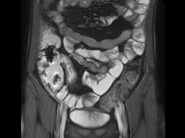
A 12-year-old boy presents to an urgent care center with severe abdominal pain, nausea, and vomiting. His height is 5 ft 3 in and weight is 99 lb (BMI 18.7). The patient has a history of chronic diarrhea and reports intermittent abdominal pain that began about 6 months ago. During this time, he has lost about 12 lb, as many foods exacerbate his symptoms, though his mother notes that even plain foods can bother his stomach. Further questioning reveals that his father has moderate Crohn disease (age of onset, 29 years), his sister has celiac disease, and the patient is of Ashkenazi Jewish descent. His body temperature is 100.2 °F. Vitamin B12, serum iron, total iron binding capacity, calcium, and magnesium are low. Stool cultures are negative. Ileocolonoscopy shows small aphthous erosions in the large intestine and in the terminal ileum.

New update focuses on acute kidney injury management in cirrhosis
Acute kidney injury (AKI) in patients with cirrhosis is potentially preventable, and there are clear steps that can be taken to manage and reverse the condition, concludes a clinical practice update from the American Gastroenterological Association.
AKI occurs in 47% of patients hospitalized with complications of cirrhosis and in approximately 30% of outpatients with cirrhosis, resulting in a total cost in the United States of $4 billion, explained Patrick S. Kamath, MD, division of gastroenterology and hepatology, Mayo Medical School, Rochester, Minn., one of the authors of this update.
Moreover, Dr. Kamath told this news organization, among patients with cirrhosis and AKI, morbidity and mortality is sevenfold higher in comparison to those without cirrhosis, and repeated episodes of AKI increase the risk of progression to chronic kidney disease.
To provide practical advice for the clinical management of patients with cirrhosis and AKI, the authors conducted an expert review of the best available published evidence and gathered expert opinion.
The update was published online in Clinical Gastroenterology and Hepatology.
Some key takeaways
Among its 14 best practice statements, it describes three situations indicative of AKI:
- A serum creatinine increase of 0.3 mg/dL or more within 48 hours, or
- A serum creatinine increase of 50% or more from baseline, which is a stable serum creatinine in the past 3 months.
- Reduction in urine output of up to 0.5 mL/kg per hour for more than 6 hours.
The update also emphasizes the importance of an accurate diagnosis, inasmuch as not all cases of AKI are due to hepatorenal syndrome (HRS), for example. It goes on to advise that the specific type of AKI be identified through medical history and physical examination, as well as with blood biochemistry, urine microscopic examination, urine chemistry, selected urinary biomarkers, and renal ultrasound.
Additionally, it underscores the need to identify and treat infections and to closely monitor fluid status.
Nancy S. Reau, MD, Rush Medical College, Chicago, who was not involved in the update, commented to this news organization that fluid status is important when giving albumin replacement therapy because of the increased risk for pulmonary edema.
She also highlighted that this update advises against transjugular intrahepatic portosystemic shunts (TIPSs) as a specific treatment for HRS-AKI, noting that, although the 2022 North American Practice-Based Recommendations for Transjugular Intrahepatic Portosystemic Shunts in Portal Hypertension do not advocate for TIPS for this indication, they also indicated that there was enough evidence to advise against it.
In other key best practice advice statements, the update advises clinicians to hold diuretics and nonselective beta blockers and to discontinue nonsteroidal anti-inflammatory drugs (NSAIDs).
‘Timely’ update
Overall, Dr. Reau believes that the update is “timely, especially in light of the recent [U.S.] approval of terlipressin, which will change our treatment options.”
This update also supports the American Association for the Study of Liver Diseases (AASLD) 2021 Practice Guidance guidelines on HRS, she added.
Zobair Younossi, MD, MPH, professor of medicine, Virginia Commonwealth University, Inova Campus, Falls Church, Va., who was not involved in writing the update, told this news organization that the document is important because of the huge increase in mortality among patients with cirrhosis and AKI.
He commented that there has been much advancement in understanding the condition, with updated nomenclature and novel medical treatments, and that this makes the update timely.
Moreover, the update will help clinicians who are involved in the care of patients with cirrhosis, he added.
Dr. Younossi said the update offers a very clearly stated algorithm for how to identify patients whose condition is easily reversible with volume repletion, in comparison with those patients who require medical treatment or even liver transplantation.
“Those things are important because that pathway gives clinicians an idea of how to do this properly,” he said.
“The key for clinicians is to make sure they understand, in the context of cirrhosis, some of the easy things that they can do to prevent AKI,” he continued. He added that the use of NSAIDs in these patients is “going to be problematic.”
Dr. Kamath has a relationship with Sequana Medical. Other authors’ relevant financial relationships are listed in the original article. Dr. Reau has relationships with Salix and Intercept. Dr. Younossi has disclosed no relevant financial relationships.
A version of this article first appeared on Medscape.com.
This article was updated 12/12/2022.
FROM CLINICAL GASTROENTEROLOGY AND HEPATOLOGY
Sexual issues common for GI patients, but docs often avoid topic
VIENNA – Sexual dysfunction in patients with gastrointestinal disorders is undermanaged, with a lack of clinician education, time constraints, and embarrassment preventing constructive discussions to improve patient care and quality of life, according to a new survey.
Overall, 71% of gastroenterologists do not ask their patients about sexual dysfunction, the survey finds.
“While patients with gastrointestinal disorders often experience sexual dysfunction, discussions around the matter are not routine in gastroenterological care,” said Marco Romano, MD, from the University of Campania “Luigi Vanvitelli,” Naples, Italy.
Romano presented the survey findings at this year’s United European Gastroenterology Week meeting.
The research shows not only a clear need for better awareness but also a need to build gastroenterologists’ confidence in addressing sexual dysfunction with their patients, Dr. Romano added.
“Most felt that sexual medicine education and improvement of communication skills within the context of their residency training might be important in order to increase the awareness of sexual dysfunction, to overcome barriers, and to improve care and quality of life for their patients,” reported Dr. Romano. “This will lead to prompt diagnosis and treatment of any sexual problems.”
Respectfully asking the patients if their gastrointestinal disorders interfere with their intimate relationships “is often considered a relief to patients who find that the gastrointestinal problem and the sexual dysfunction are interlinked,” he added.
The findings
The survey was needed because the question of whether gastroenterologists inquire about their patients’ sexual issues had never been assessed, Dr. Romano said.
The researchers sent a cross-sectional, anonymous online survey to members of the Italian Society of Gastroenterology and Digestive Endoscopy. The questionnaire, designed and informed by a literature review, consisted of 29 single multiple-choice and open-ended questions.
A total of 426 surveys were returned: 335 from experienced gastroenterologists and 91 from residents (less experienced). Of all respondents, 54.7% were men and 45.3% were women.
Even though most gastroenterologists do not ask their patients about sexual dysfunction, the majority want to learn how to manage the issue, the survey found. Of the survey respondents, 80% agreed that it would be useful for gastroenterologists to attend courses dedicated to the problem of sexual dysfunction.
Only 4% of patients report (initiate a dialogue about) the problem, the survey found. Among women aged 40-50 years, the most common complaint reported was dyspareunia (pain on intercourse). In men, the most frequent complaints reported were in the over-40s age group, with 75% citing erectile dysfunction and 45% reporting loss of libido.
The most common gastrointestinal disorders associated with sexual dysfunction are inflammatory bowel diseases (37% of cases), chronic liver diseases (28%), and irritable bowel syndrome (26%), according to the survey.
On the question of whether medications played a role in patients’ sexual dysfunction, nearly 15% of respondents said that prokinetic agents were involved, and 18% thought proton pump inhibitors affect sexual function. Both drug classes are considered responsible for sexual disturbances.
Few gastroenterologists prescribe phosphodiesterase type 5 inhibitors (PDE5i), e.g., Viagra, to treat sexual dysfunction, the survey found. Approximately 90% of respondents said that they never prescribed this class of drugs, preferring to refer patients to an andrologist. Of those who did prescribe PDE5i, significantly fewer residents did compared with experienced gastroenterologists (1.1% vs. 8.8%, respectively; P = .01).
Finally, the biggest reasons why gastroenterologists do not discuss sexual dysfunction are lack of knowledge (80%), insufficient experience (58%), time (44%), and embarrassment (30%).
Practice experience matters
There were some differences among respondents in the experienced group vs. the residents. More men were in the experienced group compared with residents (57.6% vs. 44%, respectively); mean age was 47 years vs. 29 years, respectively; and 71% had 5 or more years of experience in the experienced gastroenterologist group, whereas 78% had 1-5 years of experience among residents.
The survey found that more residents than experienced gastroenterologists “never discussed sexual dysfunction” (38.5% vs. 21.3%, respectively; P = .001) and that more residents than experienced gastroenterologists reported that “patients did not relate their sexual dysfunction to the prescribed therapy” (47.8% vs. 32.5%, respectively; P = .007).
The two groups varied regarding prescription drugs’ role in sexual dysfunction. More experienced gastroenterologists than residents felt that proton pump inhibitors (5.8% vs. 0%, respectively; P = .018) or prokinetics (19.8% vs. 9.5%, respectively; P = .028) might be responsible for some degree of sexual dysfunction.
More residents than experienced doctors felt that other (nongastroenterologic) drugs might contribute to sexual dysfunction in their patients (57.1% vs. 44.7%, respectively; P = .043).
Dr. Romano reported that fewer residents than experienced gastroenterologists referred male patients with sexual dysfunction to an andrologist (frequently/always: 28.1% vs. 44.4%, respectively; P = .004). However, more residents than experienced gastroenterologists disagreed that discussing sexual dysfunction with patients pertains only to specialists (andrologists and gynecologists; 83.5% vs. 71.2%, respectively; P = .018).
Time to step up
Asma Fikree, BMBCh, PhD, of Royal London Hospital, Barts Health NHS Trust, London, moderated the session. The survey highlights that asking patients about sexual dysfunction is an area for improvement for gastroenterologists, she said.
“We might do it in men and ask about erectile dysfunction, but we are very poor about asking in women,” Dr. Fikree noted.
The pros and cons of different medications should be discussed with patients, she said.
Gastroenterologists need to do a better job of considering how medications can lead to sexual dysfunction and interfere with quality of life, and training would help, she added.
“Some patients might not be very bothered by sexual dysfunction, but others might consider it very important,” Dr. Fikree said. “We should be considering this as part of their treatment and care.”
Dr. Romano and Dr. Fikree report no relevant financial relationships.
A version of this article first appeared on Medscape.com.
AT UEG WEEK 2022
Preexisting mental illness symptoms spiked during pandemic
“Those with preexisting mental health conditions may be particularly vulnerable to these effects because they are more susceptible to experiencing high levels of stress during a crisis and are more likely to experience isolation/despair during confinement compared to the general population,” wrote Danna Ramirez of The Menninger Clinic, Houston, and colleagues.
In a study published in Psychiatry Research , the investigators compared data from 142 adolescents aged 12-17 years and 470 adults aged 18-79 years who were admitted to an inpatient psychiatric hospital in Houston. Of these, 65 adolescents and 235 adults were admitted before the pandemic, and 77 adolescents and 235 adults were admitted during the pandemic.
Clinical outcomes were scores on the Generalized Anxiety Disorder Scale (GAD-7), the Patient Health Questionnaire (PHQ-9), the Patient Health Questionnaire for Adolescents (PHQ-A), the Difficulties in Emotion Regulation Scale–Short Form (DERS-SF), the World Health Organization Disability Assessment Scale (WHODAS), the World Health Organization Alcohol, Smoking, and Substance Involvement Screening Test (WHOASSIST), the Pittsburgh Sleep Quality Index (PSQI), the Disturbing Dream and Nightmare Severity Index (DDNSI), and the Suicide Behaviors Questionnaire–Revised (SBQ-R).
Overall, adults admitted during the pandemic had significantly higher levels of anxiety, depression, emotional dysregulation, and disability (P < .001 for all) as well as nightmares (P = .013) compared to those admitted prior to the pandemic.
Among adolescents, measures of anxiety, depression, and sleep quality were significantly higher at admission during the pandemic compared to prior to the pandemic (P = .005, P = .005, and P = .011, respectively)
Reasons for the increase in symptom severity remain unclear, but include the possibility that individuals with preexisting mental illness simply became more ill; or that individuals with symptoms delayed hospital admission out of fear of exposure to COVID-19, which resulted in more severe symptoms at admission, the researchers wrote in their discussion.
The findings were limited by several factors, including the primarily White population and the reliance on self-reports, the researchers noted. Another limitation was the lack of differentiation between patients who may have had COVID-19 before hospitalization and those who did not, so the researchers could not determine whether the virus itself played a biological role in symptom severity.
However, the results support data from previous studies and identify increased psychiatry symptom severity for patients admitted for inpatient psychiatry care during the pandemic, they said. Although resources are scarce, the findings emphasize that mental health needs, especially for those with preexisting conditions, should not be overlooked, and continuity and expansion of access to mental health care for all should be prioritized, they concluded.
The study was supported by The Menninger Clinic and The Menninger Clinic Foundation. The researchers had no financial conflicts to disclose.
FROM PSYCHIATRY RESEARCH
This brain surgery was BYOS: Bring your own saxophone
Tumor vs. saxophone: The surgical grudge match
Brain surgery is a notoriously difficult task. There’s a reason we say, “Well, at least it’s not brain surgery” when we’re trying to convince someone that a task isn’t that tough. Make one wrong incision, cut the wrong neuron, and it’s goodbye higher cognitive function. And most people appreciate thinking. Crazy, right?
One would imagine that the act of brain surgery would become even more difficult when the patient brings his saxophone and plays it randomly throughout the operation. It’s a hospital, after all, not a jazz club. Patients don’t get to play musical instruments during other surgeries. Why should brain surgery patients get special treatment?
As it turns out, the musical performance was actually quite helpful. A man in Italy had a brain tumor in a particularly complex area, and he’s left-handed, which apparently makes the brain’s neural pathways much more complicated. Plus, he insisted that he retain his musical ability after the surgery. So he and his medical team had a crazy thought: Why not play the saxophone throughout the surgery? After all, according to head surgeon Christian Brogna, MD, playing an instrument means you understand music, which tests many higher cognitive functions such as coordination, mathematics, and memory.
And so, at various points throughout the 9-hour surgery, the patient played his saxophone for his doctors. Doing so allowed the surgeons to map the patient’s brain in a more complete and personalized fashion. With that extra knowledge, they were able to successfully remove the tumor while maintaining the patient’s musical ability, and the patient was discharged on Oct. 13, just 3 days after his operation.
While we’re happy the patient recovered, we do have to question his choice of music. During the surgery, he played the theme to the 1970 movie “Love Story” and the Italian national anthem. Perfectly fine pieces, no doubt, but the saxophone solo in “Jungleland” exists. And we could listen to that for 9 hours straight. In fact, we do that every Friday in the LOTME office.
Basketball has the Big Dance. Mosquitoes get the Big Sniff
In this week’s installment of our seemingly never-ending series, “Mosquitoes and the scientists who love them,” we visit The Rockefeller University in New York, where the olfactory capabilities of Aedes Aegypti – the primary vector species for Zika, dengue, yellow fever, and chikungunya – became the subject of a round robin–style tournament.

First things first, though. If you’re going to test mosquito noses, you have to give them something to smell. The researchers enrolled eight humans who were willing to wear nylon stockings on their forearms for 6 hours a day for multiple days. “Over the next few years, the researchers tested the nylons against each other in all possible pairings,” Leslie B. Vosshall, PhD, and associates said in a statement from the university. In other words, mosquito March Madness.
Nylons from different participants were hooked up in pairs to an olfactometer assay consisting of a plexiglass chamber divided into two tubes, each ending in a box that held a stocking. The mosquitoes were placed in the main chamber and observed as they flew down the tubes toward one stocking or the other.
Eventually, the “winner” of the “tournament” was Subject 33. And no, we don’t know why there was a Subject 33 since the study involved only eight participants. We do know that the nylons worn by Subject 33 were “four times more attractive to the mosquitoes than the next most-attractive study participant, and an astonishing 100 times more appealing than the least attractive, Subject 19,” according to the written statement.
Chemical analysis identified 50 molecular compounds that were elevated in the sebum of the high-attracting participants, and eventually the investigators discovered that mosquito magnets produced carboxylic acids at much higher levels than the less-attractive volunteers.
We could go on about the research team genetically engineering mosquitoes without odor receptors, but we have to save something for later. Tune in again next week for another exciting episode of “Mosquitoes and the scientists who love them.”
Are women better with words?
Men vs. Women is probably the oldest argument in the book, but there may now be movement. Researchers have been able not only to shift the advantage toward women, but also to use that knowledge to medical advantage.

When it comes to the matter of words and remembering them, women apparently have men beat. The margin is small, said lead author Marco Hirnstein, PhD, of the University of Bergen, Norway, but, after performing a meta-analysis of 168 published studies and PhD theses involving more than 350,000 participants, it’s pretty clear. The research supports women’s advantage over men in recall, verbal fluency (categorical and phonemic), and recognition.
So how is this information useful from a medical standpoint?
Dr. Hirnstein and colleagues suggested that this information can help in interpreting diagnostic assessment results. The example given was dementia diagnosis. Since women are underdiagnosed because their baseline exceeds average while men are overdiagnosed, taking gender and performance into account could clear up or catch cases that might otherwise slip through the cracks.
Now, let’s just put this part of the debate to rest and take this not only as a win for women but for science as well.
Tumor vs. saxophone: The surgical grudge match
Brain surgery is a notoriously difficult task. There’s a reason we say, “Well, at least it’s not brain surgery” when we’re trying to convince someone that a task isn’t that tough. Make one wrong incision, cut the wrong neuron, and it’s goodbye higher cognitive function. And most people appreciate thinking. Crazy, right?
One would imagine that the act of brain surgery would become even more difficult when the patient brings his saxophone and plays it randomly throughout the operation. It’s a hospital, after all, not a jazz club. Patients don’t get to play musical instruments during other surgeries. Why should brain surgery patients get special treatment?
As it turns out, the musical performance was actually quite helpful. A man in Italy had a brain tumor in a particularly complex area, and he’s left-handed, which apparently makes the brain’s neural pathways much more complicated. Plus, he insisted that he retain his musical ability after the surgery. So he and his medical team had a crazy thought: Why not play the saxophone throughout the surgery? After all, according to head surgeon Christian Brogna, MD, playing an instrument means you understand music, which tests many higher cognitive functions such as coordination, mathematics, and memory.
And so, at various points throughout the 9-hour surgery, the patient played his saxophone for his doctors. Doing so allowed the surgeons to map the patient’s brain in a more complete and personalized fashion. With that extra knowledge, they were able to successfully remove the tumor while maintaining the patient’s musical ability, and the patient was discharged on Oct. 13, just 3 days after his operation.
While we’re happy the patient recovered, we do have to question his choice of music. During the surgery, he played the theme to the 1970 movie “Love Story” and the Italian national anthem. Perfectly fine pieces, no doubt, but the saxophone solo in “Jungleland” exists. And we could listen to that for 9 hours straight. In fact, we do that every Friday in the LOTME office.
Basketball has the Big Dance. Mosquitoes get the Big Sniff
In this week’s installment of our seemingly never-ending series, “Mosquitoes and the scientists who love them,” we visit The Rockefeller University in New York, where the olfactory capabilities of Aedes Aegypti – the primary vector species for Zika, dengue, yellow fever, and chikungunya – became the subject of a round robin–style tournament.
First things first, though. If you’re going to test mosquito noses, you have to give them something to smell. The researchers enrolled eight humans who were willing to wear nylon stockings on their forearms for 6 hours a day for multiple days. “Over the next few years, the researchers tested the nylons against each other in all possible pairings,” Leslie B. Vosshall, PhD, and associates said in a statement from the university. In other words, mosquito March Madness.
Nylons from different participants were hooked up in pairs to an olfactometer assay consisting of a plexiglass chamber divided into two tubes, each ending in a box that held a stocking. The mosquitoes were placed in the main chamber and observed as they flew down the tubes toward one stocking or the other.
Eventually, the “winner” of the “tournament” was Subject 33. And no, we don’t know why there was a Subject 33 since the study involved only eight participants. We do know that the nylons worn by Subject 33 were “four times more attractive to the mosquitoes than the next most-attractive study participant, and an astonishing 100 times more appealing than the least attractive, Subject 19,” according to the written statement.
Chemical analysis identified 50 molecular compounds that were elevated in the sebum of the high-attracting participants, and eventually the investigators discovered that mosquito magnets produced carboxylic acids at much higher levels than the less-attractive volunteers.
We could go on about the research team genetically engineering mosquitoes without odor receptors, but we have to save something for later. Tune in again next week for another exciting episode of “Mosquitoes and the scientists who love them.”
Are women better with words?
Men vs. Women is probably the oldest argument in the book, but there may now be movement. Researchers have been able not only to shift the advantage toward women, but also to use that knowledge to medical advantage.
When it comes to the matter of words and remembering them, women apparently have men beat. The margin is small, said lead author Marco Hirnstein, PhD, of the University of Bergen, Norway, but, after performing a meta-analysis of 168 published studies and PhD theses involving more than 350,000 participants, it’s pretty clear. The research supports women’s advantage over men in recall, verbal fluency (categorical and phonemic), and recognition.
So how is this information useful from a medical standpoint?
Dr. Hirnstein and colleagues suggested that this information can help in interpreting diagnostic assessment results. The example given was dementia diagnosis. Since women are underdiagnosed because their baseline exceeds average while men are overdiagnosed, taking gender and performance into account could clear up or catch cases that might otherwise slip through the cracks.
Now, let’s just put this part of the debate to rest and take this not only as a win for women but for science as well.
New AGA guidelines advise use of antiobesity medications for weight management
Adults with obesity who do not respond adequately to lifestyle interventions alone should be offered one of four suggested medications to treat obesity, with preference for semaglutide before others, according to new guidelines published by the American Gastroenterological Association in Gastroenterology.
Recommended first-line medications include semaglutide, liraglutide, phentermine-topiramate extended-release (ER), and naltrexone-buproprion ER, based on moderate-certainty evidence. Also recommended, albeit based on lower-certainty evidence, are phentermine and diethylpropion. The guidelines suggest avoiding use of orlistat. Evidence was insufficient for Gelesis100 superabsorbent hydrogel.
The substantial increase in obesity prevalence in the United States – from 30.5% to 41.9% in just the 2 decades from 2000 to 2020 – has likely contributed to increases in various obesity-related complications, wrote Eduardo Grunvald, MD, of the University of California San Diego, and colleagues. These include cardiovascular disease, stroke, type 2 diabetes mellitus, nonalcoholic steatohepatitis, obstructive sleep apnea, osteoarthritis, and certain types of cancer, such as colorectal cancer.
“Lifestyle intervention is the foundation in the management of obesity, but it has limited effectiveness and durability for most individuals,” the authors wrote. Despite a range of highly effective pharmacological therapies developed for long-term management of obesity, these agents are not widely used in routine clinical care, and practice variability is wide. There is a “small number of providers responsible for more than 90% of the prescriptions, partly due to lack of familiarity and limited access and insurance coverage,” the authors wrote.
A multidisciplinary panel of 10 experts and one patient representative, therefore, developed the guidelines by first prioritizing key clinical questions, identifying patient-centered outcomes, and conducting an evidence review of the following interventions: semaglutide 2.4 mg, liraglutide 3.0 mg, phentermine-topiramate extended-release (ER), naltrexone-bupropion ER, orlistat, phentermine, diethylpropion, and Gelesis100 superabsorbent hydrogel. The guideline panel then developed management recommendations and provided clinical practice considerations regarding each of the pharmacologic interventions.
The authors focused on adults, noting that pharmacologic treatment of childhood obesity is beyond the scope of these guidelines. The evidence synthesis yielded nine recommendations for the pharmacological management of obesity by gastroenterologists, primary care clinicians, endocrinologists, and other providers caring for patients with overweight or obesity. The target audience of the guidelines, however, includes patients and policymakers, the authors wrote.
“These guidelines are not intended to impose a standard of care, but rather, they provide the basis for rational, informed decisions for patients and health care professionals,” the authors wrote. “No recommendation can include all the unique individual circumstances that must be considered when making recommendations for individual patients. However, discussions around benefits and harms can be used for shared decision-making, especially for conditional recommendations where patients’ values and preferences are important to consider.”
The panel conducted a systematic review and meta-analysis of randomized controlled trials of Food and Drug Administration–approved obesity medications through Jan. 1, 2022. Though they primarily included studies with at least 48 weeks follow-up, they included studies with a follow-up of less than a year if one with 48 weeks’ outcomes did not exist.
The first of the nine recommendations was to add pharmacological agents to lifestyle interventions in treating adults with obesity or overweight and weight-related complications who have not adequately responded to lifestyle interventions alone. This strong recommendation was based on moderate-certainty evidence.
“Antiobesity medications generally need to be used chronically, and the selection of the medication or intervention should be based on the clinical profile and needs of the patient, including, but not limited to, comorbidities, patients’ preferences, costs, and access to the therapy,” the authors wrote. Average difference in total body weight loss with the addition of medication to lifestyle interventions was 3%-10.8%, depending on the drug. Treatment discontinuation ranged from 34 to 219 per 1,000 people in treatment groups, but adverse event rates were low.
The panel’s second recommendation suggested prioritizing of semaglutide along with lifestyle interventions based on the large magnitude of its net benefit. The remaining recommendations describes the use of each of the other medications based on their respective magnitude of effect and risk for adverse events.
Important considerations
“These medications treat a biological disease, not a lifestyle problem,” Dr. Grunvald said in a prepared statement. “Obesity is a disease that often does not respond to lifestyle interventions alone in the long term. Using medications as an option to assist with weight loss can improve weight-related complications like joint pain, diabetes, fatty liver, and hypertension.”
The authors acknowledged that cost remains a concern for the use of these therapies, especially among vulnerable populations. They also noted that the medications should not be used in pregnant individuals or those with bulimia nervosa, and they should be used with caution in people with other eating disorders. Patients with type 2 diabetes taking insulin or sulfonylureas and patients taking antihypertensives may require dosage adjustments since these obesity medications may increase risk of hypoglycemia for the former and decrease blood pressure for the latter.
The panel advised against orlistat, although it added that ”patients who place a high value on the potential small weight loss benefit and low value on gastrointestinal side effects may reasonably choose treatment with orlistat.” Those patients should take a multivitamin daily that contains vitamins A, D, E, and K at least 2 hours apart from orlistat.
The lack of available evidence for Gelesis100 oral superabsorbent hydrogel led the panel to suggest its use only in the context of a clinical trial.
The AGA will update these guidelines no later than 2025 and may issue rapid guidance updates until then as new evidence comes to light.
The guidelines did not receive any external funding, being fully funded by the AGA. The guideline chair and guideline methodologists had no relevant or direct conflicts of interest. All conflict of interest disclosures are maintained by the AGA office.
Severe pulsing headache
On the basis of the patient's presentation and described history, the likely diagnosis is migraine. By adolescence, migraine is much more common among female patients and can be connected to the menstrual cycle. The early symptoms before onset of head pain reported by this patient characterize the prodromal phase, which can occur 1-2 days before the headache, followed by the aura phase. Approximately one third of patients with migraine experience episodes with aura, like the visual disturbance described in this case.
Migraine can be diagnosed on a clinical basis, but certain neurologic symptoms with headache should be considered red flags and prompt further workup (ie, stiff neck or fever, or history of head injury or major trauma). Spontaneous internal carotid artery dissection, for example, should be investigated in the differential of younger patients who have severe headache before onset of neurologic symptoms. Patients who present with migraine are very frequently misdiagnosed as having sinus headaches or sinusitis. Relevant clinical findings of acute sinusitis are sinus tenderness or pressure; pain over the cheek which radiates to the frontal region or teeth; redness of nose, cheeks, or eyelids; pain to the vertex, temple, or occiput; postnasal discharge; a blocked nose; coughing or pharyngeal irritation; facial pain; and hyposmia. Tension-type headaches usually are associated with mild or moderate bilateral pain, causing a steady ache as opposed to the throbbing of migraines. Basilar migraine, common among female patients, is marked by vertebrobasilar insufficiency.
The American Headache Society defines migraine by the occurrence of at least five episodes. These attacks must last 4-72 hours and have at least two of these four characteristics: unilateral location, pulsating quality, moderate or severe pain intensity, and aggravation by or causing avoidance of routine physical activity. During these episodes, the patient must experience either photophobia and phonophobia or nausea and/or vomiting. If these signs and symptoms cannot be explained by another diagnosis, the patient is very likely presenting with migraine.
Identifying an effective treatment for migraines is often associated with a trial-and-error period, with an average 4-year gap between diagnosis and initiation of preventive medications. Because the patient's migraines do not seem to respond to non-steroidal anti inflammatory drugs, she may be a candidate for other treatments of mild-to-moderate migraines: nonopioid analgesics, acetaminophen, or caffeinated analgesic combinations. If attacks are moderate or severe, or even mild to moderate but do not respond well to therapy, migraine-specific agents are recommended: triptans, dihydroergotamine (DHE), small-molecule calcitonin gene-related peptide (CGRP) receptor antagonists (gepants), and selective serotonin (5-HT1F) receptor agonists (ditans).
Jasmin Harpe, MD, MPH, Headache Fellow, Department of Neurology, Harvard University, John R. Graham Headache Center, Mass General Brigham, Boston, MA
Jasmin Harpe, MD, MPH, has disclosed no relevant financial relationships.
Image Quizzes are fictional or fictionalized clinical scenarios intended to provide evidence-based educational takeaways.

An 18-year-old female patient presents with severe pulsing headache that began about 6 hours earlier. She describes feeling tired and irritable for the past 2 days and that she has had difficulty concentrating. Earlier in the day, before headache onset, she became extremely fatigued. Describing a "blinding light" in her vision, she is currently highly photophobic. The patient took four ibuprofen 2 hours ago. There is no significant medical history. She is on a regimen of estrogen-progestin and spironolactone for acne. Following advice from her primary care practitioner, she takes magnesium and vitamin B for headache prevention. The patient reports that she does not believe that she has migraines because she has never vomited during an episode. The patient explains that she has always had frequent headaches but that this is the sixth or seventh episode of this type and severity that she has had in the past year. The headaches do not seem to align with her menstrual cycle.

FMT in IBS: ‘We’ve been targeting the wrong part of the intestine’
VIENNA – , vs. it being administered into the large intestine, according to a new study.
Patients also reported an improvement in symptoms and quality of life with repeated doses of FMT (two doses, given 1 week apart), compared with a single dose in the small intestine, although statistical significance was not met.
“Administering a fecal transplant to the small intestine leads to long-term – up to 1 year in this analysis – colonization of beneficial bacteria, whereas administrating the fecal transplant to the large intestine results in the effect only lasting for the first 3 months,” said Magdy El-Salhy, MD, from the University of Bergen, Norway.
Dr. El-Salhy presented the results at the annual United European Gastroenterology Week meeting.
“It seems that bacteria in the small intestine play a more central role in IBS, as well as its associated fatigue, than bacteria in the large intestine,” Dr. El-Salhy said in an interview.
“Until now, we’ve been targeting the wrong part of the intestine,” he said.
The findings are the first to show that the small intestine is a more effective location for administering FMT than the large intestine for IBS. “It would be worthwhile doing similar [studies] in other diseases, especially in inflammatory bowel diseases,” said Dr. El-Salhy.
Researchers also didn’t expect the repeated dose to improve symptoms for a longer duration. “It really was revolutionary to see,” he added.
Some of Dr. El-Salhy’s patients have had up to 5 years of follow-up, although these results were not presented at this year’s UEG, he said.
“Around 75% of my patients have shown duration of response up to 3 years, and a few up to 5 years, on a 60-g dose from an earlier study group,” he said. “It’s an incredible result after a 10-minute treatment.”
In Dr. El-Salhy’s previous work, he found that increasing the dose from 30 g to 60 g increased the response from about 75% to about 90%. However, in this study presented, he found that increasing the dose to 90 g did not further increase the response. He also noted that while repeating the FMT dose improved symptoms and quality of life more than a single transplantation, it did not increase the response.
Targeting the small intestine
FMT has been widely investigated for the treatment of such conditions as psoriatic arthritis, Clostridioides difficile infection, and ulcerative colitis.
In this study, Dr. El-Salhy built on prior work (seven randomized controlled studies with varied outcomes) by asking whether the transplant dose increases FMT efficacy, which route of administration is more effective, and whether repeating FMT increases efficacy in patients with IBS.
A total of 186 patients were randomized to one of three groups: 90 g of frozen transplant into the large intestine (n = 62), 90 g of frozen transplant into the small intestine (n = 62), or 90 g of frozen transplant into the small intestine twice (with a 1-week interval; n = 62). FMT was administered via nasoduodenal tube and colonoscopy into the small and large intestines, respectively.
Outcomes were measured at 3, 6, and 12 months. The 12-month analysis of outcomes via patient questionnaire included 60, 61, and 60 patients, respectively.
The patient questionnaires included in the study were the IBS-SSS (a composite score of abdominal pain, duration of abdominal pain, bloating/distention, satisfaction with bowel habits, and IBS-related quality of life), the Birmingham IBS Symptom questionnaire, the Fatigue Assessment Scale questionnaire, the IBS-Quality of Life assessment, and the Short-Form Nepean Dyspepsia Index.
Fecal samples were taken and tested for bacterial loads. The bacterial profile and dysbiosis index were determined using the 16S rRNA gene.
At 3 months, patients had similar response rates, around 80%, across single dose in large intestine, single dose in small intestine, and repeat doses in small intestine.
At 6 months, the differences in response rates started to become noticeable, with 67.9% for single dose in large intestine, 71.4% for single dose in small intestine, and 86% for repeat doses in small intestine.
By 12 months, the difference in response rate between the single dose in the large and small intestines was statistically significant at 51.9% and 75.5%, respectively. The response rate to the repeat doses in the small intestine at 12 months (80.9%) was similar to that at 3 months (80.8%).
Side effects, including mild abdominal pain, diarrhea, and constipation, after FMT were seen for the first 5 days after treatment. “People who generally suffer from constipation get diarrhea after FMT and vice versa,” Dr. El-Salhy reported.
“Long-term side effects, as monitored up to 3 years, were not observed,” he added.
Treatment reduced IBS symptoms in all patient groups as measured by IBS-SSS scores. By 12 months, the score fell from around 350 to around 220 in patients who received a single dose in the large intestine, from around 300 to around 200 in patients who received a single dose in the small intestine, and from around 350 to around 170 in patients who received repeat doses in the small intestine.
Quality of life showed a statistically significant difference at 3 months between single and repeated doses in the small intestine and similarly at 6 and 12 months.
Chronic fatigue, experienced by many patients with IBS, was substantially reduced after FMT, Dr. El-Salhy noted. “This surge in energy is often more important to them than the gastrointestinal symptoms.”
Location affects bacterial success
Certain beneficial bacteria were found to thrive more when the donor transplant was administered to the small intestine than to the large intestine.
Of note, Lactobacillus species and Holdemanella biformis grew and then dropped off sharply after 3 months in patients who received a single-dose fecal transplant in the large intestine, while they grew after 3 months and continued to grow after 6 and 12 months in the groups who received a fecal transplant in the small intestine.
“We think bacteria in the small intestine have different characteristics to those in the large intestine,” Dr. El-Salhy said. “This is relatively new, because many years ago it was thought that bile acids prevented bacterial survival. Now we know lots can thrive in the small intestine.”
“It might be viral or some other component that is most effective here. We don’t know yet, but so far we have identified 11 bacteria of interest,” he added.
Broader questions
“Rather than focusing on a specific, single strain microbe as a predictor of success in a disease, the global equilibrium of microbiota is more important, and microbial ecology parameters would be interesting to assess,” remarked Gianluca Ianiro, MD, from the Università Cattolica del Sacro Cuore, Rome, who comoderated the session. “Selected survival of some bacteria through the gut may be the response.”
FMT emerged in response to the challenges posed by recurrent C. difficile infections, noted Alexander Khoruts, MD, a professor of medicine in the division of gastroenterology, hepatology, and nutrition at the University of Minnesota, Minneapolis, who was not involved in the research.
“It is much harder to achieve remodeling of the gut microbiome in non–C. difficile conditions where there is an intact and resilient indigenous microbiota,” he said in an interview. “Therefore, regimens using antibiotic preconditioning and repeated administrations of microbiota are generally more efficacious in achieving this objective.”
The specificity of the bacteria according to disease type targeted was important, said Dr. Khoruts, who has a special interest in gut microbiota.
“The big question in non–C. difficile indications is the composition of donor microbiota. It is critical that we understand the mechanisms involved in each target disease to design appropriate microbiota-based therapeutics,” he said.
Dr. Khoruts sounded a note of caution with respect to establishing the pharmacokinetic and dynamic data related to FMT, which is classified as a drug in the United States.
“It’s imperative that we develop the pharmacology discipline appropriate for this class of therapeutics, including their pharmacokinetics and pharmacodynamics, and an understanding of their potential toxicity and drug-drug interactions,” he said.
Drug distribution data are needed to determine host-microbiota interactions.
“This includes the small bowel microbiome, which continues to be woefully understudied,” Dr. Khoruts said.
Dr. El-Salhy reports no relevant financial relationships. Dr. Ianiro reports receiving personal fees for acting as speaker for Biocodex, Sofar, Malesci, and Tillotts Pharma, and for acting as consultant/advisor for Ferring Therapeutics, Biocodex, Tillotts Pharma, and Zambon. Dr. Khoruts reports he has patents pertaining to fecal microbiota separation from stool and their cryopreservation and lyopreservation.
Through the AGA Center for Gut Microbiome Research and Education, AGA is committed to keeping you up-to-speed on the latest news, research and policy updates related to the gut microbiome: www.gastro.org/microbiome.
A version of this article first appeared on Medscape.com.