User login
TNFi and increased hematologic malignancy risk in PsA: Is there a link?
Key clinical point: The overall incidence of hematologic malignancies was not higher among patients with psoriatic arthritis (PsA) receiving treatment with tumor necrosis factor inhibitors (TNFi) compared with those who were biological disease-modifying antirheumatic drug (bDMARD) naive or individuals from the general population.
Major finding: Overall, the incidence rate of hematologic malignancies was similar among patients treated with TNFi vs those who were bDMARD naive (incidence rate ratio [IRR] 0.96; 95% CI 0.68-1.35) or individuals in the general population (IRR 1.35; 95% CI 0.98-1.86).
Study details: Findings are from a cohort study including patients with PsA who either initiated first TNFi (n = 10,621) or were bDMARD-naive (n = 18,387) and matched comparators from the general population.
Disclosures: This study was supported by NordForsk and FOREUM. B Delcoigne declared being partly employed by ARTIS/Swedish Biologics Register. Several authors reported receiving grants or research support or serving as consultants for or on speaker’s bureaus of various sources.
Source: Cordtz RL et al. Haematological malignancies in patients with psoriatic arthritis overall and treated with TNF inhibitors: A Nordic cohort study. RMD Open. 2022;8(2):e002776 (Dec 23). Doi: 10.1136/rmdopen-2022-002776
Early achievement of minimal disease activity important for long-term benefits in PsA
Key clinical point: The failure to achieve minimal disease activity (MDA) in the first year after the diagnosis of psoriatic arthritis (PsA) was associated with worse health-related quality of life and health status, functional impairment, fatigue, pain, and higher anxiety and depression.
Major finding: Compared with patients who achieved sustained MDA in the first year after diagnosis, those who did not achieve MDA had higher scores for pain (estimated mean difference [β] 35.38), fatigue (β 17.88), and functional ability (β 0.81; all P < .001) and higher anxiety and depression (both P < .001) during follow-up, which persisted despite treatment intensification.
Study details: This prospective cohort study included 240 patients with newly diagnosed PsA with oligoarthritis or polyarthritis who were disease-modifying antirheumatic drug naive.
Disclosures: This study was sponsored by UCB Pharma. S Welby and AR Prickett declared being stockholders of UCB.
Source: Snoeck Henkemans SVJ et al. Importance of quick attainment of minimal disease activity for a positive impact on lives of patients with psoriatic arthritis. RMD Open. 2022;8(2):e002706 (Dec 7). Doi: 10.1136/rmdopen-2022-002706
No benefits of concomitant methotrexate in PsA patients treated with ustekinumab
Key clinical point: The addition of methotrexate or maintenance of ongoing methotrexate did not significantly enhance the efficacy of ustekinumab in patients with active psoriatic arthritis (PsA), suggesting ustekinumab as an effective therapy for PsA independent of methotrexate.
Major finding: Ustekinumab monotherapy demonstrated non-inferiority over ustekinumab+methotrexate with a comparable Disease Activity Score in 28 joints (DAS28) at week 24 (mean 2.9 vs 3.1; Mann-Whitney estimator 0.5426) and at week 52 (mean 2.8 vs 3.1; Mann-Whitney estimator 0.5461). Overall, serious adverse events occurred in 9% of patients in both treatment groups, but no deaths were reported.
Study details: Findings are from MUST, a phase 3b trial including 173 ustekinumab-naive patients with active PsA who were randomly assigned to receive ustekinumab+methotrexate or ustekinumab+placebo.
Disclosures: This study was supported by Janssen Cilag. Several authors reported receiving grants, contracts, travel support, or honoraria or fees for serving as speakers, consultants, or advisory board members for various sources.
Source: Koehm M et al. Methotrexate plus ustekinumab versus ustekinumab monotherapy in patients with active psoriatic arthritis (MUST): A randomised, multicentre, placebo-controlled, phase 3b, non-inferiority trial. Lancet Rheumatol. 2023;5(1):E14-E23 (Jan 1). Doi: 10.1016/S2665-9913(22)00329-0
Food additives may exacerbate IBD
AURORA, COLO. – Dietary additives lurking in processed foods may contribute to the development or exacerbation of inflammatory bowel disease (IBD), a leading gastroenterologist contends.
At the annual Crohn’s & Colitis Congress®, a partnership of the Crohn’s & Colitis Foundation and the American Gastroenterological Association, James D. Lewis, MD, MSCE, AGAF, of the University of Pennsylvania in Philadelphia, highlighted research from both animal and human studies pointing to certain widely used food additives such as carboxymethycellulose (CMC), polysorbate 80, and carrageenan as potential instigators in gastrointestinal inflammation.
he said.

Some additives appear to have deleterious effects on intestinal microbiota, while others may exert their baleful influence through mechanisms such as endoplasmic stress.
“It looks like some people might be a little more sensitive to additives than others, and if you were going to use any of this [research] to try and give some advice, maybe we would say that patients or first-degree relatives of people with IBD may want to avoid foods that contain high levels of additives, and if for no other reason, mothers or people with a family history of IBD might be encouraged to breastfeed to avoid early exposure to additives that are in infant formulas,” he advised.
Processed foods defined
The typical American diet may include a large proportion of processed foods, defined as “foods that have undergone biological, chemical, or physical process to improve texture, taste, or shelf life.”
Processed foods tend to be higher in fats, added sugars, and salts, and lower in fiber and intrinsic vitamins than minimally processed foods.
There is also a category of “ultraprocessed” foods, which contain little or no whole foods but are high in energy density. Many of these super(bad) foods are staples of the American diet, such as chips, hot dogs, chicken nuggets, breakfast cereal, soda, candy, and margarine. These and similar foods contribute from 25% to 50% of daily energy intake in the United States and Canada, Dr. Lewis said.
And North America is not alone, he added, pointing to a 2015 study showing that the consumption of ultraprocessed foods in Sweden increased “dramatically” from 1960 through 2010, and that this increase mirrored an increase in obesity prevalence in that nation.
Emulsifiers and thickeners
Dr. Lewis focused on emulsifiers and thickeners that are commonly added to processed foods and are, according to the Food and Drug Administration, “generally recognized as safe.”
Emulsifiers are “detergent-like molecules that stabilize mixtures of immiscible [nonhomegenous] liquids.”
Thickeners are additives that increase the viscosity of liquids without otherwise substantially changing their other properties.
In addition to the aforementioned products, other common additives include xanthan gum (a polysaccharide used as an emulsifier in salad dressings, baked goods, ice cream, and gluten-free products), maltodextrin (a sugar substitute marketed as “Splenda”), and soy lecithin (a soy derivative used as an emulsifier, stabilizer, and wetting agent).
Evidence of harm
Dr. Lewis noted that in 2013, investigators at the University of Liverpool, England, published a hypothesis suggesting that consumption of emulsifiers in processed foods may promote Crohn’s disease by increasing bacterial translocation. Their hypothesis was based in part on evidence that “very small concentrations of the emulsifier polysorbate 80 enhance bacterial translocation across intestinal epithelia. Undigested emulsifiers may increase bacterial translocation, particularly in the small intestine where the mucus layer is discontinuous. “
The authors also suggested that their hypothesis could be tested in clinical trials comparing enteral feeding with and without emulsifiers.
Other suggestive if not definitive evidence of a potential link between additives and IBD are data showing that IBD is very rare in young children.
“In your early stages of your life, you’re not consuming a lot of ultraprocessed foods. Indeed, the rate of intake of at least fast foods, which you can think of almost as a surrogate for ultraprocessed foods, goes up dramatically when people get to their teens, and this is the same time as we see, really, the big uptick in the incidence of IBD,” Dr. Lewis said.
A link between ultraprocessed food consumption and later development of IBD, primarily Crohn’s disease, is also suggested by data from the Nurses Health Study I and II and Health Professionals Follow-Up study. Among 245,112 participants with about 5.5 million person-years of follow-up, the highest vs. lowest quartile of consumption of ultraprocessed foods was associated with a 70% increase in risk for developing Crohn’s (hazard ratio 1.70, P = .0008).
Animal studies
Evidence for a possible mechanism whereby emulsifiers and thickeners cause intestinal changes comes from a study published in Nature in 2015 showing that adding CMC and PS80 to the drinking water of mice resulted in major shifts in the gut microbiota in both wild-type and interleukin 10 knockout mice, a model for IBD.
When the additives were put into the water the mice had a thinning of the mucus layer, allowing bacteria in closer proximity to the epithelium.
“When you put these into the drinking water of IL-10 knockout mice that are already predisposed to developing colitis, they were far more likely to go on to develop colitis over the course of 3 months,” Dr. Lewis said.
From mouse to man
Dr. Lewis briefly summarized results of the FRESH study that he and colleagues recently published in Gastronterology. In this trial, 16 healthy adult volunteers who agreed to stay and eat all meals at the research center were randomized to receive either an emulsifier-free diet or the identical diet enriched with 15 g of CMC daily for 11 days.
“I will comment that that’s a lot of carboxymethycellulose,” Dr. Lewis said.
The volunteers fed the CMC-enriched diet had a slight increase in abdominal discomfort after eating and a reduction in species diversity in the gut microbiota. In addition, these participants had reductions in levels of short-chain fatty acids and free amino acids, both of which are signs of a health gut environment.
“Furthermore, we identified 2 subjects consuming CMC who exhibited increased microbiota encroachment into the normally sterile inner mucus layer, a central feature of gut inflammation, as well as stark alterations in microbiota composition,” the investigators wrote.
Dr. Lewis cited a separate small study by investigators at the University of Illinois at Chicago and the University of Chicago. These investigators randomized patients with UC in remission to take supplements containing carrageenan – a seaweed-derived food additive that has been shown to cause inflammation in both in vitro and animal models – or placebo . The amount of carrageenan in the capsules was less than that found in an average daily Western diet, the authors noted.
The participants were followed with telephone calls every 2 weeks or until relapse, which was defined as an increase of 2 or more points on the Simple Clinical Colitis Activity Index (SCCAI) and intensification of treatment for UC.
Of the 12 patients who completed the study, 3 in the carrageenan group experienced relapses, compared with none of the patients in the placebo group (P = .046). The relapse occurred at 5, 32, and 42 weeks of follow-up.
Exceptions to the rule
“It’s not clear that all additives are harmful,” Dr. Lewis said, pointing to a placebo-controlled study suggesting a beneficial effect of soy lecithin in patients with UC. The additive is composed of at least 30% of phosphatidycholine, a component of intestinal mucus.
He also noted that there is an ongoing randomized, placebo-controlled trial comparing a low-additive diet to a habitual diet in 154 patients with mildly active, stable Crohn’s disease.
Session moderator Michael J. Rosen, MD, MSCI, a pediatric gastroenterologist at Stanford University Medical Center in Palo Alto, Calif., told this news organization that dietary components do appear to have an influence on the disease course in patients with IBD.
“I do think there are patients with IBD who are maybe genetically predisposed to being sensitive to certain components of diet,” he said in an interview seeking objective commentary.
“Particularly in pediatrics there are lines of evidence of diets maybe having some efficacy in treatment. It needs further study, but one commonality about those diets is that they tend to eliminate processed foods and focus on whole foods,” he said.
Dr. Lewis’ work is supported by grants from the National Institutes of Health, and from AbbVie, Takeda, Janssen, and Nestlé Health Science. He has served as a consultant to and data safety monitoring board member for several entitities. Dr. Rosen reported no conflicts of interest to disclose.
AURORA, COLO. – Dietary additives lurking in processed foods may contribute to the development or exacerbation of inflammatory bowel disease (IBD), a leading gastroenterologist contends.
At the annual Crohn’s & Colitis Congress®, a partnership of the Crohn’s & Colitis Foundation and the American Gastroenterological Association, James D. Lewis, MD, MSCE, AGAF, of the University of Pennsylvania in Philadelphia, highlighted research from both animal and human studies pointing to certain widely used food additives such as carboxymethycellulose (CMC), polysorbate 80, and carrageenan as potential instigators in gastrointestinal inflammation.
he said.
Some additives appear to have deleterious effects on intestinal microbiota, while others may exert their baleful influence through mechanisms such as endoplasmic stress.
“It looks like some people might be a little more sensitive to additives than others, and if you were going to use any of this [research] to try and give some advice, maybe we would say that patients or first-degree relatives of people with IBD may want to avoid foods that contain high levels of additives, and if for no other reason, mothers or people with a family history of IBD might be encouraged to breastfeed to avoid early exposure to additives that are in infant formulas,” he advised.
Processed foods defined
The typical American diet may include a large proportion of processed foods, defined as “foods that have undergone biological, chemical, or physical process to improve texture, taste, or shelf life.”
Processed foods tend to be higher in fats, added sugars, and salts, and lower in fiber and intrinsic vitamins than minimally processed foods.
There is also a category of “ultraprocessed” foods, which contain little or no whole foods but are high in energy density. Many of these super(bad) foods are staples of the American diet, such as chips, hot dogs, chicken nuggets, breakfast cereal, soda, candy, and margarine. These and similar foods contribute from 25% to 50% of daily energy intake in the United States and Canada, Dr. Lewis said.
And North America is not alone, he added, pointing to a 2015 study showing that the consumption of ultraprocessed foods in Sweden increased “dramatically” from 1960 through 2010, and that this increase mirrored an increase in obesity prevalence in that nation.
Emulsifiers and thickeners
Dr. Lewis focused on emulsifiers and thickeners that are commonly added to processed foods and are, according to the Food and Drug Administration, “generally recognized as safe.”
Emulsifiers are “detergent-like molecules that stabilize mixtures of immiscible [nonhomegenous] liquids.”
Thickeners are additives that increase the viscosity of liquids without otherwise substantially changing their other properties.
In addition to the aforementioned products, other common additives include xanthan gum (a polysaccharide used as an emulsifier in salad dressings, baked goods, ice cream, and gluten-free products), maltodextrin (a sugar substitute marketed as “Splenda”), and soy lecithin (a soy derivative used as an emulsifier, stabilizer, and wetting agent).
Evidence of harm
Dr. Lewis noted that in 2013, investigators at the University of Liverpool, England, published a hypothesis suggesting that consumption of emulsifiers in processed foods may promote Crohn’s disease by increasing bacterial translocation. Their hypothesis was based in part on evidence that “very small concentrations of the emulsifier polysorbate 80 enhance bacterial translocation across intestinal epithelia. Undigested emulsifiers may increase bacterial translocation, particularly in the small intestine where the mucus layer is discontinuous. “
The authors also suggested that their hypothesis could be tested in clinical trials comparing enteral feeding with and without emulsifiers.
Other suggestive if not definitive evidence of a potential link between additives and IBD are data showing that IBD is very rare in young children.
“In your early stages of your life, you’re not consuming a lot of ultraprocessed foods. Indeed, the rate of intake of at least fast foods, which you can think of almost as a surrogate for ultraprocessed foods, goes up dramatically when people get to their teens, and this is the same time as we see, really, the big uptick in the incidence of IBD,” Dr. Lewis said.
A link between ultraprocessed food consumption and later development of IBD, primarily Crohn’s disease, is also suggested by data from the Nurses Health Study I and II and Health Professionals Follow-Up study. Among 245,112 participants with about 5.5 million person-years of follow-up, the highest vs. lowest quartile of consumption of ultraprocessed foods was associated with a 70% increase in risk for developing Crohn’s (hazard ratio 1.70, P = .0008).
Animal studies
Evidence for a possible mechanism whereby emulsifiers and thickeners cause intestinal changes comes from a study published in Nature in 2015 showing that adding CMC and PS80 to the drinking water of mice resulted in major shifts in the gut microbiota in both wild-type and interleukin 10 knockout mice, a model for IBD.
When the additives were put into the water the mice had a thinning of the mucus layer, allowing bacteria in closer proximity to the epithelium.
“When you put these into the drinking water of IL-10 knockout mice that are already predisposed to developing colitis, they were far more likely to go on to develop colitis over the course of 3 months,” Dr. Lewis said.
From mouse to man
Dr. Lewis briefly summarized results of the FRESH study that he and colleagues recently published in Gastronterology. In this trial, 16 healthy adult volunteers who agreed to stay and eat all meals at the research center were randomized to receive either an emulsifier-free diet or the identical diet enriched with 15 g of CMC daily for 11 days.
“I will comment that that’s a lot of carboxymethycellulose,” Dr. Lewis said.
The volunteers fed the CMC-enriched diet had a slight increase in abdominal discomfort after eating and a reduction in species diversity in the gut microbiota. In addition, these participants had reductions in levels of short-chain fatty acids and free amino acids, both of which are signs of a health gut environment.
“Furthermore, we identified 2 subjects consuming CMC who exhibited increased microbiota encroachment into the normally sterile inner mucus layer, a central feature of gut inflammation, as well as stark alterations in microbiota composition,” the investigators wrote.
Dr. Lewis cited a separate small study by investigators at the University of Illinois at Chicago and the University of Chicago. These investigators randomized patients with UC in remission to take supplements containing carrageenan – a seaweed-derived food additive that has been shown to cause inflammation in both in vitro and animal models – or placebo . The amount of carrageenan in the capsules was less than that found in an average daily Western diet, the authors noted.
The participants were followed with telephone calls every 2 weeks or until relapse, which was defined as an increase of 2 or more points on the Simple Clinical Colitis Activity Index (SCCAI) and intensification of treatment for UC.
Of the 12 patients who completed the study, 3 in the carrageenan group experienced relapses, compared with none of the patients in the placebo group (P = .046). The relapse occurred at 5, 32, and 42 weeks of follow-up.
Exceptions to the rule
“It’s not clear that all additives are harmful,” Dr. Lewis said, pointing to a placebo-controlled study suggesting a beneficial effect of soy lecithin in patients with UC. The additive is composed of at least 30% of phosphatidycholine, a component of intestinal mucus.
He also noted that there is an ongoing randomized, placebo-controlled trial comparing a low-additive diet to a habitual diet in 154 patients with mildly active, stable Crohn’s disease.
Session moderator Michael J. Rosen, MD, MSCI, a pediatric gastroenterologist at Stanford University Medical Center in Palo Alto, Calif., told this news organization that dietary components do appear to have an influence on the disease course in patients with IBD.
“I do think there are patients with IBD who are maybe genetically predisposed to being sensitive to certain components of diet,” he said in an interview seeking objective commentary.
“Particularly in pediatrics there are lines of evidence of diets maybe having some efficacy in treatment. It needs further study, but one commonality about those diets is that they tend to eliminate processed foods and focus on whole foods,” he said.
Dr. Lewis’ work is supported by grants from the National Institutes of Health, and from AbbVie, Takeda, Janssen, and Nestlé Health Science. He has served as a consultant to and data safety monitoring board member for several entitities. Dr. Rosen reported no conflicts of interest to disclose.
AT THE CROHN’S & COLITIS CONGRESS
BC with metabolic abnormalities: No benefit of adding metformin to neoadjuvant chemotherapy
Key clinical point: Addition of metformin to the neoadjuvant docetaxel, epirubicin, and cyclophosphamide (TEC) regimen did not improve disease outcomes in patients with breast cancer (BC) and metabolic abnormalities.
Major finding: The total pathological complete response was achieved by a similar proportion of patients receiving TEC vs TEC+metformin (12.5% vs 14.6%; P = .777). Neutropenia and leucopenia, the most common grade ≥3 adverse events, were reported by 42.5% and 55.0% of patients in the TEC arm and 22.9% and 45.8% of patients in the TEC+metformin arm, respectively.
Study details: Findings are from the phase 2 NeoMET study including 92 patients with stage IIB/III BC and ≥1 metabolic syndrome component who were randomly assigned to receive six cycles of TEC or TEC+metformin.
Disclosures: This study was funded by the National Natural Science Foundation of China and other sources. The authors declared no conflicts of interest.
Source: Huang J, Tong Y et al. Neoadjuvant docetaxel, epirubicin, and cyclophosphamide with or without metformin in breast cancer patients with metabolic abnormality: Results from the randomized phase II NeoMET trial. Breast Cancer Res Treat. 2022 (Dec 16). Doi: 10.1007/s10549-022-06821-y
EMR screening in emergency department tags undiagnosed diabetes
A diabetes screening program built into an electronic medical records system identified diabetes or prediabetes in 52% of individuals flagged for abnormal hemoglobin A1c, based on data from more than 2,000 adults.
“Despite the best efforts of clinicians, researchers, and educators, the number of patients living with undiagnosed diabetes is still rising and is currently at approximately 8.5 million, and the number of people unaware of their prediabetes is approximately 77 million,” lead investigator Kristie K. Danielson, PhD, said in an interview. Screening for diabetes is critical to start treatment early, to potentially reverse prediabetes, and to prevent the long-term complications of diabetes and reduced life expectancy.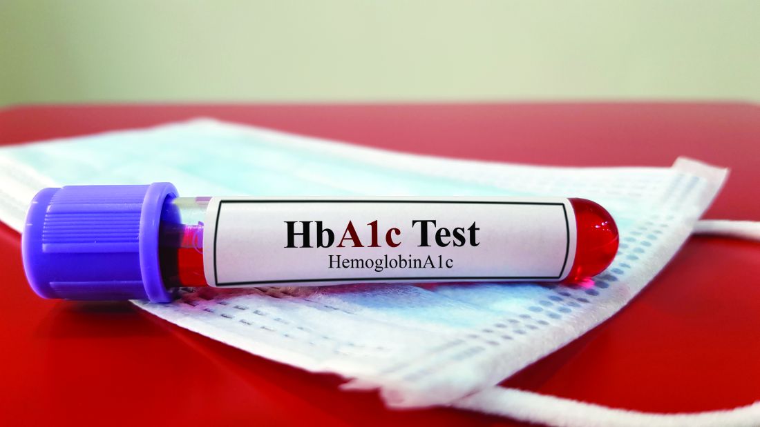
In a pilot study published in JAMA Network Open, Dr. Danielson and colleagues reviewed data from 8,441 adults who visited a single emergency department in Chicago during February–April 2021.
The EMR at the hospital contained a built-in best practice alert (BPA) that flagged patients as being at risk for type 2 diabetes based the American Diabetes Association recommendations; the identification algorithm included age 45 years and older, or those aged 18-44 years with a body mass index of 25 kg/m2 or higher, no previous history of diabetes, and no A1c measure in the last 3 years, according to the EMR.
A total of 8,441 adult patients visited the ED during the study period; 2,576 triggered BPA tests, and 2,074 had A1c results for review. Among the patients with A1c results, 52% had elevated values of 5.7% or higher. Of these, a total of 758 individuals were identified with prediabetes (A1c, 5.7%-6.4%), 265 with diabetes (A1c, 6.5%-9.9%), and 62 with severe diabetes (A1c, 10% or higher).
After testing, 352 patients with elevated A1c were contacted by the researchers. The mean age of this group was 52.2 years, 54.5% were women, and nearly two-thirds (64.8%) were non-Hispanic Black. The median income of those contacted was in the 44th percentile, and 50% had public insurance.
Most of those contacted (264 patients) were not aware of a previous diagnosis of prediabetes or diabetes; the remaining 88 had a previous diagnosis, but only 51 self-reported receiving treatment, the researchers noted.
Although the screening program successfully identified a significant number of previously undiagnosed individuals with diabetes, prediabetes, or poorly controlled diabetes, its feasibility in routine practice requires further study, the researchers wrote.
The findings were limited by several factors including the identification of patients previously diagnosed with diabetes but who were not being treated, and the potential bias toward individuals of higher socioeconomic status, the researchers noted. However, the results support further exploration of the program as a way to identify undiagnosed diabetes, especially in underserved populations.
Diabetes in underserved groups goes undetected
“We were surprised by the sheer number of people newly diagnosed with diabetes or prediabetes,” which was far greater than expected, commented Dr. Danielson of the University of Illinois at Chicago. “Clearly, we tapped into a new population that has not often been seen by primary care providers or endocrinologists, as is often the case for underserved and vulnerable individuals who visit the emergency department as a first line for health care.”
The screening alert system is straightforward to build into an existing EMR, with technical support, Dr. Danielson said. “In theory, it should be able to be incorporated into other clinical centers and emergency departments. One of the current limitations that we are seeing is that the EMR is still flagging some people already diagnosed with diabetes to be screened for diabetes.” However, “because of this, we also see this as an opportunity to identify and reach out to those with diabetes who are still underserved and not receiving the appropriate diabetes care they need.”
The study results have broader public health implications, Dr. Danielson added. “We have identified a new, large population of people with diabetes who need medical care and diabetes education. This will further add to the burden of health care and costs, and it raises the ethical question of screening and not having full resources readily available to help.
“In my opinion, the study sheds light on a significant issue that will hopefully help drive change at both a health systems and public health level locally and nationally,” she added.
“One of the significant research gaps that has emerged now is how to link these new patients to health care and diabetes education at our institution after they leave the emergency department,” said Dr. Danielson. Diabetes screening in the ED setting is “a very novel area for health system scientists, social workers, and others to now come to the table and collaborate on next steps to help our patients.”
The study was initiated by the investigators, but was supported by a grant from Novo Nordisk to two coauthors. Dr. Danielson also disclosed grant funding from Novo Nordisk during the conduct of the study.
FROM JAMA NETWORK OPEN
Microcalcifications and high breast density increase risk for breast cancer
Key clinical point: Microcalcifications and breast density, as assessed by the Breast Imaging Reporting and Data System 4th edition (BI-RADS) were associated with a significantly increased risk for breast cancer (BC).
Major finding: Microcalcification appeared to be a significant risk factor for BC irrespective of breast density (adjusted hazard ratio [aHR] 3.09; 95% CI 2.83-3.36). Both microcalcification and high breast density (BI-RADS density classification 4) were associated with a 6.65-fold (95% CI 5.59-7.72) higher risk for BC, with the risk being the highest in postmenopausal women with microcalcifications and high breast density (aHR 7.26; 95% CI 5.01-10.53).
Study details: Findings are from a retrospective cohort study including 3,910,815 women who underwent breast cancer screening, of which 58,315 women were diagnosed with BC during a median follow-up of 10.8 years.
Disclosures: This study was supported by the National Research Foundation of Korea and other sources. The authors declared no conflicts of interest.
Source: Kim S et al. Microcalcifications, mammographic breast density, and risk of breast cancer: A cohort study. Breast Cancer Res. 2022;24:96 (Dec 21). Doi: 10.1186/s13058-022-01594-0
Breast cancer: Nipple-sparing mastectomy after neoadjuvant chemotherapy is oncologically safe
Key clinical point: Nipple-sparing mastectomy (NSM), even when performed after neoadjuvant chemotherapy (NACT), resulted in a very low rate of locoregional recurrence and therefore was considered oncologically safe in women with breast cancer (BC).
Major finding: Cumulative incidences of local (P = .570), regional (P = .150), and systemic (P = .87) relapses were similar between patients who received vs did not receive NACT, with no cases of locoregional relapses being reported by the 30.3% of patients who had achieved pathological complete response in the NACT group. The rate of all complications was also similar with vs without NACT.
Study details: Findings are from a retrospective study including 112 cases of NSM after NACT in 111 women and 321 cases of primary NSM in 306 women.
Disclosures: This study was supported by Fondazione Prometeus, ONLUS, Italy. The authors declared no conflicts of interest.
Source: Meli EZ et al. Nipple-sparing mastectomy after neoadjuvant chemotherapy: Definitive results with a long-term follow-up evaluation. Ann Surg Oncol. 2023 (Jan 4). Doi: 10.1245/s10434-022-13035-5
HER2+ metastatic BC: Meta-analysis demonstrates superior efficacy of pyrotinib over lapatinib
Key clinical point: Pyrotinib plus chemotherapy outperformed lapatinib plus chemotherapy in terms of improving survival outcomes in patients with human epidermal growth factor receptor 2-positive (HER2+) metastatic breast cancer (BC) but showed a worse toxicity profile.
Major finding: Progression-free survival was significantly improved with pyrotinib vs lapatinib in patients who had received prior trastuzumab therapy (hazard ratio [HR] 0.47; P < .001) and those with trastuzumab resistance (HR 0.52; P < .001). However, the incidence of grade ≥3 diarrhea was higher with pyrotinib vs lapatinib (risk ratio 2.68; P < .001).
Study details: Findings are from a meta-analysis of five studies including 843 patients with metastatic BC who received chemotherapy with pyrotinib (n = 392) or lapatinib (n = 451).
Disclosures: This study did not receive any funding. The authors declared no conflicts of interest.
Source: Yuan Y et al. Pyrotinib versus lapatinib therapy for HER2 positive metastatic breast cancer patients after first-line treatment failure: A meta-analysis and systematic review. PLoS One. 2023;18(1):e0279775 (Jan 5). Doi: 10.1371/journal.pone.0279775
Improved survival with systemic treatment+local ablative therapy in oligometastatic BC
Key clinical point: Local ablative treatment (LAT) combined with systemic therapy demonstrated superior survival outcomes compared with systemic therapy alone and was well tolerated in patients with oligometastatic breast cancer (BC).
Major finding: LAT+systemic treatment vs only systemic treatment significantly improved progression-free survival (hazard ratio [HR] 0.35; P = .001) and overall survival (HR 0.13; P < .001). LAT was well tolerated, with only one case of grade 3 toxicity being reported.
Study details: Findings are from a retrospective single-center study including 102 patients with oligometastatic BC, of which 62 and 40 patients received systemic treatment and LAT+systemic treatment, respectively.
Disclosures: This study did not receive any funding. The authors declared no conflicts of interest.
Source: Glemarec G et al. Systemic treatment with or without ablative therapies in oligometastatic breast cancer: A single institution analysis of patient outcomes. Breast. 2022 (Dec 29). Doi: 10.1016/j.breast.2022.12.035